Serum levels of S100B from jugular bulb as a biomarker of poor prognosis in patients with severe acute brain injury
María A. Ballesteros, María I. Rubio-Lopez, María San Martín, Ana Padilla, Marcos López-Hoyos, Javier Llorca, Eduardo Miñambres
PII: S0022-510X(17)34509-4
DOI: doi:10.1016/j.jns.2017.12.017
Reference: JNS 15705
To appear in: Journal of the Neurological Sciences
Received date: 11 November 2013 Revised date: 17 November 2017 Accepted date: 17 December 2017
Please cite this article as: María A. Ballesteros, María I. Rubio-Lopez, María San Martín, Ana Padilla, Marcos López-Hoyos, Javier Llorca, Eduardo Miñambres , Serum levels of S100B from jugular bulb as a biomarker of poor prognosis in patients with severe acute brain injury. The address for the corresponding author was captured as affiliation for all authors. Please check if appropriate. Jns(2017), doi:10.1016/j.jns.2017.12.017
This is a PDF file of an unedited manuscript that has been accepted for publication. As a service to our customers we are providing this early version of the manuscript. The manuscript will undergo copyediting, typesetting, and review of the resulting proof before it is published in its final form. Please note that during the production process errors may be discovered which could affect the content, and all legal disclaimers that apply to the journal pertain.
ACCEPTED MANUSCRIPT
Serum levels of S100B from jugular bulb as a biomarker of poor prognosis in patients with severe acute brain injury
Utility of biomarkers from jugular bulb for prognosis of brain injury
María A. Ballesteros, María I. Rubio-Lopez, María San Martín, Ana Padilla, Marcos López-Hoyos
1. María A. Ballesteros. MD, PhD. (Corresponding author)
Service of Intensive Care. Marqués de Valdecilla University Hospital-IDIVAL. Avda. Valdecilla, s/n 39008 Santander Spain.
Tel 34 942 203304. Fax 34 942 203543. E-mail: [email protected]
2. María I. Rubio-Lopez. MD
Service of Intensive Care. Marqués de Valdecilla University Hospital-IDIVAL. Avda. Valdecilla, s/n 39008 Santander Spain.
Tel. 34 942 203304. Fax 34 942 203543.E-mail: [email protected]
3. María San Martín
Immunology Department. Marqués de Valdecilla University Hospital-IDIVAL. Avda. Valdecilla, s/n 39008 Santander Spain.
Tel/Fax: 34-942- 203453. E-mail: [email protected]
4. Ana Padilla. MD
Biochemistry Department. Marqués de Valdecilla University Hospital-IDIVAL. Avda Valdecilla, s/n 39008 Santander Spain.
Tel. 34 942 203304. Fax 34 942 203543.E-mail: [email protected]
5. Marcos López-Hoyos. MD, PhD
Immunology Department. Marqués de Valdecilla University Hospital-IDIVAL. University of Cantabria. Avda. Valdecilla, s/n 39008 Santander Spain.
Tel/Fax: 34-942- 203453. E-mail: [email protected]
6. Javier Llorca. MD, PhD.
Division of Preventive Medicine and Public Health, University of Cantabria, School of Medicine, Santander, Spain.
Fax 34 942 203543. E-mail: [email protected]
7. Eduardo Miñambres. MD, PhD
Service of Intensive Care. Marqués de Valdecilla University Hospital-IDIVAL. University of Cantabria. Avda. Valdecilla, s/n 39008 Santander Spain.
ACCEPTED MANUSCRIPT
The authors report no conflicts of interest. The authors are responsible for the contents and writing of the paper.
ACCEPTED MANUSCRIPT
Abstract
Aims/background: To evaluate the correlation between protein S100B concentrations
measured in the jugular bulb as well as at peripheral level and the prognostic usefulness of this marker.
Methods: A prospective study of all patients admitted to the intensive care unit with
acute brain damage was carried out. Peripheral and jugular bulb blood samples were collected upon admission and every 24 hours for three days. The endpoints were brain death diagnosis and the Glasgow Outcome Scale score after 6 months.
Results: A total of 83 patients were included. Jugular protein S100B levels were
greater than systemic levels upon admission and also after 24 and 72 hours (mean difference >0). Jugular protein S100B levels showed aceptable precision in predicting brain death both upon admission [AUC 0.67 (95% CI 0.53-0.80)] and after 48 hours [AUC 0.73 (95% CI 0.57-0.89)]. Similar results were obtained regarding the capacity of jugular protein S100B levels upon admission to predict an unfavourable outcome (AUC 0.69 (95% CI 0.56-0.79)). The gradient upon admission (jugular-peripheral levels) showed its capacity to predict the development of brain deat h [AUC 0.74 (95% CI 0.62-0.86)] and together with the Glasgow Coma Scale constituted the independent factors associated with the development of brain death.
Conclusion: Regional protein S100B determinations are higher than systemic
determinations, thus confirming the cerebral origin of protein S100B. The transcranial protein S100B gradient is correlated to the development of brain death.
Key words: Neurocritical care. Serum S100B. Acute brain injury. Brain injury
ACCEPTED MANUSCRIPT
Introduction
Acute brain damage has a high prevalence, produces important morbidity and mortality which in turn have important socioeconomic consequences [1,2]. The physiopathology of brain damage is dynamic over time, encompasses different pathways and changes according to the evolutive stage involved [3]. This may explain why there have been few advances in the management of such patients. Many of the studies in this field have been of an experimental nature and transference to a clinical setting has been characterized by small studies - multicentre trials limited by the heterogeneity of the patients included.
In the last decade there has been growing interest in the development of brain damage biomarkers that may become tools for guiding treatment, monitoring the clinical course and assessing patient prognosis [4]. In this context, different proteins have been investigated in urine, cerebrospinal fluid and serum. However, the results have not been as promising as expected. Protein S100B binds to calcium and plays a neurotrophic role at both intra and extracellular level. It is glia-speific and is essentially expressed by astrocytes, though it can also be produced by other types of cells such as adipocytes, chondrocytes, myocytes, in addition to melanoma and glioblastoma cells [5-8]. These extracerebral sources therefore may limit its usefulness as a marker. Protein S100B has been proposed as a marker of structural brain damage with potential prognostic applications. Studies have been made of its correlation to clinical outcome [9-17], the extent of damage [18], patient quality of life after brain damage [19] and the neuropsychological sequelae [19,20]. Likewise, studies have been carried out regarding the time course of this protein following neuron damage, with attempts to
ACCEPTED MANUSCRIPT
correlate it to patient prognosis [15]. Nevertheless, results have been inconclusive and even contradictory [21].
The explanation for this is that the studies carried out to date are characterized by heterogeneity of brain damage and of its severity, diversity in the biological samples used and differences in the timing of samp le collection. The vast majority of studies have used serum samples, urine samples or even cerebrospinal fluid samples yet few evaluations have been made of the concentration of S100B in cerebral venous samples obtained with catheters located in the region of the jugular bulb [22]. The importance of such samples is that they come directly from venous drainage of the brain, without interferences from other extracranial sources. One recent paper has explored the brain specificity of this protein. Pharm et al suggested that this effect is relatively insignificant, but further studies are required to end the controversy [23]. Thus the dilemma continues. Other possible markers of brain damage such as IL6 have also been evaluated in jugular samples. In this context, regional determinations were found to be significantly superior to peripheral samples and the jugular-peripheral gradient was found to be correlated to patient prognosis [24,25].
With the aim of defining the role of protein S100B obtained from cerebral venous drainage as a prognostic predictor following acute brain damage, the present study was designed to evaluate the hypothesis that regional samples yield higher protein S100B levels. The following specific objectives were established: 1) to determine whether there are differences in the levels of protein S100B according to the origin of the samples (peripheral samples obtained from the central venous catheter or regional samples obtained from the jugular bulb); 2) to assess the kinetic profiles of both determinations; and 3) to determine whether the gradient is correlated to prognosis (after 6 months and following the development of brain death).
ACCEPTED MANUSCRIPT
Material and methods Patients
A prospective observational study was made of patients with serious acute b rain damage admitted to the Intensive Care Service in “Marqués de Valdecilla” University Hospital (Santander, Spain). This is a tertiary level hospital with a 30-bed Intensive Care Unit (ICU). Patients with serious acute brain damage (head injuries or cerebral haemorrhage) were enrolled based on the following inclusion criteria: Glasgow Coma Scale score (GCS) < 9, Marshall score ≥ 3, need for mechanical ventilation, age > 14 years, blood sampling less than 8 hours after brain damage, haemodynamic stability (no shock or cardiac arrest) and the exclusion of extracranial injuries and immune diseases.
The hospital Ethics Committee approved the study and informed consent was obtained from patient relatives before inclusion in the study.
Study data
Patient data were recorded upon admission to the ICU and consisted of gender, age, pupillary response (pupillary light reflex positive in both eyes and symmetric pupils were considered normal, whereas other presentations were considered abnormal) and the Acute Physiology and Chronic Health Evaluation (APACHE) II score [26]. GCS determined by emergency services was considered. Brain death was diagnosed according to the clinical and legal criteria of Spanish law. Outcome at 6 months based on the Glasgow Outcome Score (GOS) was determined in all patients [27]. GOS was assessed blind to the serum levels. GOS 1-3 (deceased, vegetative state or severe
ACCEPTED MANUSCRIPT
disability) was regarded as an unfavourable outcome, and GOS 4-5 (moderate disability or good recovery) as a favourable outcome. The scores were determined by telephone call to patient relatives or by visiting patients in the rehabilitation ward of our centre. Medical reports were reviewed in the case of deceased patients.
Blood samples
Blood samples from the jugular bulb (regional sera), and those collected with a central venous catheter (systemic samples), were obtained simultaneously upon admission to ICU, and also 24, 48 and 72 hours after brain damage. A fiberoptic catheter (5F; Opticath 5, Abbott Laboratories, North Chicago, IL, USA) was routinely inserted into the jugular bulb on the dominant side, i.e. on the homolateral side in focal injuries [28,29]. This catheter was inserted for routine clinical management and the correct position was confirmed with lateral or AP neck X-ray. Samples were allowed to clot at room temperature and were centrifuged at 3,000 rpm for 10 minutes to separate the sera which in turn were stored at -40 ºC until use.
Measurement of protein S100B
Serum concentration of protein S100B was measured with an electrochemoluminescence test produced by Elecsys 2010 immunoassay systems (Roche Diagnostics, Mannheim, Germany). Technical specifications of the manufacturer were followed, requiring 18 minutes and a minimum of 20 μl of serum for testing purposes. The lower and upper limits of detection are 0.005-39 μg/l. Values of < 0.105 μg/l are regarded by the manufacturer as normal, based on previous studies [30,31]. The physician in charge of biochemical determinations was blinded to patient characteristics.
ACCEPTED MANUSCRIPT
Statistical analysis
Categorical variables were described using absolute value and corresponding percentage. Continuous variables were expressed as median and interquartile range (IQR). The Kolmogorov-Smirnov test was used to identify variables with normal distribution. The Spearman correlation test was used to explore the relationship between regional and systemic determinations.
Numerical values between groups were compared using the Mann-Whitney U-test, with application of the chi-squared test in the case of categorical data.
Friedman’s ANOVA was conducted for repeated measures to compare differences between the two GOS groups (unfavourable/favourable outcome) in the four S100B samples. Univariate and multivariate logistic regression analysis was performed to assess the prognostic capacity (unfavourable outcome and the development of brain death) of the clinical variables and determination of protein S100B.
Receiver operating characteristic curves were used to establish the optimum cut-off points and the sensitivity and specificity of the different determinations at different timepoints. The areas under the curve (AUC) of the different protein S100B determinations were compared using the method described by Hanley and McNeil [32]. Statistical significance was considered for p < 0.05. The SPSS version 15.0 statistical package for MS Windows was used throughout (SPSS Inc., Chicago, IL, USA).
Results
The study comprised 83 patients (26 women and 57 men) with a diagnosis of traumatic brain injury in 40 cases (48.2%) and intracranial haemorrhage in 43 (51.8%).
ACCEPTED MANUSCRIPT
All patients with traumatism had a Marshall score of ≥ 3 according to the computerized axial tomography findings. Table 1 shows demographic and clinical data of the patients included in the study. Bulb catheter placement was easy to perform and successful in all patients. This intervention required no additional time to be carried out. Mortality rate at 6 months was 55.4% (46 patients), and after this period of time 26.5% of patients showed favourable outcome (GOS 4 and 5). Brain death was diagnosed in 25 patients (30.1%). In 14 cases brain death was diagnosed in the first 72 hours.
The time course of protein S100B determinations is shown in Table 2. Two elevations (in regional samples) were recorded at the time of admission a nd 48 hours after brain damage (Figure 1). Regional and peripheral determinations showed significant correlations at all timepoints (r > 0.7; p < 0.001).
There were no differences in protein S100B values in relation to patient gender, age, type of brain damage and neurosurgical procedure at any of the timepoints in the regional or systemic samples (p > 0.05). Patients yielding an unfavourable outcome (GOS 1-3 or the development of brain death) were significantly older and in a worse serious condition (neurological and systemic). Likewise, protein S100B transcranial gradient upon admission was significantly greater in patients who developed brain death (Table 3).
Diagnostic capacity of protein S100B determinations
Gradient upon admission showed a significant capacity to predict the development of brain death [area under the ROC curve 0.739 (95% CI 0.618-0.859), p = 0.002). In relation to diagnosis of brain death, peripheral determinations upon admission, after 48 and 72 hours showed areas under the ROC curve of > 0.6 (Figure 2). Areas of > 0.6 were also recorded for regional determinations after 48 hours. In relation to diagnosis
ACCEPTED MANUSCRIPT
of unfavourable outcome (GOS 1-3), the areas under the ROC curves of the peripheral and regional samples upon admission and after 24 and 48 hours were > 0.6 (Figure 2). In relation to diagnosis of both brain death and poor prognosis, no significant differences were recorded on comparing ROC curves of peripheral and regional determinations at different timepoints (p>0.05).
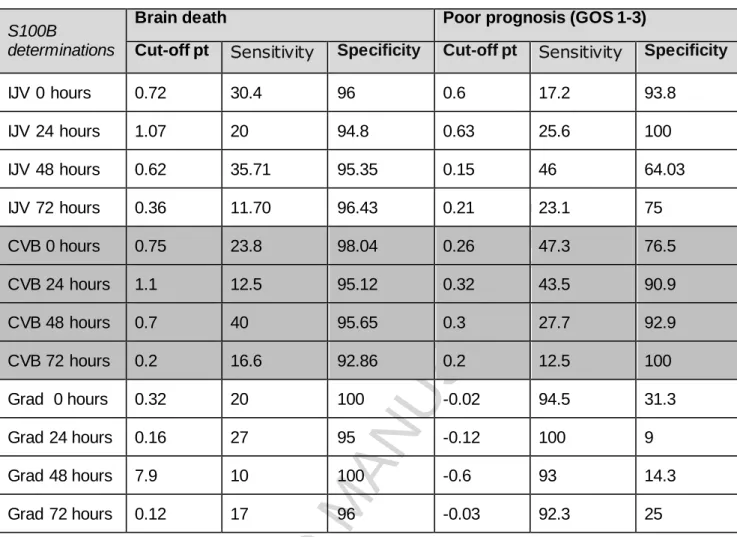
Table 4 shows the optimum cut-off points of protein S100B determinations and of the transcranial gradient in diagnosi ng development of brain death, according to the criterion of maximum sensitivity and specificity.
Univariate and multivariate analyses were made of the development of brain death, considering the main study variables (age, GCS, APACHE II, pupillary alterations and transcranial gradient upon admission), with statistically significant results for all of them. Multivariate analysis proved significant for GCS (OR 0.7, 95% CI 0.53-0.97, p = 0.017) and transcranial gradient upon admission (OR 7.16, 95% CI 1.42-36.0; p = 0.001).
However, on performing the analysis with respect to unfavourable outcome at 6 months, patient age (OR 1.055, 95% CI 1.013-1.099, p = 0.006) and the APACHE II score (OR 1.281, 95% CI 1.086 -1.511, p = 0.001) were identified as independent factors.
Discussion
This is the first study to simultaneously measure and compare protein S100B levels in regional venous blood in the brain (jugular bulb) and in systemic venous blood. To our knowledge, no other study has attempted to correlate systemic venous determinations to those obtained in cerebral venous blood samples, which offer more reliable information regarding changes occurring in the brain as a result of damage. Some studies have been carried out investigating arterial versus jugular venous samples
ACCEPTED MANUSCRIPT
[22,33]. No evaluations of the release profile in samples from the brain, which is where the protein is produced, have been made to date. This circumstance may have adversely conditioned previous studies, since extracranial protein S100B sources may limit the usefulness of peripheral determinations. One of the desirable characteristics of any potential marker is good accessibility with respect to both the technique required and to the biological sample involved. In the case of brain damage, the use o f peripheral blood samples would be ideal; however, since protein S100B can come from sources other than the brain, it is of interest to contrast peripheral determinations and measurements obtained directly from venous drainage of the brain. Our results confirm that protein S100B levels are higher in the jugular bulb samples and indicate good correlation between the two types of samples (regional and systemic). This fact ratifies the brain origin of the protein after brain damage, and shows that it meets one of the required criteria of a biomarker: specificity of the affected organ. The above in turn confirms the validity of the peripheral determinations of protein S100B in this patient cohort. The results obtained thus support the routine use of such determinations, since the obtainment of peripheral samples is feasible at any healthcare level, while regional measurements (jugular bulb catheter) or the need for cerebrospinal fluid samples are not available in all centres, pose accessibility problems, and are typically restricted to a research setting.
In addition, our findings corroborate the existence of a time pattern in protein S100B production. In the stratified analysis (GOS 1-2 vs. GOS 4-5), measurement kinetics were analogous in both types of sample, with a recorded rise in concentration 48 hours after brain damage. This behaviour is similar to that described by other authors who postulate that secondary brain damage is responsible for the observed increase detected two days after the primary brain injury, since the half-life of protein S100B is
ACCEPTED MANUSCRIPT
two hours [15]. However, other investigators have reported maximum protein S100B expression at the time of patient admission, which is followed by a decrease in levels [34]. The heterogeneity of the brain damage involved in the different series may explain these observations and might account for the sometimes conflicting findings among different authors. The behaviour of this protein underscores the relevance of follow-up of its levels over time as a reflection of secondary brain damage. Similar to findings reported in other studies, we observed no differences in protein S100B expression according to patient gender, type of brain damage or age [34].
Prognosis of patients with acute brain damage is particularly relevant in intensive care, allowing us to inform the relatives, define the best management strategy and optimize resource utilization. The results of our study indicate that determinations in regional samples are no better than determinations in systemic samples for predicting unfavourable prognosis or development of brain death. Therefore, although the systemic protein levels are lower than in the regional jugular bulb samples, they could be used as a diagnostic tool. In the literature, different S100B optimal cutoff points to predict outcome can be observed [34,35]. The cutoff points observed in our study are similar to those described by other studies, but Egea et al. proposed 0.372 μg/L as the 24-hour cutoff [36]. We would like to point out that these differences are explained by the study design and the type of brain injury. Transcranial gradient upon admission may serve as a marker of brain death, b ut the numbers are small and need to be confirmed. Development of brain death may be associated with damage of greater magnitude, i.e. very serious primary brain injuries, and thus to an increased production of protein S100B. This increased production is reflected by a significant gradient or difference between the regional and peripheral measurements, defining it as a marker of important brain damage indicative of brain death. Similar findings have been
ACCEPTED MANUSCRIPT
reported by other authors on relating the measurement of certain markers at the time of admission to patient prognosis [24,25] and to increased lesion severity upon admission. In this context, and on a speculative basis, secondary brain damage in other published series may have been responsible for brain death, and therefore the gradient measured after 48 hours may serve as the marker. Our group also detected a correlation between measurement after 48 hours and the prognosis in an in vitro model of brain damage, using regional serum samples collected on day 2 as apoptotic stimulus [37,38]. This again clearly illustrates the importance of the dynamism of brain injury and hence of secondary brain damage, and underscores the importance of the heterogeneity of the study population when it comes to drawing conclusions.
Our results show a high gradient upon admission to be associated with more serious damage and secondarily to brain death. This observation once more illustrates the heterogeneity of the characteristics of acute brain damage that constitutes a challenge for conducting studies designed to validate biomarkers for posterior application to clinical practice.
The heterogeneity of patient sample constitutes a limiting factor in our study, in the same way as clinical follow-up over 6 months and the measurement of protein S100B levels for four days.
Conclusions
The results obtained can be summarized as follows:
• Regional protein S100B levels are higher than systemic levels, confirming the brain origin of the protein.
• The time course of protein S100B levels is analogous in the regional and peripheral samples, thus confirming the suitability of using systemic samples.
ACCEPTED MANUSCRIPT
• Transcranial gradient in protein S100B concentration is correlated to the development of brain death.
• Protein S100B may be an early prognostic marker in patients with acute brain damage.
Peripheral determinations may be used in a clinical setting, given their minimally invasive nature and the lack of benefit of jugular bulb determination of S100B
Funding sources: This study has been supported by grants from the Marqués de
Valdecilla Foundation - IFIMAV (API 10/02) and the Spanish Ministry of Science - Carlos III Health Institute (PI080058). The protein S100B electrochemoluminescence assay kits were a generous donation from Roche Diagnostics, Mannheim, Germany The authors report no conflicts of interest. The authors alone are responsible for the contents and writing of the paper.
Author Disclosure Statement.
The authors report no conflicts of interest. The authors alone are responsible for the contents and writing of the paper.
ACCEPTED MANUSCRIPT
References
1. Peeters W, van den Brande R, Polinder S, Brazinova A, Steyerberg EW, Lingsma HF, et al. Epidemiology of traumatic brain injury in Europe. Acta Neurochir (Wien). 2015;157(10):1683-96.
2. Hemphill JC, Greenberg SM, Anderson CS, Becker K, Bendok BR, Cushman M, at al. Guidelines for the Management of Spontaneous Intracerebral Hemorrhage: A Guideline for Healthcare Professionals From the American Heart Association/American Stroke Association. Stroke. 2015;46(7):2032-60. 3. Masel BE, DeWitt DS. Traumatic brain injury: a disease process, not an event. J
Neurotrauma. 2010;27(8):1529-40.
4. Kövesdi E, Lückl J, Bukovics P, Farkas O, Pál J, Czeiter E, at al. Update on protein biomarkers in traumatic brain injury with emphasis on clinical use in adults and pediatrics. Acta Neurochir (Wien). 2010;152(1):1-17.
5. Wright NT, Cannon BR, Zimmer DB, Weber DJ. S100A1: Structure, Function, and Therapeutic Potential. Curr Chem Biol; 2009: 3(2):138-45.
6. Mazzini GS, Souza DO, Portela LV. The ischemic heart as an extracerebral source for S100B. Resuscitation;2009:80(1):144.
7. Harpio R, Einarsson R. S100 proteins as cancer biomarkers with focus on S100B in malignant melanoma. Clin Biochem;2004: 37(7):512-8.
8. Goncalves CA, Leite MC, Nardin P. Biological and methodological features of the measurement of S100B, a putative marker of brain injury. Clin Biochem; 2008:41(10-11):755-63.
9. da Rocha AB, Schneider RF, de Freitas GR, André C, Grivicich I, Zanoni C, et al. Role of serum S100B as a predictive marker of fatal outcome following isolated severe head injury or multitrauma in males. Clin Chem Lab Med; 2006: 44(10):1234-42.
10. Nylén K, Ost M, Csajbok LZ, Nilsson I, Hall C, Blennow K, et al. Serum levels of S100B, S100A1B and S100BB are all related to outcome after severe traumatic brain injury. Acta Neurochir (Wien).2008;150(3):221-7.
11. Bloomfield SM, McKinney J, Smith L, Brisman J. Reliability of S100B in predicting severity of central nervous system injury. Neurocrit Care. 2007; 6(2): 121-38.
12. Townend W, Ingebrigtsen T. Head injury outcome prediction: a role for protein S-100B?. Injury. 2006;37(12):1098-108.
ACCEPTED MANUSCRIPT
13. Korfias S, Stranjalis G, Boviatsis E, Psachoulia C, Jullien G, Gregson B, et al. Serum S-100B protein monitoring in patients with severe traumatic brain injury. Intensive Care Med. 2007;33(2):255-60.
14. Li N, Shen JK, Zhao WG, Cai Y, Li YF, Zhan SK. S-100B and neuron specific enolase in outcome prediction of severe head injury. Chi n J Traumatol. 2004; 7(3):156-8.
15. Raabe A, Grolms C, Sorge O, Zimmermann M, Seifert V. Serum S-100B protein in severe head injury. Neurosurgery. 1999;45(3):477-83.
16. Woertgen C, Rothoerl RD, Holzschuh M, Metz C, Brawanski A. Comparison of serial S-100 and NSE serum measurements after severe head injury. Acta Neurochir (Wien). 1997;139(12):1161-4.
17. Woertgen C, Rothoerl RD, Metz C, Brawanski A. Comparison of clinical, radiologic, and serum marker as prognostic factors after severe head injury. J Trauma. 1999;47(6):1126-30.
18. Schültke E, Sadanand V, Kelly ME, Griebel RW, Juurlink BH. Can admission S-100beta predict the extent of brain damage in head trauma patients?. Can J Neurol Sci. 2009;36(5):612-6.
19. Lima DP, Simão Filho C, Abib Sde C, de Figueiredo LF. Quality of life and neuropsychological changes in mild head trauma. Late analysis and correlation with S100B protein and cranial CT scan performed at hospital admission. Injury. 2008; 39(5):604-11.
20. de Boussard CN, Lundin A, Karlstedt D, Edman G, Bartfai A, Borg J. S100 and cognitive impairment after mild traumatic brain injury. J Rehabil Med. 2005;37(1):53-7.
21. Undén J, Astrand R, Waterloo K, Ingebrigtsen T, Bellner J, Reinstrup P, et al. Clinical significance of serum S100B levels in neurointensive care. Neurocrit Care. 2007; 6(2):94-9.
22. McKeating EG, Andrews PJ, Mascia L. Relationship of neuron specific enolase and protein S-100 concentrations in systemic and jugular venous serum to injury severity and outcome after traumatic brain injury. Acta Neurochir Suppl. 1998;71:117-9.
23. Pham N, Fazio V, Cucullo L, Teng Q, Biberthaler P, Bazarian JJ, et al. Extracranial sources of S100B do not affect serum levels. PLoS One.2010;5(9): e12691.
24. Miñambres E, Cemborain A, Sánchez-Velasco P, Gandarillas M, Díaz-Regañón G, Sánchez-González U, et al. Correlation between transcranial i nterleukin-6 gradient and outcome in patients with acute brain injury. Crit Care Med. 2003;31(3):933-8.
ACCEPTED MANUSCRIPT
25. McKeating EG, Andrews PJ, Signorini DF, Mascia L. Transcranial cytokine gradients in patients requiring intensive care after acute brain injury. B r J Anaesth. 1997;78(5):520-3.
26. Knaus WA, Draper EA, Wagner DP, Zimmerman JE. APACHE II: a severity of disease classification system. Crit Care Med. 1985;13(10):818-29.
27. Jennett B, Bond M. Assessment of outcome after severe brain damage. Lancet. 1975 Mar 1;1(7905):480-4.
28. Andrews PJ, Dearden NM, Miller JD. Jugular bulb cannulation: description of a cannulation technique and validation of a new continuous monitor. Br J Anaesth. 1991;67(5):553-8.
29. Díaz-Regañón G, Miñambres E, Holanda M, González-Herrera S, López-Espadas F, Garrido-Díaz C. Usefulness of venous oxygen saturation in the jugular bulb for the diagnosis of brain death: report of 118 patients. Intensive Care Med. 2002; 28(12):1724-8.
30. Alber B, Hein R, Garbe C, Caroli U, Luppa PB. Multicenter evaluation of the analytical and clinical performance of the Elecsys S100 immunoassay in patients with malignant melanoma. Clin Chem Lab Med. 2005;43(5):557-63. 31. Biberthaler P, Linsenmeier U, Pfeifer KJ, Kroetz M, Mussack T, Kanz KG, et al.
Serum S-100B concentration provides additional information fot the indication of computed tomography in patients after minor head injury: a prospective multicenter study. Shock. 2006;25(5):446-453.
32. Hanley JA, McNeil BJ. A method of comparing the areas under receiver operating characteristic curves derived from the same cases. Radiology. 1983;148(3): 839-843.
33. Bouvier D, Eisenmann N, Gillart T, Bonneau J, Guelon D, Schoeffler P, et al. Jugular venous and arterial concentrations of serum S100B protein in patients with severe head injury. Ann Biol Clin (Paris). 2012;70(3):269-275.
34. Murillo-Cabezas F, Muñoz-Sánchez MA, Rincón-Ferrari MD, Martín-Rodríguez JF, Amaya-Villar R, García-Gómez S, et al. The prognostic value of the temporal course of S100beta protein in post-acute severe brain injury: A prospective and observational study. Brain injury. 2010;24(4):609-19.
35. Thelin EP, Nelson DW, Bellander BM. A review of the clinical utility of ser um S100B protein levels in the assessment of traumatic brain injury. Acta Neurochir (Wien). 2017; 159(2): 209–225
36. Egea-Guerrero JJ, Murillo-Cabezas F, Gordillo-Escobar E, Rodríguez-Rodríguez A, Enamorado-Enamorado J, Revuelto-Rey J, Pacheco-Sánchez M, León-Justel A, Domínguez-Roldán JM, Vilches-Arenas A. S100B protein may detect brain death development after severe traumatic brain injury. J Neurotrauma. 2013 Oct 15;30(20):1762-9.
ACCEPTED MANUSCRIPT
37. Miñambres E, Lopez-Escribano H, Ballesteros MA, Peña M, López-Hoyos M. Apoptosis of Jurkat cells induced by serum of patients with acute severe brain injury. Intensive Care Med. 2005;31(6):791-8.
38. Ballesteros MA, López-Hoyos M, Muñoz P, Marin MJ, Miñambres E. Apoptosis of neuronal cells induced by serum of patients with acute brain injury: a new in vitro prognostic model. Intensive Care Med. 2007;33(1):58-65.
ACCEPTED MANUSCRIPT
Figure 1. Time course of protein S100B determinations at peripheral (CVB) and regional level (IJV), and of the transcranial gradient according to subsequent clinical outcome. The bars represent the median and the error bars represent first and third
quartile.
Figure 2. ROC curves of the different regional (IJV) and peripheral determinations (CVB) referred to prognosis and development of brain death. No differences bet ween curves were detected.
ACCEPTED MANUSCRIPT
Table 1. Characteristics of the study population.
Data are shown as median and interquartile or number and percentage.
APACHE II: Acute Physiology And Chronic Health Evaluation. GOS: Glasgow Outcome Score. GCS: Glasgow Coma Score. ICU : Intensive Care Medicine.
Table 2. S100B determinations depending on the origin of the samples.
IJV: internal jugular vein; CVB: central venous blood Data are shown as median (range interquartile
Table 3. Characteristics of the study population according to the prognosis and development of brain death.
Grad.: gradient (jugular level - systemic level). GCS: Glasgow Coma Score. APACHE II: Acute Physiology And Chronic Health Evaluation Poor prognosis: Glasgow Outcome Score 1 to 3 (deceased, vegetative state or severe disability). Good prognosis: Glasgow Outcome Score 4 or 5 (moderate disability with good recovery). Data are shown as median and first and third quartile. The statistical test used was Mann-Whitney U-test.
Table 4. Optimum cut-off points for the protein S100B determinations (μg/l).
IJV: internal jugular vein; Grad.: gradient (jugular level - systemic level); CVB: central venous blood; GOS: Glasgow Outcome Score
ACCEPTED MANUSCRIPT
Figure 1ACCEPTED MANUSCRIPT
Figure 2ACCEPTED MANUSCRIPT
Table 1. Characteristics of the study population.Total sample (N: 83)
Age (years) 55 (38 - 68)
Gender (females) 26 (31.3%) Type of brain damage
Head injury
Intracerebral haemorrhage
40 (48.2%) 43 (51.8%)
Neurosurgery
Intracranial pressure sensor Ventricular drainage 12 (14.5%) 36 (43.4) 15 (18.1%) APACHE II 17.1 (5.2) GCS 6 (4 8) GCS ≤ 4 21 (25.5%) Pupils Reactive Altered 47 (56.6%) 36 (43.4%) Brain death 25 (30.1%)
GOS after 6 months 1 (1 - 4) GOS 1 - 3 (poor prognosis) 61 (73.5%) GOS 4 and 5 (good recovery) 22 (26.5%) Time injury-ICU (hours) 2.3 (2,1 - 4,3)
Data are shown as median and interquartile or number and percentage.
APACHE II: Acute Physiology And Chronic Health Evaluation. GOS: Glasgow Outcome Score. GCS: Glasgow Coma Score. ICU : Intensive Care Medicine.
ACCEPTED MANUSCRIPT
Table 2. S100B determinations depending on the origin of the samples.IJV (μg/l) CVB (μg/l) Gradient 0 hours All patients (n: 83) 0.23 (0.13 - 0,49) 0.21 (0.1 - 0.4) 0.02 (0.004 - 0.06) TBI N: 40 0.30 (0.15 - 0.50) 0.25 (0.15 - 0.28) 0.02 (0.01 - 0.09) HIC 43 0.19 (0.12 - 0.43) 0.15 (0.10 - 0.34) 0.03 (0.01 - 0.06) 24 hours All patients (n: 54) 0.22 (0.13 - 0.47) 0.21 (0.1 - 0.45) 0.02 (0.004 - 0.06) TBI (N:28) 0.21 (0.13 - 0.44) 0.20 (0.11 - 0.27) 0.03 (0.005 - 0.06) HIC (N:27) 0.31 (0.14 - 0.43) 0.25 (0.11 - 0.51) 0.02 (0.01 - 0.07) 48 hours All patients (n: 54) 0.15 (0.07 - 0.25) 0.14 (0.07 - 0.27) 0.01 (0.01 - 0.04) TBI (N:28) 0.15 (0.1 - 0.3) 0.14 (0.1 - 0.31) 0.01 (0.02 - 0.04) HIC (N:27) 0.14 (0.05 - 0.44) 0.09 (0.07 - 0.23) 0.03 (0.01 - 0.022) 72 hours Al patients (n :24) 0.11 (0.06 - 0.19) 0.07 (0.03 - 0,15) 0.01 (0.004 - 0.06) TBI (N:15) 0.14 (0.10 - 0.26) 0.10 (0.04 - 0.25) 0.01 (0.005 - 0.7) HIC (N:9) 0.09 (0.06 - 0.16) 0.06 (0.03 - 0.13) 0.012 (0.006 0.04)
IJV: internal jugular vein; CVB: central venous blood Data are shown as median (range interquartile)
ACCEPTED MANUSCRIPT
Table 3. Characteristics of the study population according to the prognosis and development of brain death.
Grad.: gradient (jugular level - systemic level). GCS: Glasgow Coma Score. APACHE II: Acute Physiology And Chronic Health Evaluation Unfavourable outcome: Glasgow Outcome Score 1 to 3 (deceased, vegetative state or severe disability). Favourable outcome: Glasgow Outcome Score 4 or 5 (moderate disability with good recovery).
Data are shown as median and first and third quartile. The statistical test used was Mann-Whitney U-test. Unfavourable outcome N=61 Favourable outcome N=22 p Brain death N=25 Survivors N=58 p Age (years) 57 (50 -69) 37 (22-53) 0.001 57 (45-69) 53 (37-66) 0.153 GCS 5 (4 7) 7 (6-9) 0.004 4 (4-5) 7 (5-9) 0.001 APACHE II 19 (16-21) 12 (10-14) 0.001 19 (17-23) 16 (12-20) 0.002 S100B Grad 0 hours (μg/l) 0.02 (0.007-0.6) 0.02 (0.002-0.05) 0.130 0.05 (0.02- 0.17) 0.01 (-0.002- 0.05) 0.002 S100B Grad 24 hours (μg/l) 0.004 (-0,01 - 0,04) 0.03 (0.004 -0.7) 0.983 0.01 (0.04 - 0.18) 0.02 (-0.003 - 0.05) 0.915 S100B Grad 48 hours (μg/l) 0.005 (-0.01 - 0.02) 0.005 (0,001 - 0,04) 0.584 0.02 (-0.06- 0.06) 0.01 (-0.01 - 0.02) 0.824 S100B Grad 72 hours (μg/l) 0.01 (0.005 - 0.06) 0.008 (0.001 - 0,03) 0.405 0.02 (0.003 - 0.06) 0.01 (0.004 - 0.06) 0.910
ACCEPTED MANUSCRIPT
Table 4. Optimum cut-off points for the protein S100B determinations (μg/l).
S100B
determinations
Brain death Poor prognosis (GOS 1-3)
Cut-off pt Sensitivity Specificity Cut-off pt Sensitivity Specificity
IJV 0 hours 0.72 30.4 96 0.6 17.2 93.8 IJV 24 hours 1.07 20 94.8 0.63 25.6 100 IJV 48 hours 0.62 35.71 95.35 0.15 46 64.03 IJV 72 hours 0.36 11.70 96.43 0.21 23.1 75 CVB 0 hours 0.75 23.8 98.04 0.26 47.3 76.5 CVB 24 hours 1.1 12.5 95.12 0.32 43.5 90.9 CVB 48 hours 0.7 40 95.65 0.3 27.7 92.9 CVB 72 hours 0.2 16.6 92.86 0.2 12.5 100 Grad 0 hours 0.32 20 100 -0.02 94.5 31.3 Grad 24 hours 0.16 27 95 -0.12 100 9 Grad 48 hours 7.9 10 100 -0.6 93 14.3 Grad 72 hours 0.12 17 96 -0.03 92.3 25
IJV: internal jugular vein; Grad.: gradient (jugular level - systemic level); CVB: central venous blood; GOS: Glasgow Outcome Score
ACCEPTED MANUSCRIPT
Highlights Regional protein S100B determinations are higher than systemic determinations, confirming the cerebral origin of protein S100B.
The transcranial protein S100B gradient is related to the development of brain death.