The PcACE1 transcription factor from Phanerochaete chrysosporium contains a Cys and Ser rich transactivation domain
6
0
0
Texto completo
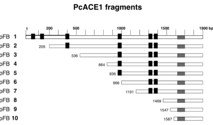
(2) 352. BULL ET AL. Biol Res 44, 2011, 351-355. assay to the promoter of the yeast metallothionein in the presence of copper, but not in its absence (Canessa et al., 2008). The PcACE1 amino acid sequence contains the same three Cys motifs present at the amino terminus of Ace1, Amt1 and Crf1, and additional three. Table 1 shows the amino acid sequence of the six Cys motifs present in the amino acid sequence of PcACE1. The fourth Cys motif, C-X-C-X3-C-X-C-X2-C-X2-H, is highly similar to C1 from Mac1, and at the carboxy terminus of GRISEA and Cuf1, transcription factors that are active at low copper concentration (Rutherford and Bird, 2004). Another unique finding is that PcACE1 contains 68% serines within the amino acid sequence (S)5-H-(S)3-H-R-S between amino acid residues 546 and 564, an unusually high number. In this report we present evidence that the fifth and sixth Cys-rich motifs from PcACE1 and the Ser-rich motif located further downstream are involved in transcription transactivation. METHODS Strains and culture media. S. cerevisiae YM335::RY171 (a gal4-536 ura3-52 ade2-101 lys2801 His3-200met) is a derivative from YM335 having a GAL1lacZ fusion gene integrated at the URA3 locus and has been previously described (Ma and Ptashne, 1987). Yeast cells were grown in YPD (10 g yeast extract, 10 g bactopeptone and 2% dextrose or glucose for 1 lt) or synthetic defined media without His (SC-His) (Johnston, 1994). SC-His medium was prepared as follows: Mix 1: To 500 ml of water, 6.7 g yeast nitrogen base (without amino acids), 10 mg adenine, 40 mg uracil and 50 mg Tyr, were added. Mix 2: To 100 ml of water, 200 mg Arg, 600 mg Ile, 600 mg Leu, 400 mg Lys, 100 mg Met, 600 mg Phe, 1g Thr and 400 mg Trp, were added. Mix 3: 435 ml of sterile water was added to Mix 1 plus 10 ml of Mix 2 and 5 ml of 1M Na2HPO4 (pH 7).The carbon sources for the defined media, where appropriate, were glucose, glycerol, ethanol and/or galactose at 2% final concentration (Johnston, 1994).. activation domain in different transcription factors, for instance Ace1 (Hu et al., 1990), Mac1 (Graden and Winge, 1997), and proto-oncogenes c-rel (Bull et al., 1990) and c-relB (Ryseck et al., 1992), among others. Vector pMA424 (Ma and Ptashne, 1987) contains the ADH promoter followed by the DNA binding domain of the yeast transcription factor GAL4 (amino acids 1-147) and the ADH termination domain. The last two are separated by a BamH1 site. The plasmid also contains a HIS3 gene for selection of the transformed yeast (Ma and Ptashne, 1987). PcACE1 cDNA cloned in pET21a (+) between Nde1 (5’) and BamH1 (3’) sites was kindly provided by Dr. Rafael Vicuña. Oligonucleotides containing BamH1 sites were designed for site-directed mutagenesis in different places of PcACE1 cDNA. Care was taken to keep the coding sequence in frame with the GAL4 DNA-binding domain. Oligonucleotides and position are shown in Table 2. PCR amplification of PcACE1 cDNA was performed with Taq Platinum DNA polymerase in 25 μl solution. PcACE1 fragments were ligated into the unique BamH1 site of pMA424. Recombinant plasmids containing fragments of PcACE1 cDNA were named pFB1 to pFB10. Figure 1 shows a schematic representation of the 10 different PcACE1 fragments obtained. In this figure, the position of the Cys motifs are highlighted in black and the position of the Ser motif in gray (amino acids 546-564). Yeast transformation. Yeast cells auxotrophic for His and carrying the promoter of GAL1 in the genome, followed by the reporter gene β-galactosidase, were transformed with the previously obtained recombinant plasmids. Later, the reporter activity was analyzed in each yeast transformant (Ma and Ptashne, 1987). One isolated colony of S. cerevisiae YM335::RY171 was grown in YPD culture medium for 16 hr at 30ºC. 100 ml of YPD medium was inoculated with 1 ml of saturated culture that was obtained the night before. The culture was grown until Abs600. Plasmid constructions. PcACE1 fragments. The method designed by Ma and Ptashne (1987) was used. Several groups have been able to localize the transcriptional. 200. 1. 500. 1000. 1500. 1900 bp. pFB 1 pFB 2 pFB 3. TABLE 1 Localization and sequence of the Cys motifs in the amino acid sequence of PcACE1. pFB 4 p pFB 5 pFB 6 pFB 7 pFB 8. Position (aa). Amino acid sequence. pFB 9 pFB 10. 1. 11-25. C-E-T-C-I-K-G-H-R-S-S-N-C-K-H. 2. 43-61. C-D-H-C-R-E-L-R-K-T-K-Q-V-H-V-K-C-V-C. 3. 126-136. C-T-C-K-T-T-G-I-C-N-C. 4. 298-312. C-D-C-G-P-N-C-A-C-P-G-C-V-I-H. 5. 430-439. C-K-C-P-H-R-V-C-A-C. 6. 450-457. C-T-C-P-S-C-N-H. The positions of the amino acid residues (aa) are shown, together with the respective amino acid sequence. Cys and His residue pairs are in bold.. 205 536 864 936 966 1191 1469 1547 1587. Figure 1. A diagram of PcACE1 cDNA fragments obtained by PCR and cloned into the BamHI site of pMA424 is shown. The names of clones obtained (pFB1 to pFB10) are shown to the left; numbers on the top of the figure correspond to length in base pairs of complete PcACE1 cDNA. All fragments share 3’ end of cDNA and differ in the 5’ end. Numbers immediately before each fragment correspond to the nucleotide at which the fragment begins. The Cys motifs are in black, and the position of the Ser motif in gray (amino acids 546-564). The length of each box is not to scale..
(3) 353. BULL ET AL. Biol Res 44, 2011, 351-355. 0.4-0.6, divided in 4 sterile tubes and incubated on ice, after which they were centrifuged for 7.5 min at 4,400 rpm in Sorvall SS34 rotor, followed by a centrifugation in a microfuge for 1 min at 14,000 rpm. The supernatant was discarded and the cells were resuspended in 1 ml sterile water. This was done twice. Then, they were suspended in 1 ml of 0.1 M lithium acetate. After another centrifugation, the cells were resuspended in 0.4 ml of 0.1 M lithium acetate. Aliquots of 100 μl were placed in new tubes, and the following reagents were added: 240 μl 50% PEG 3,500; 36 μl 1 M lithium acetate; 25 μl denatured salmon sperm DNA (10 mg/ml) and 90 ng of supercoiled pFB plasmid. After one min vortexing, tubes were incubated for 30 min at 28°C, followed by incubation at 42°C for 15 min with gentle agitation. Afterwards, tubes were centrifuged at 14,000 rpm for 1 min; the supernatant was discarded and the pellet was washed twice with 1 ml of sterile water. Part of the supernatant was discarded. The remaining yeast cells were plated on petri dishes containing SC-His and 2% glucose. Colonies were visualized after incubation at 28°C for 3 days. Induction of transformed yeast cells and determination of b-galactosidase activity. Transformed yeast cells were cultured in liquid SC–HIS and 2% glucose medium for 18 hrs. The medium was then changed to the induction medium (SC-His, glycerol, ethanol and galactose) and incubated for 4 hr at 30ºC.The induced cells were concentrated to 1 ml by centrifugation and the medium was changed to 1 ml Buffer Z (60 mM Na2HPO4, 40 mM NaH2PO4, 10 mM KCl, 1 mM MgSO4 and 2.7 ml b-mercapto-ethanol). Yeast cells were lysed by addition of 70 ml of 0.1% SDS and 100 ml chloroform, and vortexed for 10 sec. Then, 200 μl of 4 mg/ ml o-nitrophenyl-β-D-galactopyranose (substrate) were added, incubated for 5 min at 30ºC. The reaction was stopped with 0.5 ml 1 M sodium carbonate. After centrifugation for 3 min, the product (o-nitrophenol) was quantified by absorbance at 420. nm as Yocum et al. (1984). The determination of each sample was repeated 3 to 6 times. Enzyme activity was determined using the following formula: β-galactosidase specific activity (units) = Abs 420 x 1000/ time (min) x vol (ml) x ABS 600. The standard errors were within 10%. All DNA manipulations were performed with standard procedures (Sambrook et al., 1989). The plasmid constructs were verified by restriction enzyme mapping and confirmed by double-stranded sequencing (Macrogen, Korea). RESULTS AND DISCUSSION. Any transcription factor contains at least two domains; the DNA binding domain and the transcription transactivation domain (Ptashne, 1988). Both domains are key to a functional transcription factor. Interestingly, Ma and Ptashne (1987) demonstrated that these modules can be interchanged between two different transcription factors. Each DNA binding fragment retains its properties and binds to the same nucleotide sequence recognized by the original transcription factor in the promoter of the target gene. This finding allowed us to determine the localization of transactivation domains of several transcription factors, using the DNA binding domain of a well-known transcription factor such as GAL4. The amino acid sequence of transcription activation domains can be highly variable; for instance, transcription factor Sp1 has a transactivation domain rich in glutamines, and CTF1 is rich in prolines. VP16 of Herpes simplex virus contains hydrophobic and aromatic residues. On the other hand, GCN4 contains acidic residues in the transactivation domain. NF-kB p65 needs to be phosphorylated in order to be active (Schmitz et al., 1995). The transcription activation domain of Mac1 maps to the C1 Cys-rich domain (Graden and Winge, 1987).. TABLE 2 Oligonucleotides utilized for site-directed Mutagenesis Name. Nucleotide sequence. Position (bp). RV172. 5’GCCGTCAGGGATCCCTCCAGGCTTCGAGT. 942. RV173. 5’GACTACAGCCAGCGGATCCGAGCCCCTATTGCA. 1471. RV174. 5’TTCTGCTGCGACAGGGATCCCTCGCGTGTCACTCA. 1593. RV176. 5’GGATCCTTAGAATATCCGTGGACTGCCATCGTCA. 1902-1869. RV177. 5’TCGCCGGAATTCCGGGGATCC. pMA424. RV178. 5’AGGATCCTAGTCGGTGAGAA. 1. RV179. 5’TCCGGCGCAGGGATCCCCTCGTCG. RV180. 5’AAACAGACGGATCCTTCCACGT. 537. RV181. 5’CCTCCCACCGGGATCCCTGCTGCGA. 865. RV183. 5’ATGTGGCAGTCCTGGATCCTCTGGCCCAAT. 1191. RV184. 5’TCCGTTAGGGATCCGGGGCGTCTT. 1548. RV203. 5’AACGAATGAACGGGATCCCTTCCACT. 1061. 202. The BamH1 site in each oligonucleotide is underlined. All oligonucleotides are FORWARD, except RV176, which is REVERSE..
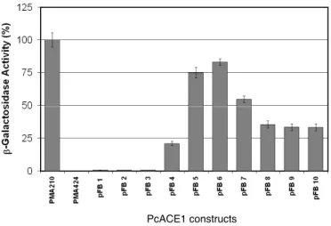
(4) 354. BULL ET AL. Biol Res 44, 2011, 351-355. We decided to map the transactivation domain of the transcription factor PcACE1 using the method described by Ma and Ptashne (1987). Full-length PcAce1 or partial sequences of it were fused to the GAL4 DNA binding domain and assayed for transcription activation. Figure 2 shows the percentage of b-galactosidase activity obtained with each yeast transformant. 100% was defined as activity of the full-length GAL4 yeast transcription factor (positive control). pFB1, pFB2 and pFB3, the largest open reading frames, showed no reporter activity. However, when the first 850 bp of the 5’ terminal sequences of PcACE1 were deleted (clones pFB4, pFB5, pFB6 and pFB7), transactivation activity was present, suggesting that an inhibitory region had been eliminated. The C terminal half of the protein functions as a transcription activator. The highest reporter activity was obtained with clones pFB5 and pFB6, yielding about 80% of the positive control. In this region, the PcACE1 fragments contain the Cys motifs 5 and 6 and the Ser-rich motif. Surprisingly, Mac1, which is active only at very low copper concentrations, contains a similar transcription activating domain at the Cys-rich region (Graden and Winge, 1997). Therefore, these results indicate that this type of Cys motif can function as a transcription activating domain not only in transcription factors that respond to minimal copper, but also in some that respond to high copper concentrations, as in this case. pFB8, pFB9 and pFB10 have about 30% transactivation activity. Interestingly, this region only has the Ser-rich motif and lacks Cys motifs. These results suggest that there could be two transactivating regions in PcACE1, one containing the Cys motifs and a second containing the Ser-rich motif. It would be interesting to look for phosphorylation in Ser. There are examples of activation by phosphorylation, such as Mac1 (Heredia et al., 2001) or C-terminal domain of the largest subunit from yeast RNA polymerase II (Kim et al., 2009).. Why are the pFB1, pFB2 and pFB3 clones inactive, in spite of the presence of all Cys and Ser-rich motifs? There are other examples in which the full-length transcription factor is inactive, in spite of the presence of the transactivation domain, for instance the proteins c-REL (Bull et al., 1990) and c-RELB (Ryseck et al., 1992). There are a few possible explanations for this result. First, Ace1 from S. cerevisiae is active only in the presence of copper (Furst and Hamer, 1989). Copper may cause a conformation change in PcACE1 by forming a cuprous thiolate, similar to Ace1 (Brown et al., 2002) and Mac1 (Jensen and Winge, 1998). It could also be that the addition of the DNA binding domain of GAL4 (147 amino acids), produces a steric hindrance that prevents the correct folding of the protein. On the other hand, pFB1 contains two different DNA binding sequences, one from GAL4, and the other from PcACE1. Each would try to bind to its target promoter, and the binding would be not productive. In the case of pFB2 and pFB3, there could be a competition for other necessary regulators. Site-directed mutagenesis of crucial amino acids will be able to determine the important amino acids. Taken together, in this work, we show the transactivation domain of PcACE1 maps to the carboxy terminus, where two conserved Cys- and Ser-rich motifs are localized. Future experiments will help to better understand the transcriptional mechanisms involved in fungal copper homeostasis. These data contribute to the understanding of how the copper levels are regulated in a basidiomycete fungus. ACKNOWLEDGEMENTS. This work was supported by the FONDECYT N°1085236 and VRI 18/2010 grants. We thank Rafael Vicuña for the PcACE1 clone. The comments of Luis F. Larrondo and Paulo Canessa are greatly appreciated. REFERENCES. PcACE1 constructs. Figure 2. β-galactosidase activity of yeast transformed with clones pFB1 to pFB10. Plasmids expressing the GAL4-PcACE1 chimeras (pFB1 to pFB10), wild type GAL4 (pMA210) or GAL4 DNA binding domain (pMA424) were introduced into a yeast strain lacking a functional GAL4 gene but bearing a Gal80 gene and an integrated GAL1::lacZ fusion gene (YM335::RY171). β-galactosidase activities were measured from cells grown in the presence of galactose, glycerol and ethanol. Each experiment was done 3 to 7 times except pFB6 and pFB7, which is the mean of three different clones.. BALAMURUGAN K, SCHAFFNER W (2006) Copper homeostasis in eukaryotes: Teetering on a tightrope. BB Acta 1763: 737-746. BEAUDOIN J, LABBÉ S (2001) The fission yeast copper-sensing transcription factor Cuf1 regulates the copper transporter gene expression through an AceI/Amt1-like recognition sequence. J Biol Chem 276: 15472-15480. BORGHOUTS C, SCHECKHUBER CQ, STEPHAN O, OSIEWACZ HD (2002) Copper homeostasis and aging in the fungal model system Podospora anserina: differential expression of PaCtr3 encoding a copper transporter. Int J Biochem Cell Biol 34: 1355-1371. BROWN KR, KELLER GL, PICKERING IJ, HARRIS HH, GEORGE GN, WINGE DR (2002) Structures of the cuprous-thiolate clusters of the Mac1 and Ace1 transcriptional activators. Biochem 41: 6469-6476. BULL P, MORLEY KL, HOEKSTRA MF, HUNTER T, VERMA IM (1990) The mouse c-rel protein has an N-terminal regulatory domain and a C-terminal transcriptional transactivation domain. Mol Cell Biol 10: 5473-5485. CANESSA P, ÁLVAREZ JM, POLANCO, R, BULL P, VICUÑA R (2008) The copper-dependent ACE1 transcription factor activates the transcription of the mco1 gene from the basidiomycete Phanerochaete chrysosporium. Microbiol 154: 491-499. DAMERON CT, WINGE DR, GEORGE GN, SANSONE M, HU S, HAMER D (1991) A copper-thiolate polynuclear cluster in the ACE1 transcription factor. Proc Natl Acad Sci USA. 88: 6127-6131. FARRELL RA, THORVALDSEN JL, WINGE DR (1996) Identification of the Zn(II) site in the copper-responsive yeast transcription factor, AMT1: a conserved Zn module. Biochem 35: 1571-1580. GARCÍA S, PRADO M, DÉGANO R, DOMÍNGUEZ A (2002) A copperresponsive transcription factor, CRF1, mediates copper and cadmium resistance in Yarrowia lipolytica J Biol Chem 277: 37359-37368. G E O R G AT S O U E , M AV R O G I A N N I S L A , F R A G I A D A K I S D , ALEXANDRAKI D (1997) The yeast Fre1p/Fre2p cupric reductases.
(5) BULL ET AL. Biol Res 44, 2011, 351-355. facilitate copper uptake and are regulated by the copper-modulated Mac1p.activator. J Biol Chem 272:13786-13792. GRADEN JA, WINGE DR (1997) Copper-mediated repression of the activation domain in the yeast Mac1p transcription factor. Proc Natl Acad Sci USA. 94: 5550-5555. HEREDIA J, CROOKS M, ZHU Z (2001) Phosphorylation and Cu + coordination-dependent DNA binding of the transcription factor Mac1p in the regulation of copper transport. J Biol Chem 276: 8793–8797. HU S, FÜRST P, HAMER D (1990) The DNA and Cu binding functions of ACE1 are interdigitated within a single domain. New Biol 2: 544-555. JENSEN LT, WINGE DR (1998) Identification of a copper-induced intramolecular interaction in the transcription factor Mac1 from Saccharomyces cerevisiae. EMBO J 17: 5400-5408. JOHNSTON JR (1994) Molecular Genetics of yeast: a practical approach. 2nd Ed Oxford University Press, NY NY pp 121-134. JUNGMANN J, REINS HA, LEE J, ROMEO A, HASSETT R, KOSMAN D, JENTSCH S (1993) MAC1, a nuclear regulatory protein related to Cudependent transcription factors is involved in Cu/Fe utilization and stress resistance in yeast. EMBO J 12: 5051-5056. KIM M, SUH H, CHO E-J, BURATOWSKI S (2009) Phosphorylation of the yeast Rpb1 C-terminal domain at serines 2, 5, and 7. J Biol Chem 284: 26421–26426. MA J, PTASHNE M (1987) Deletion analysis of GAL4 defines two transcriptional activating segments. Cell 48: 847-853. POLANCO R, CANESSA P, RIVAS A, LARRONDO LF, LOBOS S, VICUÑA R (2006) Cloning and functional characterization of the gene encoding. 355. the transcription factor ACEI in the basidiomycete Phanerochaete chrysosporium. Biol Res 39: 641-648. PTASHNE M (2005) Regulation of transcription: from lambda to eukarytes. TIBS 30: 275-279. RUTHERFORD JC, BIRD AJ (2004) Metal-responsive transcription factors that regulate iron, zinc, and copper homeostasis in eukaryotic cells. Eukar Cell 3: 1-13. RYSECK RP, BULL P, TAKAMIYA M, BOURS V, SIEBENLIST U, DOBRZANSKI P, BRAVO R (1992) RelB, a new Rel family transcription activator that can interact with p50-NF-κB. Mol Cell Biol 12: 674-684. SAMBROOK J, FRITSCH EF, MANIATIS T (1989) Molecular cloning. A laboratory manual, 2nd Edition Cold Spring Harbor Laboratory Press. SCHMITZ ML, DOS SANTOS SILVA MA, BAEUERLE PA (1995) Transactivation domain 2 (TA2) of p65 NF-kappa B: similarity to TA1 and phorbol ester-stimulated activity and phosphorylation in intact cells. J Biol Chem 270: 15576-15584. THIELE DJ (1988) ACE1 regulates expression of the Saccharomyces cerevisiae metallothionein gene. Mol Cell Biol 8: 2745-2752. TURNER RB, SMITH DL, ZAWROTNY ME, SUMMERS MF, POSEWITZ, MC, WINGE DR (1998) Solution structure of a zinc domain conserved in yeast copper-regulated transcription factors. Nat Struct Biol 5: 551-555. YOCUM RR, HANLEY S, WEST R, PTASHNE M (1984) Use of lacZ fusions to delimit regulatory elements of the inducible divergent GAL1-GAL10 promoter in Saccharomyces cerevisiae. Mol Cell Biol 4: 1985-1998. ZHOU PB, THIELE DJ (1993) Copper and gene regulation in yeast. BioFactors 4: 105-115..
(6)
(7)
Figure
Documento similar