Altered lipid metabolism in a
Drosophila
model
of Friedreich’s ataxia
Juan A. Navarro
1, Elisabeth Ohmann
1, Diego Sanchez
2, Jose´ A. Botella
1, Gerhard Liebisch
3,
Marı´a D. Molto´
4, Marı´a D. Ganfornina
2, Gerd Schmitz
3and Stephan Schneuwly
1,∗1
Institute of Zoology, Universitaetsstrasse 31, University of Regensburg, 93040 Regensburg, Germany, 2
Departamento de Bioquı´mica y Biologı´a Molecular y Fisiologı´a, Instituto de Biologı´a y Gene´tica Molecular, C/Sanz y Fore´s s/n, Universidad de Valladolid-CSIC, 47003 Valladolid, Spain,3Institute for Clinical Chemistry and Laboratory Medicine, University of Regensburg, Franz-Josef-Strauß-Allee 11, 93053 Regensburg, Germany and4Department of Genetics, Universidad de Valencia, CIBERSAM, 46100 Burjassot, Valencia, Spain
Received February 17, 2010; Revised April 8, 2010; Accepted May 1, 2010
Friedreich’s ataxia (FRDA) is the most common form of autosomal recessive ataxia caused by a deficit in the mitochondrial protein frataxin. Although demyelination is a common symptom in FRDA patients, no multicel-lular model has yet been developed to study the involvement of glial cells in FRDA. Using the recently
estab-lished RNAi lines for targeted suppression of frataxin in Drosophila, we were able to study the effects of
general versus glial-specific frataxin downregulation. In particular, we wanted to study the interplay between lowered frataxin content, lipid accumulation and peroxidation and the consequences of these effects on the sensitivity to oxidative stress and fly fitness. Interestingly, ubiquitous frataxin reduction leads to an increase in fatty acids catalyzing an enhancement of lipid peroxidation levels, elevating the intracellular toxic poten-tial. Specific loss of frataxin in glial cells triggers a similar phenotype which can be visualized by accumulat-ing lipid droplets in glial cells. This phenotype is associated with a reduced lifespan, an increased sensitivity to oxidative insult, neurodegenerative effects and a serious impairment of locomotor activity. These symp-toms fit very well with our observation of an increase in intracellular toxicity by lipid peroxides.
Interestingly, co-expression of aDrosophilaapolipoprotein D ortholog (glial lazarillo) has a strong protective
effect in our frataxin models, mainly by controlling the level of lipid peroxidation. Our results clearly support a strong involvement of glial cells and lipid peroxidation in the generation of FRDA-like symptoms.
INTRODUCTION
Friedreich’s ataxia (FRDA) is an autosomal recessive neurodegenerative disease affecting the central (CNS) and per-ipheral (PNS) nervous systems (1,2). The FRDA neuropathol-ogy typically presents early degeneration of large sensory neurons in the dorsal root ganglia (DRG), followed by degener-ation of sensory posterior columns, spinal-cerebellar tracts and cortical-spinal motor tracts together with atrophy of the large sensory fibers in peripheral nerves (reviewed in 3) and grumose degeneration in the dentate nucleus (4). FRDA is also characterized by hypertrophic cardiomyopathy that becomes the main cause of death among the patients (5) and by accumulation of labile iron in patient’s mitochondria (6).
FRDA is caused by a decreased expression of the mitochon-drial protein frataxin (7,8). Frataxin deficiency leads to a dys-function of the respiratory chain complexes and Krebs cycle components mainly due to an inappropiate Fe – S cluster syn-thesis, therefore provoking a bioenergetic failure and the sub-sequent cell death (9–11).
Frataxin-deficient cells are hypersensitive to oxidative insult pointing to oxidative stress as a key factor in FRDA pathology (12–17). Furthermore, samples from FRDA patients normally show increased levels of oxidative stress markers such as malondialdehyde (MDA, a lipid peroxidation product), 8-hydroxy-2′-deoxyguanosine (an indicator of oxidized DNA) and higher glutathione transferase activity (18–20, respectively).
∗To whom correspondence should be addressed. Tel:+49 9419433067; Fax:+49 9419433325; Email: stephan.schneuwly@biologie.uni-regensburg.de
#The Author 2010. Published by Oxford University Press. All rights reserved. For Permissions, please email: journals.permissions@oxfordjournals.org
Human Molecular Genetics, 2010 1–13 doi:10.1093/hmg/ddq183
HMG Advance Access published May 19, 2010
at Universitaetsbibliothek Regensburg on May 21, 2010
http://hmg.oxfordjournals.org
(Fig.2A). The level of different phospholipids [choline (PC), phosphatidylglycerin (PG) and phosphatidyl-inositol (PI)] did not show any relevant increase (Fig.2B).
In order to determine whether such an accumulation was due to a general increase of FAs or only to a contribution of some specific species, we carried out a GC/MS analysis of FAs. Figure 1.Lipid droplet accumulation in frataxin-deficient glial cells. (AandD) 35-day-oldRepo-GAL4/+controls. (B,CandE) 35-day-oldRepo-GAL4/
fhRNAi-1. (F) 35-day-old UAS-GLaz/+;Repo-GAL4/fhRNAi-1. (B) Frataxin-deficient brains displayed degenerative defects in the form of extensive outer chiasm vacuolization (arrowhead). (C) The appearance of lipid-like droplets is denoted with white arrows. (D and E) Ultrastructural analysis revealed accumu-lation of lipid bodies in glial cells of frataxin-deficient brains. (F) Altered properties of glial-lipid deposits after co-expression ofGLaz. (G) GC/MS analysis of FAs fromDrosophilaheads. Frataxin deficiency results in a remarkable increase of C14:0, C16:1 and C18:1.GLazis unsuccessful in restoring the amount of these three FAs to control levels. Significance in (G) was determined by one-way ANOVA withpost hocNewman– Keuls (∗∗∗P,0.001 and∗P,0.05). Error bar represents standard error. FA, fatty acid. The scale bar represents 50mm (A and B); 25mm (C) and 2.5mm (D – F).
Human Molecular Genetics, 2010 3
at Universitaetsbibliothek Regensburg on May 21, 2010
http://hmg.oxfordjournals.org
In the L3 samples, we were able to detect a profile consisting of 19 different FAs. Eight of them were extremely low represented (,6‰) and were not considered in our study. Myristic acid (C14:0), palmitic acid (C16:0), palmitoleic acid (C16:1), oleic acid (C18:1) and linoleic acid (C18:2) were the most abundant acids (each of them representing.10% of total FA composition). In addition, FAs with chains containing more than 20 carbons were not detected. Figure2C and D shows that frataxin deficiency induced a general accumulation of FAs independently of their relative abundance compared with control flies. In agreement with the glial results (Fig.1G), the increase was higher in the saturated and monounsaturated FAs (from 65 up to 160%) in contrast to only slight increases detected in the polyunsaturated FAs (25%).
Taken together, these results suggest that loss of frataxin triggers a general accumulation of FAs either by an increased synthesis or by a defective catabolism. This finding is in agree-ment with our observation of accumulation of lipid-like dro-plets in glial cells of frataxin-deficient flies.
Overexpression ofGLazcounteracts the accumulation of FAs in frataxin knock-down flies
GLaz is an important protein involved in antioxidant defense and lipid metabolism in glial cells and therefore a good candi-date for attenuating the frataxin-dependent glial phenotype. A first hint for such an interaction came from the ultrastructural
analysis, which shows a modification of the quality of lipid droplets in the glial cells of frataxin knock-down flies that co-express GLaz (compare Fig. 1E with F). In agreement with this observation, lipid analysis of L3 larvae that ubiqui-tously co-expressed GLaz showed a significant reduction of free FA content but no changes in TAG or phospholipids (Fig. 2A and B). To study any specific effects of GLaz on FA content, a GC/MS approach was performed identifying a biased action ofGLaz with an exclusive and partial recovery of monounsaturated FAs, but not saturated or polyunsaturated FAs (Fig.2C and D).
To exclude that the partial rescue ofGLazis due to a GAL4 dilution effect using multiple UAS constructs in one genotype, expression levels were compared by semi-quantitative real-time PCR. Both genotypes, single frataxin interference (da-GAL4/fhRNAi-1) and co-expression of GLaz (UAS-GLaz/+;da-GAL4/fhRNAi-1) showed a similar frataxin level of 10% relative to control values (data not shown).
Our results clearly indicate thatGLazis capable of partially rescuing the frataxin lipid phenotype by selectively reducing the increased amount of monounsaturated FAs.
Glial function of frataxin in lifespan and oxidative stress
Next, we investigated the impact of the glial function of fra-taxin on fly survival. Enhanced sensitivity to oxidative insult is a hallmark in FRDA models, hence we examined whether Figure 2.Lipid analysis of frataxin interference lines detects a drastic increase in free FAs partially rescued byDrosophila GLaz. (AandB) Analysis of neutral and phospholipid levels by thin-layer chromatography in late L3 larvae. The values are expressed as a percentage of control (da-GAL4/+). Frataxin knock-down (da-GAL4/fhRNAi-1) mainly triggers accumulation of free FAs when compared with controls. These accumulations are partially rescued byGLazco-expression (UAS-GLaz/+;da-GAL4/fhRNAi-1). (CandD) GC/MS analysis of FA content. (C) Comparison of the most abundant FA in our samples. (D) Comparison of low-level FA (,10%). Significance was determined by one-way ANOVA withpost hocNewman–Keuls (∗∗∗P,0.001;∗∗P,0.01 and∗P,0.05). Error bar rep-resents standard error. FFA, free fatty acid; TAG, triacylglycerides; PC, phosphatidylcholine; PG, phosphatidylglycerin; PI, phosphatidylinositol; FA, fatty acid.
at Universitaetsbibliothek Regensburg on May 21, 2010
http://hmg.oxfordjournals.org
reducing frataxin expression in glial cells was sufficient to exacerbate the sensitivity to oxidative stress. Hyperoxia has been proven to be a relevant system to re-create an unbalanced oxidative status (50). As can be seen in Fig.3A, frataxin inter-ference (Repo-GAL4/fhRNAi-1) under oxygen-induced stress resulted in a drastic reduction of mean (73%) and maximum lifespan (55%) when compared with controls. Shortening of mean (25%) and maximum (32%) lifespan was also revealed by survival analysis under normal conditions (Fig.3B).
These results indicate that frataxin is essential for a correct function of glial cells. In particular, absence of glial frataxin increased sensitivity of this cell type towards reactive oxygen species (ROS), leading to premature death of the flies.
Depletion of frataxin expression in glial cells affects nervous system integrity
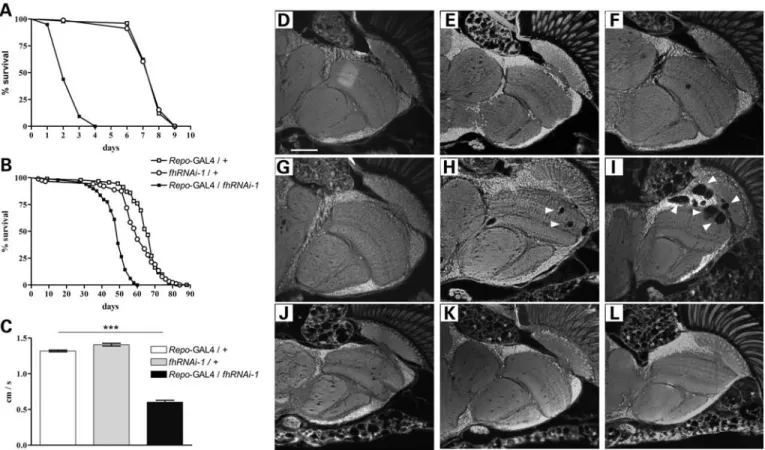
In order to study the effect of frataxin downregulation on the integrity and function of theDrosophilanervous system, loco-motor activity and brain degeneration of Repo-GAL4/ fhRNAi-1flies were analyzed.
Loss of frataxin provoked a 55% reduction in the climbing ability of 10-day-old flies (Fig.3C). Moreover, brain analysis
showed an age-dependent degeneration that was first detected at day 20 and further increased at day 30 (Fig. 3G – I). The vacuolization is mainly focused on medulla, lamina and outer chiasm (Fig. 3I, arrowheads), which is consistent with a location of tissue enriched in glial cells. In addition, an ultra-structural analysis showed that the lipid droplets described in Figure1can be detected before the presence of vacuolization (data not shown).
Our results suggest that lack of frataxin initiates a glial dys-function that correlates with the decrease in negative geotaxis performance concomitant with a lipid accumulation finally culminating in cell death.
GLazoverexpression partially rescues loss of frataxin phenotypes in glial cells
Next, we examined the rescue efficiency ofGLaz in glia. As shown in Figure 4A and B, GLaz was able to clearly extend the lifespan of frataxin-deficient flies under oxidative stress as well as under normal conditions (50 and 25%, respectively). Furthermore, GLaz could significantly improve the climbing performance of the affected individuals at 5 and 10 days (Fig. 4C). The same behavioral experiments were conducted Figure 3.Physiological and behavioral effects of glial frataxin depletion. (A) Lifespan under hyperoxia (99.5% O2). Downregulation of frataxin in glia strongly
enhances susceptibility to oxidative stress. (B) Lifespan under normoxia. Pan-glial reduction offhexpression shortens the mean and maximum lifespan compared with control flies. (C) Negative geotaxis experiment with 10-day-old individuals. Loss of frataxin in glia strongly reduced walking ability. (D–L) Autofluor-escent paraffin brain sections. (D – F,Repo-GAL4/+controls); (G – I,Repo-GAL4/fhRNAi-1) and (J – L, fhRNAi-1/+controls). (D, G and J, 5-day-old flies); (E, H and K, 20-day-old flies) and (F, I and L, 35-day-old flies). Frataxin downregulation in glial cells induces age-dependent brain vacuolization focused on the outer chiasm, lamina and medulla (arrowheads). Statistical differences between survival curves in (A) and (B) were analyzed using the Kaplan – Meier test, and exclusively Repo-GAL4/fhRNAi-1 showed a statistical significant behavior (P,0.001). Significance in (C) was determined by one-way ANOVA withpost hocNewman– Keuls (∗∗∗P,0.001). Error bar represents standard error. The scale bar represents 50mm.
Human Molecular Genetics, 2010 5
at Universitaetsbibliothek Regensburg on May 21, 2010
http://hmg.oxfordjournals.org
infhRNAi-2flies, which showed similar results, although with a strongerGLazrescue effect (Fig.4D – F).
In order to exclude possible GAL4 dilution artifacts due to the presence of a second UAS line, negative geotaxis exper-iments and survival under hyperoxia were also carried out with co-expression of a nuclear GFP construct (UAS-Stinger). No significant differences were observed in both experiments compared with frataxin interference (Fig.4A and C).
Next, we asked whether GLaz, when overexpressed in glial cells, is also able to partially rescue the FA phenotype induced by a reduction of frataxin levels. By means of the GC/MS technique, we found that GLaz failed to reduce the amount of the monounsaturated FAs C16:1 and C18:1 in the head samples (Fig. 1G). As can be seen in Figure 1F, the ultrastructural analysis by electron microscopy of UAS-GLaz/+;Repo-GAL4/fhRNAi-1 brains also displayed the vacuolization and the lipid vesicles observed in Repo-GAL4/fhRNAi-1 individuals (Fig. 1E), with the peculiarity that the vesicles seemed empty after prep-aration. De Martinoet al. (51) carefully studied the influence of distinct plastic embedding media and staining procedures on the lipid morphology in light and electron microscopy and they discovered that the binding affinity of osmium tetrox-ide varies proportionally with the oxidation status of the FAs.
This would suggest thatGLazfunction has an influence on the nature of the lipids stored in the vesicles and that the oxidation state of these lipids might be altered by the co-expression of GLazin glial cells.
These results show thatGLazis able to increase the lifespan and the locomotor ability of frataxin-deficient flies without directly reducing the excess of FAs in the glial cells and there-fore indicating that there is not necessarily a direct link between lipid accumulation and behavioral phenotypes.
Reduction of frataxin expression promotes lipid peroxidation
Human ApoD and Drosophila GLaz have been reported to repress lipid peroxides (39,41,43). Moreover, lipid peroxidation products such as MDA have been regularly found in FRDA patients’ samples (18). Therefore, we asked whether frataxin interference also increased lipid peroxidation levels in Drosophila. In order to elucidate the mechanism by which GLaz improved frataxin-deficient conditions, we studied the effect of systemic frataxin reduction on lipid peroxidation. As reported in Llorenset al. (15), a moderate ubiquitous reduction of frataxin (fhRNAi-2) was enough to bypass any pre-adult leth-ality and shows some typical features of FRDA, such as Figure 4.Lifespan and behavioral rescue offh-deficientDrosophilabyGLazoverexpression. (AandD) Lifespan experiments under hyperoxia conditions (99.5% O2)
using eitherfhRNAi-1(A) orfhRNAi-2(D) flies.GLazis able to partially protect frataxin-deficientDrosophilaagainst oxidative insult although no benefit is detected in control flies. (BandE) Lifespan under standard conditions usingfhRNAi-1(B) orfhRNAi-2(E) flies. Co-expression ofGLazshows an extension of mean and maximum lifespan of flies with reduced frataxin. (CandF) Negative geotaxis experiments of 5- and 10-day-old flies usingfhRNAi-1(C) orfhRNAi-2(F) flies.GLazalleviates the locomotor deficits in frataxin knock-down individuals. Survival curves were analyzed using the Kaplan–Meier test. Significance in (C) and (F) was determined by one-way ANOVA withpost hocNewman– Keuls (∗∗∗P,0.001). Error bar represents standard error.
at Universitaetsbibliothek Regensburg on May 21, 2010
http://hmg.oxfordjournals.org
enhanced sensitivity to oxidative stress, together with reduced aconitase activity and shortened lifespan.
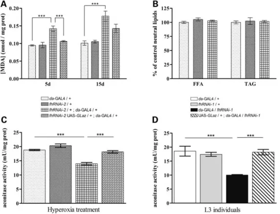
As shown in Figure5A, the total amount of the MDA product was notably increased in 5- and 15-day-old frataxin-deficient flies. Nevertheless, neither young (7-day-old, Fig. 5B) nor older (30-day-old, data not shown) frataxin-deficient flies present significant alterations in the TAG or free FA content. Interestingly, co-expression ofGLazshowed an almost complete rescue of this phenotype, with a strong reduction (30%) of lipid peroxide accumulation in young flies (5-day-old). A mild decrease (9%) was observed in older individuals (15-day-old).
These results suggest that lack of frataxin besides inducing hypersensitivity to oxidative insult also increases the pro-duction of oxidative radicals by itself. Our data also indicate that the abnormal peroxidation of lipids in frataxin knock-down flies is independent of lipid accumulation. In addition, GLazis able to reduce the increased lipid peroxide generation, although in a time-restricted manner. Therefore, the increase in lipid peroxidation correlates well with the phenotypes described in glia, and GLaz was able to rescue the effects driven by the loss of frataxin.
GLazrestores aconitase activity in frataxin-deficient larvae and adult flies
One of the most characteristic biochemical defects associated with a loss of frataxin is the reduction of aconitase activity
(9,15,33). Therefore, we asked whether co-expression of GLazwas able to restore the enzymatic activity in a frataxin-deficient environment. This hypothesis was tested in two different experimental scenarios: on the one hand, in L3 larvae with a strong reduction of frataxin produced by the fhRNAi-1 transgene (17); on the other hand, in young adult flies with a milder reduction of frataxin caused by the fhRNAi-2transgene combined with oxidative stress (15). We used the da-GAL4 driver because it provides a more wide-spread expression facilitating the detection of changes in aco-nitase activity. Loss of frataxin provoked a 28% decrease in aconitase activity in hyperoxia-treated adult flies (Fig. 5C) and 46% in larvae (Fig.5D).GLazwas able to restore aconi-tase activity effectively, up to levels comparable with control flies (Fig.5C and D).
Taken together, these data indicate that preserving the redox balance is a major mechanism by whichGLazco-expression is able to counteract some of the effects caused by frataxin deficiency.
DISCUSSION
FRDA, the most frequent form of hereditary ataxia in the Cau-casian population, is produced by a deficit in the mitochondrial protein frataxin. Although big efforts are being made using animal, cell culture and yeast models, the function of frataxin still remains elusive and controversial. Nevertheless, using Figure 5. GLaz decreases lipid peroxidation levels and restores aconitase activity in frataxin-deficient flies. (A) Frataxin-deficient adults (fhRNAi-2/+;
da-GAL4/+) display an increase in lipid peroxides that can be restored to control levels byGLazin 5-day-old flies (5d) but not in 15-day-old flies (15d). (B)fhRNAi-2/+;da-GAL4/+do not induce any significant changes in TAG or FFA content. (CandD)GLazsignificantly protects aconitase from ROS-mediated inactivation in frataxin-deficient L3 (caused byfhRNAi-1transgene) and adult flies (caused byfhRNAi-2transgene). Significance was determined by one-way ANOVA withpost hocNewman– Keuls (∗∗∗P,0.001). Error bar represents standard error. FFA, free fatty acid; TAG, triacylglycerides.
Human Molecular Genetics, 2010 7
at Universitaetsbibliothek Regensburg on May 21, 2010
http://hmg.oxfordjournals.org
mitochondrial iron accumulation by Yfh1p, a putative homolog of frataxin.Science,276, 1709 – 1712.
74. Pandolfo, M. and Koenig, M. (1998) Friedreich’s ataxia. In Wells, R.D. and Warren, S.T. (eds),Genetic Instabilities and Hereditary Neurological Diseases, Academic Press, Texas A&M University, Houston, USA, pp. 371 – 398.
75. Lu, C., Schoenfeld, R., Shan, Y., Tsai, C., Hammock, B. and Cortopassi, G. (2009) Frataxin deficiency induces Schwann cell inflammation and death.Biochim. Biophys. Acta,1792, 1052 – 1061.
76. Sanchez, D., Ortega-Cubero, S., Akerstro¨m, B., Herrera, M., Bastiani, M.J. and Ganfornina, M.D. (2008) Molecular interactions of the neuronal GPI-anchored lipocalin Lazarillo.J. Mol. Recognit.,21, 313 – 323. 77. Do Carmo, S., Fournier, D., Mounier, C. and Rassart, E. (2009) Human
apolipoprotein D overexpression in transgenic mice induces insulin resistance and alters lipid metabolism.Am. J. Physiol. Endocrinol. Metab., 296, E802– E811.
78. Hull-Thompson, J., Muffat, J., Sanchez, D., Walker, D.W., Benzer, S., Ganfornina, M.D. and Jasper, H. (2009) Control of metabolic homeostasis by stress signaling is mediated by the lipocalin NLaz.PLoS Genet.,5, e1000460.
79. Thomas, E.A. and Yao, J.K. (2007) Clozapine specifically alters the arachidonic acid pathway in mice lacking apolipoprotein D.Schizophr. Res.,89, 147 – 153.
80. Lechner, M., Wojnar, P. and Redl, B. (2001) Human tear lipocalin acts as an oxidative-stress induced scavenger of potentially harmful lipid peroxidation products in a cell culture system.Biochem. J.,356, 129 – 135. 81. Kakhlon, O., Manning, H., Breuer, W., Melamed-Book, N., Lu, C.,
Cortopassi, G., Munnich, A. and Cabantchik, Z.I. (2008) Cell functions impaired by frataxin deficiency are restored by drug-mediated iron relocation.Blood,112, 5219 – 5227.
82. Zanella, I., Derosas, M., Corrado, M., Cocco, E., Cavadini, P., Biasiotto, G., Poli, M., Verardi, R. and Arosio, P. (2008) The effects of frataxin silencing in HeLa cells are rescued by the expression of human mitochondrial ferritin.Biochim. Biophys. Acta,1782, 90 – 98.
83. O’Neill, H.A., Gakh, O., Park, S., Cui, J., Mooney, S.M., Sampson, M., Ferreira, G.C. and Isaya, G. (2005) Assembly of human frataxin is a mechanism for detoxifying redox-active iron.Biochemistry,44, 537 – 545. 84. Gakh, O., Park, S., Liu, G., Macomber, L., Imlay, J.A., Ferreira, G.C. and Isaya, G. (2006) Mitochondrial iron detoxification is a primary function of
frataxin that limits oxidative damage and preserves cell longevity.Hum. Mol. Genet.,15, 467 – 479.
85. Adinolfi, S., Iannuzzi, C., Prischi, F., Pastore, C., Iametti, S., Martin, S.R., Bonomi, F. and Pastore, A. (2009) Bacterial frataxin CyaY is the gatekeeper of iron – sulfur cluster formation catalyzed by IscS.Nat. Struct. Mol. Biol.,16, 390 – 396.
86. Chantrel-Groussard, K., Geromel, V., Puccio, H., Koenig, M., Munnich, A., Ro¨tig, A. and Rustin, P. (2001) Disabled early recruitment of antioxidant defenses in Friedreich’s ataxia.Hum. Mol. Genet.,10, 2061 – 2067.
87. Jiralerspong, S., Ge, B., Hudson, T.J. and Pandolfo, M. (2001) Manganese superoxide dismutase induction by iron is impaired in Friedreich ataxia cells.FEBS Lett.,509, 101 – 105.
88. Paupe, V., Dassa, E.P., Goncalves, S., Auche`re, F., Lo¨nn, M., Holmgren, A. and Rustin, P. (2009) Impaired nuclear Nrf2 translocation undermines the oxidative stress response in Friedreich ataxia.PLoS ONE,4, e4253. 89. Van Dijk, W., Do Carmo, S., Rassart, E., Dahlba¨ck, B. and Sodetz, J.M.
(2006) The plasma lipocalins a1-acid glycoprotein, apolipoprotein D, apolipoprotein M and complement protein C8g. In Akerstro¨m, B., Borregaard, N., Flower, D.R. and Salier, J.P. (eds),Lipocalins, Landes Bioscience, Georgetown, TX, pp. 140 – 166.
90. Do Carmo, S., Jacomy, H., Talbot, P.J. and Rassart, E. (2008) Neuroprotective effect of apolipoprotein D against human coronavirus OC43-induced encephalitis in mice.J. Neurosci.,28, 10330– 10338. 91. He, X., Jittiwat, J., Kim, J.H., Jenner, A.M., Farooqui, A.A., Patel, S.C.
and Ong, W.Y. (2009) Apolipoprotein D modulates F2-isoprostane and 7-ketocholesterol formation and has a neuroprotective effect on organotypic hippocampal cultures after kainate-induced excitotoxic injury.Neurosci. Lett.,455, 183 – 186.
92. Perrimon, N. and Mathey-Prevot, B. (2007) Matter arising: off-targets and genome-scale RNAi screens inDrosophila. Fly(Austin),1, 1 – 5. 93. Botella, J.A., Ulschmid, J.K., Gruenewald, C., Moehle, C., Kretzschmar,
D., Becker, K. and Schneuwly, S. (2004) The Drosophila carbonyl reductase sniffer prevents oxidative stress-induced neurodegeneration.
Curr. Biol.,14, 782 – 786.
94. Bligh, E.G. and Dyer, W.J. (1959) A rapid method of total lipid extraction and purification.Can. J. Biochem. Physiol.,37, 911 – 917.
Human Molecular Genetics, 2010 13
at Universitaetsbibliothek Regensburg on May 21, 2010
http://hmg.oxfordjournals.org