Antifibrogenic effect in vivo of low doses of insulin like growth factor I in cirrhotic rats / Inma Castilla Cortázar [et al ]
11
0
0
Texto completo
(2) 186. B. Muguerza et al. / Biochimica et Biophysica Acta 1536 (2001) 185^195. sion as well as the enzymatic activities such as prolyl hydroxylase (EC 1.14.11.2; proline 2-oxoglutarate dioxygenase) [5,6]. On the other hand, it is known that Ito cells transformed into myo¢broblast are the most important cellular source of hepatic ¢brosis [6]. Insulin-like growth factor-I (IGF-I) is a growth factor with anabolic e¡ects [7], which is mainly produced by hepatocytes. Patients with advanced liver cirrhosis show low serum concentrations of this hormone [8^12]. Previous works have shown bene¢cial e¡ects of low doses of IGF-I on experimental cirrhosis (nutritional status [13], malabsorption [14^16], osteopenia [17] and hypogonadism [18]) including liver function tests and oxidative liver damage [19]. This growth factor has been considered a potential factor involved in ¢brogenesis both in vitro and in vivo [20^ 26]. The reported role for IGF-I as stimulator of Ito cell proliferation in vitro and the observation of increased expression of IGF receptors during the process of ¢brogenesis support this opinion [20^26]. We have previously shown that the administration of low doses of IGF-I to CCl4 -cirrhotic rats improves liver function and reduces ¢brosis [19]. To gain more insight into the mechanisms behind the hepatoprotective e¡ects of IGF-I, we have explored the e¡ect of this treatment on mechanisms leading to ¢brosis. Hence, histopathology, evaluation of transformed Ito cells into myo¢broblasts, hydroxyproline content, prolyl hydroxylase activity and collagen types I and III mRNA expression were assessed. 2. Materials and methods 2.1. Induction of liver cirrhosis All the experimental procedures were performed in conformity with the Guide for the Care and Use of Laboratory Animals (7th edn., National Academy Press, Washington, DC). Forty-eight male Wistar rats (3 weeks old, weighing about 130^150 g) were used for this study. Twenty-four of them were randomly allocated to two control groups. Liver cirrhosis was induced in the remaining 24 rats by inhalation of CCl4 (Merck, Darmstadt, Germany). The organic solvent was administered twice a week, with a progressively increasing exposure time, ranging from 1 to 5 min, during an 11 week period as. previously described [14,19]. To accelerate the development of cirrhosis, phenobarbital (Luminal, Bayer, Leverkusen, Germany) was added to drinking water (400 mg/l) beginning 1 week before the ¢rst CCl4 exposure and throughout the entire period of cirrhosis induction [27]. Animals were housed in cages placed in a room with a 12 h light^darkness cycle and constant humidity and temperature (20³C). Both food (standard semipuri¢ed diet for rodents, purchased from B.K. Universal, Sant Vicent dels Horts, Spain) and water were given ad libitum. Healthy control rats (CO) were subjected to the same protocol excluding phenobarbital administration and CCl4 exposure. 2.2. Study design Ten days after stopping CCl4 administration, the study period was initiated. Rats were randomly assigned to receive either vehicle (saline) (group CI, n = 12) or recombinant human (rh) IGF-I (2 Wg IGF-I/100 g bw/day, in two divided doses) (group CI+IGF, n = 11) subcutaneously for 14 days. Healthy rats were also randomly assigned to receive vehicle (CO, n = 12) or IGF-I (CO+IGF-I, n = 12) at the same dosage as the CI+IGF group. Animals were sacri¢ced by decapitation 24 h after administering the last dose. Livers were dissected out. Tissue sample from the left major liver lobe was processed (¢xed in Bouin solution) for histological examination. Tissue specimens were immediately frozen by immersion in liquid N2 and stored at 380³C. The inclusion criteria for the study were established cirrhosis with ¢brous septa delimiting regenerative nodules. None of the cirrhotic rats in this series had ascites. 2.3. Histological degree of ¢brosis and hydroxyproline content In liver sections stained with Masson's trichrome, semiquantitative assessment of ¢brosis was blindly performed using a numerical scoring system based on the number, length and thickness of ¢brous septa as previously described [19] that explored the whole preparation. The length of the septa (examined at 80U magni¢cation) was assessed as follows: 1 point,. BBADIS 62033 30-5-01 Cyaan Magenta Geel Zwart.
(3) B. Muguerza et al. / Biochimica et Biophysica Acta 1536 (2001) 185^195. minimal grade ¢brosis that can be observed in normal livers; 4 points, septa con£uent between portal tracts and between portal tracts and central veins; and 2 or 3 points, intermediate lengths of septa observed. The width of the ¢brous septa was calculated at 150U magni¢cation, scoring 4 points when the mean value of the thickness of nine septa (three periportal, three perivenous and three perinodular) was 75^55 Wm, 3 points when it was 55^40 Wm and 2 points when it was approx. 40^30 Wm. The number of septa was scored as 4 points when there were numerous septa extending into the nodules, thus dissecting a small number of hepatocytes forming micronodules, 2^3 points when septa penetrating into nodules were less numerous surrounding bigger nodules, and 1 point when there was no formation of micronodules inside macronodules. Each preparation (four ¢elds, at magnifying lens) was evaluated by two di¡erent observers, receiving a maximum of 12 points each time. The arithmetic mean of the two scores was taken as the ¢nal score. Liver hydroxyproline levels were determined as a rough index of collagen content by multiplying the hydroxyproline content by the factor 7.46 [28]. Brie£y, 50 mg aliquots of liver tissue were hydrolyzed for 22 h at 110³C in 6 N HCl, and hydroxyproline content was quanti¢ed by HPLC using the Pico-Tag method (Waters, Milford, MA, USA) for amino acid analysis (coe¤cient inter- and intraassay variation = 1% and 0.05% respectively; detection limits 6 40 pmol). Hydrolysate, 25 Wl, was derivatized with phenyl isothiocyanate at pH 9^10 to produce phenyl thiocarbamyl amino acid derivatives. After derivatization, samples were dried under vacuum and redissolved with 0.01 M disodium hydrogen phosphate in acetonitrile (pH 7.4). Finally, samples were introduced into a 300U3.9 mm HPLC chromatographic column (Waters), with a 10 Wm particle stationary phase, at 46³C. An automatic injection system was used to introduce samples in the column. As a mobile phase, 70 mM sodium acetate in acetonitrile/methyl alcohol/water (45:45:10) pH 6.46 was used. Commercially available standard amino acid solutions were processed similarly, and used as external standard to calculate hydroxyproline concentrations in the experimental samples. Hydroxyproline content was expressed as Wmol/mg liver protein. Liver protein concentration was deter-. 187. mined in liver tissue homogenates by Bradford's method [29]. 2.4. Enzymatic activity of prolyl hydroxylase Prolyl hydroxylase was measured in liver homogenates by the method of Hutton et al. [30] with slight modi¢cations as described previously [31]. This method measures the tritiated water formed when tritiated proline (40 Ci/mmol, New England Nuclear, Boston, MA, USA) present in a 3,4 (n) proline-labeled polypeptide substrate is hydroxylated to hydroxyproline. Brie£y, this technique involves the homogenization of the frozen liver sample in a solution of sucrose 0.25 M, EDTA 10 WM, dithiothreitol 1 mM, 50 Wg/ml phenylmethylsulfonyl £uoride, 0.1% (w/v) Triton X-100, and 50 mM Tris^HCl bu¡er (pH 7.2) a 4³C, in an Elvehjem^Potter apparatus. The incubation was carried out with 200 Wl of the aliquots of the supernatants that were centrifuged at 500Ug for 5 min, 150 Wl [3 H]proline-labeled substrate prepared in isolated chick embryo connective tissue and 800 Wl Tris^HCl bu¡er (pH 7.2), containing 1 mM ferrous ammonium sulfate, 2 g/l denaturalized bovine serum albumin, 0.4 g/l catalase (2U106 u/l), 2 mM K-ketoglutarate, 0.1 mM dithiothreitol, and 50 mM ascorbic acid. The reaction was incubated aerobically for 30 min at 30³C and stopped with 100 Wl of 50% trichloroacetic acid. The tritiated water produced was separated by vacuum distillation and counted in a scintillation counter. A blank obtained by the addition of homogenization bu¡er instead of sample was used in each determination. Prolyl hydroxylase activity in each homogenate and blank was measured in duplicate. Total protein concentrations in homogenates were measured by the method of Bradford [29]. The results were expressed as cpm/mg protein. 2.5. RNA extraction and hybridization RNA extraction was performed by homogenization in 4 M guanidinium thiocyanate followed by phenol chloroform extraction. The RNA was spectrophotometrically measured and 25 Wg/lane were then analyzed by formaldehyde agarose electrophoresis and transferred by capillary blotting to nylon ¢lters (Gene Screen, New England Nuclear). Ethid-. BBADIS 62033 30-5-01 Cyaan Magenta Geel Zwart.
(4) 188. B. Muguerza et al. / Biochimica et Biophysica Acta 1536 (2001) 185^195. Fig. 1. Histopathologic ¢ndings in liver biopsy (4 Wm section; Masson's trichrome stain; original magni¢cation 150U) from: CI, untreated cirrhotic rat; CI+IGF, cirrhotic rat treated with IGF-I. Liver hydroxyproline content in these two animals was 4.71 and 3.66 Wg/mg protein for CI and CI+IGF, respectively.. ium bromide staining was used to assess the relative amount and the intact nature of the RNA. Subsequent probing of the Northern blot hybridizations was performed using cDNAs 32 P-labeled by random priming using a commercially available kit (Amersham, UK). The cDNAs used include GADPH as an internal control, K1(I) collagen and K1(III) collagen. Nylon membranes were prehybridized and hybridized at 42³C in 50% formamide containing 6USSC, 50 mM sodium phosphate pH 7.0, 1 mM EDTA, 1UDenhardt's solution, 50 Wg/ml sheared single-stranded salmon sperm DNA, and 10% dextran sulfate. Prehybridization was performed for 4 h and hybridization for 16 h in the presence of 1U106 cpm/ml cDNA. All the blots were washed under stringent conditions at 65³C. Autoradiograms were developed using Kodak XAR-5 ¢lm and intensifying screens at 380³C. After each hybridization step, the blot was washed with 50% formamide in 1USSC at 75³C for 45 min to remove the probe. Quanti¢cation was performed on scanned X-ray ¢lms of Northern blots corrected by GADPH mRNA in the same RNA preparations.. 2.5.1. Immunostaining for K-actin When injured, liver Ito cells assume a ¢broblastlike morphology expressing K-actin. These transformed cells produce large amounts of collagen. Immunohistochemical staining of K-actin in para¤n sections (4 Wm) was performed using an avidin^biotin peroxidase technique as described by Shu el al. [32] with a few modi¢cations. The primary antibody anti-K-actin was obtained from Bio Genex Laboratories (San Roman, USA). Negative controls were performed by omission of the antigen retrieval procedure. The positive staining was estimated blindly in the entire preparation using a numerical score from 1 to 4 points as follow: 1 point, staining exclusively localized in smooth muscle, around the vessels that can be observed in normal livers; 4 points, large lines of immunostaining along the ¢brous septa; 2 or 3 points, little stain deposition or intermediate lengths of lines, respectively. 2.6. Statistical analysis Data are expressed as mean ( þ S.E.M.). To assess. BBADIS 62033 30-5-01 Cyaan Magenta Geel Zwart.
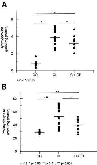
(5) B. Muguerza et al. / Biochimica et Biophysica Acta 1536 (2001) 185^195. 189. the homogeneity among the di¡erent groups of rats a Kruskal^Wallis test was used, followed by multiple post-hoc comparisons using Mann^Whitney U tests with Bonferroni adjustment. A regression model was ¢tted considering malondialdehyde (MDA) concentration and prolyl hydroxylase activity as the dependent and independent variables, respectively. Any P value less than 0.05 was considered to be statistically signi¢cant. Calculations were performed with SPSSW in v.6.0. program. 3. Results 3.1. Liver histology and liver hydroxyproline content All rats from groups CI and CI+IGF showed, at the end of the study, micronodular or macromicronodular cirrhosis. The histological score of ¢brosis was signi¢cantly lower in CI+IGF than in CI (9.1 þ 0.5 vs. 10.4 þ 0.2; P 6 0.05) (Fig. 1). In accordance with this observation, hydroxyproline content (Wmol/mg protein) was also signi¢cantly lower in hydroxyproline cirrhotic rats treated with IGF-I than in CI animals (3.2 þ 0.3 vs. 3.8 þ 0.3, P 6 0.05) and signi¢cantly higher in these two groups than in healthy controls (0.7 þ 0.1, P 6 0.01, both) (Fig. 2A). Similar values were found for the two control groups (CO+IGF = 0.69 þ 0.06 Wmol hydroxyproline/mg protein). Accordingly, no histopathological ¢ndings were observed in the CO+IGF group. Therefore, no further mechanisms leading to ¢brosis were studied in this group. 3.2. Prolyl hydroxylase activity High hepatic levels of prolyl hydroxylase activity (cpm/mg protein) were observed in CI rats as compared with controls (CI = 76.75 þ 5.75; CO = 32.73 þ 2.22; P 6 0.01). However, this enzymatic activity was signi¢cantly reduced in CI+IGF as compared with untreated cirrhotic rats (CI+IGF = 66.76 þ 6.38; P 6 0.05) (Fig. 2B). 3.3. Expression of mRNAs coding for type I and III collagens The expression of mRNAs coding for K1(I) and. Fig. 2. (A) Liver hydroxyproline content, in the three main experimental groups: CO, healthy controls; CI, untreated cirrhotic group; CI+IGF, cirrhotic animals treated with IGF-I. (B) Activity of prolyl hydroxylase (cpm/mg protein) in the three groups. Hepatic levels of prolyl hydroxylase activity were higher in untreated cirrhotic rats (CI) that in controls (CO) (P 6 0.01). This enzymatic activity was signi¢cantly reduced in cirrhotic animals treated with IGF-I (CI+IGF) as compared with CI (P 6 0.05). *P 6 0.05, **P 6 0.01, ***P 6 0.001.. K1(III) chain collagens was investigated by Northern blot. The absorbance values scanned from X-ray ¢lms of Northern blots were corrected for GADPH mRNA used as blot control (collagen K1(I): CO = 2.20 þ 0.43; CI = 5.87 þ 1.10; CI+IGF = 3.75 þ 0.53, P 6 0.01 CO vs. cirrhotic groups, and P 6 0.05 CI vs. CI+IGF group; and collagen K1(III) CO = 5.51 þ 1.02; CI = 33.93 þ 5.54; CI+IGF = 24.50 þ 4.93,. BBADIS 62033 30-5-01 Cyaan Magenta Geel Zwart.
(6) 190. B. Muguerza et al. / Biochimica et Biophysica Acta 1536 (2001) 185^195. Fig. 3. Northern blot analysis: levels of collagen K1(I) mRNA in liver from healthy controls (CO, n = 12), untreated cirrhotic rats (CI, n = 12) and the cirrhotic group treated with IGF-I (CI+IGF, n = 11). Expression of collagen K1(I) was 2.67-fold higher in the CI group than in the CO group, and 1.70-fold in CI+IGF animals. *P 6 0.05, **P 6 0.01.. P 6 0.01 CO vs. cirrhotic groups, and P 6 0.05 CI vs. CI+IGF animals). As shown in Figs. 3 and 4, there were di¡erences in the expression of K1(I) and K1(III) collagens in both cirrhotic groups as compared to the CO group. However, the expression of these mRNAs was lower in the CI+IGF-I group than in the CI group. The same ¢lters were successively hybridized with K1(I) and K1(III) collagen cDNA. Thus, the values are not directly comparable. 3.4. Immunohistochemistry for K-actin The hepatic distribution of K-actin, as a marker of myo¢broblasts, was performed using immunohistochemistry, because myo¢broblasts are considered the most important source of collagen [6,33,34]. In liver sections from all CO animals, K-actin immunostaining was localized exclusively in myocytes, surrounding the vessels (Fig. 5, CO, score: 1.0 þ 0.0). In the livers from the CI group immunostaining was also found in the ¢brous septa and in many instances these were con£uent (between portal tracts and around the central veins), and the immunostaining was as strong as in myocyte cells (Fig. 5, CI,. score: 2.7 þ 0.3 points). However, K-actin immunostaining was much lower in the CI+IGF than in the CI group (Fig. 5, CI+IGF, score: 2.1 þ 0.2). Semiquantitative scoring for K-actin showed signi¢cant di¡erences between controls and cirrhotic groups (P 6 0.001) and between untreated and IGF-treated rats (P 6 0.05). 3.5. Lipid peroxidation index: correlation between malondialdehyde and prolyl hydroxylase activity In order to ¢nd a relationship between parameters of ¢brogenesis and oxidative liver damage, MDA, an index of lipid peroxidation [35,36], was assessed as described in a previous work [19]. Hepatic levels of MDA (nmol/g tissue) were increased in CI rats as compared with the control group (CI = 158 þ 35; CO = 40 þ 3; P 6 0.01) and as was previously reported in a similar protocol [19], this marker of lipid peroxidation was again signi¢cantly reduced in CI+IGF as compared with CI (CI+IGF = 57 þ 6 nmol/g tissue, P 6 0.01). A signi¢cant and direct correlation between hepatic MDA and prolyl hydroxylase activity was found (r = 0.57, P 6 0.01).. BBADIS 62033 30-5-01 Cyaan Magenta Geel Zwart.
(7) B. Muguerza et al. / Biochimica et Biophysica Acta 1536 (2001) 185^195. 191. Fig. 4. Northern blot analysis: levels of collagen K1(III) mRNA in liver from healthy controls (CO, n = 12), untreated cirrhotic rats (CI, n = 12) and the cirrhotic group treated with IGF-I (CI+IGF, n = 11). Expression of collagen K1(III) was 6.16-fold higher in the CI group and 4.44-fold in the CI+IGF animals, than in the CO group. *P 6 0.05, **P 6 0.01.. 4. Discussion This study shows that several mechanisms leading to ¢brogenesis in rats with CCl4 -induced cirrhosis are reduced with low doses of IGF-I over a 2 week period. These mechanisms, such as prolyl hydroxylase activity, transformation of Ito cells into myo¢broblasts and mRNA expression of the most abundant types (K1(I) and K1(III)) of collagen, appear to be relevant in the development of ¢brosis in the liver. In fact, the histopathological scores of ¢brosis and hydroxyproline content were lower in cirrhotic rats treated with IGF-I than in untreated cirrhotic animals. In a previous protocol, we reported similar histological ¢ndings [19]. The above mentioned work suggested hepatoprotective e¡ects on the liver in cirrhotic rats which received a short-term course of. IGF-I. We observed a decrease in lipid peroxidation associated with an increase of antioxidant enzyme activities in hepatic tissue [19]. Interestingly enough, the present study demonstrates that IGF-I not only decreases hepatic collagen content as was previously shown [19], but also its synthesis, reducing both prolyl hydroxylase activity and collagen mRNA expression. Although the mechanism of IGF-I action remains hypothetical, the present study provides evidence of the anti¢brogenic e¡ect in vivo of IGF-I, at these low doses. This anti¢brogenic e¡ect exerted by IGF-I in the CCl4 model of liver cirrhosis seems to be related to the antioxidant activities displayed by this hormone. Much evidence has been accumulated in recent years supporting the hypothesis that oxidative stress plays a major role in the pathogenesis of liver injury.. BBADIS 62033 30-5-01 Cyaan Magenta Geel Zwart.
(8) 192. B. Muguerza et al. / Biochimica et Biophysica Acta 1536 (2001) 185^195. BBADIS 62033 30-5-01 Cyaan Magenta Geel Zwart.
(9) B. Muguerza et al. / Biochimica et Biophysica Acta 1536 (2001) 185^195. 193. Fig. 5. Immunohistochemistry for K-actin in liver histologic sections (4 Wm). The immunostaining was focused around the vessel in healthy controls (CO). More extensive zones were stained in untreated cirrhotic animals (CI), suggesting a major number of Ito cells transformed into myo¢broblasts (P 6 0.01 vs. CO). The stain for K-actin was signi¢cantly less in the cirrhotic group treated with IGF-I (CI+IGF, P 6 0.05 vs. CI group).. 6. Although a causal relationship between oxidative damage and hepatocellular injury still remains a matter of debate [37^42], in vitro experiments have demonstrated that lipid peroxidation can cause cytopathic changes and trigger gene transcription [37]. Lipid peroxidation seems to upregulate the expression and synthesis of ¢brogenic cytokines [44]. It has been shown that lipid peroxidation products stimulate the expression of collagen genes in myo¢broblasts [37,45] and the activity of hepatic prolyl hydroxylase [38,43]. The activation of hepatic stellate cells is mediated by oxidative stress [43]. Ito cells are perisinusoidal cells thought to be a major source of collagen in normal and ¢brotic livers [6]. These cells (isolated in culture or in injured liver) assume a ¢broblast-like morphology expressing K-actin. These transformed cells produce large amounts of procollagen type I and also type III [6,47]. In the present study, a higher hepatic lipid peroxidation, expression of collagen mRNAs, prolyl hydroxylase activity, and hydroxyproline content were found in untreated cirrhotic rats as compared to cirrhotic animals treated with IGF-I. In addition, a more extensive K-actin staining was present in the livers from the untreated cirrhotic group. The diminution of mRNA collagen was observed in both collagen K1(I) and K1(III). In untreated cirrhotic animals the expression of collagen K1 type I was 2.67 times higher than in the controls and it was only 1.7 times that in treated cirrhotic animals. Collagen K1 type I is the most important component of extracellular matrix in the cirrhotic liver [6,46]. In addition, the expression of collagen K1 type III was 6.16 times higher in untreated cirrhotic animals than in controls and only 4.44 times higher in CI+IGF rats. Because prooxidant hepatocellular injury and resulting ¢brogenesis occur in a diversity of human liver diseases [9,11,46], and the mentioned previous data indicated that IGF-I, at low doses, protects the liver against oxidative damage [19], this antioxidant mechanism of IGF-I could explain the anti¢brogenic. actions described in the present study. These ¢ndings are in apparent opposition with the reported role of IGF-I as a stimulator of Ito cell proliferation in vitro [20^26] and with the observation of increased expression of IGF receptors during the process of ¢brogenesis [25]. A problem in interpreting these data is the reported low expression of IGF-I receptors in hepatocytes that makes it di¤cult to explain the biological action of IGF-I in this tissue. However, there are some data that may contribute to clarify this point. Regenerating hepatocytes express IGF-I receptors [50] and in addition we have recently observed a reduction in GHR mRNA levels in the liver of cirrhotic rats and ^ interestingly ^ a higher expression after IGF-I treatment [51]. Improved responses of the liver parenchyma to GH could be behind the e¡ects of IGF-I treatment [51]. On the other hand, several works have reported that IGF-I induces hepatocyte growth factor (HGF) production by liver cells [48]. Since HGF may have important roles in liver regeneration and induces anabolic e¡ects on cirrhotic liver [48,49], the described bene¢cial actions of IGF-I could be secondary to HGF synthesis. In fact, transduction with HGF gene suppressed the increase of transforming growth factor-L1 (TGF-L1), which plays an essential part in the progression of liver cirrhosis, inhibited ¢brogenesis and hepatocyte apoptosis, and produced the complete resolution of ¢brosis in the cirrhotic liver, thereby improving the survival rate of rats with this severe illness [49]. The mechanism that couples these two growth factors (HGF and IGF-I) remains to be elucidated. In summary, this study demonstrates that the administration of IGF-I, at low doses, in vivo, not only decreases hepatic collagen deposition but also its production, acting as an anti¢brogenic agent at two levels: decreasing prolyl hydroxylase activity and mRNA expression of collagen. This work provides new evidence of the bene¢cial e¡ect of IGF-I supplementation in experimental liver cirrhosis. These re-. BBADIS 62033 30-5-01 Cyaan Magenta Geel Zwart.
(10) 194. B. Muguerza et al. / Biochimica et Biophysica Acta 1536 (2001) 185^195. sults provide an experimental basis for further studies aiming at exploring the potential of IGF-I in the treatment of human cirrhosis.. [14]. [15]. Acknowledgements The authors wish to thank Dr. Dan Edwall from Pharmacia and Upson for providing rhIGF-I used in this study; Mr. Guillermo Ezpeleta, from the Department of Pathology of the Hospital of Navarra, for his help in histological analysis of liver specimens; and the `Gobierno de Navarra', Mr. J. Celaya, Mr. I. Sanz and `Fundaciön Echëbano' for ¢nancial collaboration. Finally, we are deeply indebted to Mrs. M.P. Redin and C. Chocarro for their expert secretarial and technical assistance. This study was supported by the Program `I+D, Comisiön Interministerial de Ciencia y Tecnolog|¨a (CICYT), Gobierno de Espan¬a', SAF 99/0072.. [16]. [17]. [18]. [19]. [20] [21] [22] [23]. References [1] H.O. Conn, C.E. Atterbury, in: L. Schi¡, E.R.J. Schi¡ (Eds.), Cirrhosis, Lippincott, Philadelphia, PA, 1987, pp. 725^864. [2] M. Rojkind, M.A. Giambrone, L. Biempica, Gastroenterology 76 (1979) 710^719. [3] D.M. Bissel, S.L. Friedman, J.J. Maher, F.J. Roll, Hepatology 11 (1990) 488^498. [4] J. Risteli, K.I. Kivirikko, Biochem. J. 144 (1974) 115^122. [5] K.I. Kivirikko, R. Myllyla, Ann. NY Acad. Sci. 460 (1985) 187^201. [6] F.R. Weiner, M.A. Giambrone, M.J. Czaja, A. Shah, G. Annoni, S. Takahashi, M. Eghbali, M.A. Zern, Hepatology 11 (1990) 111^117. [7] J.I. Jones, D.R. Clemmons, Endocr. Rev. 1 (1995) 3^34. [8] R.M. Schimpf, D. Lebrec, M. Donadieu, Acta Endocrinol. 86 (1977) 355^362. [9] N. Hattori, H. Kurahachi, K. Ikekubo, T. Ishihara, K. Moridera, M. Hino, Y. Saiki, H. Imura, Metab. Clin. Exp. 41 (1992) 377^381. [10] S. MÖller, U. Becker, A. Juul, N.E. Skakkeb×k, E. Christensen, and EMALD Group, Hepatology 23 (1996) 1073^ 1078. [11] S. MÖller, M. Gronbaek, K. Main, U. Becker, N.E. Skakkebaek, J. Hepatol. 17 (1993) 315^320. [12] A. Caufriez, P. Reding, D. Urbain, J. Goldstein, G. Copinschi, J. Endocrinol. Invest. 14 (1991) 317^321. [13] A. Picardi, A. Costa de Oliveira, B. Muguerza, A. Tosar,. [24] [25]. [26]. [27] [28] [29] [30] [31]. [32] [33] [34] [35] [36]. J. Quiroga, I. Castilla-Cortäzar, S. Santidriän, J. Prieto, J. Hepatol. 24 (1996) 267^279. I. Castilla-Cortäzar, A. Picardi, J. Ainzua, A. Tosar, E. Urdaneta, M. Pascual, M. Garc|¨a, J. Quiroga, J. Prieto, Am. J. Physiol. Gastrointest. Liver Physiol. 276 (1999) G37^G42. I. Castilla-Cortäzar, J. Prieto, E. Urdaneta, M. Pascual, M. Nün¬ez, E. Zudaire, M. Garc|¨a, J. Quiroga, S. Santidriän, Gastroenterology 113 (1997) 1180^1187. A. Pascual, I. Castilla-Cortäzar, E. Urdaneta, J. Quiroga, M. Garc|¨a, A. Picardi, J. Prieto, Am. J. Physiol. Gastrointest. Liver Physiol. 279 (2000) G319^G324. A. Cemborain, I. Castilla-Cortäzar, M. Garc|¨a, J. Quiroga, B. Muguerza, A. Picardi, J. Prieto, J. Hepatol. 28 (1998) 122^131. I. Castilla-Cortäzar, M. Garc|¨a, J. Quiroga, N. Diez, F. Diez-Caballero, A. Calvo, M. Diaz, J. Prieto, Hepatology 31 (2000) 592^600. I. Castilla-Cortäzar, M. Garc|¨a, B. Muguerza, J. Quiroga, R. Përez, S. Santidriän, J. Prieto, Gastroenterology 113 (1997) 1682^1691. R.H. Goldstein, C.F. Poliks, P.F. Pilch, B.D. Smith, A. Fine, Endocrinology 124 (1989) 964^969. M. Pinzani, H.E. Abboud, D.C. Aron, Endocrinology 127 (1990) 2343^2349. A.M. Gressner, A. Brenzel, T. Vossmeyer, Liver 13 (1993) 86^94. E.M. Zimmermann, R.B. Sartor, R.D. McCall, M. Pardo, D. Bender, K. Lund, Gastroenterology 105 (1993) 399^409. A.M. Gressner, B. Lahme, A. Brenzel, Hepatology 22 (1995) 1507^1518. A. Brenzel, O.H. Weiner, A.M. Gressner, in: K. Wake, E. Wisse, D.L. Knook (Eds.), Cells of the Hepatic Sinusoid, vol. 5, The Kup¡er Cell Foundation, Leiden, 1995, pp. 386^389. G. Svegliati-Baroni, F. Ridol¢, A. Di Sario, A. Casini, L. Marucci, G. Gaggiotti, P. Orlandoni, G. Macarri, L. Perego, A. Benedetti, F. Folli, Hepatology 29 (1999) 1743^1751. K. Chatamra, E. Proctor, Br. J. Exp. Pathol. 62 (1981) 283^ 288. J. George, G. Chandrakasan, Biochim. Biophys. Acta 1292 (1996) 215^222. M.M. Bradford, Anal. Biochem. 72 (1976) 248^254. J.J. Hutton, A.L. Tappel, S. Underfriend, Anal. Biochem. 16 (1966) 384^394. M. Torres-Salinas, A. Parës, J. Caballer|¨a, W. Jimënez, D. Heredia, M. Bruguerz, J. Rodës, Gastroenterolgy 90 (1986) 1241^1246. S.Y. Shu, G. Ju, L.Z. Fan, Neurosci. Lett. 85 (1988) 169^ 171. M.G. Bachem, D. Meyer, R. Melchior, K.-M. Sell, A.M. Gressner, J. Clin. Invest. 89 (1991) 19^27. A.M. Gressner, M.G. Bachem, Ann. Biol. Clin. 52 (1994) 205^226. J.M.C. Gutteridge, Clin. Chem. 41 (1995) 1819^1828. H. Esterbauer, R.J. Schaur, J. Zollner, Free Radic. Biol. Med. 11 (1991) 82^128.. BBADIS 62033 30-5-01 Cyaan Magenta Geel Zwart.
(11) B. Muguerza et al. / Biochimica et Biophysica Acta 1536 (2001) 185^195 [37] P. Bedossa, K. Houglum, C. Trautwein, A. Holstege, M. Chojkier, Hepatology 19 (1994) 1262^1271. [38] S. Yamada, M. Yamada, Y. Murawaki, C. Hirayama, Biochem. Pharmacol. 40 (1990) 1015^1029. [39] R. Nordmann, C. Ribiere, H. Rouach, Free Radic. Biol. Med. 12 (1992) 219^240. [40] B.G. Rosser, G.J. Gores, Gastroenterology 108 (1995) 252^ 275. [41] J.M. McCord, Clin. Biochem. 26 (1993) 351^357. [42] C.S. Lieber, Gastroenterology 106 (1994) 1085^1105. [43] A.J. McCullough, in: W.G. Rector (Ed.), Complications of Crhonic Liver Disease, Mosby Year Book Inc., St. Louis, MO, 1992, pp. 182^211. [44] A. Gimënez, A. Parës, S. Alië, J. Camps, R. Deulofeu, J. Caballer|¨a, J. Rodës, J. Hepatol. 21 (1994) 292^298. [45] J. Camps, T. Bargallo, A. Gimenez, S. Alie, J. Caballeria, A. Parës, J. Joven, L. Masana, J. Rodës, J. Clin. Sci. 83 (1992) 695^700.. 195. [46] G. Poli, M. Parola, Free Radic. Biol. Med. 22 (1997) 287^ 305. [47] K.S. Lee, M. Buck, K. Houglum, M. Chojkier, J. Clin. Invest. 96 (1995) 2461^2468. [48] S. Skrtic, V. Wallenius, S. Ekberg, A. Brezel, A.M. Gressner, J.O. Jansson, J. Hepatol. 30 (1999) 115^124. [49] T. Ueki, Y. Kaneda, H. Tsutsui, K. Nakanishi, Y. Sawa, R. Morishita, K. Matsumoto, T. Nakamura, H. Takahashi, E. Okamoto, J. Fujimoto, Nat. Med. 2 (1999) 226^230. [50] J.F. Caro, J. Poulos, O. Ittoop, W.J. Pories, E.G. Flickinger, M.K. Sinha, J. Clin. Invest. 81 (1988) 976^981. [51] E. Mirpuri, E. Garc|¨a-Trevijano, I. Castilla-Cortäzar, C. Berasain, J. Quiroga, C. Rodr|¨guez-Ortigosa, J.M. Mato, J. Prieto, M.A. Avila, Am. J. Physiol. (2001) submitted for publication.. BBADIS 62033 30-5-01 Cyaan Magenta Geel Zwart.
(12)
Figure


Documento similar
(A) Relative GH receptor (GHR) protein levels in control rats (C); rats receiving acute central administration of insulin (CI); pair-fed rats (PF); pair-fed rats treated with
The response of renal artery segments to ET-1, and the expression of ET-1 and its receptors in arterial tissue in 8- and 24-month-old rats, ei- ther fed ad libitum or subjected to
Haemodynamic Parameters in the Perfused Hearts Before I/R coronary in the perfused rats, coronary perfusion pressure, maximal dP/dt and heart rate were similar in the rats from
We find that hepatic GRK2 protein levels are upregulated in different mouse models of steatosis and, conversely, we demonstrate that the development of NAFLD
The main aim of the present study was to evaluate the effect of kale seeds germination under periodical application of low UV-B doses in the main phytochemicals content (GLs,
The expansionary monetary policy measures have had a negative impact on net interest margins both via the reduction in interest rates and –less powerfully- the flattening of the
Jointly estimate this entry game with several outcome equations (fees/rates, credit limits) for bank accounts, credit cards and lines of credit. Use simulation methods to
In our sample, 2890 deals were issued by less reputable underwriters (i.e. a weighted syndication underwriting reputation share below the share of the 7 th largest underwriter
