A Combined Experimental and Theoretical Study on the Formation of Ag Filaments on β Ag 2 MoO 4 Induced by Electron Irradiation
23
0
0
Texto completo
(2) energy dispersive spectroscopy (EDS) characterization. To complement experimental results, chemical stability, structural and electronic aspects have been studied systematically using first-principles electronic structure theory within a QTAIM framework. The Ag nucleation and formation on -Ag2MoO4 is a result of structural and electronic changes of the AgO4 tetrahedral cluster as a constituent building block of Ag2MoO4, consistent with Ag metallic formation. The formation of Ag filament transforms the -Ag2MoO4 semiconductor from n to p type concomitant with the appearance of Ag defects.. 1. Introduction. Metal tungstates and molybdates form a family of multicomponent metal oxide compounds, and many studies have been made regarding their interesting structures, intriguing physical and chemical properties, as well as a wide range of applications in photoluminescence, optical fibers, scintillator materials, photochromism, humidity sensors, multiferroic materials, and catalysts.1-19 One member of the molybdate family, silver molybdate (Ag2MoO4) has been a target of intense investigation for its chemical stability at elevated temperatures and subsequent high-temperature lubricating properties20as well as its potential applications in electrochemical devices and gas-sensing,20-24and in surface enhanced Raman scattering techniques.23,25 Several studies have been made to obtain Ag2MoO4 based materials as flower-like,26 nanoparticles21 and wire-like nanostructures.22Low dimensional Ag2MoO4 nanostructures have been obtained by Nagaraju et al. who reported the synthesis of.
(3) nanorods/nanowires/multipods and the photoluminescence of microrods;27 while Feng et al. have been synthetized as ultra long orthorhombic silver trimolybdate nanowires28. Qian et al.29have reported the microwave-assisted hydrothermal synthesis of cube-like Ag-Ag2MoO4 with visible-light photocatalytic activity. Bao et al.30reported on a roomtemperature synthesis of Ag nanoparticles decorated with silver molybdate nanowires using a solution-based chemical reaction method. Fodjo et al.25have published a facile hydrothermal technique to synthesize Ag2MoO4 at relatively low temperatures (80 ºC and 20 ºC). Tang et al.31have obtained Ag2Mo2O7/chitin composite gel-membranes which were fabricated incorporating Ag2Mo2O7 in the regenerated chitin gel-membranes via a green pathway. Cheng et al.22 studied photoswitches of one-dimensional Ag2MO4 (M = Cr, Mo, and W) and noticed the formation of silver nanoparticles at the surface of Ag2MoO4 under electron beam irradiation, as already previously reported during TEM observation of other silver nanowires.32 The ability of chemists and physicists to interact with atoms, molecules, and solids at the quantum level; and the spatial scales of atoms and chemical bonds and on temporal scales of electron and nuclear motion has gained increasing sophistication through the development of precise photon and electron probes. In particular, electron probes have precise spatial resolution, which has enabled imaging of single atoms and atomic lattice contrast on surfaces and in solids. Scanning electron microscopy (SEM), scanning transmission electron microscopy (STEM), and transmission electron microscopy (TEM) are well recognized techniques that provide unique capabilities for in situ imaging and control of nanoscale phenomena.33-37.
(4) In recent years, papers on monitoring local structural and electronic changes generated by the electron beam within TEM have been started to appear, and TEM is a very powerful tool for the observation, fabrication and manipulation of nanostructures with the advantages of precise nanoscale control,38,39 in which high energy electrons transmit through the specimen and provide useful electronic structure information of the samples based on a variety of electron-solid interactions.40Applying this technique, novel phenomena have been discovered in the process of TEM observation, such as nanoscale phase/shape transformation,41inorganic nanostructure modification,42colloid nanocrystal growth and self assembly,36,43-47lithium encapsulation and release in LiCl nanoshells and nanotubes,48nanostructure fabrication,49coalescence,50nanoscale crystallization and some growth processes which occur only in the beam’s presence.36,44,46,47,51,52Different studies have quantified the role of the electron beam in affecting the chemical reactions being characterized.44,52 In particular, electron beam irradiation allows the preparation of noble and transition metal nanoparticles;43,53Pattabi et al. have synthesized silver nanoparticles with enhanced antibacterial activity by using electron beam irradiation.54During imaging, the electron beam interacts with the sample causing a novel phenomenon that occurs only in the beam´s presence. In particular, an accurate understanding of the interactions between the electron beam and the material is essential to account for, suppress, and exploit the beam effects. First principle calculations are essential in supporting the experimental observation to provide a deeper understanding of atomic and electronic structure that is already being exploited to explain their innovative properties and allow efficient exploration of new materials. In this context, very recently, the formation of Ag filaments.
(5) on α-Ag2WO4 induced by electron irradiation55 and their bactericidal56 and photoluminescent applications,57as well their application as an efficient ozone sensor have been reported.58 The crystal structure Ag2MoO4 obtained by Wyckoff59can be found in two forms: -Ag2MoO4 has a tetragonal structure while -Ag2MoO4 is cubic with a spinel structure.59-61 The -phase irreversibly transforms to the most stable -phase upon heating above ambient temperature. Arora et al.60have studied the behavior of the cubic spinel phase of Ag2MoO4 at high pressure as characterized by X-ray diffraction and Raman spectroscopy, and, in this context, our group has studied the effect of pressure on -Ag2MoO462as a continuation of previous articles on the MgAl2O4 normal spinel and Zn2SnO4 inverse spinel structures.63,64 Very recently, we have investigated a correlation between theoretical calculations and experimental data to explain the electronic structure and optical properties of -Ag2MoO4 microcrystals.65 Due to the novelty associated to the observed nucleation and early evolution of Ag filaments on Ag2MoO4 crystals, driven by an accelerated electron beam from an electronic microscope under high vacuum. Here, we report on a comprehensive and combined experimental and theoretical work to understand the observed nucleation and early evolution of Ag filaments on -Ag2MoO4 crystals, driven by an accelerated electron beam from an electronic microscope under high vacuum. We have studied the geometric and electronic structure of -Ag2MoO4, and have derived a mechanism sequence using electron irradiation as relevant to early events for the formation and growth of Ag filaments from AgO6 and MoO4 clusters, as constituent polyhedra of -Ag2MoO4. Quantum theory of atoms in molecules (QTAIM) developed by Bader and.
(6) collaborators66-68allows the analysis of the experimental or theoretical electron density distribution, (r), in a solid, and the study of properties of(r) to reveal the bonding interactions in the crystal system, and the nature of these interactions. The electronic charge of each atom is evaluated using the Bader charge analysis within the QTAIM framework, which is a way of dividing molecules or solids into atoms based on electronic charge density. These computations envisaged in this study are thought to answer the questions: What happens with the electron excess as it approaches the surface and bulk of. Ag2MoO4? How are the electrons distributed in this material and how can it is related with the structural and electronic evolution? How does the strength of the Ag-O and MoO bonds change after electron irradiation Ag2MoO4? We shall discuss how the analysis, provided from both experimental and theoretical results, of the structural and electronic structure of -Ag2MoO4 allows us to explain the Ag nucleation process. The discussion will address the details of image acquisition and analysis and will provide a guide to interpret experimental results.. 2. Results and discussion. Figure 1 shows the XRD patterns of -Ag2MoO4 which were prepared using the microwave-assisted hydrothermal method. All of the diffraction peaks of samples can be indexed as cubic structure without any deleterious phases, and with a space group of (Fd m) and point-group symmetry (. ) and are in agreement with the standard values of. Inorganic Crystal Structure Data (ICSD) base no. 2889161.
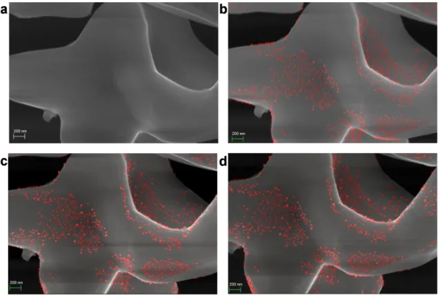
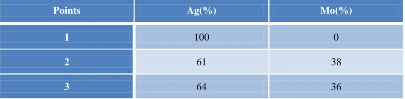
(7) Figure 2 shows the Raman spectrum of the as-prepared Ag2MoO4 between 100 -1000 cm-1. It is possible to verify the presence of four Raman active modes which indicates the structural order at short-range. The active Raman mode (Eg) located at 279 cm-1 is corresponds to the external structure vibrations which correspond to octahedral [AgO6] clusters, while Raman mode (F2g) found at 354 cm-1 is related to the torsional vibration of the [O–Mo–O] bonds present in the tetrahedral [MoO4] clusters. Finally, the active Raman mode (A1g) found at 873 cm-1 corresponds to the symmetric stretching vibration of the [←O←Mo→O→] bonds clusters present in the tetrahedral [MoO4] clusters.23,65 Figure 3 shows a time-resolved series of FE-SEM images obtained under high vacuum (1 × 10⁻⁵ Pa) during the growth of Ag filaments stimulated by the electron beam on the Ag2MoO4 surface. Figure 3 a shows a FE-SEM image of the crystals that were acquired after a rapid approach and focus adjustment (time zero). At this point, the image reveals a small amount of Ag on the surface. Figure 3 b shows that after 2 min of exposure to a 30 kV electron beam, the particle shows some regions with Ag on the surface. It is interesting to notethat the modifications, even small, occur after a few seconds of exposure. After 6 min of exposure, Figure 3 d, the metallic Ag seems to reach a limit point of growth. Further information, a short video (video S1) of the Ag growth is in the Supplementary Information. Figure 4 illustrates the TEM images with EDS characterization. Results of the composition are shown in Table 1. Therefore the end of the region that emerged due to the electron exposure (point 1) confirmed the composition of 100% of Ag in relation of Mo. Furthermore, it is possible to see the crystallographic planes revealing the Ag.
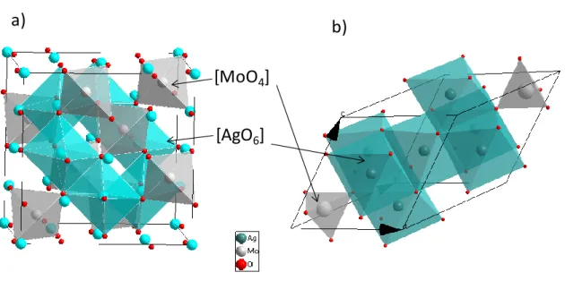
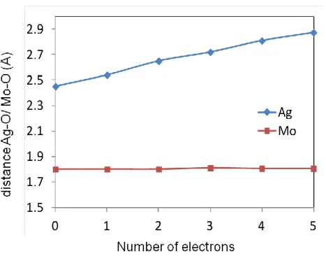
(8) crystallinity. The region near the interface and inside the particles, points 2 and 3, consisted of about 64% of Mo to 36% of Ag. These results reinforce the fact that the Ag atoms come from the interior deep regions of the particles. This behavior provokes the presence of Ag defects inside -Ag2MoO4 material and this semiconductor changes from n to p type. Figure 5 a) shows the conventional cubic unit cell containing 8 formula units. Mo ions (grey atoms) occupy tetrahedral 8a sites while Ag ions (blue atoms) reside at the octahedral 16d position. Oxygen atoms (red atoms) stay at 32e positions. Arora et al.60performed powder XRD measurements at ambient pressure obtaining a single-phase material with a lattice parameter of 9.313 Å. Our calculations at ambient pressure yield a lattice parameter of 9.427 Å which is only 1.2% greater than the experimental value.62 The primitive unit cell is depicted in Figure 5 b), with only 2 formula units. In sense to understand the quantum phenomenon observed experimentally of electron absorption we introduced electrons, separately, up to ten in the cluster constituent of the cubic unit cell of -Ag2MoO4. The values for the distances Ag-O and Mo-O as a function of the number of electrons, N, are shown in Figure 6. In the case of Ag-O distances there is a pronounced increase with the addition of electrons. An analysis of the results presented in Figure 7 shows that the Ag atoms of the [AgO6] clusters are the more prone to be reduced. At N=4, practically all Ag atoms are reduced whereas, there is a minor decrease of the electron density is sensed for Mo atoms (that form [MoO4] clusters) (a decrease of 0.2 units at N=4). Therefore, the electron excess imposed in the material is transferred from one cluster to another through the.
(9) lattice network and the Ag formation process involves [AgO6] adjacent clusters, and to a minor extent the [MoO4] clusters. Bond critical points densities and its Laplacian: The calculations of the charge density, ρbcp, at the (3,-1) bond critical points (BCP) as well as its Laplacian, 2ρbcp, in Ag-O bonds for [AgO6] units are presented in Table 2. The effect of adding electrons to the material produces striking differences in the values of the Laplacian and charge density at the (3,-1) BCP. Thus, it is worth noting that the Ag-O bonds considerably reduce their Laplacian and charge density values as the number of electrons are added indicating that these bonds become less strong in favor of the formationof metallic Ag.. 3. Conclusions. The dynamics and the state properties of excess electrons, provided by an electron beam of TEM, have attracted widespread interest due to the fundamental importance of this process in physics, chemistry, and biology. The electron beam irradiation in TEM provokes the synthesis of nanomaterials and investigating their morphology, structure, and chemical transformation, which is of importance for the development of novel nanostructures, especially for those that cannot be obtained using conventional chemical and physical methods. Electron–material interactions offer an alternative approach to control the electronic structure and thus the chemical and physical properties, and the corresponding activities. In particular, the addition of electrons generated large change in the charge.
(10) density e resulting in bond modifications. As a consequence, a wide range of different chemistries can be initiated by electron–material interactions. We report the in situ growth of Ag on -Ag2MoO4, never observed before, by electron beam irradiation, and this work provides a fundamental perspective on the chemistry initiated by electrons on Ag2MoO4 via a non-classical mechanism. This insight is relevant to apparently unrelated topics such as surface functionalisation or electron beam induced deposition processes. Moreover, mutual awareness of the common concepts of these different fields will also help to solidify the broader understanding of electron-initiated process because each field, and others, can provide examples that reveal principles of common interest. Electron-initiated chemistry therefore remains an active and dynamic research area and many more interesting discoveries and applications can be expected. The present study communicates intimate details on the internal processes and these findings raise fundamental questions about the physical nature of this phenomenon itself. The results derived from the present study are based on the information provided by the joint analysis of experimental and theoretical work to provide a deeper insight and understanding of increasingly complex functional materials, and can be considered as a clear example of a complex modeling paradigm as was introduced by Billinge and Levin.75. 4. Experimental procedure. 4.1. Synthesis of β-Ag2MoO4.
(11) β-Ag2MoO4 microcrystals were prepared at room temperatures without the use of any surfactant by a simple precipitation method. The typical β-Ag2MoO4 crystals synthesis procedure is described as follows: 1×10⁻³mols of molybdate sodium dihydrate (Na2MoO4.2H2O; 99.5% purity, Sigma-Aldrich) and 2×10⁻³mols of silver nitrate (AgNO3; 99.8% purity, Sigma-Aldrich) were each separately dissolved in 50 mL of deionized water. The silver solution was dripped into the molybdate solution under stirring. The resulting suspensions were washed with deionized water several times to remove remaining ions and dried at 60ºC for 12 h.. 4.2. Experimental characterizations of β-Ag2MoO4. The β-Ag2MoO4 crystals were structurally characterized by X-ray diffraction (XRD) patterns using a diffractometer model LabX XRD-6000 (Shimadzu®, Japan) with Cu-Kα radiation (λ = 1.5406 Å) in the 2θ range from 20° to 80° in the normal routine with a scanning velocity of 1°/min. Micro-Raman measurements were recorded using the (Horiba; Jobin-Yvon, France) model LabRAM HR 800 mm. The shapes, sizes, visualization of Ag filaments and element distribution of the βAg2MoO4 microcrystals were observed with a field-emission scanning electron microscope (FEG-SEM) model Supra 35-VP, Carl Zeiss, Germany operated at 20 kV, and transmission electron microscopy (TEM) operating at 200 KV, and Electron Dispersion Spectroscopy (EDS); model Tecnai G2TF20, FEI..
(12) 4.3.Theoretical calculations. First-principles total-energy calculations were carried out within the periodic density-functional-theory (DFT) framework using the VASP program.69,70 The KohnSham equations have been solved by means of the Perdew, Burke, and Ernzerhof exchange-correlation functional,69 and the electron-ion interaction as described by the projector-augmented-wave pseudopotentials.71,72 Due to the well known limitations of standard DFT in describing the electronic structure of “strongly-correlated” compounds, a correction to the PBE wavefunctions are adopted (PBE+U) with the inclusion of a repulsive on-site Coulomb interaction U following the methodof Dudarev et al.73. Eventually the value of the Hubbard parameter was tested and a value of U = 6 eV was used. The plane-wave expansion was truncated at a cut-off energy of 400 eV and the Brillouin zones have been sampled through Monkhorst-Pack special k-points grids that as sure geometrical and energetic convergence for the Ag2MoO4 structures considered in this study. The keyword NELECT has been used in order to increase the number of electrons in the bulk structure, and all the crystal structures are optimized simultaneously on both the volume of the unit-cell and the atomic positions.The relationship between charge density topology and elements of molecular structure and bonding was noted by Bader.66,74This relationship, Bader’s quantum theory of atoms in molecules (QTAIM),6668. is now a well-recognized tool used to analyze electron density, to describe interatomic. interactions, and to rationalize chemical bonding. The different strong and weak interactions between two atoms can be determined unequivocally from QTAIM calculations. According to the standard QTAIM framework, concepts such as (3,-1) bond.
(13) critical points (BCPs), their respective bond paths (BPs), and L(r) = −∇2ρ(r) maps can be analyzed to reveal the nature of these interactions. A BCP serves as the origin of gradient lines of the electron density that define the interatomic surface or zero-flux surface—a surface that unambiguously defines the boundary between the two atoms and identifies the atomic basin, i.e., the electron density “belonging” to a specific atom.. Acknowledgments. The authors are grateful to Prometeo/2009/053 (GeneralitatValenciana), Ministerio de Economia y Competitividad (Spain), CTQ2012-36253-C03-02, Spanish Brazilian. program. (PHB2009-0065-PC),. FAPESP. (Project. 2013/07296-2. and. 2012/14468-1), INCTMN/FAPESP (Project 2008/57872-1), INCTMN/CNPq (Project 573636/2008-7 and Grant 150753/2013-6) for financially supporting this research. We also acknowledge the ServeiInformática, UniversitatJaume I, for the generous allotment of computer time.. (1) V. B. Mikhailik, H. Kraus, G. Miller, M. S. Mykhaylyk, D. Wahl, J. Appl. Phys. 2005, 97, 083523. (2) F. Amano, A. Yamakata, K. Nogami, M. Osawa, B. Ohtani, J. Am. Chem. Soc. 2008, 130, 17650. (3) K. Fukuda, K. Akatsuka, Y. Ebina, R. Ma, K. Takada, I. Nakai, T. Sasaki, Acs Nano 2008, 2, 1689..
(14) (4) M. Shang, W. Z. Wang, H. L. Xu, Cryst. Growth Des. 2009, 9, 991. (5) S. Y. Song, Y. Zhang, Y. Xing, C. Wang, J. Feng, W. D. Shi, G. L. Zheng, H. J. Zhang, Adv. Funct. Mater. 2008, 18, 2328. (6) Y. G. Su, L. P. Li, G. S. Li, Chem. Mater. 2008, 20, 6060. (7) D. Ye, D. Z. Li, W. J. Zhang, M. Sun, Y. Hu, Y. F. Zhang, X. Z. Fu, J. Phys. Chem. C 2008, 112, 17351. (8) L. W. Zhang, Y. J. Wang, H. Y. Cheng, W. Q. Yao, Y. F. Zhu, Adv. Mater. 2009, 21, 1286. (9) Y. Zhao, Y. Xie, X. Zhu, S. Yan, S. X. Wang, Chem. Eur. J. 2008, 14, 1601. (10) L. Zhou, W. Z. Wang, H. L. Xu, S. M. Sun, Cryst. Growth Des. 2008, 8, 3595. (11) Y. X. Zhou, Q. Zhang, J. Y. Gong, S. H. Yu, J. Phys. Chem. C 2008, 112, 13383. (12) L. Gracia, V. M. Longo, L. S. Cavalcante, A. Beltran, W. Avansi, M. S. Li, V. R. Mastelaro, J. A. Varela, E. Longo, J. Andres, J. Appl. Phys. 2011, 110, 043501. (13) V. M. Longo, L. Gracia, D. G. Stroppa, L. S. Cavalcante, M. Orlandi, A. J. Ramirez, E. R. Leite, J. Andres, A. Beltran, J. A. Varela, E. Longo, J. Phys. Chem. C 2011, 115, 20113. (14) E. Orhan, F. M. Pontes, C. D. Pinheiro, E. Longo, P. S. Pizani, J. A. Varela, E. R. Leite, T. M. Boschi, A. Beltran, J. Andres, J. Eur. Ceram. Soc. 2005, 25, 2337. (15) N. M. Batra, S. K. Arora, T. Mathews, J. Mater. Sci. Lett. 1988, 7, 254. (16) M. R. D. Bomio, R. L. Tranquilin, F. V. Motta, C. A. Paskocimas, R. M. Nascimento, L. Gracia, J. Andres, E. Longo, J. Phys. Chem. C 2013, 117, 21382. (17) W. S. Cho, M. Yashima, M. Kakihana, A. Kudo, T. Sakata, M. Yoshimura, J. Am. Ceram. Soc. 1997, 80, 765..
(15) (18) F. Lei, B. Yan, H. H. Chen, Q. Zhang, J. T. Zhao, Cryst. Growth Des. 2009, 9, 3730. (19) J. C. Sczancoski, L. S. Cavalcante, N. L. Marana, R. O. da Silva, R. L. Tranquilin, M. R. Joya, P. S. Pizani, J. A. Varela, J. R. Sambrano, M. S. Li, E. Longo, J. Andres, Curr. Appl. Phys. 2010, 10, 614. (20) W. Gulbinski, T. Suszko, Wear 2006, 261, 867. (21) S. Bhattacharya, A. Ghosh, Phys. Rev. B 2007, 75, 092103. (22) L. Cheng, Q. Shao, M. W. Shao, X. W. Wei, Z. C. Wu, J. Phys. Chem. C 2009, 113, 1764. (23) E. Y. Liu, W. Z. Wang, Y. M. Gao, J. H. Jia, Tribol. Lett. 2012, 47, 21. (24) A. Sanson, F. Rocca, C. Armellini, S. Ahmed, R. Grisenti, J. Non-Cryst. Solids 2008, 354, 94. (25) E. K. Fodjo, D.-W. Li, N. P. Marius, T. Albert, Y.-T. Long, J. Mater. Chem. A 2013, 1, 2558. (26) D. P. Singh, B. Sirota, S. Talpatra, P. Kohli, C. Rebholz, S. M. Aouadi, J. Nanopart. Res. 2012, 14, 781. (27) G. Nagaraju, G. T. Chandrappa, J. Livage, Bull. Mater. Sci. 2008, 31, 367. (28) M. Feng, M. Zhang, J.-M. Song, X.-G. Li, S.-H. Yu, Acs Nano 2011, 5, 6726. (29) L. Z. Qian, C. X.Tai, X. Z. Ling, Sci. China Chem. 2013, 56, 443 (30) Z. Y. Bao, D. Y. Lei, J. Dai, Y. Wu, Appl. Surf. Sci. 2013, 287, 404. (31) H. Tang, A. Lu, L. Li, W. Zhou, Z. Xie, L. Zhang, Chem. Eng. J. 2013, 234, 124. (32) X. J. Cui, S. H. Yu, L. L. Li, L. Biao, H. B. Li, M. S. Mo, X. M. Liu, Chem. Eur. J. 2004, 10, 218..
(16) (33) N. de Jonge, F. M. Ross, Nat. Nanotechn. 2011, 6, 695. (34) R. F. Egerton, P. Li, M. Malac, Micron 2004, 35, 399. (35) L. R. Parent, D. B. Robinson, T. J. Woehl, W. D. Ristenpart, J. E. Evans, N. D. Browning, I. Arslan, Acs Nano 2012, 6, 3589. (36) T. J. Woehl, J. E. Evans, L. Arslan, W. D. Ristenpart, N. D. Browning, Acs Nano 2012, 6, 8599. (37) T. J. Woehl, K. L. Jungjohann, J. E. Evans, I. Arslan, W. D. Ristenpart, N. D. Browning, Ultramicroscopy 2013, 127, 53. (38) D. Tham, C. Y. Nam, J. E. Fischer, Adv. Mater. 2006, 18, 290. (39) M. Y. Yen, C. W. Chiu, F. R. Chen, J. J. Kai, C. Y. Lee, H. T. Chiu, Langmuir 2004, 20, 279. (40) Y. Xu, L. Shi, X. Zhang, K. Wong, Q. Li, Micron 2011, 42, 290. (41) A. H. Latham, M. E. Williams, Langmuir 2008, 24, 14195. (42) J. H. Warner, Adv. Mater. 2008, 20, 784. (43) J. U. Kim, S. H. Cha, K. Shin, J. Y. Jho, J. C. Lee, J. Am. Chem. Soc. 2005, 127, 9962. (44) J. E. Evans, K. L. Jungjohann, N. D. Browning, I. Arslan, Nano Lett. 2011, 11, 2809. (45) J. Park, H. Zheng, W. C. Lee, P. L. Geissler, E. Rabani, A. P. Alivisatos, Acs Nano 2012, 6, 2078. (46) J. M. Yuk, J. Park, P. Ercius, K. Kim, D. J. Hellebusch, M. F. Crommie, J. Y. Lee, A. Zettl, A. P. Alivisatos, Sci. 2012, 336, 61. (47) H. Zheng, R. K. Smith, Y.-w. Jun, C. Kisielowski, U. Dahmen, A. P. Alivisatos, Sci. 2009, 324, 1309..
(17) (48) W. Neng, M. Martini, W.-n. Su, X. Ling, L.-t. Sun, Y.-t. Shen, Crystengcomm 2013, 15, 7872. (49) D. Wan, M. Matteo, W. N. Su, L. Xu, L. T. Sun, Y. T. Shen, CrystEngComm 2013, 15, 7872. (50) Y. Liu, X.-M. Lin, Y. Sun, T. Rajh, J. Am. Chem. Soc. 2013, 135, 3764. (51) H.-G. Liao, L. Cui, S. Whitelam, H. Zheng, Sci. 2012, 336, 1011. (52) K. W. Noh, Y. Liu, L. Sun, S. J. Dillon, Ultramicroscopy 2012, 116, 34. (53) S. Sepulveda-Guzman, N. Elizondo-Villarreal, D. F. A. Torres-Castro, X. Gao, J. P. Zhou, M. Jose-Yacaman, Nanotechn. 2007, 18, 335604. (54) R. M. Pattabi, K. R. Sridhar, S. Gopakumar, B. Vinayachandra, M. Pattabi, Int. J. Nanoparticles 2010, 3, 53. (55) E. Longo, L. S. Cavalcante, D. P. Volanti, A. F. Gouveia, V. M. Longo, J. A. Varela, M. O. Orlandi, J. Andres, Sci. Rep. 2013, 3, 1676. (56) V. M. Longo, C. C. D. Foggi, M. M. Ferrer, A. F. Gouveia, R. S. André, W. Avansi, C. E. Vergani, A. L. Machado, J. Andrés, L. S. Cavalcante, A. C. Hernandes, E. Longo, J. Phys. Chem. A 2014 doi:10.1021/jp410564p. (57) E. Longo, D. P. Volanti, V. R. M. Longo, L. Gracia, I. A. C. Nogueira, M. A. P. Almeida, A. N. Pinheiro, M. M. Ferrer, L. S. Cavalcante, J. Andr s, J. Phys. Chem. C 2014, doi:10.1021/jp408167v. (58) L. F. da Silva, A. C. Catto, W. J. Avansi, L. S. Cavalcante; J. Andrés, K. Aguir, V. R. Mastelaro, E. Longo, Nanoscale 2014, 6, 4058. (59) R. W. G. Wyckoff, J. Am. Chem. Soc. 1922, 44, 1994. (60) A. K. Arora, R. Nithya, S. Misra, T. Yagi, J. Solid State Chem. 2012, 196, 391..
(18) (61) J. Donohue, W. Shand, J. Am. Chem. Soc. 1947, 69, 222. (62) A. Beltran, L. Gracia, E. Longo, J. Andrés, J. Phys. Chem. C 2014, 118, 3724. (63) L. Gracia, A. Beltran, J. Andres, J. Phys. Chem. C 2011, 115, 7740. (64) L. Gracia, A. Beltran, J. Andres, R. Franco, J. M. Recio, Phys. Rev. B 2002, 66, 224114. (65) A. F. Gouveia, J. C. Sczancoski, M. M. Ferrer, A. S. Lima, M. R. M. C. Santos, M. S.. Li,. R.. S.. Santos,. E.. Longo,. L. S.. Cavalcante,. Inorg.. Chem.. 2014,. doi:10.1021/ic500335x. (66) R. F. W. Bader, Atoms in molecules : a quantum theory; Clarendon Press Oxford New York, 1990. (67) C. F. Matta, R. J. Boyd, The quantum theory of atoms in molecules; Wiley-VCH: Weinheim, 2007. (68) P. L. A. Popelier, Atoms in molecules : an introduction; Prentice Hall: Harlow, 2000. (69) G. Kresse, J. Furthmuller, Comput. Mater. Sci. 1996, 6, 15. (70) G. Kresse, J. Hafner, Phys. Rev. B 1994, 49, 14251. (71) G. Kresse, D. Joubert, Phys. Rev. B 1999, 59, 1758. (72) J. P. Perdew, K. Burke, M. Ernzerhof, Phys. Rev. Lett. 1996, 77, 3865. (73) S. L. Dudarev, G. A. Botton, S. Y. Savrasov, C. J. Humphreys, A. P. Sutton, Phys. Rev. B 1998, 57, 1505. (74) P. Hohenberg, W. Kohn, Phys. Rev. B1964, 136, B864. (75) S. J. L. Billinge, I. Levin, Sci. 2007, 316, 561..
(19) Figure 1: XRD of a Ag2MoO4 sample.. Figure 2: Raman spectrum of Ag2MoO4..
(20) Figure 3: Time-resolved FE-SEM images of Ag2MoO4..
(21) Figure 4: TEM image of Ag2MoO4.. a). b) [MoO4] [AgO6]. Figure 5: Spinel structure of -Ag2MoO4; a) conventional cell and b) primitive cell..
(22) Figure 6: Ag-O and Mo-O distances as a function of the number of electrons added, N.. Figure 7: Bader charge density of Ag, Mo and O centers (of [AgO6] and [MoO4] clusters) as a function of the number of electrons added. q() represents the number of valence electrons minus the calculated charge density..
(23) Table 1: Ag and Mo relationship shown by EDS. Points. Ag(%). Mo(%). 1. 100. 0. 2. 61. 38. 3. 64. 36. Table 2.Charge density at the (3,-1) BCPs and its Laplacian in Ag-O bonds for [AgO6] clusters as a function of the number of electrons added, N. Ag-O N. ρbcp. 2ρbcp. 0. 0.26. 3.21. 1. 0.16. 1.93. 2. 0.11. 1.18. 3. 0.08. 0.79. 4. 0.06. 0.61.
(24)
Figure

+2
Documento similar
So, we have combined the analysis by two different techniques such as x-ray absorption spectroscopy (XAS) and neutron powder diffraction (NPD) to extend our understanding on
The behavior of the samples in the trap depended on the nanoparticle struc- ture. Ag/Ag 2 S aggregates were held in the trap for minutes, while Ag 2 S nanoparticles were pushed out
In the previous sections we have shown how astronomical alignments and solar hierophanies – with a common interest in the solstices − were substantiated in the
As a first step in our study, we characterized the mid-IR high-ionization emission lines produced by AGN and star-formation for a large sample of local galaxies, includ- ing
We have characterized the structural and magnetic properties of these platelets by Mössbauer spectroscopy, x-ray transmission microscopy (TXM), transmission electron microscopy
X-ray powder diffraction (XRPD) pattern of the product obtained by high temperature thermolysis of EDAB.. The Raman spectrum of the EDAB thermolysis product is shown in Figure 5,
Effect of high hydrostatic pressure and thermal processing on the nutritional quality and enzyme activity of fruit smoothie... Effects of combined treatment of
The Dwellers in the Garden of Allah 109... The Dwellers in the Garden of Allah
