Biological Research
versión impresa ISSN 0716-9760Biol. Res. vol.44 no.1 Santiago 2011
http://dx.doi.org/10.4067/S0716-97602011000100002
Biol Res 44: 7-15, 2011
ARTICLES
Sublethal concentrations of waterborne copper induce
cellular stress and cell death in zebrafish embryos and
larvae
Pedro P Hernandez1#, Cristian Undurraga1#, Viviana E. Gallardo1, Natalia
Mackenzie1, Miguel L Allende1* and Ariel E Reyes2,3,4
1 FONDAP Center for Genome Regulation, Facultad de Ciencias, Universidad de Chile,
Casilla 653 Santiago, Chile.
2 Laboratorio de Biología del Desarrollo, Facultad de Ciencias Biológicas, Universidad
Andrés Bello, Santiago, Chile.
3 Facultad de Ciencias de la Salud, Universidad Diego Portales, Santiago, Chile. 4 Millennium Institute for Fundamental and Applied Biology, Santiago, Chile.
ABSTRACT
Copper is an essential ion that forms part of the active sites of many proteins. At the same time, an excess of this metal produces free radicals that are toxic for cells and organisms. Fish have been used extensively to study the effects of metals, including copper, present in food or the environment. It has been shown that different metals induce different adaptive responses in adult fish. However, until now, scant information has been available about the responses that are induced by waterborne copper during early life stages of fish. Here, acute toxicity tests and LC50 curves have been
generated for zebrafish larvae exposed to dissolved copper sulphate at different concentrations and for different treatment times. We determined that the larvae incorporate and accumulate copper present in the medium in a concentration-dependent manner, resulting in changes in gene expression. Using a transgenic fish line that expresses enhanced green fluorescent protein (EGFP) under the hsp70
Key words: acute exposure, apoptosis, copper, early life stage, heat shock, protein-70, zebrafish.
INTRODUCTION
Multicellular organisms have developed sophisticated strategies to maintain a proper balance of copper and other metals in the body. To date, substantial information has been accumulated on the effects of metals like copper, cadmium, mercury and other heavy metals in organisms (Blechinger et al., 2002b; Coronado et al., 2001; Dave and Xiu 1991; Gravenmier et al., 2005; Szebedinszky et al., 2001). Two disorders in humans are associated with either lack or excess of copper in the organism. In Menkes disease, intestinal copper transport is blocked (Ambrosini and Mercer 1999; Camakaris et al., 1999; Mercer and Llanos 2003), while in Wilson's disease there is an excess of copper in the organism due to a failure in excretion of the metal from hepatic cells (Harris 2001; Mercer and Llanos 2003). Disorders in copper metabolism can be emulated in animal models, for example, using knockout mice or mutant zebrafish recovered in genetic screening. The absence of the high affinity copper transporter, Ctr1, causes early embryonic death in mice homozygous for this mutation (Kuo et al., 2001; Lee et al., 2001). Heterozygous animals are viable, but show increased
sensitivity to low copper levels in the diet and accumulate subnormal copper levels in some tissues (Kuo et al., 2001; Lee et al., 2001). The zebrafish Ctr1 protein, which has 70% identity with its human cognate, is also essential during early development, as loss of function of this gene causes larvae to die three days after fertilization (Mackenzie et al., 2004). Recently, the identification of a zebrafish hypomorphic mutation in the ortholog of the Menkes disease gene (atp7a) was reported
(Mendelsohn et al., 2006; Madsen et al., 2008; Madsen and Gitlin, 2008). This mutant shows a notochord abnormality, a lethal defect for the developing embryo.
Zebrafish have been widely used for detection of heavy metal contamination, making this animal a convenient biological water contaminant sensor (Blechinger et al., 2002b; Chan and Cheng 2003; Dave and Xiu 1991; Li et al., 2004; Ribeyre et al., 1995; Rougier et al., 1996). However, there is little information on the effects of copper on early fish development and its potential toxicity at sub-lethal concentrations (around 5 µM of Cu) (Hernandez et al., 2006; Sandahl et al., 2007; Seok et al., 2006). In adult fish, copper uptake is largely regulated by the gills (Grosell et al., 2004; Kamunde et al., 2003; Matsuo et al., 2004; Taylor et al., 2003), but in developing embryos, prior to gill development; other mechanisms could account for this process.
manner. In sub-lethal doses, copper elicits a cellular stress response and induces cell apoptosis in tissues involved in copper uptake and metabolism. Our studies suggest that long-term exposure to low doses of copper in the aquatic environment can affect embryonic health and, ultimately, survival in fish.
MATERIALS AND METHODS
Zebrafish maintenance
A breeding colony of AB wild type zebrafish (Danio rerio) was maintained at 28.5o C on a 14:10 hour light: dark cycle (Westerfield, 1995). All embryos used were collected by natural spawning and staged according to Kimmel et al., (1995) and were raised at 28°C in E3 medium (5 mM NaCl, 0.17 mM KCl, 0.33 mM CaCl2, 0.33 mM MgSO4, and
0.1% Methylene Blue (Merk, Darmstadt, Germany) in Petri dishes as described by Haffter et al., (1996). We express the larval ages in hours post-fertilization (hpf) or days post-fertilization (dpf). Copper was added as CuSO4 (Merk, Darmstadt, Germany)
to E3 medium, larvae were exposed for the required times and copper concentrations and subsequently rinsed a minimum of three times in fresh medium before processing for further analysis.
Mortality curves and determination of LC50 values
Embryos or larvae were exposed to different concentrations of copper sulphate. For the LC50 calculation, 72 hpf larvae were exposed to 0, 1, 5, 10, 25, 50, 100, 250 or 500 µM CuSO4 dissolved in E3 medium. The larvae were maintained in the medium and the
number of dead larvae were counted and removed at 1, 2, 3, 6, 18, 24, 30, 42, 48, 54, 66, 71, 78, 90 and 96
h during incubation with the metal. For each concentration of copper, 15 larvae were incubated in Petri dishes (10 cm, with 40 ml of medium) and at each time point we recorded the number of dead larvae and calculated the mean from three separate experiments; results were expressed as the percentage of dead larvae with respect to the starting number of fish. The medium was changed once a day as normal
maintenance, maintaining the corresponding copper concentration or control E3 medium. Then, we plotted the data and calculated LC50 values using a 95% confidence test, Trimmed Spearman-Karber software, version 1.5 (Hamilton et al., 1977).
Determination of copper uptake and accumulation in larvae
We determined copper accumulation in the larvae using two methods: measuring the uptake of 64Cu and quantifying the total copper content in an atomic absorption
spectrometer (ASS).
Uptake of
64Cu in zebrafish larvae
72 hpf zebrafish larvae (10 larvae per sample, in triplicate) were exposed to 10 mM
64CuSO
4 (specific activity of 1.6 mCi/ mg, Chilean Nuclear Energy Commission)
64Cu was quantified in a beta-counter (Packard Tricarb 2100TR liquid scintillation
analyzer) and calibrated to a standard curve of radiolabeled CuSO4 dissolved in
metal-free water. To quantify the protein in each sample, we took triplicate samples for each incubation time determined the protein concentrations using the Bradford method.
Accumulation of copper in larvae
To quantify the level of copper in the larvae, 72hpf larvae were exposed to
concentrations of 0, 1, 5, 10, 25, or 50 CuSO4 for 6 hrs. Then the larvae were washed
8 time with 1 ml of metal-free water (10 larvae per sample, in triplicate) and then the larvae were digested with concentrated ultrapure nitric acid (1: 1) overnight at 60°C. Copper content were determined by atomic absorption spectrometer (AAS) equipped with a graphite furnace (SIMAA 6100, Perkin Elmer, Shelton, CT). MR-CCHEN-002 (Venus clams) and DOlt-2 (Dogfish liver) preparations were used as reference materials to validate the mineral analyses. Proteins of each sample were determined using the Bradford method in triplicate samples.
Exposure of the hsp70:egfp transgenic zebrafish line to waterborne
copper
In order to obtain information on the stress response induced by acute exposure of zebrafish to copper, we incubated transgenic Tg(hsp70:egfp) zebrafish in different concentrations of copper sulphate. 72 hpf larvae were exposed for 2 h to 100, 200, and 400 µM CuSO4 dissolved in E3 growth medium (n=65 to 70 larvae each
concentration). We analyzed EGFP expression under UV light to detect the activation of the hsp70 promoter compared to control animals. The larvae were maintained in Petri dishes and E3 medium was renewed daily. After incubation, the animals were
incubated in E3 copper-free medium for 24 h and the accumulation of EGFP was monitored over the next five days. The larvae were observed under UV fluorescence in a Leica MZ12.5 dissecting scope and recorded with an Optronics 60800 camera. We recorded the visible expression of EGFP in five tissues during the assays: gills, olfactory pit, liver, spinal cord and brain.
Immunohistochemistry
To further analyze the organ-specific expression of EGFP induced by copper exposure in the Tg(hsp70:egfp) larvae, we carried out immunostains against the reporter protein. 72 hpf transgenic larvae were exposed for 2 h to 300 | M CuSO4 dissolved in
E3 Medium, washed and then maintained in E3 medium for 72 hours. Then the
detection was performed using rabbit anti-GFP polyclonal antibody (Santa Cruz Biotechnology Inc., Santa Cruz, CA, USA, SC-8334, 1:500), incubated overnight at 4°C. An alkaline phosphatase (AP)-conjugated goat anti-rabbit IgG (H+L) was used as the secondary antibody (KPL, Inc, Gaitherburg, MD, USA, Cat. N°375-1506, 1:500), incubated for 1 h at room temperature. The stain was developed by incubation for 15 min with 0.4 mg/ml NBT (Nitro Blue Tetrazolium Chloride, Roche Diagnostic, Manheim, Germany), 0.19 mg/ ml BCIP (5-bromo-4-chloro-3-indolylphosphate, toluidine salt, Roche, Diagnostic, Manheim, Germany) in 100 mM Tris buffer (pH 9.5) and 50 mM MgSO4.
After immunostaining, all sections were dehydrated in ascending concentrations of ethanol, cleared with xylene, and coverslipped with Cytoseala mounting media (Stephen Scientific, NJ, USA).
Whole-mount TUNEL staining
To detect apoptotic cells induced by copper we incubated 72 hpf larvae with 300 | M copper for 2 h. The embryos were then rinsed with E3 copper-free medium and maintained for 24 or 72 h and fixed. The larvae were assayed for whole-mount
terminal deoxynucleotidyl transferase-mediated dUTP nick-end labeling (TUNEL) using an in situ cell death detection kit (Roche, Diagnostic, Manheim, Germany, Cat.
N°11684809910). The TUNEL assay was performed according to a previously described method (Kozlowski et al., 2005) with some modifications. After the treatment larvae were fixed overnight at 4°C in 4% of paraformaldehyde in PBS. The samples were washed three times for 15 min in PBST (0.1% Tween-20 in PBS) and dehydrated in methanol 100% at -20°C for 24 h. To rehydrate the samples following methanol incubation, each was rehydrated for 5 min with a methanol/PBST gradient until
reaching 100% PBST. Then, larvae were digested with Proteinase K (GIBCO, Carlsbad, CA, concentration 10 mg/ ml) for 30 min at room temperature. Digestion was stopped by washing in PBST twice for 5 min and re-fixed in 4% paraformaldehyde for 20 min at room temperature, followed by three washes with PBST for 5 min each and one wash in terminal deoxynucleotidyl transferase (TdT) buffer for 10 min. Then the larvae were pre-incubated with TdT enzyme with labeled nucleotides (DIG-dUTP) at 4°C for 60 min, and then incubated successively at 37°C for 1 h. After the enzymatic labeling, the larvae were maintained at 4°C in blocking solution for 2-3 h and overnight at 4°C with anti-DIG (1:2000 in blocking solution). After incubation, the embryos were washed in PBST for 20 min four times and pre-incubated with AP buffer (three times for 5 min). The samples were stained using a solution containing NBT/BCIP in AP buffer. The reaction was stopped by transferring the larvae to PBST and washed 3 times for 5 min. The embryos where observed in a stereomicroscope (Leica MZ12.5) and photographed with Optronic camera.
Data analyses
Variables were tested in triplicates, and experiments were repeated at least twice. One-way ANOVA was used to test differences in mean values, and Turkey's post-hoc test was used for comparisons (InStat program from GraphPad Prism). Differences were considered significant if p < 0.05.
Effect of copper on larval survival
We determined the lethality curves for copper sulphate dissolved in embryo medium (E3) by incubating 72 hpf larvae in different concentrations of the metal and
measuring mortality up to 96 hours after initiation of the treatment (7 days post-fertilization, dpf). Mortality was recorded at different times (1, 2, 3, 6, 18, 24, 30, 42, 48, 54, 66, 71, 78, 90 and 96 hours after initiation of treatment) by counting larvae that showed evident necrosis, absence of heart beat or failure to move with mechanical stimulation. Fish were counted and the percentage of dead larvae with respect to the initial number was calculated. In control samples of fish (no copper added to the medium), a mortality of approximately 10% was observed after 72 hours of incubation (6 dpf), which increased to 20% at 96 hours postincubation (7 dpf) (Figure 1A). Larvae incubated in the range of 1 to 10 µM of copper show mortality rates similar to those of the control larvae. At 25 µM CuSO4 we observe 100% mortality at 90 hours
post-incubation (hpi). The fish treated with 50 and 100 of CuSO4 show 100% mortality at
Copper incorporation and accumulation in zebrafish larvae
In order to determine whether dissolved copper present in the medium is uptaken and accumulated into developing zebrafish, we incubated 72 hpf larvae with 10 µM 64CuSO
4
for different lengths of time and incorporated radioactivity was measured. When compared to untreated larvae, we found that radiolabeled copper is uptaken from the medium into the 64CuSO4 treated larvae and this incorporation increases with
incubation time (Figure 2A). While copper uptake was not significant between 5 and 30 min compared to control fish, after 60 and 120 minutes of incubation, copper uptake was significantly higher compared to control larvae (one-way ANOVA, p<0.01).
Additionally, we determined if copper present in the medium accumulated in the larvae. The copper uptake measured by AAS show that larvae incubated with 1 to 25 µM of CuSO4 do not show an increase in copper content (Figure 2B). However at 50 µM
Exposure to copper induces hsp70 gene promoter activation in
transgenic zebrafish larvae
Cells respond to metabolic stress by inducing the expression of a number of genes including those of the heat shock response such as Heat Shock Proteins (HSPs). We took advantage of the availability of an hsp70:egfp transgenic zebrafish line,
protein (EGFP) in an almost identical pattern to that of the endogenous hsp70 gene either during normal development, after heat shock (Halloran et al., 2000), or after cadmium exposure (Blechinger et al., 2002b).
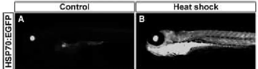
We exposed transgenic 72 hpf larvae to a 2 h pulse of high copper-loaded media (Figure 3A). Control larvae show a basal expression in the lens as previously reported (Halloran et al., 2000; Blechinger et al., 2002a) (Figure 3A), while heat-shocked larvae show the characteristic pattern of EGFP label throughout the body (Supplementary Figure 1). Exposure to low concentrations of copper (below 100µM CuSO4) did not
appreciably modify the levels of detectable EGFP compared to control fish. Transient exposure to 100µM CuSO4, induces expression of EGFP in gills and olfactory pits, and
weak expression in brain, liver and spinal cord (Figure 3B and 3E); this expression increases at 200 ^iM of CuSO4 and fluorescence appears in the olfactory epithelium
and spinal cord (Figure 3C and 3F). Treatment with a higher concentration of copper (400µM CuSO4) induces strong expression in spinal cord, liver and brain (Figure 3D
and 3G). We documented the expression of EGFP in the different larval tissues for each concentration at different exposure times by counting the larvae in each sample that showed label in these tissues and expressing this value as a percentage of the total number of larvae (Figure 3 E-G). These results show that induction of the hsp70
Figure 3. The Hsp70 promoter is induced by copper in a dose-dependent manner. Transgenic Tg(hsp70:egfp) zebrafish larvae of 72 hpf were exposed transiently to a 2 h pulse with 0 µM (A), 100 µM (B), 200 µM (C) and 400 µM (D) of waterborne copper (as CuSO4 ) in the medium. Larvae
To analyze the expression of EGFP in higher detail, 72 hpf transgenic larvae were exposed to 300 µM copper for 2 h and processed for EGFP immunodetection at 72 h after copper exposure (Figure 4). Observations of living control larvae under
fluorescent illumination shows expression of EGFP exclusively in the lens (Figure 4A). In contrast, exposed larvae show expression in olfactory pit, gills, liver, spinal cord and brain (Figure 4B). Anti-GFP immunohistochemistry in sectioned slices of copper
exposed fish shows expression of EGFP in the retina and also in the telencephalon (r and t, respectively, Figure 4C). Interestingly, posterior transverse sections evidence the expression of EGFP in the spinal cord (sc), pronephric glomeruli (pg), pronephric ducts (pd), pneumatic duct (pnd), stomach (s), liver (L) and intestine (i) (Figure 4D and 4E).
Figure 4. Tissue-specific stress response after treatment with high concentrations of waterborne copper: 72 hpf Tg(hsp70:egfp)
observed under UV light 72 hours after copper exposure. Control animals show background EGFP fluorescence only in the lens (A). In treated larvae, expression of EGFP was evident in olfactory
epithelium (oe), gills (g), central nervous system (cns), and liver (B). EGFP detection was also carried out by immunohistochemistry in sections by using an antibody against EGFP and a secondary antibody conjugated to alkaline phosphatase (AP). Sections show EGFP expression in retina (r), telencephalon (t), liver (L), stomach (s), pronephric ducts (pd), pneumatic duct (pnd), pronephric glomeruli (pg), spinal cord (sc) and intestine (i) (C to E). Sections were counterstained with eosine after developing the alkaline phosphatase activity; the corresponding transverse sections are indicated in B. In A and B pictures are lateral views, anterior to left. In C, D and E are transverse sections, dorsal is up; y, yolk.
Cell death in zebrafish larvae exposed to copper
Having established the expression pattern of the hsp70 gene in larvae exposed to copper, we investigated the spatial distribution of apoptotic cells in copper-treated larvae after acute exposure treatments by using the TUNEL assay. Larvae exposed for 2 hours to 300µM CuSO4 and processed by TUNEL 72 h after exposure show an
increase of apoptotic staining in the central nervous system (cns), gills (ga) and also strong labeling in the pronephros (prn) (Figure 5B and 5D) compared to control larvae at the same stages (Figure 5A and 5C).
A variety of studies have shown that fish, as well as other aquatic organisms, are particularly sensitive to pollutants and heavy metals in the early stages of their life cycle (Herkovits et al., 1998; Hernandez et al., 2006; Linbo et al., 2006; Witeska et al., 1995). However, some studies have shown that fish eggs are more resistant than young fish to the toxic effects of copper sulfate. In fact, experiments using zebrafish as an animal model show that adult zebrafish are more sensitive to copper than embryos (Palmer et al., 1998). These authors calculated a 4-day median lethal concentration 50 (LC50) for adults (five to nine months old) of 1.90 mM and a 12-day LC50 for embryos of 2.2 mM. Nevertheless, copper toxicity in fishes varies among species and depends on the physical and chemical characteristics of the water (Kamunde et al., 2003).
In our study, acute toxicity tests of waterborne copper were performed in zebrafish larvae (72 hpf) and the LC50 values for dissolved copper sulphate were calculated. Our lethality curves indicate that there is an increase in the mortality rate as the copper concentration is increased, as well as when the incubation with waterborne copper is longer. We found that at concentrations lower than 10 µM of copper (approximately 0.63 mg/L of copper) the larvae present similar mortality rates to controls. However at concentrations higher than 25 µM (1.6 mg/L) the mortality was significant increased. Calculations of the LC50 value for each exposure time show a logarithmic dependence between the LC50 and time of exposure. Furthermore, the LC50 value calculated for larvae exposed to copper continuously for 4 days was 13.82 µM, showing that larvae are more resistant to waterborne copper than adults, where this value was calculated at 1.9 µM (Palmer et al., 1998).
To determine whether waterborne copper is incorporated in larvae, we measured copper uptake and loading by using radiolabeled copper and AAS, respectively. Our data indicate that copper is incorporated into larvae in a time-dependent manner and also that the metal accumulates in larvae depending on its concentration in the
medium. The primary uptake pathway of trace metals in fish is the food, under normal dietary and waterborne conditions (Kamunde et al., 2003; Kjoss et al., 2005; Craig et al., 2009), even though some reports suggest that copper and iron can also be taken up by the gills (Clearwater et al., 2002; Grosell and Wood, 2002; Kamude et al., 2003). In rainbow trout diet contributes more than 90% of the body burden of this metal. However, gill-uptake contributes importantly to the body Cu load when the dietary source of copper is deficient (Kamunde et al., 2003). As the larvae in our tests are not feeding, the mechanism of entry is limited to the body surface, especially exposed tissues not covered by the epidermis (gills, olfactory epithelium,
mechanosensory organs). Copper could be entering cells via the same mechanisms that mediate entry in adult tissues, Na+ dependent transporters, copper transporter-1
(Ctrl) and the divalent metal transporter 1 (Dmtl). We have previously shown that Ctrl
is expressed in zebrafish during early embryogenesis, providing a possible transport mechanism at this stage (Mackenzie et al., 2004).
Based in our data of mortality assays and LC50 values we designed experiments to examine whether copper is capable of inducing the expression of the hsp70 gene, a chaperone whose expression is activated by heat shock. This protein has been reported to be induced by a variety of heavy metals including Zn, Cd, Hg, Ag and Cu (Murata et al., 1999; Williams et al., 1990) and this activation is mediated by the heat shock transcription factor HSF-1 (Mosser et al., 1988; Murata et al., 1999). Recent work has shown that copper can activate the human hsp70 promoter in mosaic transgenic fish (Seok et al., 2006), and previous reports show that heat shock and cadmium induce the expression of EGFP in Tg(hsp70:egfp) fish expressing EGFP driven by the hsp70
2007; Matz and Krone, 2007). Using these transgenic fish, we found that waterborne copper induces EGFP expression in a concentration-dependent manner. The histological analysis in sections shows that the expression induced by copper is localized mainly to the head and, especially, the brain. Moreover, waterborne copper induces EGFP
expression in organs such as the spinal cord, pronephric glomeruli, pronephric ducts, pneumatic duct, liver, stomach and intestine. The liver, intestine and renal tissues have been shown to accumulate copper in toadfish and rainbow trout after waterborne copper exposure (Grosell et al., 2004; Kamunde et al., 2003), suggesting that this selective accumulation of copper in these tissues could be a protection mechanism against internal copper toxicity. Studies using primary cultures of rainbow trout hepatocytes exposed to CuSO4 (0-200 mM) resulted in both a dose-dependent
elevation of hsp70 expression and cell death by apoptosis (Feng et al., 2003). Moreover, in vitro studies show that copper exposure is also able to induce the
expression of metallothioneins (MTs) (Cheuk et al., 2008), small cysteine-rich proteins with high affinity for heavy metals, that could complex the copper ions and protect the cells from toxicity. Our results were similar to those previously reported for cadmium-induced EGFP expression (Blechinger et al., 2002a; 2007). In our experiments we found additional expression sites such as the brain and spinal cord in fish exposed to high copper concentrations, and also that appearance of the marker in some tissues depended strongly on the incubation time. While EGFP was induced in the brain only at the highest concentration tested, it could reveal that metal toxicity at this
concentration overwhelms the physiological capacity of the larva to prevent its dissemination. Nonetheless, expression in the brain appeared to decrease over time, possibly indicating progressive recovery of the liver or other detoxifying systems. The gills and olfactory epithelium showed EGFP expression in the larvae exposed to 100 µM copper from the earliest time point analyzed, 24 hpf. Interestingly MT is expressed in these tissues during normal embryogenesis of zebrafish (Chen et al., 2004),
suggesting that these tissues could be "primed" for protection against accumulation of copper or other metals. Prolonged incubation induced the expression of EGFP in liver (where MT is not expressed in developing fish, Chen et al., 2004). Our results also differ from those previously reported, in which mosaic transgenic fish carrying EGFP under the control of the exogenous human hsp70 promoter were exposed to copper (Seok et al., 2006). These authors show mosaic EGFP expression in gills, skin
epithelium, auditory epithelium and myotubes. The differences can be explained by the fact that these authors used fish transiently expressing EGFP from the human hsp70
promoter and obtained mosaic expression of EGFP. This is in contrast to the stable transgenic Tg(hsp70:egfp) line used in our experiments that expresses EGFP in the entire embryo. In addition, these authors reported a calculated LC50 of 1.2 µM of copper in contrast to the value of 13.82 µM calculated in our experiments, a difference of an order of magnitude (Seok et al., 2006). There are several possible explanations for these differences such as the strain of fish used (AB strain in our study versus a non-typified strain), ion composition of the media (E3 in our experiments versus
Ringer's solution) and water hardness (Long et al., 2004; Van Genderen et al., 2007).
feed. At 96 hpf, the digestive organs are formed and liver cells start secreting bile at 72-96 hpf (Pack et al., 1996). As no hsp70 response was observed until 96-125 hpf in these tissues, we suggest that the mechanisms that regulates the distribution of copper in the body appears concomitantly with vascularization and organogenesis.
High intracellular copper has been described as toxic to the cells via generation of reactive oxygen species (ROS) (Pourahmad and O'Brien, 2000). A major cytotoxic role for ROS includes the activation of the apoptosis pathway and some studies clearly implicate a role for ROS in copper-induced apoptosis in vitro (Ma et al., 1998;
Pourahmad and O'Brien, 2000; Zhai et al., 2000). Heat shock proteins are key players in the cellular stress response process and crucial for defending cells from copper toxicity (Ma et al., 1998). Our TUNEL assays in larvae acutely exposed to 300 µM (18.9 mg/L) copper show that waterborne copper induces apoptosis in tissues including the pronephros, brain and gills, organs that also showed expression of EGFP in transgenic Tg(hsp70:egfp) larvae after similar copper treatments. Stress in these tissues followed by cell death is a likely consequence of the exposure to copper. However, we did not detect TUNEL staining in the liver, an organ that showed a strong stress response by visualization of the reporter protein. In contrast, the proneprhos showed the strongest TUNEL staining, even though EGFP expression in this organ was only detected by immunostaining. These observations suggest that in some tissues, such as liver tissue, the stress response induced by copper allows cell survival, probably by strong
induction of MT expression, while other organs, such as the pronephros, fail to show a stress response and manifest high levels of cell death.
The data above indicate that copper is toxic for zebrafish larvae because the metal is incorporated and accumulates in diverse tissues, as is suggested by the expression of EGFP in Tg(hsp70:egfp) larvae and TUNEL assays after copper treatments. Our results provide evidence that copper can be lethal to developing fish when it is present in the water, because of the deleterious effects on several key organs.
In this work we observed that when transgenic zebrafish larvae are exposed to external copper, the hsp70:egfp transgene was expressed in different tissues in a dose-dependent manner. Thus, this resource represents an excellent system to evaluate how the larva copes with external copper and how it is able to restrict the distribution of accumulating metal to different tissues. However, it is not capable of detecting the subtle effects induced by low doses of waterborne copper, limiting its potential use as a live biosensor tool. Finally, our findings show that the most sensitive organs to stress induced by waterborne copper are the central nervous system and the liver, even though the most affected in terms of cell death are the gills and
pronephros. Cell death is likely to be elicited by the induction of ROS, providing a possible immediate cause for the death of exposed larvae.
ACKNOWLEDGEMENTS
We thank the following colleagues for valuable resources and reagents: John Kuwada for the Tg(hsp70:egfp) transgenic fish; Miguel Arredondo for absorption spectroscopy studies and Francisca Reyes for help with radioactive Cu assays. This work was supported by grants from ICM (P06-039F to M.A. and P04-071F to A.R.), FONDAP (15090007 to M.A.), FONDECYT (1070867 to M.A. and 1095128 to A.E.R.), and the International Copper Association.
NOTES
# These authors contributed equally to this work.
* Corresponding author: Miguel L. Allende, Facultad de Ciencias Universidad de Chile, Casilla 653 Santiago, Chile. E-mail: allende@uchile.cl, Tel: 978 7390, Fax: 56-2-276 3802.
Received: 18-12-2009. In Revised form: 16-09-2010. Accepted: 17-12-2010.
REFERENCES
AMBROSINI L, MERCER JF (1999) Defective copper-induced trafficking and localization of the Menkes protein in patients with mild and copper-treated classical Menkes disease. Hum Mol Genet 8: 1547-1555. [ Links ]
BLECHINGER SR, EVANS TG, TANG PT, KUWADA JY, WARREN JT, JR., KRONE PH (2002) The heat-inducible zebrafish hsp70 gene is expressed during normal lens development under non-stress conditions. Mech Dev 112: 213-215. [ Links ]
BLECHINGER SR, KUSCH RC, HAUGO K, MATZ C, CHIVERS DP, KRONE PH (2007) Brief embryonic cadmium exposure induces a stress response and cell death in the
developing olfactory system followed by long-term olfactory deficits in juvenile zebrafish. Toxicol Appl Pharmacol 224: 72-80. [ Links ]
BLECHINGER SR, WARREN JT, JR., KUWADA JY, KRONE PH (2002) Developmental toxicology of cadmium in living embryos of a stable transgenic zebrafish line. Environ Health Perspect 110: 1041-1046. [ Links ]
CAMAKARIS J, VOSKOBOINIK I, MERCER JF (1999) Molecular mechanisms of copper homeostasis. Biochem Biophys Res Commun 261: 225-232. [ Links ]
CHAN PK, CHENG SH (2003) Cadmium-induced ectopic apoptosis in zebrafish embryos. Arch Toxicol 77: 69-79. [ Links ]
CHEN WY, JOHN J A, LIN C H, LIN HF, WU SC, LIN CH, CHANG CY. (2004) Expression of metallothionein gene during embryonic and early larval development in zebrafish. Aquat Toxicol 69: 215-227. [ Links ]
CHEUK WK, CHAN PC, CHAN KM (2008) Cytotoxicities and induction of metallothionein (MT) and metal regulatory element (MRE)-binding transcription factor-1 (MTF-1) messenger RNA levels in the zebrafish (Danio rerio) ZFL and SJD cell lines after exposure to various metal ions. Aquat Toxicol 89: 103-112. [ Links ]
CORONADO V, NANJI M, COX DW (2001) The Jackson toxic milk mouse as a model for copper loading. Mamm Genome 12: 793-795. [ Links ]
CRAIG PM, GALUS M, WOOD CM, MCCLELLAND GB (2009) Dietary iron alters
waterborne copper-induced gene expression in soft water acclimated zebrafish (Danio rerio). Am J Physiol Regul Integr Comp Physiol 296: R362-373. [ Links ]
DAVE G, XIU RQ (1991) Toxicity of mercury, copper, nickel, lead, and cobalt to embryos and larvae of zebrafish, Brachydanio rerio. Arch Environ Contam Toxicol 21: 126-134. [ Links ]
DRUMMOND IA, MAJUMDAR A, HENTSCHEL H, ELGER M, SOLNICA-KREZEL L, SCHIER AF, NEUHAUSS SC, STEMPLE DL, ZWARTKRUIS F, RANGINI Z, DRIEVER W, FISHMAN MC (1998) Early development of the zebrafish pronephros and analysis of mutations affecting pronephric function. Development 125: 4655-4667. [ Links ]
FENG Q, BOONE AN, VIJAYAN MM (2003) Copper impact on heat shock protein 70 expression and apoptosis in rainbow trout hepatocytes. Comp Biochem Physiol C Toxicol Pharmacol 135C: 345-355. [ Links ]
GRAVENMIER JJ, JOHNSTON DW, ARNOLD WR (2005) Acute toxicity of vanadium to the threespine stickleback, Gasterosteus aculeatus. Environ Toxicol 20: 18-22. [ Links ]
GROSELL M, MCDONALD MD, WALSH PJ, WOOD CM (2004) Effects of prolonged copper exposure in the marine gulf toadfish (Opsanus beta) II: copper accumulation, drinking rate and Na+/K+ -ATPase activity in osmoregulatory tissues. Aquat Toxicol 68: 263-275. [ Links ]
GROSELL M, WOOD CM (2002) Copper uptake across rainbow trout gills: mechanisms of apical entry. J Exp Biol 205: 1179-1188. [ Links ]
HAFFTER P, GRANATO M, BRAND M, MULLINS MC, HAMMERSCHMIDT M, KANE DA, ODENTHAL J, VAN EEDEN FJ, JIANG YJ, HEISENBERG CP, KELSH RN, FURUTANI-SEIKI M, VOGELSANG E, BEUCHLE D, SCHACH U, FABIAN C, NUSSLEIN- VOLHARD C (1996) The identification of genes with unique and essential functions in the development of the zebrafish, Danio rerio. Development 123: 1-36. [ Links ]
HALLORAN MC, SATO-MAEDA M, WARREN JT, SU F, LELE Z, KRONE PH, KUWADA JY, SHOJI W (2000) Laser-induced gene expression in specific cells of transgenic zebrafish. Development 127: 1953-1960. [ Links ]
HAMILTON MA, RUSSO RC, THURSTON RV (1977) Trimmed Spearman-Karber method for estimating median lethal concentrations in toxicity bioassays. Environ Sci Technol 11: 714-719; Correction 712(714):417 (1978). [ Links ]
HERKOVITS J, CARDELLINI P, PAVANATI C, PEREZ-COLL CS (1998) Cadmium uptake and bioaccumulation in Xenopus laevis embryos at different developmental stages. Ecotoxicol Environ Saf 39: 21-26. [ Links ]
HERNANDEZ PP, MORENO V, OLIVARI FA, ALLENDE ML (2006) Sub-lethal
concentrations of waterborne copper are toxic to lateral line neuromasts in zebrafish (Danio rerio). Hear Res 213: 1-10. [ Links ]
KAMUNDE CN, PYLE GG, MCDONALD DG, WOOD CM (2003) Influence of dietary sodium on waterborne copper toxicity in rainbow trout, Oncorhynchus mykiss. Environ Toxicol Chem 22: 342-350. [ Links ]
KJOSS VA, GROSELL M, WOOD CM (2005) The influence of dietary Na on Cu
accumulation in juvenile rainbow trout exposed to combined dietary and waterborne Cu in soft water. Arch Environ Contam Toxicol 49: 520-527. [ Links ]
KOZLOWSKI DJ, WHITFIELD TT, HUKRIEDE NA, LAM WK, WEINBERG ES (2005) The zebrafish dog-eared mutation disrupts eya1, a gene required for cell survival and differentiation in the inner ear and lateral line. Dev Biol 277: 27-41. [ Links ]
KRONE PH, BLECHINGER SR, EVANS TG, RYAN JA, NOONAN EJ, HIGHTOWER LE (2005) Use of fish liver PLHC-1 cells and zebrafish embryos in cytotoxicity assays. Methods 35: 176-187. [ Links ]
KUO YM, ZHOU B, COSCO D, GITSCHIER J (2001) The copper transporter CTR1 provides an essential function in mammalian embryonic development. Proc Natl Acad Sci U S A 98: 6836-6841. [ Links ]
LEE J, PROHASKA JR, THIELE DJ (2001) Essential role for mammalian copper
transporter Ctr1 in copper homeostasis and embryonic development. Proc Natl Acad Sci U S A 98: 6842-6847. [ Links ]
LI WH, CHAN PC, CHAN KM (2004) Metal uptake in zebrafish embryo-larvae exposed to metal-contaminated sediments. Mar Environ Res 58: 829-832. [ Links ]
LINBO TL, STEHR CM, INCARDONA JP, SCHOLZ NL (2006) Dissolved copper triggers cell death in the peripheral mechanosensory system of larval fish. Environ Toxicol Chem 25: 597-603. [ Links ]
LONG KE, VAN GENDEREN EJ, KLAINE SJ (2004) The effects of low hardness and pH on copper toxicity to Daphnia magna. Environ Toxicol Chem 23: 72-75. [ Links ]
MA Y, CAO L, KAWABATA T, YOSHINO T, YANG BB, OKADA S (1998) Cupric
nitrilotriacetate induces oxidative DNA damage and apoptosis in human leukemia HL-60 cells. Free Radic Biol Med 25: 568-575. [ Links ]
MADSEN EC, GITLIN JD (2008) Zebrafish mutants calamity and catastrophe define critical pathways of gene-nutrient interactions in developmental copper metabolism. PLoS Genet 4: e1000261. [ Links ]
MADSEN EC, MORCOS PA, MENDELSOHN BA, GITLIN JD (2008) In vivo correction of a Menkes disease model using antisense oligonucleotides. Proc Natl Acad Sci U S A 105: 3909-3914. [ Links ]
MATSUO AY, PLAYLE RC, VAL AL, WOOD CM (2004) Physiological action of dissolved organic matter in rainbow trout in the presence and absence of copper: sodium uptake kinetics and unidirectional flux rates in hard and softwater. Aquat Toxicol 70: 63-81. [ Links ]
MATZ CJ, KRONE PH (2007) Cell death, stress-responsive transgene activation, and deficits in the olfactory system of larval zebrafish following cadmium exposure. Environ Sci Technol 41: 5143-5148. [ Links ]
MENDELSOHN BA, YIN C, JOHNSON SL, WILM TP, SOLNICA-KREZEL L, GITLIN JD (2006) Atp7a determines a hierarchy of copper metabolism essential for notochord development. Cell Metab 4: 155-162. [ Links ]
MERCER JF, LLANOS RM (2003) Molecular and cellular aspects of copper transport in developing mammals. J Nutr 133: 1481S-1484S. [ Links ]
MOSSER DD, CARON AW, BOURGET L, MERIIN AB, SHERMAN MY, MORIMOTO RI, MASSIE B (2000) The chaperone function of hsp70 is required for protection against stress-induced apoptosis. Mol Cell Biol 20: 7146-7159. [ Links ]
MOSSER DD, THEODORAKIS NG, MORIMOTO RI (1988) Coordinate changes in heat shock element-binding activity and HSP70 gene transcription rates in human cells. Mol Cell Biol 8: 4736-4744. [ Links ]
MURATA M, GONG P, SUZUKI K, KOIZUMI S (1999) Differential metal response and regulation of human heavy metal-inducible genes. J Cell Physiol 180: 105-113. [ Links ]
PACK M, SOLNICA-KREZEL L, MALICKI J, NEUHAUSS SC, SCHIER AF, STEMPLE DL, DRIEVER W, FISHMAN MC (1996) Mutations affecting development of zebrafish digestive organs. Development 123: 321-328. [ Links ]
PALMER FB, BUTLER CA, TIMPERLEY MH, EVANS CW (1998) Toxicity to embryo and adult zebrafish of copper complexes with two malonic acids as models for dissolved organic matter. Environ Toxicol Chem 17: 1538-1545. [ Links ]
POURAHMAD J, O'BRIEN PJ (2000) A comparison of hepatocyte cytotoxic mechanisms for Cu2+ and Cd2+. Toxicology 143: 263-273. [ Links ]
RIBEYRE F, AMIARD-TRIQUET C, BOUDOU A, AMIARD JC (1995) Experimental study of interactions between five trace elements--Cu, Ag, Se, Zn, and Hg--toward their
ROUGIER F, MENUDIER A, BOSGIRAUD C, NICOLAS JA (1996) Copper and zinc exposure of zebrafish, Brachydanio rerio (Hamilton-Buchaman): effects in
experimental Listeria infection. Ecotoxicol Environ Saf 34: 134-140. [ Links ]
SANDAHL JF, BALDWIN DH, JENKINS JJ, SCHOLZ NL (2007) A sensory system at the interface between urban stormwater runoff and salmon survival. Environ Sci Technol 41: 2998-3004. [ Links ]
SEOK SH, BAEK MW, LEE HY, KIM DJ, NA YR, NOH KJ, PARK SH, LEE HK, LEE BH, RYU DY, PARK JH (2007) Arsenite-induced apoptosis is prevented by antioxidants in
zebrafish liver cell line. Toxicol In Vitro 21: 870-877. [ Links ]
SEOK SH, PARK JH, BAEK MW, LEE HY, KIM DJ, UHM HM, HONG JJ, NA YR, JIN BH, RYU DY (2006) Specific activation of the human HSP70 promoter by copper sulfate in mosaic transgenic zebrafish. J Biotechnol 126: 406-413. [ Links ]
STANKIEWICZ AR, LACHAPELLE G, FOO CP, RADICIONI SM, MOSSER DD (2005) Hsp70 inhibits heat-induced apoptosis upstream of mitochondria by preventing Bax translocation. J Biol Chem 280: 38729-38739. [ Links ]
SZEBEDINSZKY C, MCGEER JC, MCDONALD DG, WOOD CM (2001) Effects of chronic Cd exposure via the diet or water on internal organ-specific distribution and
subsequent gill Cd uptake kinetics in juvenile rainbow trout (Oncorhynchus mykiss). Environ Toxicol Chem 20: 597-607. [ Links ]
TAYLOR LN, WOOD CM, MCDONALD DG (2003) An evaluation of sodium loss and gill metal binding properties in rainbow trout and yellow perch to explain species
differences in copper tolerance. Environ Toxicol Chem 22: 2159-2166. [ Links ]
TSAI JW, LIAO CM (2006) A dose-based modeling approach for accumulation and toxicity of arsenic in tilapia Oreochromis mossambicus. Environ Toxicol 21: 8-21. [ Links ]
VAN GENDEREN E, GENSEMER R, SMITH C, SANTORE R, RYAN A (2007) Evaluation of the Biotic Ligand Model relative to other site-specific criteria derivation methods for copper in surface waters with elevated hardness. Aquat Toxicol 84: 279-291. [ Links ]
WESTERFIELD (1995) The Zebrafish Book, fourth ed. University of Oregon Press, Eugene. [ Links ]
WILLIAMS GT, MORIMOTO RI (1990) Maximal stress-induced transcription from the human HSP70 promoter requires interactions with the basal promoter elements independent of rotational alignment. Mol Cell Biol 10: 3125-3136. [ Links ]
WITESKA M, JEZIERSKA J, CHABER J (1995) The influence of cadmium on common carp embryos and larvae. Aquaculture 129: 129-132. [ Links ]
© 2014 Sociedad de Biología de Chile
Canadá 253, piso 3º, Dpto. F.
PO Box 16164
Santiago - Chile
Tel.: (56-2) 22093503
Fax: (56-2) 22258427