Transient receptor potential canonical 3 (TRPC3) channels are required for hypothalamic glucose detection and energy homeostasis
11
0
0
Texto completo
(2) diabetes.diabetesjournals.org. channels play in controlling neuronal excitability, several groups have focused on identifying the channels responsible for nutrient and hormone sensing. This effort has highlighted KATP channels as key players in neuronal glucose sensing (11). However, even if the role of the KATP channel is undeniable, we and others have pointed out that additional channels might be critical for glucose sensing (11–13). More specifically, we found that the opening of channels exhibiting a nonselective cation conductance (NSCC channels) is involved in the glucose response of a subpopulation of GE neurons in the MBH (12). Transient receptor potential (TRP) and, particularly, TRP canonical (TRPC) channels represent one of the largest families of NSCC channels (14). A link between TRPC channels and the control of energy metabolism has been proposed based on studies showing that TRPC channels mediate some of the effects of leptin and insulin on hypothalamic neuronal activity (15–17). Nevertheless, the role of TRPC channels in hypothalamic glucose detection and central control of energy homeostasis remains unknown. Of note, TRPC3 channel activity is regulated by ROS, suggesting that these channels may be involved in glucose sensing in GE neurons of the MBH (18,19). Therefore, the goal of the current study was to determine whether TRPC3 channels are part of the glucose-sensing machinery in GE neurons of the MBH and to assess their contribution in central glucose effects. RESEARCH DESIGN AND METHODS Animals. Male outbred Sprague-Dawley rats or inbred C57BL/6J mice were purchased from Charles River Laboratoire France. TRPC3-deficient (TRPC3 knockout [KO]); control wild-type (TRPC3 WT), same genetic background; and TRPC3lox/lox (exon 7 of Trpc3 flanked by LoxP sites) mice were provided by the Comparative Medicine Branch of the National Institute of Environmental Health Sciences (Research Triangle Park, NC) (20). Animals were housed in a controlled environment (12-h light/dark cycle, lights on at 7:00 A.M.), with food and water available ad libitum. All procedures were performed in agreement with European Directive 2010/63/UE and approved by the French Ministry of Research and the local ethics committee of the University of Burgundy (C2EA Grand Campus Dijon number 105; agreement number 02404.02).. Chrétien and Associates. 315. RNA equivalent 40–70 ng). Sequences or references of primers for SYBR Green and TaqMan assays are presented in Supplementary Table 1. Western Blots. Lysates were prepared by homogenizing brain in radioimmunoprecipitation assay buffer containing protease inhibitors by using a TissueLyser (QIAGEN). Lysates were diluted in Laemmli buffer, and samples (200 mg of protein) were loaded onto a 4–15% TGX precast gel (Bio-Rad). The gel was fast transferred onto polyvinylidene fluoride membrane by using Trans-Blot Turbo (Bio-Rad). Membranes were blocked in 3% blocking reagent (Bio-Rad) and then incubated with homemade rabbit anti-TRPC3 (1:100, overnight at 4°C [21]). Secondary immunodetection was carried out with anti-rabbit IgG-horseradish peroxidase (1:50,000, 1 h at room temperature), and the chemiluminescent signal was acquired on a ChemiDoc station (Bio-Rad). The specificity of the antibody was confirmed by using TRPC3 KO as presented in Supplementary Fig. 1. Virus Injection. TRPC3lox/lox mice (8–10 weeks old in which exon 7 of Trpc3 is flanked by LoxP sites [20]) were anesthetized with isoflurane and placed on a stereotaxic apparatus (Kopf Instruments). Adeno-associated virus expressing Cre and green fluorescent protein (AAV-Cre/GFP) or GFP (AAV-GFP) under the cytomegalovirus (AAV-Cre/GFP) or CBA (chicken b-actin) (AAV-GFP) promoter were injected bilaterally into the MBH (stereotaxic coordinates relative to bregma: 21.4 mm anteroposterior, +0.4 mm lateral, and 25.6 mm dorsoventral from the dura at a rate of 50 nL/min for 5 min per side). The plasmid CBA.nls-myc-Cre/eGFP expressed the myc-nls-Cre-GFP fusion protein (22). AAV-Cre/GFP or AAV-GFP serotype 2/9 (6 3 1011 genomes/mL) were produced by the viral production facility of INSERM UMR 1089 (Nantes, France). Glucose and Insulin Tolerance Tests. Glucose tolerance test (GTT) and insulin tolerance test (ITT) were performed in 5-h–fasted mice by measuring blood glucose (Accu-Chek; Roche) from the tail vein after oral glucose (2 g/kg) or intraperitoneal (IP) insulin (0.3 units/kg) administration. Blood samples were collected for insulin measurements during the oral GTT (OGTT) by using a bead-based AlphaLISA Human Insulin Detection Kit (PerkinElmer).. RT-PCR. Central Glucose-Induced Insulin Secretion. After extraction (RNeasy; QIAGEN), mRNA quality and quantity were assessed by microelecrophoresis (Experion; Bio-Rad). Reverse transcription (QuantiTect Reverse Transcription Kit; QIAGEN) was performed on standardized amounts of starting RNA for all samples in each study. Quantitative PCR (qPCR) was carried out on a StepOnePlus thermocycler (Applied Biosystems) by using either SYBR Green (Fast SYBR Green Master Mix; Applied Biosystems) or hydrolysis probes (TaqMan Gene Expression Master Mix; Applied Biosystems) on standardized cDNA (starting. As previously described (23), a silastic catheter was implanted into the left-side carotid artery in the cranial direction in anesthetized (pentobarbital 60 mg/kg) mice or rats and secured in place with sutures. Despite its potential role in autonomic output, we use pentobarbital because it is, to our knowledge, the only approved anesthetic that does not affect blood glucose level (24,25). Fifteen minutes after the surgery, a bolus of glucose (mice: 25 mg/kg in 30 mL over 30 s; rats: 9 mg/kg in 100 mL over 60 s; osmolarity adjusted to 300–310 mOsmol/L with NaCl).
(3) 316. TRPC3: New Actor in Brain Glucose Detection. was administered through the catheter. Blood samples were collected from the tail vein to measure blood glucose and plasma insulin levels. Food Intake. Food intake was measured by hand or by using the BioDAQ food intake monitoring system (Research Diets) in singly housed animals. Three measurements of 24-h food intake were averaged per mouse over 1 week. Fasting-Refeeding Tests in Response to Increased Brain or IP Glucose Level. Before brain glucose injection, animals (8–10 weeks old) were anesthetized with isoflurane and placed on a stereotaxic apparatus (Kopf Instruments) to implant a guide cannula (Plastics One) into the right-side lateral ventricle (stereotaxic coordinates relative to bregma: 20.5 mm anteroposterior, +1 mm lateral, and 22.1 mm dorsoventral from the dura). Seven days after the surgery, naive or cannulated animals were deprived of food for 18 h with ad libitum access to water. Glucose was administered through the guide cannula (125 mg in 2 mL at a rate of 0.4 mL/min; sucrose used to control osmolarity effects) or IP (2 g/kg; NaCl 0.9% used as control) 1 h before the onset of the dark cycle. Access to food was restored 30 min after intracerebroventricular injection, and food intake was measured for 6 h. Calcium Imaging on Primary Culture of Dissociated MBH Neurons. Dissociated MBH neurons were prepared from brain of 4- to 5-week-old TRPC3 WT or KO mice or rats as described previously (26–28). Briefly, MBH explants were dissected from 250-mm brain slices cut with a Vibroslice (Leica). MBH cells were mechanically dissociated with papain by using the Neural Tissue Dissociation Kit (MACS; Miltenyi Biotec) plated on poly-L-lysine (50 mg/mL)–coated glass coverslips in astrocyte-conditioned media overnight before calcium imaging experiments. Astrocyte-conditioned media were prepared as previously described (29) from MBH astrocytes plated on poly-L-lysine (50 mg/mL)–coated dishes and maintained in Neurobasal-A media (Invitrogen) containing (in mmol/L) 2.5 D-glucose, 1 L-lactate, 0.23 L-pyruvate, and 0.5 L-glutamine; 10% FBS; 100 units/mL penicillin/streptomycin; and 10 mg/mL gentamycin supplemented with 1% B-27 (Invitrogen). MBH neurons were loaded for 20 min with 0.5 mmol/L Fura-2 acetoxymethyl ester (Molecular Probes) in Hepesbuffered balanced salt solution containing 2.5 mmol/L D-glucose supplemented with 0.002% pluronic acid. After incubation, cells were mounted in a thermostatically regulated microscope chamber (37°C) mounted on an inverted microscope (Olympus IX70). Fura-2 fluorescence images, acquired every 10 s by alternating excitation at 340 and 380 nm and emissions (420–600 nm), were collected by using a cooled, charge-coupled device camera with TiLL Photonics Live Acquisition software. Values for the 340/380 nm fluorescence ratio, representative of [Ca2+]i, were obtained after correction for background fluorescence values.. Diabetes Volume 66, February 2017. GE neurons were identified in response to a 2.5–10 mmol/L increase in glucose level. 10 mmol/L glucose is higher than levels found in brain areas such as the hypothalamus (30). Nevertheless, in view of the presence of fenestrated capillaries in the ventral part of the ARC (31), we could speculate that glucose level is locally closer to blood level and rises to .5 mmol/L. Changes in [Ca2+]i were quantitated by calculating the integrated area under the curve (AUC) of each glucose response with the Live Acquisition software. Neurons were considered as GE if the increase in [Ca2+]i in response to a 2.5–10 mmol/L increased glucose level occurred between 2 and 20 min of treatment, had an amplitude .0.1 (Dratio 340/380), lasted at least 30 s, and was transient. At the end of each recording, neuronal excitability was verified by measuring Ca2+ response to 50 mmol/L KCl. Neurons that showed no response to KCl were not considered for analysis. We verified that the overall excitability of MBH GE neurons (amplitude of the calcium response to KCl) was not affected by any TRPC or KATP channel modulators or antioxidants and in neurons isolated from TRPC3 KO mice (data not shown). Analysis of each experiment was obtained from at least three independent cultures prepared from at least six animals. Expression of Data and Statistics. Data are expressed as mean 6 SEM. Intergroup comparisons were performed by ANOVA with Bonferroni post hoc tests or Student t test as described in the figure legends. GraphPad Prism 5 software was used for the statistical analyses. P , 0.05 was set as significant. RESULTS TRPC3 Channels Are Expressed in the MBH. The first aim of the study was to determine whether TRPC3 channels are expressed in the MBH, where GE neurons are located. TRPC3 channels are known to be expressed in several peripheral tissues, including muscles, liver, and white adipose tissue (32). Within the brain, high TRPC3 expression has been reported in the cerebellum and cortex (33). In the hypothalamus, expression of TRPC3 channels has been detected but not investigated in specific nuclei (33). We found that through qPCR and Western blotting that TRPC3 channels are expressed in the MBH (Fig. 1) as well as in other brain regions and peripheral tissues. MBH TRPC3 Channels Are Critical for the Regulation of Body Weight and Food Intake. The role of TRPC3 channels in the control of energy homeostasis was investigated by using whole-body TRPC3deficient (TRPC3 KO) mice. TRPC3 KO mice have impaired motor coordination, but the impact of TRPC3 deficiency on metabolic status, to our knowledge, has never been investigated (20). We found that TRPC3 KO mice have increased body weight associated with increased nocturnal food intake (Fig. 2A and B). Change in food intake is not associated with changes in the expression level of MBH neuropeptides that regulate food intake (Fig. 2C). In.
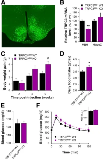
(4) diabetes.diabetesjournals.org. Chrétien and Associates. 317. Figure 1—TRPC3 channels are expressed in the MBH. A: TRPC3 mRNA expression obtained by RT-qPCR from cerebellum (Cereb.), MBH, cortex, hippocampus (Hippo.), EWAT, liver, and tibialis anterior (Tib. Ant.) samples (n = 6). B: Representative Western blot of Cereb. and MBH against TRPC3. a.u., arbitrary units.. addition, TRPC3 KO mice showed an increased fasting blood glucose level associated with mild glucose intolerance (Fig. 2D and E). However, the mild glucose intolerance in TRPC3 KO mice was not associated with alteration of insulin levels during the IPGTT (Supplementary Fig. 2A) or with insulin sensitivity (Supplementary Fig. 2B and C). The decrease in blood glucose level during an ITT (Supplementary Fig. 2B) or the quantification of phosphorylation level of the protein AKT (a marker of insulin signaling) in skeletal muscle, epididymal white adipose tissue (EWAT), or liver (Supplementary Fig. 2C) (34) were not statistically. different between TRPC3 KO and WT mice. These data suggest that TRPC3 channels are involved in the control of food intake, body weight, and glucose homeostasis, which agrees with TRPC3 expression in metabolically active tissues and organs (32). To investigate the role of TRPC3 channels expressed in the MBH in the control of energy and glucose homeostasis, we selectively invalidated TRPC3 expression in this area by using Cre expressing AAV injected in the MBH of TRPC3lox/lox mice (TRPC3MBH KO mice) (Fig. 3A). TRPC3 gene expression is significantly reduced in the MBH of. Figure 2—TRPC3-deficient mice present increased body weight and food intake associated with impaired glucose homeostasis. A: Body weight of TRPC3 WT (n = 48) or KO (n = 45) mice. B: Nocturnal and diurnal food intake of TRPC3 WT (n = 11) or KO (n = 6) mice. C: MBH mRNA expression of neuropeptide Y (NPY), agouti-related peptide (AgRP), proopiomelanocortin (POMC), and cocaine- and amphetamineregulated transcript (CART) from TRPC3 WT (n = 9) and KO (n = 14) mice. D: Fasting blood glucose level of TRPC3 WT (n = 37) or KO (n = 37) mice. E: Blood glucose levels during an OGTT (2 g/kg) and the AUC (inset) in TRPC3 WT (n = 10) and KO (n = 8) mice. **P < 0.01 vs. WT unpaired t test; #P < 0.05 vs. WT, two-way ANOVA with Bonferroni post hoc test. a.u., arbitrary units..
(5) 318. TRPC3: New Actor in Brain Glucose Detection. Diabetes Volume 66, February 2017. Central TRPC3 Channels Are Required for Central Glucose Detection. We then tested whether MBH TRPC3 channels are involved in brain glucose detection. To this end, we measured food intake and insulin secretion in response to increased brain glucose levels in TRPC3 KO mice. As previously reported (35), intracerebroventricular injection of glucose decreased food consumption in WT mice (Fig. 4A). The anorectic effect of central glucose was completely blunted in TRPC3 KO mice (Fig. 4A). Similarly, the effect of glucose on food intake in response to an IP injection of glucose was blunted in TRPC3 KO mice (Fig. 4B). Of note, the anorectic effect of IP glucose injection also was reduced in the TRPC3MBH KO mice (Supplementary Fig. 4). As previously described, the delivery of a glucose bolus to the brain through an intracarotid injection that does not change peripheral blood glucose triggered a rapid and transient increase in plasma insulin through activation of the hypothalamic-pancreatic axis (23,36). The current data show that insulin secretion induced by intracarotid glucose administration is blunted in TRPC3 KO mice compared with WT controls (Fig. 4C). To further highlight the importance of TRPC3 channels in MBH glucose detection, we selectively inhibited their activity by injecting the TRPC3 inhibitor pyrazole-3 (Pyr3) into the MBH of rats before the intracarotid glucose bolus (Fig. 4D). Pyr3 inhibited intracarotid glucose-induced insulin secretion (Fig. 4D). Together, the findings demonstrate that MBH TRPC3 channels are required for brain glucose detection and the associated regulation of food intake and insulin secretion. Figure 3—Inhibition of MBH TRPC3 expression increases food intake and body weight gain. A: Representative photomicrograph showing GFP expression after AAV-cre/GFP injection in the MBH of TRPC3lox/lox mice. B: MBH and hippocampus (HippoC) TRPC3 mRNA expression in TRPC3lox/lox mice injected in the MBH with AAV-cre/GFP (TRPC3MBH KO; n = 5) or AAV-GFP (TRPC3MBH WT; n = 6). C–F: Body weight gain post–AAV injection, average daily food intake measured between weeks 5 and 6 post–AAV injection, fasting blood glucose level, and blood glucose levels during an IPGTT (2 g/kg) and AUC (F inset) of TRPC3MBH WT (n = 19) or KO (n = 19) mice. *P < 0.05, **P < 0.001 vs. WT, unpaired t test; #P < 0.05 vs. WT, two-way ANOVA with Bonferroni post hoc test. a.u., arbitrary units.. TRPC3MBH KO mice compared with controls (Fig. 3B). TRPC3MBH KO mice showed an increased body weight gain 6 weeks after AAV injection associated with an increased food intake during the 6th week (Fig. 3C and D). The increased body weight gain is significantly correlated with the reduction of TRPC3 expression (Pearson r = 20.71, P = 0.01) (Supplementary Fig. 3). Change in body weight gain in TRPC3MBH KO mice, however, was not associated with changes in fasting blood glucose level or glucose tolerance (Fig. 3E and F). Together, these data show that MBH TRPC3 channels are critical for food intake and body weight control but do not seem to directly regulate glucose homeostasis.. TRPC3 Channels Participate in the Response of MBH GE Neurons to Glucose. We performed wide-field single-cell imaging of [Ca2+]i by using a Fura-2 calcium probe in freshly dissociated MBH cells to study their direct response to glucose as described previously (26,27,37,38). Through this approach, we found that ;9% of MBH neurons tested were GE neurons because they harbored a transient increase in [Ca2+]i in response to a rise in glucose level from 2.5 to 10 mmol/L (Supplementary Fig. 5). Of note, the responses of MBH GE to two consecutive 2.5–10 mmol/L increases in glucose level were similar in terms of magnitude (Supplementary Fig. 5B and C). The glucose response latency, however, was shorter for the second increase in glucose level (Supplementary Fig. 5D). To determine the role of TRPC3 channels in response to glucose in MBH GE neurons, we first studied these neurons in dissociated MBH cells from TRPC3 KO or WT mice. The number of MBH GE neurons recorded did not differ between mice (TRPC3 WT 6.1 6 0.5%, KO 5.9 6 0.4%; P . 0.05). However, the glucose response magnitude was significantly decreased in TRPC3 KO mice (Fig. 5A–C). The latency of GE neurons in response to glucose also was significantly increased in MBH cells from TRPC3 KO mice (Fig. 5D). These data suggest that TRPC3 channels regulate the responsiveness of MBH GE neurons..
(6) diabetes.diabetesjournals.org. Chrétien and Associates. 319. Figure 4—MBH TRPC3 channels are required for brain glucose detection. A: Food intake in response to intracerebroventricular (ICV) infusion of glucose or sucrose (lateral ventricle, 0.125 mg in 2 mL infused over 5 min) in 18-h fasted TRPC3 WT (sucrose: n = 6; glucose: n = 5) and TRPC3 KO (sucrose: n = 6; glucose: n = 6) mice. B: Food intake in response to IP infusion of glucose (2 g/kg) or NaCl (0.9%) in 18-h fasted TRPC3 WT (NaCl: n = 7; glucose: n = 8) or TRPC3 KO (NaCl: n = 6; glucose: n = 7) mice. C: Experimental protocol, blood glucose, and plasma insulin (Dinsulin secretion vs. T0) in response to an intracarotid glucose injection (25 mg/kg) in TRPC3 WT (n = 8) and KO (n = 13) mice. D: Experimental protocol, blood glucose, or plasma insulin (Dinsulin secretion vs. T0) in response to an intracarotid glucose injection (9 mg/kg) toward the brain in control rats (n = 9) or infused in the MBH with the TRPC3 inhibitor Pyr3 (3 mmol/L; n = 8). *P < 0.05, **P < 0.01 vs. WT or vehicle, two-way ANOVA with Bonferroni post hoc test; #P < 0.05 vs. T0, one-way ANOVA with Bonferroni post hoc test.. We confirmed these data by using a pharmacological approach in dissociated cells isolated from rat MBH. The results showed that the nonselective TRPC channel inhibitor SKF96365 (39) strongly inhibited glucose responses in all GE neurons tested (n = 27 of 27 GE neurons tested) (Fig. 6A and D). SKF96365 also significantly increased the latency of the glucose response of GE neurons (Fig. 6E). Moreover, the TRPC3 channel inhibitor Pyr3 (39) reduced the response to glucose in ;70% of GE neurons (n = 26 of 37 GE neurons tested) (Fig. 6B and D). Of note, the TRPC3 activator 1-oleoyl-2-acetyl-rac-glycerol (OAG) (40) mimicked glucose response without any change in glucose levels in ;70% of the GE neurons (n = 13 of 20 GE neurons tested) (Fig. 6C and D). The. latency of the glucose response of GE neurons is not affected by Pyr3 or OAG (Fig. 6E). Together, these data show that TRPC3 is a key component of the glucose-sensing machinery in MBH GE neurons. ROS but Not KATP Channels Are Necessary for the Response to Increased Glucose Level by MBH GE Neurons. We and others previously showed that a subpopulation of MBH GE neurons increase their electrical activity in response to increased glucose levels through the opening of an NSCC channel rather than through a KATPdependent pathway (12,13). We confirmed these data by showing that the KATP channel opener diazoxide does not.
(7) 320. TRPC3: New Actor in Brain Glucose Detection. Diabetes Volume 66, February 2017. Figure 5—TRPC3-deficient mice present an impaired MBH GE neuron response to increased glucose. A and B: Representative calcium imaging traces of MBH GE neuron response from TRPC3 WT and TRPC3 KO mice. C and D: Quantification of glucose response magnitude (AUC) and latency in response to a 2.5–10 mmol/L glucose increase in TRPC3 WT (n = 34 GE neurons in 576 total cells) and TRPC3 KO (n = 32 GE neurons in 651 total cells) mice. Four independent cultures. *P < 0.05, unpaired t test.. significantly inhibit the magnitude of glucose response in MBH GE neurons (Supplementary Fig. 6). Our group previously demonstrated that MBH ROS production is important for brain glucose detection and the regulation of food intake and insulin secretion (7,8). Thus, we tested whether ROS mediates the response of MBH GE neurons to increased glucose. The responses to glucose in ;95% of GE neurons were robustly blocked by a cocktail of the antioxidants Trolox/glutathione (GSH) (n = 26 of 27 GE neurons tested) or the H2O2-decomposing enzyme catalase (n = 13 of 14 GE neurons tested) (Fig. 7A–C). The antioxidants did not alter the latency of the glucose response of GE neurons (Fig. 7D). Together, these data demonstrate that ROS, particularly H2O2, but not KATP channels is necessary for the detection of increased glucose levels by MBH GE neurons. DISCUSSION. The detection of changes in glucose level depends on several key processes, including transport, metabolism, and membrane excitability, that are unique to glucose and different from neurotransmitters and hormones. Several components of the glucose sensing machinery have already been highlighted, including GLUT2, sodiumcoupled glucose cotransporters, glucokinase, KATP channels, Na+, and K+ ATPase (37,41–45). The current study highlights that the TRPC3 channel constitutes a new player in glucose sensing. Evidence has suggested that TRPC. channels are involved in the detection of some hormone and nutrient sensing (11). For instance, studies have shown that the effects of leptin or insulin on ARC neurons depend on TRPC channels (15–17). Nevertheless, the physiological role of MBH TRPC channels in energy homeostasis has not been investigated in vivo. In the current study, we provide physiological evidence that MBH TRPC3 channels contribute to the central control of energy homeostasis and neuronal glucose detection. We demonstrate that TRPC3 channels are expressed in the MBH, a key region of the hypothalamus involved in metabolic sensing. However, none of the commercial or homemade TRPC3 antibodies gave a specific immunohistochemical signal in brain sections (data not shown). Thus, the identity of TRPC3-expressing neurons is yet to be determined. Of particular interest is whether TRPC3expressing neurons are preferentially located in the ARC or VMN within the MBH. Nevertheless, we can speculate that TRPC3-mediated glucose sensing operates preferentially in ARC GE neurons because work from Routh and colleagues (3) has shown that VMN GE neurons do not directly detect increased glucose .5 mmol/L. The current data show that TRPC3 deficiency in the MBH or whole body leads to an increase in food intake and body weight, thus suggesting that TRPC3 regulates energy balance. Of note, whole-body TRPC3 KO mice were also characterized by mild glucose intolerance and increased basal blood glucose level, a phenotype not.
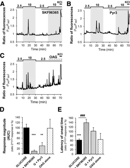
(8) diabetes.diabetesjournals.org. Chrétien and Associates. 321. Figure 6—TRPC3 channels are required for GE neuron response to increased glucose. A–C: Representative calcium imaging traces of MBH GE neuron response to a 2.5–10 mmol/L glucose increase in the presence or not of the nonselective TRPC channel inhibitor SKF96365 (5 mmol/L) or TRPC3 channel inhibitor Pyr3 (1 mmol/L) or activated by the TRPC3 activator OAG (10 mmol/L). D and E: Quantification of glucose response magnitude (AUC) and latency of the second response to a 10 mmol/L increased glucose level in the presence of solvent (glucose alone) or TRPC channel modulators of cells presenting a residual response. **P < 0.01, ***P < 0.001 vs. first glucose response, paired t test; ###P < 0.001 vs. glucose, one-way ANOVA with Bonferroni post hoc test. Nonsignificant P > 0.05. G, glucose.. observed in TRPC3MBH KO mice. This suggests that TRPC3expressing MBH neurons could specifically regulate food intake, whereas peripheral or extra-MBH TRPC3-expressing cells could control glucose homeostasis. Consistent with other studies, we found that TRPC3 is expressed in muscle, liver, and EWAT. Although the role of TRPC3 in liver and EWAT is unknown (46), studies have suggested that TRPC3 regulates insulin signaling and glucose uptake in muscle (47,48). We found that the glucose intolerance observed in TRPC3 KO mice is not associated with alteration in insulin sensitivity or secretion. Consistent with this latter parameter, TRPC3 has never been found, to our knowledge, in pancreatic b-cells, which could support an eventual role in insulin secretion. The role of TRPC3 in insulin signaling is, however, more controversial. In the current study, we show that the decrease in blood glucose level. during an ITT or the phosphorylation of AKT in response to insulin in muscles, white adipose depots, or liver is not altered in TRPC3 KO mice. These data are not consistent with previous studies showing that TRPC3 channels influence insulin signaling in vitro in muscle cells (47,48). The role of TRPC3 channels possibly is not dominant in muscle cells in vivo or that TRPC3 deficiency is compensated by other mechanisms. Further studies are required to assess the role of TRPC3 in glucose homeostasis in specific peripheral tissues and organs. We found that the anorectic effect of glucose as well as the intracarotid glucose-induced insulin secretion are impaired in TRPC3-deficient mouse models. This latter result is not consistent with the unaltered insulin secretion observed in TRPC3 KO mice during the OGTT. The intracarotid glucose-induced insulin secretion lasts only 1–3 min. Thus, we hypothesize that affecting only the.
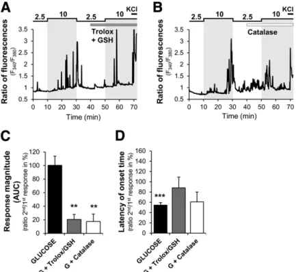
(9) 322. TRPC3: New Actor in Brain Glucose Detection. Diabetes Volume 66, February 2017. Figure 7—ROS is involved in GE neuron response to increased glucose. A and B: Representative calcium imaging traces of MBH GE neuron in response to a 2.5–10 mmol/L glucose increase in the presence or not of a cocktail of nonselective Trolox/GSH antioxidants (Trolox 0.2 mmol/L + GSH 0.1 mmol/L or the selective H2O2-removing enzyme catalase (4,000 units/mL). C and D: Quantification of glucose response magnitude and latency of the second response to a 10 mmol/L increased glucose level in the presence of solvent (glucose alone) or antioxidants of cells presenting a residual response. **P < 0.01, ***P < 0.001 vs. first glucose response, paired t test. Nonsignificant P > 0.05 vs. glucose, one-way ANOVA with Bonferroni post hoc test. G, glucose.. brain-driven insulin secretion has only a minimal effect on plasma insulin level when direct pancreatic glucose-induced insulin secretion is triggered during the GTT. In addition, oral administration of glucose induces incretin secretion by the gut, which potentiates glucose-stimulated insulin secretion and might hide phenotypic differences between WT and KO mice. Nevertheless, these in vivo data from TRPC3 KO mice are supported by in vitro calcium imaging recordings showing that the glucose response of MBH GE neurons is impaired when TRPC3 expression or activity is altered. Together, these data demonstrate for the first time that TRPC3 channels are required for hypothalamic glucose detection and the associated regulation of food intake and insulin secretion. We show that ROS production is necessary for the response of MBH GE neurons to glucose. H2O2 seems to be the important ROS entity because of the impact of the H2O2-decomposing enzyme catalase on the glucose response of GE neurons. This agrees with previous studies from our group showing that H2O2 is produced in the MBH in response to increased brain glucose levels and is necessary for glucose modulation of food intake and insulin secretion (7,8). These results also are consistent with the general role of ROS in the hypothalamic regulation of energy and glucose homeostasis (9,49). The fact that ROS production is necessary for GE neuron response to glucose implicates a metabolism-. dependent signaling pathway involving multiple cascades. The necessity of several steps could explain why the latency of GE neurons to respond to glucose is relatively long. However, a discrepancy exists between the present in vitro and in vivo data. We found that the intracarotid glucose injection increases insulin secretion within 1–3 min, whereas GE neurons seem to respond to increased glucose over a longer period. Thus, the longer latency in the in vitro recordings might reflect the limitation of calcium recordings in dissociated cells in addition to the necessary metabolic steps necessary to increase the electrical activity of GE neurons. However, a question remains about how ROS modulates TRPC3 activity. Studies have shown that H2O2 increases TRPC3 channel activity (50,51). However whether H2O2 directly regulates TRPC3 channel activity through the oxidation of cysteine thiol groups or whether H2O2 stimulates a redox-sensitive signaling pathway that leads to increased TRPC3 activity is unclear. H2O2 may indirectly activate TRPC3 channels through the activation of phospholipase C (50), which agrees with the current data showing that OAG, an analog of diacylglycerol (DAG) generated by phospholipase C, mimics the glucose response in MBH GE neurons. In view of the current data, we cannot rule out that other channels might be involved in glucose responses in GE neurons. Indeed, their response to glucose is not.
(10) diabetes.diabetesjournals.org. totally blunted in TRPC3-deficient mice. In addition, pharmacological inhibition of TRPC3 activity does not impair the glucose response of all MBH GE neurons tested. Of note, the KATP channel opener diazoxide did not affect the glucose response of MBH neurons, supporting the concept that KATP channels are not involved in GE neuron response to glucose as previously shown (12). One possible explanation could be that other TRPC isoforms contribute to glucose responses in GE neurons. Indeed, the nonselective TRPC channels inhibitor SKF96365 impaired glucose responses in all GE neurons tested, suggesting that other TRPC channels may be involved. To support this hypothesis, other TRPC channels have been shown to be redox sensitive [e.g., TRPC4, C6 (19)]. TRPC3 also has been shown to heterodimerize with TRPC4 channels to form a heteromeric redox-sensitive complex in HEK293 and porcine aortic endothelial cells (18). We found that TRPC4 and C6 channels are expressed in the MBH (Supplementary Fig. 7). Thus, these channels may be potential candidates for involvement in the glucose response of GE neurons along with the TRPC3 isoform. We did not observe, however, changes in TRPC4 and C6 expression in the MBH of TRPC3 KO mice, suggesting that TRPC3 deficiency is not compensated by other isoforms (Supplementary Fig. 7). Further investigations are required to assess whether TRPC4 or C6 channels are also involved in glucose sensing. Another question is whether TRPC3 channels are involved in the detection of other metabolic signals besides glucose. We anticipate that these channels may be part of free fatty acid (FFA)– and/or insulin-sensing mechanisms because increased brain FFA or insulin level raises hypothalamic ROS production (52,53). The role of TRPC3 channels in insulin signaling is reinforced by insulin having been shown to activate some MBH neurons through TRPC channels (17). The idea that TRPC3 channels might be involved in FFA sensing is also supported by the current results that show that OAG, a DAG analog, increases the activity of MBH GE neurons and that the incubation of hypothalamic neurons with FFA increases the production of DAG in the presence of high glucose levels (54). As such, glucose may stimulate the generation of DAG in the presence of FFA, which in turn may directly potentiate the response to glucose of MBH GE neurons by activating TRPC3 channels. In conclusion, the current study demonstrates that TRPC3 channels are required at the cellular level to mediate the effect of increased glucose on MBH GE neurons through a mechanism that depends on ROS production. We also show that TRPC3 channels are necessary in vivo for the regulation of food intake and insulin secretion in response to increased brain glucose levels. Together, the results show that TRPC3 channels are novel players in central glucose sensing and regulation of energy balance.. Acknowledgments. The authors thank Thierry Alquier (Montreal Diabetes Research Center, University of Montreal, Montreal, Quebec, Canada) for insightful comments and advice on the manuscript; Richard Palmiter (University of. Chrétien and Associates. 323. Washington, Seattle, WA) and Serge Luquet (University Paris Diderot, Paris, France) for providing the plasmid sequence of the AAV-cre/GFP; and A. Lefranc, L. Decocq, E. Lalarme, and A. Mathou (Centre des Sciences du Goût et de l’Alimentation, Dijon, France) for animal care. Funding. C.Chr. was supported by a salary award from Institut National de la Recherche Agronomique. This work was supported by L’Agence Nationale de la Recherche grants ANR-13-JVS1-0003-01 to A.B. and ANR-11-BSV1-0007 to C.L., Deutsche Forschungsgemeinschaft grants SFB 894 to V.F. and TRR-152 to J.H., and National Institutes of Health Intramural Research Program grant Z01ES-101684 to L.B. This work was also supported by grants from the Marie Skłodowska-Curie actions–Seventh Framework Programme (CIG NeuROSenS PCIG09-GA-2011-293738 to X.F.) and Société Francophone du Diabète (to X.F.). Duality of Interest. No potential conflicts of interest relevant to this article were reported. Author Contributions. C.Chr., C.F., F.L., S.G., C.Che., S.C., X.B., R.B., K.L., R.S., E.N., A.L., J.G., and X.F. researched data. C.Chr. and X.F. wrote the manuscript. C.Chr., A.B., C.L., L.P., and X.F. contributed to the discussion. C.Chr., Z.B.A., A.B., V.F., J.H., C.M., L.B., C.L., L.P., and X.F. reviewed and edited the manuscript. X.F. is the guarantor of this work and, as such, had full access to all the data in the study and takes responsibility for the integrity of the data and the accuracy of the data analysis.. References 1. Schwartz MW, Woods SC, Porte D Jr, Seeley RJ, Baskin DG. Central nervous system control of food intake. Nature 2000;404:661–671 2. Fioramonti X, Contié S, Song Z, Routh VH, Lorsignol A, Pénicaud L. Characterization of glucosensing neuron subpopulations in the arcuate nucleus: integration in neuropeptide Y and pro-opio melanocortin networks? Diabetes 2007;56:1219–1227 3. Song Z, Levin BE, McArdle JJ, Bakhos N, Routh VH. Convergence of preand postsynaptic influences on glucosensing neurons in the ventromedial hypothalamic nucleus. Diabetes 2001;50:2673–2681 4. Oomura Y, Ono T, Ooyama H, Wayner MJ. Glucose and osmosensitive neurones of the rat hypothalamus. Nature 1969;222:282–284 5. Oomura Y, Ooyama H, Sugimori M, Nakamura T, Yamada Y. Glucose inhibition of the glucose-sensitive neurone in the rat lateral hypothalamus. Nature 1974;247:284–286 6. Lam TK, Gutierrez-Juarez R, Pocai A, Rossetti L. Regulation of blood glucose by hypothalamic pyruvate metabolism. Science 2005;309:943–947 7. Leloup C, Magnan C, Benani A, et al. Mitochondrial reactive oxygen species are required for hypothalamic glucose sensing. Diabetes 2006;55:2084–2090 8. Carneiro L, Allard C, Guissard C, et al. Importance of mitochondrial dynamin-related protein 1 in hypothalamic glucose sensitivity in rats. Antioxid Redox Signal 2012;17:433–444 9. Andrews ZB, Liu ZW, Walllingford N, et al. UCP2 mediates ghrelin’s action on NPY/AgRP neurons by lowering free radicals. Nature 2008;454:846–851 10. Toda C, Kim JD, Impellizzeri D, Cuzzocrea S, Liu ZW, Diano S. UCP2 regulates mitochondrial fission and ventromedial nucleus control of glucose responsiveness. Cell 2016;164:872–883 11. Sohn JW. Ion channels in the central regulation of energy and glucose homeostasis. Front Neurosci 2013;7:85 12. Fioramonti X, Lorsignol A, Taupignon A, Pénicaud L. A new ATP-sensitive K+ channel-independent mechanism is involved in glucose-excited neurons of mouse arcuate nucleus. Diabetes 2004;53:2767–2775 13. Yang XJ, Kow LM, Funabashi T, Mobbs CV. Hypothalamic glucose sensor: similarities to and differences from pancreatic beta-cell mechanisms. Diabetes 1999;48:1763–1772 14. Nilius B, Voets T. TRP channels: a TR(I)P through a world of multifunctional cation channels. Pflugers Arch 2005;451:1–10 15. Qiu J, Fang Y, Rønnekleiv OK, Kelly MJ. Leptin excites proopiomelanocortin neurons via activation of TRPC channels. J Neurosci 2010;30:1560–1565 16. Qiu J, Fang Y, Bosch MA, Rønnekleiv OK, Kelly MJ. Guinea pig kisspeptin neurons are depolarized by leptin via activation of TRPC channels. Endocrinology 2011;152:1503–1514.
(11) 324. TRPC3: New Actor in Brain Glucose Detection. 17. Qiu J, Zhang C, Borgquist A, et al. Insulin excites anorexigenic proopiomelanocortin neurons via activation of canonical transient receptor potential channels. Cell Metab 2014;19:682–693 18. Poteser M, Graziani A, Rosker C, et al. TRPC3 and TRPC4 associate to form a redox-sensitive cation channel. Evidence for expression of native TRPC3-TRPC4 heteromeric channels in endothelial cells. J Biol Chem 2006;281:13588–13595 19. Song MY, Makino A, Yuan JX. Role of reactive oxygen species and redox in regulating the function of transient receptor potential channels. Antioxid Redox Signal 2011;15:1549–1565 20. Hartmann J, Dragicevic E, Adelsberger H, et al. TRPC3 channels are required for synaptic transmission and motor coordination. Neuron 2008;59:392–398 21. Meissner M, Obmann VC, Hoschke M, et al. Lessons of studying TRP channels with antibodies. In TRP Channels. Chapter 6. Zhu MX, Ed. Boca Raton, FL, CRC/Taylor & Francis, 2011 22. Wu Q, Palmiter RD. GABAergic signaling by AgRP neurons prevents anorexia via a melanocortin-independent mechanism. Eur J Pharmacol 2011;660:21–27 23. Fergusson G, Ethier M, Guévremont M, et al. Defective insulin secretory response to intravenous glucose in C57Bl/6J compared to C57Bl/6N mice. Mol Metab 2014;3:848–854 24. Saha JK, Xia J, Grondin JM, Engle SK, Jakubowski JA. Acute hyperglycemia induced by ketamine/xylazine anesthesia in rats: mechanisms and implications for preclinical models. Exp Biol Med (Maywood) 2005;230:777–784 25. Pénicaud L, Ferré P, Kande J, Leturque A, Issad T, Girard J. Effect of anesthesia on glucose production and utilization in rats. Am J Physiol 1987;252: E365–E369 26. Dunn-Meynell AA, Routh VH, Kang L, Gaspers L, Levin BE. Glucokinase is the likely mediator of glucosensing in both glucose-excited and glucose-inhibited central neurons. Diabetes 2002;51:2056–2065 27. Kang L, Routh VH, Kuzhikandathil EV, Gaspers LD, Levin BE. Physiological and molecular characteristics of rat hypothalamic ventromedial nucleus glucosensing neurons. Diabetes 2004;53:549–559 28. Vazirani RP, Fioramonti X, Routh VH. Membrane potential dye imaging of ventromedial hypothalamus neurons from adult mice to study glucose sensing. J Vis Exp 2013;(81):e50861 29. Fioramonti X, Marsollier N, Song Z, et al. Ventromedial hypothalamic nitric oxide production is necessary for hypoglycemia detection and counterregulation. Diabetes 2010;59:519–528 30. Routh VH, Hao L, Santiago AM, Sheng Z, Zhou C. Hypothalamic glucose sensing: making ends meet. Front Syst Neurosci 2014;8:236 31. Ciofi P. The arcuate nucleus as a circumventricular organ in the mouse. Neurosci Lett 2011;487:187–190 32. Birnbaumer L. The TRPC class of ion channels: a critical review of their roles in slow, sustained increases in intracellular Ca(2+) concentrations. Annu Rev Pharmacol Toxicol 2009;49:395–426 33. Hartmann J, Konnerth A. TRPC3-dependent synaptic transmission in central mammalian neurons. J Mol Med (Berl) 2015;93:983–989 34. Badin PM, Vila IK, Louche K, et al. High-fat diet-mediated lipotoxicity and insulin resistance is related to impaired lipase expression in mouse skeletal muscle. Endocrinology 2013;154:1444–1453 35. Lanfray D, Arthaud S, Ouellet J, et al. Gliotransmission and brain glucose sensing: critical role of endozepines. Diabetes 2013;62:801–810. Diabetes Volume 66, February 2017. 36. Atef N, Ktorza A, Pénicaud L. CNS involvement in the glucose induced increase of islet blood flow in obese Zucker rats. Int J Obes Relat Metab Disord 1995;19:103–107 37. Kang L, Dunn-Meynell AA, Routh VH, et al. Glucokinase is a critical regulator of ventromedial hypothalamic neuronal glucosensing. Diabetes 2006;55:412–420 38. Kohno D, Gao HZ, Muroya S, Kikuyama S, Yada T. Ghrelin directly interacts with neuropeptide-Y-containing neurons in the rat arcuate nucleus: Ca2+ signaling via protein kinase A and N-type channel-dependent mechanisms and cross-talk with leptin and orexin. Diabetes 2003;52:948–956 39. Harteneck C, Gollasch M. Pharmacological modulation of diacylglycerolsensitive TRPC3/6/7 channels. Curr Pharm Biotechnol 2011;12:35–41 40. Hofmann T, Obukhov AG, Schaefer M, Harteneck C, Gudermann T, Schultz G. Direct activation of human TRPC6 and TRPC3 channels by diacylglycerol. Nature 1999;397:259–263 41. Bady I, Marty N, Dallaporta M, et al. Evidence from glut2-null mice that glucose is a critical physiological regulator of feeding. Diabetes 2006;55:988–995 42. Kurita H, Xu KY, Maejima Y, et al. Arcuate Na+,K+-ATPase senses systemic energy states and regulates feeding behavior through glucose-inhibited neurons. Am J Physiol Endocrinol Metab 2015;309:E320–E333 43. O’Malley D, Reimann F, Simpson AK, Gribble FM. Sodium-coupled glucose cotransporters contribute to hypothalamic glucose sensing. Diabetes 2006;55: 3381–3386 44. Parton LE, Ye CP, Coppari R, et al. Glucose sensing by POMC neurons regulates glucose homeostasis and is impaired in obesity. Nature 2007;449:228–232 45. Leloup C, Orosco M, Serradas P, Nicolaïdis S, Pénicaud L. Specific inhibition of GLUT2 in arcuate nucleus by antisense oligonucleotides suppresses nervous control of insulin secretion. Brain Res Mol Brain Res 1998;57:275–280 46. Feng S, Li H, Tai Y, et al. Canonical transient receptor potential 3 channels regulate mitochondrial calcium uptake. Proc Natl Acad Sci U S A 2013;110: 11011–11016 47. Fauconnier J, Lanner JT, Sultan A, et al. Insulin potentiates TRPC3-mediated cation currents in normal but not in insulin-resistant mouse cardiomyocytes. Cardiovasc Res 2007;73:376–385 48. Lanner JT, Bruton JD, Assefaw-Redda Y, et al. Knockdown of TRPC3 with siRNA coupled to carbon nanotubes results in decreased insulin-mediated glucose uptake in adult skeletal muscle cells. FASEB J 2009;23:1728–1738 49. Leloup C, Casteilla L, Carrière A, et al. Balancing mitochondrial redox signaling: a key point in metabolic regulation. Antioxid Redox Signal 2011;14:519–530 50. Groschner K, Rosker C, Lukas M: Role of TRP channels in oxidative stress. Novartis Found Symp 2004;258:222–230; discussion 231–225, 263–226 51. Groschner K, Hingel S, Lintschinger B, et al. Trp proteins form store-operated cation channels in human vascular endothelial cells. FEBS Lett 1998;437:101–106 52. Benani A, Troy S, Carmona MC, et al. Role for mitochondrial reactive oxygen species in brain lipid sensing: redox regulation of food intake. Diabetes 2007;56: 152–160 53. Jaillard T, Roger M, Galinier A, et al. Hypothalamic reactive oxygen species are required for insulin-induced food intake inhibition: an NADPH oxidasedependent mechanism. Diabetes 2009;58:1544–1549 54. Taïb B, Bouyakdan K, Hryhorczuk C, Rodaros D, Fulton S, Alquier T. Glucose regulates hypothalamic long-chain fatty acid metabolism via AMP-activated kinase (AMPK) in neurons and astrocytes. J Biol Chem 2013;288:37216–37229.
(12)
Figure


+3
Documento similar