Development and application of methodologies for non targeted metabolomics in animal models of lung injury / a dissertation by Shama Naz; supervisor Coral Barbas Arribas and co supervisor Antonia García Fernández
235
0
0
Texto completo
(2)
(3) UNIVERSITY OF SAN PABLO CEU Faculty of Pharmacy Department of Chemistry and Biochemistry Centre of Metabolomics and Bioanalysis. Development and Application of Methodologies for Non-targeted Metabolomics in Animal Models of Lung Injury PhD Dissertation Shama Naz Madrid, 2014.
(4)
(5) This work was conducted at the Centre of Metabolomics and Bioanalysis, Department of Chemistry and Biochemistry, Faculty of Pharmacy, University San Pablo CEU, under the direction supervision of Dr. Coral Barbas, Professor of Analytical Chemistry and cosupervised by Dr. Antonia García, Senior Lecturer of Analytical Chemistry.. Dr. Coral Barbas and Dr. Antonia García, supervisor and co-supervisor of this work, express their agreement to present the same on the ground that meets the requirements and the original contribution to the specified subject.. Fdo. Dr. Coral Barbas. Fdo. Dr. Antonia García.
(6)
(7) ACKNOWLEDGEMENTS All praises and thanks are due to ALLAH, for the successful completion of this dissertation. The success and final outcome of this dissertation required a lot of guidance and assistance from many people and I am extremely fortunate to have got this all along the completion of my graduate study. I first and foremost thank my supervisor Dr. Coral Barbas and co-supervisor Dr. Antonia García for their cordial support and mentorship throughout my graduate career. Their understanding, encouragement and personal guidance have been of great help in overcoming many difficulties, which arose throughout my PhD period. I extend a special thanks to the fellowship authority Marie Curie Initial Training Network (European Union Seventh Framework Program, FP7/2007-2013, the grant agreement number 264864) for providing me the prestigious financial sustenance for my entire graduate period. I warmly thank the University of San Pablo CEU, Madrid, Spain, for giving me the opportunity to study here. I especially appreciate Dr. Francisco Javier Rupérez for his companionship and insightful discussions of pathway analysis in metabolomics. I am truly grateful for the collaboration, support, and friendship of all the members of the Center for Metabolomics and Bioanalysis Laboratory. They have made this graduate school experience remarkably enjoyable. I would not forget to thank Jesús Ruiz-Cabello, Jose Angel Lorente and Nicolas Nin from the Hospital University of Getafe, Madrid for their wonderful collaboration, support and guidance till the completion of this project. I am endlessly thankful to all my family for bearing me and helping me in completing my graduate career. I owe my loving thanks to my husband and son, without their encouragement, understanding and above all their smiley faces, it would have been impossible to finish this work.. vii.
(8)
(9) DEDICATION To my husband Zia and my son Zarif, their unwavering love and support made this endeavor possible..
(10)
(11) TABLE OF CONTENTS Acknowledgements……………………………………………………………………………….... vii. List of abbreviations……………………………………………………………………………….. xiii. Introduction………………………………………………………………………………………….. 1. Objectives……………………………………………………………………………………………. 33. Outlines……………………………………………………………………………………………….. 37. Chapter-1…………………………………………………………………………………………...... 43. A manuscript on ʺMethod validation strategies involved in non-targeted metabolomics ʺ...... 47. A manuscript on ʺMethod development and validation for rat serum fingerprinting with CEMS: Application to ventilator induced lung injury studyʺ.…………………………..…................ 49. Chapter-2……………………………………………………………………………………………... 51. A manuscript on ʺA metabolomic approach to the pathogenesis of ventilator induced lung injuryʺ........................................................................................................................................ 55. Chapter-3……………………………….…………………………………………………..………... 57. A manuscript on ʺAnalytical protocols based on LC-MS, GC-MS and CE-MS for nontargeted metabolomics of biological tissues............................................................................. 61. A manuscript on ʺMultiplatform analytical methodology for metabolic fingerprinting of lung tissueʺ....................................................................................................................................... 63. Chapter-4……………………………………………………………………………………………... 65. A manuscript on ʺUnveiling the effect of ventilation in an animal model of sepsis through a multiplatform lung fingerprinting approachʺ…………………………………..…………………... 69. Summary…………………………………………………………………………………….……….. 71. Conclusion………………………………………………………………………………………….... 79. xi.
(12)
(13) LIST OF ABBREVIATIONS ALI: Acute lung injury ARDS: Acute respiratory distress syndrome CE: Capillary electrophoresis CE-MS: Capillary electrophoresis - mass spectrometry ESI: Electrospray Ionization GC: Gas chromatography GC-MS: Gas chromatography - mass spectrometry ICU: Intensive care unit LC: Liquid chromatography LC-MS: Liquid chromatography - mass spectrometry LLE: Liquid Liquid Extraction LOD: Limit of detection LOQ: Limit of quantitation MS: Mass spectrometry MVDA: Multivariate data analysis NMR: Nuclear magnetic resonance spectroscopy OPLS-DA: Orthogonal partial least square- discriminant analysis PCA: Principal component analysis PLS-DA: Partial least square- discriminant analysis TOF: Time of flight QC: Quality control SI-ALI: Sepsis induced acute lung injury SPE: Solid Phase Extraction VILI: Ventilator induced lung injury xiii.
(14)
(15)
(16)
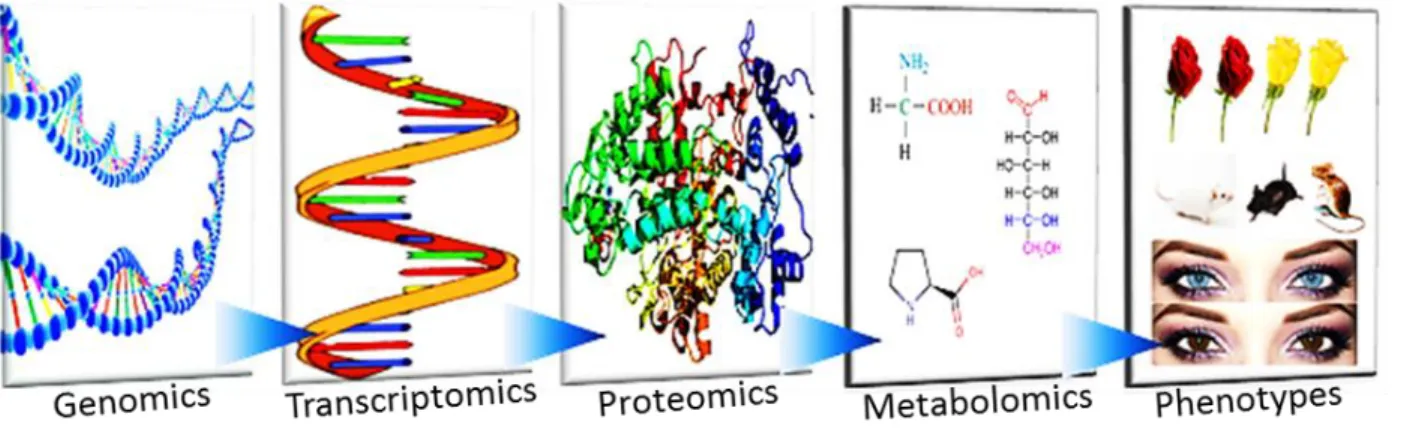
(17) Introduction 2014 Metabolomics background: The term “metabolome” has been defined as the complete set of metabolites synthesized by an organism, reflecting the closest non-structural phenotype. To understand the mechanism of a biological system and their potential dysregulation in any diseased conditions, the study of metabolism plays a dynamic role. However, “metabolome” comprises a huge number of compounds (are mainly of low molecular mass, usually < 1,500 Da) but differ in many other chemical and physical properties (1, 2). The field metabolomics, which is defined as the study of metabolites, is growing exponentially in the post genomic era with advances in analytical instrumentation, chemometric tools and online database system. Consequently, metabolomics has gained considerable attention in biomedical research as practiced independently to study metabolite interaction or in combination with genomics, transcriptomics, and/or proteomics data to study biological systems in a holistic manner (e.g. systems biology) (3). In the evolution of “omics” cascade, metabolomics helps to answer the questions what has happened and what is going to happen in any altered biological metabolism (Figure 1). Metabolomics involved in monitoring the ultimate product of gene expression which is the metabolite. Metabolites are organic compounds that may not be directly encoded in the genome, and their biosynthesis often involves a diversity of enzymes. Furthermore, metabolites are stoichiometrically interrelated, which results in more complex metabolic networks that do not exist in the case of transcripts or proteins. So the application of metabolomics approach may offer the valuable and functional information that is crucial to system biology (3-5).. Figure 1: The OMICS Cascade. The metabolomics is complementary to transcriptomics and proteomics. Metabolome captures the functional or physiological state of a system and provides a link between genotypes and phenotypes.. Nicholson Group at The Imperial College of London and the Fiehn group at Max-Planck Institute of Molecular Plant Physiology, coined the terms “metabonomics” and "metabolomics" in 1999 and 2002,. 3.
(18) Introduction 2014 respectively (6, 7). However, now-a-days these two terms are being used indistinctly. Different strategies have emerged for tackling the enormous data generated from the metabolomics study. However, the exact definition of each strategy is still in debate. All the available approaches for metabolomics are presented in Table 1. In order to avoid any conflict in this dissertation, the available metabolomics strategies are divided in two major categories such as targeted and non-targeted.. Table 1: Available strategies for metabolomics analysis Term. Definition. Metabolomics. Complete quantitative and qualitative analysis and identification of all metabolites present in an organism or a biological system, under a given set of conditions (4).. Metabonomics. The quantitative detection of endogenous metabolites that are vigorously altered in a living system in response to pathophysiological stimuli or genetic modification (6).. Global metabolomics. The identification and quantification of a set of metabolites related. profiling. through their metabolic pathway(s) or similarities in their chemistry (8).. Metabolomics fingerprinting. Unbiased global analysis of all possible metabolites present in crude samples or simple cellular extracts using high-throughput techniques providing all the metabolite information present in the given set of sample (9, 10).. Metabolomics footprinting. The analysis of metabolites secreted from the intracellular complement of an organism (or biological system) into its extracellular medium or matrix. This approach is commonly used in microbial metabolomics (11, 12).. Target analysis. Quantitative analysis of one or few pre-defined metabolites related to a specific metabolic/diseased condition (13).. Targeted approach focuses on some or a group of metabolites to be analyzed. Usually these metabolites are related to specific pathways, or often directly related to genetic perturbations. Targeted approach allows us to quantify the concentration of the metabolite under study, employing robust and sensitive methods. This strategy is very useful in hypothesis testing and addressing well defined biological questions. One of the main limitations of this approach is, it does not provide the global metabolite information of a system. Thus information related to other metabolites can be lost with this approach.. 4.
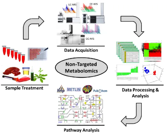
(19) Introduction 2014 Non-targeted approach attempts to detect as many metabolites as possible in a given set of sample. This approach yields a global picture of the metabolic state of any system of interest. The non-targeted approach measures the signal of all possible chemical entity named as “feature”.. The significant. increasing or decreasing pattern of these features, in the comparison of two or more groups, is the ultimate goal of non-targeted metabolomics. These differentiating features then can be further interrogated and structurally identified, leading to new ideas regarding the alteration in biochemical processes responsible for the observed differences. Hence, this class of experiment is often referred as “hypothesis generating”. Both approaches can participate in the quest for novel biomarkers. The level of glucose and cholesterol in blood and creatinine level in serum, describes the value of even a single biomarker. Metabolomics is not only limited to discover biomarkers, however the whole profiling could aid in understanding the mechanism of the specific disease states. For example, evaluating related biochemical pathways in response to drug treatment will give a more complete description of feedback mechanisms than a single biomarker can deliver.. Non-targeted metabolomics workflow: “Metabolome” of any biological system can be very diverse with metabolites of complex and divergent physicochemical properties. The fundamental of non-targeted approach is to cover a broad metabolic picture through single or in combination of complementary analytical systems along with various extraction protocols. Non-targeted metabolomics approach comprises of four principal stages: (i) sample treatment, (ii) data acquisition employing single or multiplatform techniques, (iii) data processing and statistical analysis using chemometrics followed by compound identification and (iv) chemical marker identification through pathway interpretation. All these four steps are remarkably interrelated and as depicted in Figure 2, each of the steps may contain several sub-steps. Unlike any other analytical approach, one of the critical steps in non-targeted metabolomics is the transformation of any kind sample to an appropriate solution suitable for instrumental analysis which will contribute in covering an array of metabolite classes. Sample preparation stage constitutes a series of experimental stages, choosing a sample specific to the biological question to be answered, sample collection, applying quenching steps and trying different metabolite extraction protocols. Throughout this step, care must be taken to avoid the introduction of any form of unwanted variability that would significantly affect the outcome of the analysis. No single solvent or extraction procedure can extract all metabolite classes from a given sample. Thus the involvement of two/more extraction protocols and solvents could enhance the range (14-17).. 5.
(20) Introduction 2014 Data acquisition (instrumental analysis) follows the sample preparation step and requires advanced analytical techniques as the ultra-complexity of samples for metabolomics analysis makes it impossible to technologically separate, quantify and identify every metabolite within a biological sample. Several analytical platforms are being used in metabolomics either alone or in combination of two or three (1820). Prior to any sample analysis, analytical method development and its validation comes in forehand.. Figure 2: General workflow involved in non-targeted fingerprinting approach, from sample treatment to pathway analysis.. Sample analysis generates huge data sets in terms of molecular feature. Therefore informatics and statistics are essential tools for processing metabolomics data sets (21, 22). Numerous software packages have been developed to aid with data processing (data pre-processing, data pre-treatment and statistical modelling of the primary data) in an automated manner. Data pre-processing and pre-treatment includes centring, scaling, transformation, filtering and above all aid in ‘cleaning’ the data to focus on biologically relevant information (23). The cleaned data are then subjected to statistical analysis which provides model-based descriptions of the biological variation in the system under study. These statistical models specifically single out representative metabolites of interest which can further be chemically or structurally identified through online based database information and finally in a definitive manner by MS/MS or injecting authentic standards.. 6.
(21) Introduction 2014 The final step in non-targeted approach is relating these identified metabolites with their respective pathways. Though not 100% complete but online data bases provides metabolite information along with their related pathway information. All the metabolites are very much related in the biological system however in a complex manner. So the alteration in one of them could alter the entire network and thus the reason behind any biological dysregulation can be uncovered.. Selection of sample type: Non-targeted approach has been applied on several sample types from non-invasive to highly invasive, such as urine, blood, plasma/serum, saliva, cerebrospinal fluid, cell lines, exhaled breath condensate, plants, or various tissues (20, 24-33). The biological question being asked influences the chosen type of sample. Each of the matrices has their own strengths and weaknesses. Most widely used sample types are, urine and plasma/serum due to their ease of collection and minimally invasive nature (17, 34). These sample type has been used extensively in metabolomics study so their protein, lipid and metabolite composition are very much well known. Urine is being the most attractive due to its low levels of proteins and cellular material and normalization of total metabolite content using creatinine is well described (17, 35, 36). Urine and plasma/serum are integrated bio-fluids. This offers the simultaneous advantage of reflecting both localized and systemic changes; however, it can be difficult to identify the origins of the observed metabolic changes. Plasma/serum also possesses a number of analytical challenges, especially higher protein concentrations mean samples require deproteinization prior to analysis (17). Although many metabolites are found in bio-fluids but not all affects or changes in the same direction at tissue level or vice versa which has been established by several studies (32, 37). Metabolic changes in specific disease state are first seen in at the tissue site. Moreover, pairwise comparison of diseased and control tissue regions could provide strong markers. Hence, tissue metabolomics can take a big part in non-targeted metabolomics research (20, 38-40). Sample treatment as well pose a challenging part in non-targeted metabolomics. An ideal sample-pre-treatment protocol for non-targeted approach should be: (i) non-selective, (ii) simple and fast with the minimal number of steps, (iii) reproducible, and (iv) incorporate a metabolism-quenching step, if required. Overall, the goal of the sample-preparation procedure is to transform the sample reproducibly into a format that is compatible with the given analytical platform while maintaining the original metabolite composition of the sample as unique as possible. The characteristics and applications of the mostly used biological samples are as follows-. Urine In mammals, the kidneys extract the soluble waste from the body system and the excess of water, named as urine. Apart from waste and water, urine contains a lot of other low molecular weight compounds in. 7.
(22) Introduction 2014 high concentration such as urea, inorganic salts, creatinine, ammonia, organic acids and various others. Since ancient period, urine has given considerable attention as a diagnostic bio-fluid. Bioanalysis of urine are being performed in clinical diagnosis routinely for glucose, bilirubin, ketone bodies, nitrates, leukocyte esterase, specific gravity, hemoglobin, and protein. Being relatively simple, typically protein free and easily accessible bio-fluid, urine has been applied to various non-targeted metabolomics approach using one or multiplatform techniques such as nuclear magnetic resonance spectroscopy (NMR), liquid chromatography-mass spectrometry (LC-MS), gas chromatography-mass spectrometry (GC-MS), capillary electrophoresis-mass spectrometry (CE-MS) and so on (24, 41-44). Several non-targeted analysis also used to study a variety of renal conditions, such as bladder, ovarian and kidney diseases (45-48). In non-kidney related diseases urinary metabolomics has also been applied (49). Recently a mostly complete urine metabolome has been explained by Bouatra S. et. al. using quantitative multiplatform metabolomics (50). Almost 3079 detectable metabolites in human urine (both exogenous and endogenous) were proposed. Relative to other bio-fluids, such as cerebrospinal fluid or saliva, urine contains significantly more compounds with significant chemical diversity (51, 52). Moreover, the human urine metabolome is a subset of the human serum metabolome, both in terms of composition and chemical diversity (26). The use of metabolomics through examination of patient urine is an ideal means to study disease prognosis and diagnosis. Urine metabolomics has potential utility in non-targeted metabolomics as well for biomarker discovery (47). Since urine has potential importance in non-targeted metabolomics, so urine sample preparation is also being studying (42). Once urinary biomarkers are discovered and validated, they could conceivably be used for prognosis as well as to predict response to targeted therapies as obtaining urine is always more feasible than gaining access to tumor tissue.. Blood (plasma/serum) Blood serves as a connecting bridge in all body systems through tissue and cells, as all the molecules that are being secreted, excreted or discarded by different tissues and cells are transported to blood in response to different physiological needs or stresses. Blood contains a wide variety of chemically diverse low molecular weight compounds of the metabolome. Most of today’s diagnosis are based on blood (plasma/serum) analysis because it is evident fact that tissue lesions, organ dysfunctions and pathological states can alter both the chemical and protein composition of blood plasma/ serum (53). Though plasma and serum are derived from blood, however compound composition is of little different (54). Plasma is the liquefied component of unclotted blood usually obtained with EDTA treatment and contains clotting agents and other components (55). On the other hand, serum is the fluid component of clotted blood (contains proteins, cells and other components, except clotting factors and fibrinogen). Both plasma and serum are aqueous solutions containing a variety of substances including proteins and peptides (such as albumins, globulins, lipoproteins, enzymes and hormones), nutrients (such as. 8.
(23) Introduction 2014 carbohydrates, lipids and amino acids), electrolytes, organic wastes and variety of other small organic molecules suspended or dissolved in them. Blood plays a critical role in transporting and exchanging nutrients, gases, hormones, regulates the pH and ion composition, excerts defensive mechanism against toxins/pathogens and most of all stabilize body temperature (55). Being an important and easily accessible biological fluid, plasma/serum has been applied to various non-targeted metabolomics approach using one or multiplatform techniques such as NMR, LC-MS, GC-MS, CE-MS and so on (18, 19, 56-58). Several non-targeted analyses also used to study a variety of diseases. A database of the human serum metabolome (a complete set of 4229 compounds) has already been introduced (26). Accordingly efforts have been made to develop proper sample treatment in order to get reliable and fruitful results (17). Unlike urine, serum/plasma metabolomics has also potential utility in non-targeted metabolomics for biomarker discovery. The identified biomarkers can be used for prognosis as well as to predict response to targeted therapies as obtaining blood is minimally invasive and more feasible than gaining access to tumor tissue.. Tissue Tissue metabolomics has many advantages over bio-fluids. The metabolic modifications and the upstream regulations are first seen in tissue. Moreover, the pairwise comparison of tissue taken from diseased region and non-diseased region could reflect the interactions despite of any individual differences. Global determination of metabolite concentrations in tissues provide novel anatomical aspects of pathological conditions that cannot be obtained from target-specific fluid measurements. Providing more relevant information than systemic bio-fluids, tissue metabolomics has a great importance in biomedical research. So far, many studies have already shown the applicability on a variety of human/animal tissues for metabolomics including liver, kidney, lung, brain and spleen from both rodents and other models (20, 32, 59-61). One of the main challenges lies in tissue metabolomics is its collection and treatment to make it appropriate for instrumental analysis. An extensive review of the up-to date published researches based on the challenges related to MS based non-targeted animal/human tissue metabolomics is one of the focuses of this present study. The accepted review manuscript, describing the non-targeted tissue metabolomics has been attached with Chapter-3.. Analytical platforms: The expanding field of non-targeted metabolomics is growing exponentially in recent years by the advances in analytical methods of high-resolution NMR, MS coupled to separation techniques such as LC, GC, CE, Fourier transform-infrared spectroscopy or direct infusion (62-67). Of these, NMR and MS coupled to LC and GC are the most extensively applied.. 9.
(24) Introduction 2014 NMR is a rapid, non-destructive technique, requires minimum sample preparation and thus lessens the chance of sample loss or the introduction of variability during sample preparation (68). Prior NMR analysis, sample derivatization or analyte separation is not required. Hence NMR analysis is independent of analyte polarity. In NMR, the signal intensity for all compounds is dependent on the molar concentrations and reproducibility is high. However the sensitivity is relatively low (micromolar range) and required sample amount is of very high (69). Although NMR can yield detailed information on the quantities and identities of metabolites present in a biological sample, the chemical elucidation of NMRdetected compounds can be highly complex as a result of overlapping signals. Moreover, NMR analysis is sensitive to the chemical environment (pH, ionic strength, temperature, etc.) of the sample and the differential sensitivity of metabolites to the chemical environment, which hampers the quality of NMR analyses for complex samples (68). However, recent development of cryogenic probes, higher strength superconducting magnets, miniaturized radio frequency coils and multidimensional techniques are being developed in order to improve the sensitivity and resolution of NMR (68-70). Compared to NMR, MS methods are more prevalent in metabolomics for measuring metabolites in complex biological samples, because they allow reliable metabolite identification with high sensitivity (typically pg level). The developments in LC, GC and CE have significantly broadened the applicability of MS-based metabolomics. However, the comparatively long sample preparation steps and analytical separation of the metabolites before it is directed into the mass analyzer can make the analysis time consuming. Fourier transform coupled to MS provides extremely high resolution and mass accuracy (71). However, this instrument is relatively very expensive which has limited its use to date. Current challenges involved in the development of MS-based metabolomics include the development of more robust methods for chromatographic separation and reduction of matrix effects, including ion suppression, which can cause widely varying signal intensities. Single analytical approach is not sufficient to cover the entire metabolome. Thus multiplatform applications are necessary to extend the coverage. To obtain this goal, MS detection coupled to separation techniques are becoming the most relevant tools along with NMR. Multiplatform non-targeted metabolomics has been applied on different biological specimens for non-targeted analysis. The importance of multiplatform approach has been well demonstrated by several studies (19, 20, 26). The main objective of non-targeted metabolomics study is to obtain as much as analyte signals as possible. In order to attain this goal, advanced analytical tools with characteristics of high degree of sensitivity, selectivity, throughput ability and comprehensiveness are desired. The features of most widely used analytical techniques are presented in Table 2. One particular technique is not sufficient for the analysis of all compounds, thus a selection of two from the table would give more coverage than a single one. Otherwise any one form of separation will inherently introduce a bias towards the analytes being detected.. 10.
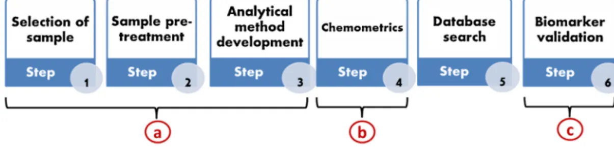
(25) Introduction 2014 Table 2: Most widely used analytical techniques used in non-targeted metabolomics analysis.* Technique. . Sensitivity. Throughput. Comprehensiveness. NMR. Low. Low–high. Low–high. LC-MS. High. High. High. GC-MS. High. High. High. CE-MS. Medium. Medium. High. Information obtained from (72). Method development and validation The development of a suitable method is the pre-requisite for any analytical platform and non-targeted metabolomics is not out of this benchmark. Prior analyzing, in any instrumental platform, a valid and robust method needs to be developed and validated. Analytical procedures play a critical role in acquiring corresponding analyte signal for further data analysis in non-targeted approach. Validation of analytical method should demonstrate that they are suitable for their intended use. Validation should be founded on method development performed beforehand that suggests the suitability and robustness of the method. The steps of method development and method validation depend upon the type of method being developed. A proper method should have the following characteristics● Specificity: ability to measure desired analyte in a complex mixture. ● Accuracy: agreement between measured and real value. ● Linearity: proportionality of measured value to concentration. ● Precision: agreement between series of measurements. ● Range: concentration interval where method is precise, accurate, and linear. ● Detection limit: lowest amount of analyte that can be detected. ● Quantitation limit: lowest amount of analyte that can be measured. ● Robustness: reproducibility under normal but variable laboratory conditions.. Method for non-targeted metabolomics are being developed mainly taking into consideration the most important criteria, which is broader metabolite coverage. Although for targeted metabolomics there are several guidelines available, for non-targeted metabolomics analysis scientists are using alternative approaches to validate the method following one/two from the using-. 11.
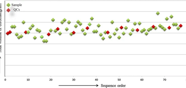
(26) Introduction 2014 a. Validation criteria used in targeted metabolomics in terms of linearity, accuracy, precision, limit of detection and limit of quantitation. b. One of the main limitations in high-throughput analysis is the drift in both chromatographic and MS performance. To control and monitor this type of drawbacks use of quality control samples (QCs) has been introduced in non-targeted analysis. QCs are a pool of sample made from the biological test samples to be studied, or a representative bulk control sample which should be assessed against predefined criteria to enable acceptance or rejection of the analytical run. c.. Use of external standards, are very common in pharmaceutical industry in order to check the reliability of the methodology.. d. An alternative validation strategy is the statistical model validation. The acquired data from instrumental analysis generate huge data sets which are usually expressed in class models applying chemometrics tools. Two approaches are available for validation: test-set and resampling method. Another widely used method validation strategy is the prediction of QCs in the chemometrics models. The clustered prediction of QCs describes that the separations between the groups are not due to the instrumental variations but due to the sample itself. One of the primary objectives of this dissertation is to develop and validate analytical methodologies for serum and lung tissue which has been described in chapter 1 and 3 respectively. The available strategies involved in MS based non-targeted metabolomics till date has been discoursed. A review has been accomplished on the available validation strategies for non-targeted metabolomics methods and included in Chapter-1 pointing the objective of this dissertation.. Data analysis High-throughput non-targeted metabolomics analysis generates extremely large volume of data. Hence automated software is needed in order to understand and handle these large data sets. The automated software helps to identify peaks from raw data, align them among different samples and identify each metabolite. Afterwards, informatics and statistics are essential tools for reprocessing metabolomics data sets (55). Metabolomics data analysis comprises of data pre-processing, data pre-treatment and statistical analysis (modeling) of the primary data. The main target of metabolomics study is to obtain variations in the data sets due to their biological means. However non-biological, technical or instrumental variations are as well very prone in any analytical platforms. In order to clean these unexpected variations, data mining (pre-processing and pre-treatment) plays an important role. Most frequently applied tools in metabolomics data mining are: alignment, normalization, centering, scaling, filtering and mathematical transformation of the raw data (23). Alignment helps in assigning the obtained peaks, which appears in all samples at the same mass (m/z) and time of the same feature, finally listing all possible components as represented by spectral data. Peak alignment is necessary to perform in order to. 12.
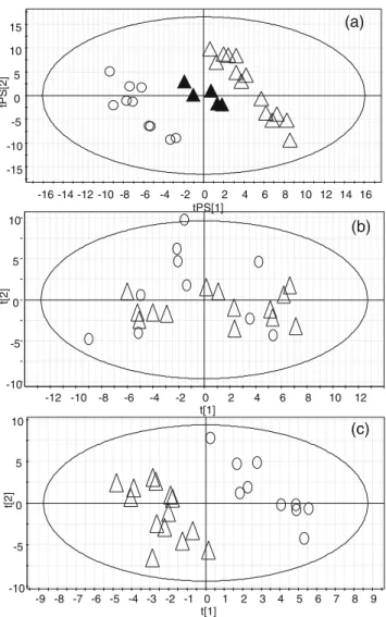
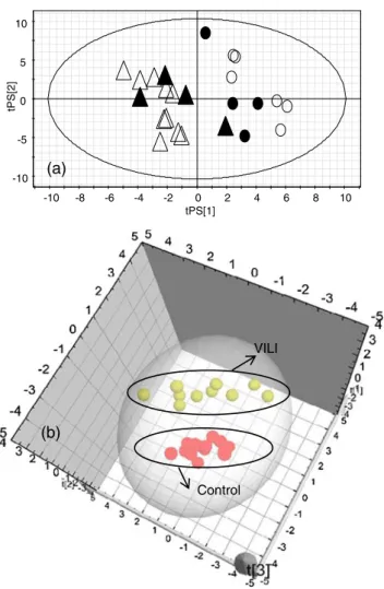
(27) Introduction 2014 compare data across samples. Data normalization minimizes the differences in the detection between samples arising from variation in the concentration. Several data normalization strategies are available such as the use of internal standards, MS total signal, MS total useful signal, sample volume and median fold change normalization (35, 73). The aim of logarithmic data transformation is to reduce the influence of potential outliers and to transform the data matrix into a more Gaussian-type distribution (74). Moreover, transformation increases the weight of low-intensity metabolites and compresses the upper end of the measurement scale (75). Scaling of the data is desirable to adjust the importance assigned to the elements of the data in fitting the model. By scaling, the weight of each variable is adjusted with a scaling factor estimated by either a dispersion criterion or a size measure. Unit variance scaling and Parscaling are two of the most prevailing scaling types used for non-targeted metabolomics studies (76, 77). Generally centring is applied in combination with scaling. Mean centring converts concentrations to fluctuations and variables are centred around zero (23). The final step in data mining is the data filtration. Applying data filtering, potential biomarkers can be selected by retaining only masses that appear in all samples within the groups. The frequently used filtering allows features to be filtered out based on their presence/absence. It is a crucial point for studies carried out on experimental groups under various conditions such as treatment or diet, because metabolites coming from the treatment, which contribute to discriminate the groups but are not interesting for the study, can be eliminated by retaining only those compounds present in all samples in all groups. Depending on the analytical technique, different software packages are available to perform data mining in an automated manner (23). The cleaned data are then subjected to statistical analysis with the help of chemometric tools, which provides model-based descriptions of the biological variation in the system under study. The chemometric tool uses mathematics, statistics and formal logic (a) to design or select optimal experimental procedures, (b) to provide maximum relevant chemical information by analyzing chemical data and (c) to obtain knowledge about chemical systems (78). Two different statistical analyses are being used in non-targeted metabolomics data analysis: univariate and multivariate data analysis (MVDA). The univariate data analysis assumes that the response of variables is influenced by only one factor. In a complex disease, different biologic pathways simultaneously governed by multiple variables are involved and compromised. The traditional statistical analysis tends to transform all problems into univariate problems, even those that are inherently multivariate. For this reason, MVDA is a proper statistical tool for the interpretation of data coming from a metabolomics study. It summarizes data tables with many variables and few observations and works reducing the number of variables and classifying the data into groups (55, 79). MVDA provides statistical models specifically single out representatives of metabolites of interest (annotated peaks), which can further be chemically or structurally identified in a definitive manner. The first step of MVDA is the creation of the X matrix, where the samples are included in rows, and all the variables (the compounds) in columns. Subsequently, it is always necessary a data pre-treatment (scaling and transformation) for improving relevant information. After this, data are analyzed through the. 13.
(28) Introduction 2014 unsupervised principal component analysis (PCA) and the supervised partial least squares regressiondiscriminant analysis (PLS-DA) and orthogonal partial least squares-discriminant analysis (OPLS -DA). To avoid the risk of over fitting, as the results found after MVDA are sensitive to chance-correlations, statistical model needs to be validated. Validation tools, such as (a) permutation test and (b) crossvalidation, are most widely used to provide an objective assessment of the performance and stability of a model. (a) Permutation Test: A permutation test is used to assess the significance of a classification. The class assignment can be permuted several times and for each permutation, a model between the data and the permuted class-assignment can be built. The discrimination between classes of the model based on the permutated class-assignment is compared to the discrimination of the model based on the original classification (55). (b) Cross-Validation: In cross-validation the first step is to divide the samples into several groups. These groups are then sequentially withheld while the remaining samples are used to build cross validated models. The withheld samples for each model are predicted and the predictive ability is measured and this process repeated until all the samples are withheld and predicted. The data statistics provides statistically significant masses for a corresponding compound which is/are involved in the differentiation of study group. These significant masses are then finally search against online data bases, which provides information about the mass with their related corresponding compound(s). Several online databases (free and commercial) are available which provides identification of the significant masses, chemically or structurally in a definitive manner. Two types of databases are available: (i) databases that contain metabolic pathways based on information from the literature which has been integrated and curated manually and (ii) databases that contain raw or processed data from analytical system. Examples of some online databases are: Kyoto Encyclopedia of Genes and Genomics (http://www.genome.jp/kegg/), gatersleben.de),. Reactome. MetaCyc. (http://MetaCyc.org),. (http://www.reactome.org),. The. MetaCrop Human. (http://metacrop.ipk-. Metabolome. Database. (http://www.hmdb.ca), Small Molecular Pathway Database (http://www.smpdb.ca), Golm Metablome Database (http://csbdb.mpimp-golm.mpg.de/gmd.html), METLIN (http://metlin.scripps.edu), MassBank (www.massbank.jp), Metaboanalyst 2.0 http://www.metaboanalyst.ca and Mouse Multiple Tissue Metabolome Database (http://mmmdb.iab.keio.ac.jp) (61, 80-89). There are also several commercial pathway databases available which contain integrated knowledge and well-studied pathways, e.g. Cell Signal. pathways. (www.cellsignal.com),. (http://www.sigmaaldrich.com/lifescience/. Sigma–. cell-biology/learning-center.html),. (http://www.ambion.com), and ProteinLounge (http://www.proteinlounge.com/).. 14. Aldrich Ambion. pathways pathways.
(29) Introduction 2014 Application to pulmonary diseases Pulmonary diseases are prevalent and one of the major causes of global morbidity and mortality (90-92). The principle pulmonary diseases are acute respiratory disease syndrome/acute lung injury (ARDS/ALI), chronic obstructive pulmonary disease, asthma, pulmonary arterial hyper tension, pulmonary embolism, cystic fibrosis, lung cancer and particularly infectious disease including tuberculosis. The exact mechanism of disease onset are still unknown, however environmental exposures have been found to be directly involved in the development of lung diseases (93, 94). At present several quantitative and semiquantitative tests are involved in pulmonary disease diagnosis such as radiological examinations, spirometry, sputum analysis and, more recently, exhaled nitric oxide, carbon monoxide testing etc. (9598). A range of markers are also available to improve clinical diagnosis but unfortunately they have very low specificity and they are often incapable of identifying and diagnosing specific disease sub-phenotypes (34, 99). However, MS-based techniques have been used to measure leukotriene B4, a marker of inflammation and 8-isoprostane, a marker of oxidative stress (100, 101). A number of challenges are still there which need to be addressed to improve diagnosis, understand and treatment of lung diseases. The promising technique metabolomics have successfully applied and classified several respiratory diseases, including asthma, chronic obstructive pulmonary disease, ARDS, ALI, pulmonary embolism, lung cancer and cystic fibrosis (102-108). With the application of metabolomics it was possible to tentatively identify distinct areas of metabolism and the pathways that characterize the individual disease metabolic phenotypes,. proving. the. fact. that. metabolomics. approaches. can. play. a. central. role. in. diagnosing/characterizing pulmonary diseases.. Acute lung injury ALI/ARDS, is characterized by the onset of clinically significant hypoxemia and diffuse bilateral pulmonary infiltrations. ALI/ARDS is an important cause of pulmonary and non-pulmonary morbidity in patients who survive hospitalization (109). Due to the inherent heterogeneity of the disease along with the consistent lack of correlation between biochemical markers, pathophysiologic variables and clinical outcomes, the search for ALI biomarkers are being hampered. Figure 3 explains the pathogenesis of lung injury obtained from Ware and Matthay (109). Usually normal lung epithelium is composed on flat and cuboidal type of cells, which comprises 90 % and 10% respectively of the alveolar surface area. Compared to cuboidal, flat type cells are more prone to injury. Upon injury the normal epithelial fluids transport disrupted and contribute to epithelial flooding (110, 111). Following those neutrophils starts adhering to the injured capillary endothelium and marginating through the interstitium into the air space. Air space contains alveolar macrophage which secrets cytokines, interleukin-1, 6, 8, and 10, and tumor necrosis factor α, which act locally to stimulate chemotaxis and activate neutrophils. Neutrophils involves in releasing oxidants, proteases, leukotrienes and platelet-activating factor (109). With the improved. 15.
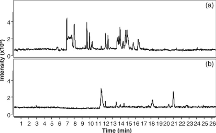
(30) Introduction 2014 understanding of ALI pathophysiology, interleukin-1β and tumor necrosis factor-α have been evaluated to check their diagnostic or prognostic capability as a biomarker. These inflammatory mediators can be detected in the distal airspaces of the lung in ALI/ARDS patients (112-114). Most recently investigators have documented the influence of the coagulation system in ALI/ARDS. ALI is characteristically heterogeneous, depending on the origin it can be several types such as endothelial injury, epithelial Injury, neutrophil-dependent lung injury, ventilator induced lung injury (VILI) and sepsis induced lung injury (SI-ALI) (115-119). Following the aim of this dissertation, a brief discussion about VILI and SI-ALI are as follows.. Figure 3. The normal alveolus (left-hand side) and the injured alveolus in the acute phase of acute lung injury and the acute respiratory distress syndrome (right-hand side). Obtained from Ware and Matthay (109).. (i) Ventilator-Induced Lung Injury (VILI): In clinics, from several decades mechanical ventilation has been used as the important part of basic life support. However several potential complication has been identified with the use of mechanical ventilation (120).. There are evidences that mechanical. ventilation at high volumes and pressures can injure the lung, causing increased permeability pulmonary edema in both uninjured and injured lung (121, 122). Thus, VILI has received much attention in both experimental and clinical field. This harmful effect of ventilation has been hypothesized by the fact that. 16.
(31) Introduction 2014 the capillary stress failed due to alveolar over-inflation. More recently, cyclic opening and closing of atelectatic alveoli during mechanical ventilation have been shown to involve in developing lung injury independently of alveolar over-inflation. Alveolar over-inflation together with the repeated collapse and reopening of alveoli can initiate a cascade of pro-inflammatory cytokines (123). In intensive care units (ICU) patients with ALI/ARDS might require ventilation at traditional tidal volumes. The use of ventilation may affect the uninjured alveoli along with the injured one, promoting further lung injury and finally contribute to multi-organ failure (123). Several other techniques has been applied in order to evaluate the exact molecular mechanism of ALI, however there are no evidences of non-targeted metabolomics application on VILI models of ALI. One of the aims of this dissertation is to apply non-targeted metabolomics methodologies on serum samples of VILI and healthy rat models applying CE-MS and LCMS. The application and findings of the applied methodologies are described in Chapter-1 and Chapter-2.. (ii) Sepsis-Induced Lung Injury (SI-ALI): Sepsis is a potentially deadly medical condition associated with an extremely complex chain of systematic inflammatory and anti-inflammatory processes due to the presence of a known or suspected severe infection, trauma, blood loss, and perforated neoplasms (124). The characteristic effects of sepsis are extreme inflammatory changes, profound hypotension, hypoxemia, and lethal tissue damage, which ultimately proceed to organ dysfunction (125, 126). Despite extensive research, sepsis remains one of the causes for death and the mortality rate remains higher (approximately 30%) in ICU (127). Generally sepsis affected patients are treated in ICU and to support their lung function they may require mechanical ventilation. There are evidences that approximately 74% of sepsis affected patients admitted in ICU consider sepsis as the primary risk factor for ALI (119). The accurate progression of ALI in patients whether induced by mechanical ventilation or sepsis is still not known. One of the reasons is the cause of ALI which is extremely heterogeneous, leading to difficulties in phenotyping patients as well as monitoring and treatment. Currently there are several cell-specific biomarkers available such as cytokines and their receptors, surfactant protein D, coagulation factors such as protein C and plasminogen activators (128). Nevertheless, these biomarkers mainly help to understand the pathogenesis of the disease but not the progression. On the other hand the role of mechanical ventilation during sepsis is not known: whether the lung injury is the effect of ventilation or sepsis by itself. The molecular mechanism behind SI-ALI is still under investigation. Several metabolomics study has been performed on sepsis and ALI induced by sepsis, separately (129, 130). The molecular changes could aid in finding the involvement of ventilation or sepsis in SI-ALI. Moreover, the proved fact is that tissue provides more site specific information. Considering all these, another aim of this dissertation is the application multiplatform tissue metabolomics on the lung tissue of sepsis, VILI and SI-ALI, to find out exact mechanisms. Chapter-4 explains the results of the applied multiplatform methodology on lung tissue of rat models of sepsis, VILI and SI-ALI.. 17.
(32) Introduction 2014 Animal models for lung injury For more than a hundred years, virtually every medical breakthrough in human and animal health has been the direct result of research using animals. The use of animals in biomedical research is essential to the development of new and more effective methods for diagnosing and treating diseases that affect both humans and animals. Animals are being used in biomedical research to learn more about health problems and to assure the safety of new medical treatments. Medical researchers need to understand health problems before they can develop the ways to treat them. Some diseases and health problems involve processes that can only be studied in living organisms. Animals are the good research subjects for a variety of reasons. Animals are biologically similar to humans. In fact, chimpanzees share more than 99% of DNA and mice share more than 98% DNA with humans. Therefore, animals are susceptible to many of the same health problems as humans. Animals have a shorter life cycle than humans and as a result, they can be studied throughout their whole life span or across several generations. In addition, scientists can easily control the environment around animals (diet, temperature, lighting), which would be difficult to do with humans. A variety of animals provide very useful models for the study of diseases afflicting both humans and animals, including rats, mice, birds, rabbits, guinea pigs, sheep, fish, frogs, pigs, birds, dogs, cats, primates, among others. Approximately 95% of the animal model based research includes the use of rats and mice. Ideally, an “animal model of ALI” should be a model in which this pathologic triad is reproduced. Many different modeling strategies have been developed in an attempt to reproduce the features of human ALI in animals. From a practical standpoint, there are four general types of model systems: 1. Direct induction of lung injury using a noxious stimulus. These include the intratracheal or intranasal administration of bacteria or bacterial products such as lipopolysaccharides, the administration of an acid such as hydrochloric acid or of gastric particulates to reproduce aspiration, the administration of high inspired fractions of oxygen, depletion of surfactant by serial lavage with 0.9% sodium chloride, the induction of ischemia/reperfusion by clamping the hilum (131-133). 2. Another direct way of inducing lung injury is the exposure to mechanical stretch using mechanical ventilation with high tidal volumes, which is called VILI (18). 3. There are some indirect ways of inducing lung injury in animal model. This category includes models based on reproducing sepsis, such as cecal ligation and puncture (CLP), the administration of intravenous bacteria or lipopolysaccharide and mesenteric ischemia/reperfusion. This category also includes the oleic acid model, with attempts to reproduce the release of oleic acid from bone marrow in patients with multiple bone fractures (134).. 18.
(33) Introduction 2014 4. Combination models. To better reproduce human ALI/ARDS, different injury strategies can be combined; most commonly these include saline lavage followed by mechanical ventilation, or CLP followed by hemorrhage. No single animal model reproduces all the histopathological elements of ALI satisfactorily. Thus, when choosing an animal model of ALI, it is important to consider the key features of ALI that will be tested by the hypothesis of the study, and then choose a model that reproduces those features. Concentrating on the hypothesis of this dissertation, sepsis, VILI and SI-ALI rat models were chosen as a model for ALI to apply the developed non-targeted metabolomics approaches.. The originality of this dissertation This dissertation contains several originalities: (i). An extensive review on methods for human/animal based tissue metabolomics.. (ii). A critical review on the available validation strategies used for MS-based non-targeted metabolomics.. (iii). A CE-MS based methodology for non-targeted serum analysis including sample treatment, and validation. Following method development, a complete fingerprinting of rat serum was performed for the first time using CE-MS.. (iv). A multiplatform method (LC-MS, GC-MS and CE-MS) for lung tissue fingerprinting was designed using a minimum amount of sample. It was developed and validated along with sample treatment. The multiplatform method was then used for a complete mouse lung fingerprinting.. (v). All the developed and validated methods for serum and lung were applied on the rat models of lung injury (VILI+SI-ALI) for the first time.. (vi). Involvement of collagen remodeling, oxidative stress, energy metabolism, carnitine biosynthesis and cholinoenergic pathway in the pathogenesis of ALI.. (vii). Association of ceramide and sphingomyelin pathways in the pathogenesis of ALI.. (viii). The effect of ventilation was more prominent compared to sepsis in ALI.. (ix). Many of the serum and lung metabolites changes in opposite directions for ALI.. 19.
(34) Introduction 2014 References: 1.. Lindon JC, Nicholson JK, Holmes, E. The handbook of metabonomics and metabolomics.. Elsevier; 2006. 2.. Oliver SG, Winson MK, Kell DB, Baganz F. Systematic functional analysis of the yeast genome.. Trends in biotechnology 1998;16:373-378. 3.. Schilmiller AL, Pichersky E, Last RL. Taming the hydra of specialized metabolism: How systems. biology and comparative approaches are revolutionizing plant biochemistry. Current opinion in plant biology 2012;15:338-344. 4.. Fiehn O. Combining genomics, metabolome analysis, and biochemical modelling to understand. metabolic networks. Comparative and functional genomics 2001;2:155-168. 5.. Schilling CH, Schuster S, Palsson BO, Heinrich R. Metabolic pathway analysis: Basic concepts. and scientific applications in the post-genomic era. Biotechnology progress 1999;15:296-303. 6.. Nicholson JK, Lindon JC, Holmes E. 'Metabonomics': Understanding the metabolic responses of. living systems to pathophysiological stimuli via multivariate statistical analysis of biological NMR spectroscopic data. Xenobiotica; the fate of foreign compounds in biological systems 1999;29:1181-1189. 7.. Fiehn O. Metabolomics--the link between genotypes and phenotypes. Plant molecular biology. 2002;48:155-171. 8.. Bottcher C, Roepenack-Lahaye EV, Willscher E, Scheel D, Clemens S. Evaluation of matrix. effects in metabolite profiling based on capillary liquid chromatography electrospray ionization quadrupole time-of-flight mass spectrometry. Analytical chemistry 2007;79:1507-1513. 9.. Allard E, Backstrom D, Danielsson R, Sjoberg PJ, Bergquist J. Comparing capillary. electrophoresis-mass spectrometry fingerprints of urine samples obtained after intake of coffee, tea, or water. Analytical chemistry 2008;80:8946-8955. 10.. Kapur B, Hershkop S, Koren G, Gaughan V. Urine fingerprinting: Detection of sample tampering. in an opiate dependency program. Therapeutic drug monitoring 1999;21:243-250. 11.. Kaderbhai NN, Broadhurst DI, Ellis DI, Goodacre R, Kell DB. Functional genomics via metabolic. footprinting: Monitoring metabolite secretion by escherichia coli tryptophan metabolism mutants using FTIR and direct injection electrospray mass spectrometry. Comparative and functional genomics 2003;4:376-391. 12.. Kell DB, Brown M, Davey HM, Dunn WB, Spasic I, Oliver SG. Metabolic footprinting and systems. biology: The medium is the message. Nature reviews Microbiology 2005;3:557-565.. 20.
(35) Introduction 2014 13.. Yoshida H, Yamazaki J, Ozawa S, Mizukoshi T, Miyano H. Advantage of LC-MS metabolomics. methodology targeting hydrophilic compounds in the studies of fermented food samples. Journal of agricultural and food chemistry 2009;57:1119-1126. 14.. Luque-Garcia JL, Neubert TA. Sample preparation for serum/plasma profiling and biomarker. identification by mass spectrometry. Journal of chromatography A 2007;1153:259-276. 15.. Vuckovic D. Current trends and challenges in sample preparation for global metabolomics using. liquid chromatography-mass spectrometry. Analytical and bioanalytical chemistry 2012;403:1523-1548. 16.. Beckonert O, Keun HC, Ebbels TM, Bundy J, Holmes E, Lindon JC, Nicholson JK. Metabolic. profiling, metabolomic and metabonomic procedures for NMR spectroscopy of urine, plasma, serum and tissue extracts. Nature protocols 2007;2:2692-2703. 17.. Dunn WB, Broadhurst D, Begley P, Zelena E, Francis-McIntyre S, Anderson N, Brown M,. Knowles JD, Halsall A, Haselden JN, Nicholls AW, Wilson ID, Kell DB, Goodacre R, Human Serum Metabolome C. Procedures for large-scale metabolic profiling of serum and plasma using gas chromatography and liquid chromatography coupled to mass spectrometry. Nature protocols 2011;6:1060-1083. 18.. Naz S, Garcia A, Rusak M, Barbas C. Method development and validation for rat serum. fingerprinting with CE-MS: Application to ventilator-induced-lung-injury study. Analytical and bioanalytical chemistry 2013;405:4849-4858. 19.. Izquierdo-Garcia JL, Naz S, Nin N, Rojas Y, Erazo M, Martinez-Caro L, Garcia A, de Paula M,. Fernandez-Segoviano P, Casals C, Esteban A, Ruiz-Cabello J, Barbas C, Lorente JA. A metabolomic approach to the pathogenesis of ventilator-induced lung injury. Anesthesiology 2014;120:694-702. 20.. Naz S, Garcia A, Barbas C. Multiplatform analytical methodology for metabolic fingerprinting of. lung tissue. Analytical chemistry 2013;85:10941-10948. 21.. Weljie AM, Newton J, Mercier P, Carlson E, Slupsky CM. Targeted profiling: Quantitative analysis. of 1H NMR metabolomics data. Analytical chemistry 2006;78:4430-4442. 22.. Liland KH, Almoy T, Mevik BH. Optimal choice of baseline correction for multivariate calibration of. spectra. Applied spectroscopy 2010;64:1007-1016. 23.. van den Berg RA, Hoefsloot HC, Westerhuis JA, Smilde AK, van der Werf MJ. Centering, scaling,. and transformations: Improving the biological information content of metabolomics data. BMC genomics 2006;7:142. 24.. Lindon JC, Keun HC, Ebbels TM, Pearce JM, Holmes E, Nicholson JK. The consortium for. metabonomic toxicology (comet): Aims, activities and achievements. Pharmacogenomics 2005;6:691699.. 21.
(36) Introduction 2014 25.. Pechlivanis A, Chatziioannou AC, Veskoukis AS, Kouretas D, Mougios V, Theodoridis GA. gc-ms. analysis of blood for the metabonomic investigation of the effects of physical exercise and allopurinol administration on rats. Journal of chromatography B, Analytical technologies in the biomedical and life sciences 2014. 26.. Psychogios N, Hau DD, Peng J, Guo AC, Mandal R, Bouatra S, Sinelnikov I, Krishnamurthy R,. Eisner R, Gautam B, Young N, Xia J, Knox C, Dong E, Huang P, Hollander Z, Pedersen TL, Smith SR, Bamforth F, Greiner R, McManus B, Newman JW, Goodfriend T, Wishart DS. The human serum metabolome. PloS one 2011;6:e16957. 27.. Cuevas-Cordoba B, Santiago-Garcia J. Saliva: A fluid of study for omics. Omics : a journal of. integrative biology 2014;18:87-97. 28.. O'Sullivan A, Willoughby RE, Mishchuk D, Alcarraz B, Cabezas-Sanchez C, Condori RE, David. D, Encarnacion R, Fatteh N, Fernandez J, Franka R, Hedderwick S, McCaughey C, Ondrush J, PaezMartinez A, Rupprecht C, Velasco-Villa A, Slupsky CM. Metabolomics of cerebrospinal fluid from humans treated for rabies. Journal of proteome research 2013;12:481-490. 29.. Canuto GA, Castilho-Martins EA, Tavares M, Lopez-Gonzalvez A, Rivas L, Barbas C. CE-ESI-. MS metabolic fingerprinting of leishmania resistance to antimony treatment. Electrophoresis 2012;33:1901-1910. 30.. Krug S, Kastenmuller G, Stuckler F, Rist MJ, Skurk T, Sailer M, Raffler J, Romisch-Margl W,. Adamski J, Prehn C, Frank T, Engel KH, Hofmann T, Luy B, Zimmermann R, Moritz F, Schmitt-Kopplin P, Krumsiek J, Kremer W, Huber F, Oeh U, Theis FJ, Szymczak W, Hauner H, Suhre K, Daniel H. The dynamic range of the human metabolome revealed by challenges. FASEB journal : official publication of the Federation of American Societies for Experimental Biology 2012;26:2607-2619. 31.. Farag MA, Gad HA, Heiss AG, Wessjohann LA. Metabolomics driven analysis of six nigella. species seeds via UPLC-QTOF-MS and GC-MS coupled to chemometrics. Food chemistry 2014;151:333-342. 32.. Ganti S, Taylor SL, Abu Aboud O, Yang J, Evans C, Osier MV, Alexander DC, Kim K, Weiss RH.. Kidney tumor biomarkers revealed by simultaneous multiple matrix metabolomics analysis. Cancer research 2012;72:3471-3479. 33.. Denkert C, Bucher E, Hilvo M, Salek R, Oresic M, Griffin J, Brockmoller S, Klauschen F, Loibl S,. Barupal DK, Budczies J, Iljin K, Nekljudova V, Fiehn O. Metabolomics of human breast cancer: New approaches for tumor typing and biomarker discovery. Genome medicine 2012;4:37. 34.. Atzei A, Atzori L, Moretti C, Barberini L, Noto A, Ottonello G, Pusceddu E, Fanos V.. Metabolomics in paediatric respiratory diseases and bronchiolitis. The journal of maternal-fetal & neonatal. 22.
(37) Introduction 2014 medicine : the official journal of the European Association of Perinatal Medicine, the Federation of Asia and Oceania Perinatal Societies, the International Society of Perinatal Obstet 2011;24 Suppl 2:59-62. 35.. Warrack BM, Hnatyshyn S, Ott KH, Reily MD, Sanders M, Zhang H, Drexler DM. Normalization. strategies for metabonomic analysis of urine samples. Journal of chromatography B, Analytical technologies in the biomedical and life sciences 2009;877:547-552. 36.. Wagner S, Scholz K, Sieber M, Kellert M, Voelkel W. Tools in metabonomics: An integrated. validation approach for LC-MS metabolic profiling of mercapturic acids in human urine. Analytical chemistry 2007;79:2918-2926. 37.. Yonezawa K, Nishiumii S, Kitamoto-Matsuda J, Fujita T, Morimoto K, Yamashita D, Saito M,. Otsuki N, Irino Y, Shinohara M, Yoshida M, Nibu K. Serum and tissue metabolomics of head and neck cancer. Cancer genomics & proteomics 2013;10:233-238. 38.. Budczies J, Brockmoller SF, Muller BM, Barupal DK, Richter-Ehrenstein C, Kleine-Tebbe A,. Griffin JL, Oresic M, Dietel M, Denkert C, Fiehn O. Comparative metabolomics of estrogen receptor positive and estrogen receptor negative breast cancer: Alterations in glutamine and beta-alanine metabolism. Journal of proteomics 2013;94:279-288. 39.. Song H, Wang L, Liu HL, Wu XB, Wang HS, Liu ZH, Li Y, Diao DC, Chen HL, Peng JS. Tissue. metabolomic fingerprinting reveals metabolic disorders associated with human gastric cancer morbidity. Oncology reports 2011;26:431-438. 40.. Zhang Y, Zhang A, Yan G, Cheng W, Sun H, Meng X, Liu L, Xie N, Wang X. High-throughput. metabolomic approach revealed the acupuncture exerting intervention effects by perturbed signatures and pathways. Molecular bioSystems 2014;10:65-73. 41.. Jacobs DM, Spiesser L, Garnier M, de Roo N, van Dorsten F, Hollebrands B, van Velzen E,. Draijer R, van Duynhoven J. SPE-NMR metabolite sub-profiling of urine. Analytical and bioanalytical chemistry 2012;404:2349-2361. 42.. Want EJ, Wilson ID, Gika H, Theodoridis G, Plumb RS, Shockcor J, Holmes E, Nicholson JK.. Global metabolic profiling procedures for urine using UPLC-MS. Nature protocols 2010;5:1005-1018. 43.. Law WS, Huang PY, Ong ES, Ong CN, Li SF, Pasikanti KK, Chan EC. Metabonomics. investigation of human urine after ingestion of green tea with gas chromatography/mass spectrometry, liquid chromatography/mass spectrometry and (1)H NMR spectroscopy. Rapid communications in mass spectrometry : RCM 2008;22:2436-2446. 44.. Balderas C, Ruperez FJ, Ibanez E, Senorans J, Guerrero-Fernandez J, Casado IG, Gracia-. Bouthelier R, Garcia A, Barbas C. Plasma and urine metabolic fingerprinting of type 1 diabetic children. Electrophoresis 2013;34:2882-2890.. 23.
(38) Introduction 2014 45.. Issaq HJ, Nativ O, Waybright T, Luke B, Veenstra TD, Issaq EJ, Kravstov A, Mullerad M.. Detection of bladder cancer in human urine by metabolomic profiling using high performance liquid chromatography/mass spectrometry. The Journal of urology 2008;179:2422-2426. 46.. Kim K, Aronov P, Zakharkin SO, Anderson D, Perroud B, Thompson IM, Weiss RH. Urine. metabolomics analysis for kidney cancer detection and biomarker discovery. Molecular & cellular proteomics : MCP 2009;8:558-570. 47.. Kind T, Tolstikov V, Fiehn O, Weiss RH. A comprehensive urinary metabolomic approach for. identifying kidney cancerr. Analytical biochemistry 2007;363:185-195. 48.. Pasikanti KK, Esuvaranathan K, Ho PC, Mahendran R, Kamaraj R, Wu QH, Chiong E, Chan EC.. Noninvasive urinary metabonomic diagnosis of human bladder cancer. Journal of proteome research 2010;9:2988-2995. 49.. Slupsky CM, Steed H, Wells TH, Dabbs K, Schepansky A, Capstick V, Faught W, Sawyer MB.. Urine metabolite analysis offers potential early diagnosis of ovarian and breast cancers. Clinical cancer research : an official journal of the American Association for Cancer Research 2010;16:5835-5841. 50.. Bouatra S, Aziat F, Mandal R, Guo AC, Wilson MR, Knox C, Bjorndahl TC, Krishnamurthy R,. Saleem F, Liu P, Dame ZT, Poelzer J, Huynh J, Yallou FS, Psychogios N, Dong E, Bogumil R, Roehring C, Wishart DS. The human urine metabolome. PloS one 2013;8:e73076. 51.. Mandal R, Guo AC, Chaudhary KK, Liu P, Yallou FS, Dong E, Aziat F, Wishart DS. Multi-platform. characterization of the human cerebrospinal fluid metabolome: A comprehensive and quantitative update. Genome medicine 2012;4:38. 52.. Takeda I, Stretch C, Barnaby P, Bhatnager K, Rankin K, Fu H, Weljie A, Jha N, Slupsky C.. Understanding the human salivary metabolome. NMR in biomedicine 2009;22:577-584. 53.. Grant GH, Butt WR. Immunochemical methods in clinical chemistry. Advances in clinical. chemistry 1970;13:383-466. 54.. Liu L, Aa J, Wang G, Yan B, Zhang Y, Wang X, Zhao C, Cao B, Shi J, Li M, Zheng T, Zheng Y,. Hao G, Zhou F, Sun J, Wu Z. Differences in metabolite profile between blood plasma and serum. Analytical biochemistry 2010;406:105-112. 55.. Massart DLV, B. G.; Buydens, L. M. Handbook of chemometrics and qualimetrics part A. Elsevier. Science; 1997. 56.. Foxall PJ, Spraul M, Farrant RD, Lindon LC, Neild GH, Nicholson JK. 750 mhz 1H-NMR. spectroscopy of human blood plasma. Journal of pharmaceutical and biomedical analysis 1993;11:267276.. 24.
(39) Introduction 2014 57.. Zelena E, Dunn WB, Broadhurst D, Francis-McIntyre S, Carroll KM, Begley P, O'Hagan S,. Knowles JD, Halsall A, Consortium H, Wilson ID, Kell DB. Development of a robust and repeatable uplcms method for the long-term metabolomic study of human serum. Analytical chemistry 2009;81:13571364. 58.. Cho SS, Hyndman AG. The ontogeny of transferrin receptors in the embryonic chick retina: An. immunohistochemical study. Brain research 1991;549:327-331. 59.. Huang Q, Yin P, Wang J, Chen J, Kong H, Lu X, Xu G. Method for liver tissue metabolic profiling. study and its application in type 2 diabetic rats based on ultra performance liquid chromatography-mass spectrometry. Journal of chromatography B, Analytical technologies in the biomedical and life sciences 2011;879:961-967. 60.. Hori S, Nishiumi S, Kobayashi K, Shinohara M, Hatakeyama Y, Kotani Y, Hatano N, Maniwa Y,. Nishio W, Bamba T, Fukusaki E, Azuma T, Takenawa T, Nishimura Y, Yoshida M. A metabolomic approach to lung cancer. Lung cancer 2011;74:284-292. 61.. Sugimoto M, Ikeda S, Niigata K, Tomita M, Sato H, Soga T. Mmmdb: Mouse multiple tissue. metabolome database. Nucleic acids research 2012;40:D809-814. 62.. Lindon JC, Holmes E, Nicholson JK. Metabonomics: Systems biology in pharmaceutical research. and development. Current opinion in molecular therapeutics 2004;6:265-272. 63.. Wagner S, Scholz K, Donegan M, Burton L, Wingate J, Volkel W. Metabonomics and biomarker. discovery: Lc-ms metabolic profiling and constant neutral loss scanning combined with multivariate data analysis for mercapturic acid analysis. Analytical chemistry 2006;78:1296-1305. 64.. Thysell E, Surowiec I, Hornberg E, Crnalic S, Widmark A, Johansson AI, Stattin P, Bergh A,. Moritz T, Antti H, Wikstrom P. Metabolomic characterization of human prostate cancer bone metastases reveals increased levels of cholesterol. PloS one 2010;5:e14175. 65.. Yamatani H, Takahashi K, Yoshida T, Soga T, Kurachi H. Differences in the fatty acid metabolism. of visceral adipose tissue in postmenopausal women. Menopause 2014;21:170-176. 66.. Lin S, Liu H, Kanawati B, Liu L, Dong J, Li M, Huang J, Schmitt-Kopplin P, Cai Z. Hippocampal. metabolomics using ultrahigh-resolution mass spectrometry reveals neuroinflammation from alzheimer's disease in crnd8 mice. Analytical and bioanalytical chemistry 2013;405:5105-5117. 67.. Kirwan JA, Broadhurst DI, Davidson RL, Viant MR. Characterising and correcting batch variation. in an automated direct infusion mass spectrometry (DIMS) metabolomics workflow. Analytical and bioanalytical chemistry 2013;405:5147-5157. 68.. Kim HK, Choi YH, Verpoorte R. NMR-based metabolomic analysis of plants. Nature protocols. 2010;5:536-549.. 25.
(40) Introduction 2014 69.. Kim HK, Choi YH, Verpoorte R. NMR-based plant metabolomics: Where do we stand, where do. we go? Trends in biotechnology 2011;29:267-275. 70.. Okazaki Y, Saito K. Recent advances of metabolomics in plant biotechnology. Plant. biotechnology reports 2012;6:1-15. 71.. Brown SC, Kruppa G, Dasseux JL. Metabolomics applications of FT-ICR mass spectrometry.. Mass spectrometry reviews 2005;24:223-231. 72.. Tugizimana FP, L.; Dubery, I. Plant metabolomics: A new frontier in phytochemical analysis.. South African Journal of Science 2013;109:Art. #0005. 73.. Veselkov KA, Vingara LK, Masson P, Robinette SL, Want E, Li JV, Barton RH, Boursier-Neyret C,. Walther B, Ebbels TM, Pelczer I, Holmes E, Lindon JC, Nicholson JK. Optimized preprocessing of ultraperformance liquid chromatography/mass spectrometry urinary metabolic profiles for improved information recovery. Analytical chemistry 2011;83:5864-5872. 74.. Steuer R, Morgenthal K, Weckwerth W, Selbig J. A gentle guide to the analysis of metabolomic. data. Methods in molecular biology 2007;358:105-126. 75.. Morgenthal K, Wienkoop S, Wolschin F, Weckwerth W. Integrative profiling of metabolites and. proteins: Improving pattern recognition and biomarker selection for systems level approaches. Methods in molecular biology 2007;358:57-75. 76.. Boccard J, Veuthey JL, Rudaz S. Knowledge discovery in metabolomics: An overview of ms data. handling. Journal of separation science 2010;33:290-304. 77.. Trygg J, Holmes E, Lundstedt T. Chemometrics in metabonomics. Journal of proteome research. 2007;6:469-479. 78.. Garcia A, Barbas C. Gas chromatography-mass spectrometry (GC-MS)-based metabolomics.. Methods in molecular biology 2011;708:191-204. 79.. Rubingh CM, Bijlsma S, Derks EP, Bobeldijk I, Verheij ER, Kochhar S, Smilde AK. Assessing the. performance of statistical validation tools for megavariate metabolomics data. Metabolomics : Official journal of the Metabolomic Society 2006;2:53-61. 80.. Kanehisa M, Goto S, Furumichi M, Tanabe M, Hirakawa M. Kegg for representation and analysis. of molecular networks involving diseases and drugs. Nucleic acids research 2010;38:D355-360. 81.. Caspi R, Altman T, Billington R, Dreher K, Foerster H, Fulcher CA, Holland TA, Keseler IM,. Kothari A, Kubo A, Krummenacker M, Latendresse M, Mueller LA, Ong Q, Paley S, Subhraveti P, Weaver DS, Weerasinghe D, Zhang P, Karp PD. The metacyc database of metabolic pathways and enzymes and the biocyc collection of pathway/genome databases. Nucleic acids research 2014;42:D459-471.. 26.
(41) Introduction 2014 82.. Grafahrend-Belau E, Weise S, Koschutzki D, Scholz U, Junker BH, Schreiber F. Metacrop: A. detailed database of crop plant metabolism. Nucleic acids research 2008;36:D954-958. 83.. Croft D, O'Kelly G, Wu G, Haw R, Gillespie M, Matthews L, Caudy M, Garapati P, Gopinath G,. Jassal B, Jupe S, Kalatskaya I, Mahajan S, May B, Ndegwa N, Schmidt E, Shamovsky V, Yung C, Birney E, Hermjakob H, D'Eustachio P, Stein L. Reactome: A database of reactions, pathways and biological processes. Nucleic acids research 2011;39:D691-697. 84.. Wishart DS, Knox C, Guo AC, Eisner R, Young N, Gautam B, Hau DD, Psychogios N, Dong E,. Bouatra S, Mandal R, Sinelnikov I, Xia J, Jia L, Cruz JA, Lim E, Sobsey CA, Shrivastava S, Huang P, Liu P, Fang L, Peng J, Fradette R, Cheng D, Tzur D, Clements M, Lewis A, De Souza A, Zuniga A, Dawe M, Xiong Y, Clive D, Greiner R, Nazyrova A, Shaykhutdinov R, Li L, Vogel HJ, Forsythe I. HMDB: A knowledgebase for the human metabolome. Nucleic acids research 2009;37:D603-610. 85.. Jewison T, Su Y, Disfany FM, Liang Y, Knox C, Maciejewski A, Poelzer J, Huynh J, Zhou Y, Arndt. D, Djoumbou Y, Liu Y, Deng L, Guo AC, Han B, Pon A, Wilson M, Rafatnia S, Liu P, Wishart DS. Smpdb 2.0: Big improvements to the small molecule pathway database. Nucleic acids research 2014;42:D478484. 86.. Kopka J, Schauer N, Krueger S, Birkemeyer C, Usadel B, Bergmuller E, Dormann P, Weckwerth. W, Gibon Y, Stitt M, Willmitzer L, Fernie AR, Steinhauser D. [email protected]: The golm metabolome database. Bioinformatics 2005;21:1635-1638. 87.. Smith CA, O'Maille G, Want EJ, Qin C, Trauger SA, Brandon TR, Custodio DE, Abagyan R,. Siuzdak G. METLIN: A metabolite mass spectral database. Therapeutic drug monitoring 2005;27:747751. 88.. Horai H, Arita M, Kanaya S, Nihei Y, Ikeda T, Suwa K, Ojima Y, Tanaka K, Tanaka S, Aoshima K,. Oda Y, Kakazu Y, Kusano M, Tohge T, Matsuda F, Sawada Y, Hirai MY, Nakanishi H, Ikeda K, Akimoto N, Maoka T, Takahashi H, Ara T, Sakurai N, Suzuki H, Shibata D, Neumann S, Iida T, Tanaka K, Funatsu K, Matsuura F, Soga T, Taguchi R, Saito K, Nishioka T. Massbank: A public repository for sharing mass spectral data for life sciences. Journal of mass spectrometry : JMS 2010;45:703-714. 89.. Xia J, Psychogios N, Young N, Wishart DS. Metaboanalyst: A web server for metabolomic data. analysis and interpretation. Nucleic acids research 2009;37:W652-660. 90.. Mukherjee AB, Zhang Z. Allergic asthma: Influence of genetic and environmental factors. The. Journal of biological chemistry 2011;286:32883-32889. 91.. Carraro S, Rezzi S, Reniero F, Heberger K, Giordano G, Zanconato S, Guillou C, Baraldi E.. Metabolomics applied to exhaled breath condensate in childhood asthma. American journal of respiratory and critical care medicine 2007;175:986-990.. 27.
Figure

+7

Documento similar
• Development of an application for the generation of broadcasted news abstracts integrating on-line techniques for segment classification video skimming and abstract presentation
(Helsinki). How cells respond to interferons. Establishment of an IL-2 independent, human T-cell line possessing only the p70 IL-2 receptor. The expression of the Hodgkin's
110 Based on the evidences previously described, GC- -MS was the configuration selected for the 111 development and validation of a method for the absolute quantitation of lactic
Validation of the index for inclusion questionnaire for parents of non-university education students
The Index for Inclusion for families of non-university education students was shown to be a robust and adequate psychometric instrument to assess the degree of development of
The authors developed a simple and rapid method based on liquid chromatography in tandem with mass spectrometry (LC/MS-MS) with solid phase extraction (SPE)
No obstante, como esta enfermedad afecta a cada persona de manera diferente, no todas las opciones de cuidado y tratamiento pueden ser apropiadas para cada individuo.. La forma
To this end, field (greenhouse) and laboratory studies were performed in tomato samples hoping to monitor the degradation of chlorantraniliprole into TPs.. Altacor® was diluted
644 mass spectral and retention index libraries for metabolomics based on quadrupole and time-of- 645 flight gas chromatography/mass spectrometry. G., Controlling the quality of
