Angiotensin II Independent Upregulation of Cyclooxygenase 2 by Activation of the (Pro) Renin Receptor in Rat Renal Inner Medullary Cells
7
0
0
Texto completo
(2) 444 Hypertension February 2013 inner medulla and urine supports a functional role of the PRR and soluble PRR for local generation of AngII.,16,1718 However, the effects of PRR activation on COX-2 expression in the renal medulla remain unclear. To test the hypothesis that PRR activation enhances COX-2 expression independently of angiotensin II type 1 receptor (AT1R) in primary cultured inner medullary (IM) cells, we examined the effects of AT1R or PRR activation on the stimulation of ERK1/2 pathway and COX-2 expression. By knockingdown PRR using short hairpin RNA (shRNA) technology, we examined the direct role of PRR activation on COX-2 expression in rat renal IM cells.. Materials and Methods Primary Cultures of Rat IM Cells Long-term primary cultures of rat renal IM cells were prepared as described previously17 (see expanded Methods in the online-only Data Supplement).. Immunocolocalization of PRR and COX-2 Methanol-fixed cultured rat IM cells were stained with specific antibodies (AT1R, aquaporin-2 [AQP-2], COX-2, PRR, Anion exchanger-2, a marker for intercalated cells, and Tenascin C, a marker for interstitial cells) to evaluate cell type population. Non-specific cross-reaction between COX-2 and COX-1 antibody was ruled out by Western blot using a COX-2 protein standard. To further confirm our findings in the cultured cells, we performed immunofluorescence studies in rat kidney sections (3 μm) using anti-PRR and anti– COX-2 antibodies (see expanded Methods in the online-only Data Supplement).. Quantitative Real-Time Polymerase Chain Reaction COX-2 mRNA expression levels in primary cultured IM cells were measured using TaqMan PCR system using the following primers: 5′- TCCGTAGAAGAACCTTTTCC -3′ (sense); 5′- GGAGTCTGGAACATTGTGAA -3′ (antisense), and 5′-6FAM- GGAAATAAGGAGCTTCCTGA -BHQ1-3′ (fluorogenic probe). Data were normalized against β-actin mRNA as previously described.17. Western Blot Analysis Protein quantification was performed using a rabbit COX-2 antibody (Cayman, Ann Arbor, MI), a mouse anti-phospho-p44/42 ERK1/2 antibody (Thr202/Tyr204), and a rabbit anti-total ERK antibody (Cell Signaling Technology, Beverly, MA). Densitometric analyses were performed by normalization against β-actin.. PRR Knockdown in Primary Cultures of Rat Renal IM Cells Using shRNA IM cells were transfected with 1 µg of plasmids (pGeneClip hMGFP Vector) using Lipofectamine LTX (Invitrogen, Carlsbad, CA) for 36 hours. Efficiency of transfection was confirmed by green fluorescent protein detection (see expanded Methods in the online-only Data Supplement). Scramble shRNA sequence was used as a negative control.. Statistical Analyses An average number of 6 to 8 independent observations were performed for each treatment. Data were evaluated by the Grubb test followed when appropriate by paired and unpaired Student t test or by 1-way ANOVA with Tukey post test. For mRNA and protein data, control levels were defined as 100%. Significance was defined as P<0.05. Results are expressed as mean±SEM. Nonsignificant difference is represent as NS.. Results PRR and COX-2 are Coexpressed in Interstitial and Intercalated Cells To examine the cell type specific localization of PRR and COX-2, primary cultured rat IM CD cells and paraffin embedded rat kidney sections (3 µm) were assessed by immunofluorescence using specific antibodies. As shown in Figure 1A–1I, in long-term primary rat IM CD culture cells (5 days), a subpopulation of cells was positive for AQP-2 a marker of principal cells (red; 1A) whereas they all expressed AT1R on the cell membrane (red; 1B). In addition, few cells resulted positive for COX-2 (green; 1C) and PRR (green, 1D). Cells expressing COX-2 (green; 1E) were positive for AT1R (red; 1E) but negative for AQP-2 (red; 1F), indicating that AT1R colocalizes with COX-2 in interstitial cells (1E), as previously described.19,20 AQP-2 positive cells (red) did not express Tenascin C (green; 1G), a well-known marker of interstitial cells; nor anion exchanger type 1 (red and AQP-2 in green; 1H), a marker of intercalated typeA cells. Interestingly, PRR (red) and COX-2 (green) were coexpressed by similar cell types (1I). In addition, in the rat kidney sections, PRR was found coexpressed in tubular intercalated type-A cells with anion exchanger type 1 (PRR in green and anion exchanger type 1 in red; 1J) and with COX-2 in the CD (PRR in red and COX-2 in green; 1N) indicating that COX-2 is immunoexpressed by the intercalated cells of the CD. Peroxidase reaction using a COX-2 antibody confirms this observation in inner medullary tissues (1K). Furthermore, COX-2 (red, 1O) and PRR (green, 1P) were colocalized in the interstitial cells of the rat kidney inner medulla (1Q, merged colors). In true consecutive rat kidney sections (1L, 1M) it was observed that PRR-positive expressing cells (green, 1L) were AQP-2–negative (red, 1M), indicating that PRR is not expressed by the principal cells. As described previously,21 COX-2 and COX-1 colocalize in intercalated cells. Cells stained only for COX-1 were also observed, corresponding to principal cells as described22 (see online-only Data Supplement). PRR and COX-2 were coexpressed in tubular (Figure 1M, arrowheads) and interstitial cells (Figure 1M, arrows) in rat kidney sections.. AngII Increases COX-2 Expression in Cultured Rat IM Cells Via AT1R. The maximum response for COX-2 protein expression was obtained using a single dose of AngII (100 nmol/L) after 6 hours, whereas AngII content in the media was reduced ≈20% after 3 to 6 hours. No effects were observed on COX-1 protein levels (see online-only Data Supplement). AngII (100 nmol/L) increased COX-2 mRNA (AngII: 191±3 % versus vehicle: 100±4 %; P<0.05; n=8, Figure 2A) and protein levels (AngII: 170±9 % versus vehicle: 100±10 %; P<0.05; n=8, Figure 2B). These effects were blocked by 1 μmol/L candesartan (AngII+Cand: 103±5 % versus vehicle: 100±10 %; n=6, P=NS), indicating that AngIIdependent stimulation of COX-2 in IM cells is mediated by AT1R activation..
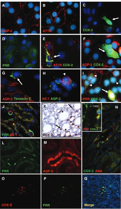
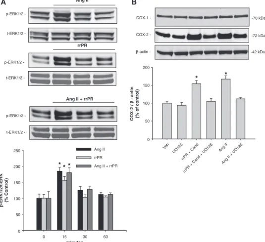
(3) Gonzalez et al PRR Stimulates COX-2 in the Inner Medulla 445. A. B. C. AQP-2. AT1R. COX-2. D. E. F. AT1R COX-2. PRR. AQP-2 COX-2. G. H. I. AQP-2 Tenascin C. AE-1 AQP-2. PRR COX-2. J. K. 100X. PRR COX-2. PRR AE-1. COX-2. L. M. PRR. AQP-2. COX-2 PRR. O. P. Q. COX-2. PRR. Merge. COX-2 Expression Is Stimulated Independently by PRR and AT1R via ERK1/2 Pathway in Rat IM Cells To stimulate PRR in IM cells, we used a nanomolar dose of a rat recombinant prorenin (rrPR).8 As shown in Figure 3A, AngII (100 nmol/L) and rrPR (100 nmol/L) treatments both independently increased p-ERK1/2 at 15 minutes (AngII: 185±12 %; rrPR 156±12 % versus vehicle: 100±13 %; n=6, P<0.05). Combined AngII and rrPR treatments caused. N. Figure 1. Characterization of long-term rat inner medullary (IM) cells (A–I). IM cells show specific immunoexpression of AQP-2 (red; A), AT1R (red; B), COX2 (green; C), and PRR (green; D). AT1R (red) colocalizes with COX-2 (green) in the same type of cells (E), indicating that interstitial cells (arrows) strongly stained for COX-2 (green) also coexpress AT1R (red) in the plasma membrane, as previously described (E). Cells expressing AQP-2 (red; F), the principal cells, do not coexpress COX-2 (arrow, green; F). Tenascin C (arrow, green; G), a marker for interstitial cells, does not colocalize with AQP-2 (red; E). Likewise, anion exchanger type 1 (AE-1; red; H), a known immunomarker for intercalated type-A cells (arrowheads), does not colocalizes with AQP-2 (green; H). Evidence for the presence of PRR (red; I) colocalizing with COX-2 (arrowheads, green; I) is shown in I. Nuclei were counterstained with DAPI (4′,6-diamidino-2-phenylindole; blue). In addition, in normal rat kidney sections (3 µm) immunofluorescence studies demonstrate that PRR (apical green immunoreactivity) and AE-1 (basolateral red immunoreactivity) colocalize in tubular intercalated type-A cells (J). K, Specific immunoreactivity for COX-2 in the interstitial cells (arrows) and some tubular cells (arrowheads) in the rat inner renal medulla (brown chromogen, 3,3′-Diaminobenzidine DAB). In consecutive kidney sections (L and M), is evident that PRR (green; L) is immunoexpressed by negative AQP-2–expressing cells (red; M). N, Examples of the colocalization of PRR (red; cell membrane) with COX-2 (green; intracellular localization) in the intercalated cells of the collecting ducts. In addition, this panel contains a high resolution microphotograph (left upper corner, 100×, oil immersion) for clear details. The lower panels O to Q show microphotographs (20×magnification) of a rat kidney section stained using dual immunofluorescence for COX-2 (red; O) and PRR (green; P) counterstained with DAPI (blue fluorochrome) demonstrate that COX-2 and PRR also colocalized in interstitial cells (merge, Q). Images were visualized using a Nikon Eclipse 50i immunofluorescence microscope and microphotographs were captured using a digital camera Nikon DS-U2/L2. AT1R indicates angiotensin II type 1 receptor; AQP-2, aquaporin; COX, cyclooxygenase-2; PRR, (pro)renin receptor.. a similar response at 15 minutes, without an additive effect (AngII+rrPR: 180±15 %; n=6, P<0.05). To test whether COX-2 expression was upregulated by PRR or AT1R activation via ERK1/2 pathway, IM cells were treated with the ERK1/2 inhibitor UO126 (10 μmol/L). Treatment with rrPR plus candesartan at 1 μmol/L (AT1R blocker) increased COX-2 protein levels (rrPR: 154±8 % versus vehicle: 100±5 %; n=6, P<0.05) to a similar extent observed in IM cells treated with AngII (AngII: 167±9 % versus vehicle: 100±5 %;.
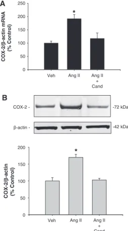
(4) 446 Hypertension February 2013 A. levels. PRR-shRNA did not affect the upregulation of COX-2 mediated by AngII (mRNA: 165±10%; n=6, P<0.05, protein: 158±10%; n=6, P<0.05).. COX-2/β-actin mRNA (% Control). 250. *. 200 150. Discussion. 100 50 0 Veh. Ang II. Ang II + Cand. B COX-2 -. -72 kDa. β-actin -. -42 kDa. COX-2/β-actin (% Control). 200. *. 150. 100. 50. 0 Veh. Ang II. Ang II + Cand. Figure 2. Cyclooxygenase-2 (COX-2) mRNA (A) and protein levels (B) levels were augmented after 6 h of angiotensin II (AngII) treatment; candesartan (Cand; 1 μmol/L) blocks this effect. n=6 to 8; *P<0.05. Veh indicates vehicle.. n=6, P<0.05). Importantly, upregulation of COX-2 by rrPR or AngII treatments was prevented by ERK1/2 inhibitor UO126 (rrPR+UO126: 105±8 %; AngII+UO126: 112±3 %; n=6, P=NS; Figure 3B).. PRR Knockdown Suppresses the PRR-Mediated Increases in COX-2 Expression in Rat IM Cells Target cells for transfections were interstitial and epithelial tubular cells, and PRR-shRNA reduced PRR protein expression compared with cells transfected with green fluorescent protein-scramble shRNA (47±4 % versus vehicle: 100±7 %; n=6, P<0.05; see online-only Data Supplement). Additional sets of cells were treated using the same dose of rrPR or AngII as in previous experiments and compared with cells transfected with PRR-shRNA. As shown in Figure 4A and 4B, rrPR treatment in the presence of candesartan increased COX-2 mRNA (160±7% versus vehicle: 100±4%; n=6, P<0.05) and protein (165±5% versus vehicle: 100±10%; n=6, P<0.05) levels. No additive effects were observed by combining AngII+rrPR (202±24% versus vehicle: 100±4%; n=6, P<0.05). AngII stimulation of COX-2 mRNA (191±3% versus vehicle: 100±4%; n=6, P<005) and protein (170±9% versus vehicle: 100±10%; n=6, P<0.05) levels were prevented by candesartan. Importantly, PRR-shRNA transfection of IM cells avoids the stimulatory effect of rrPR on COX-2 mRNA (84±10% versus vehicle: 100±4%; n=6, P=NS) and protein (108±5% versus vehicle: 100±10%; n=6, P<0.05). The present study demonstrates that COX-2 expression is upregulated in rat kidney IM cells by activation of either PRR or AT1R via MAPK/ERK1/2 pathway. This study also shows that long-term primary cultured rat IM cells are composed of CD epithelial cells and interstitial cells, and most importantly the novel finding that COX-2 and PRR colocalize in interstitial and CD type-A intercalated cells. Previous studies in rats and mice have shown that AngII stimulates renin and prorenin synthesis and secretion by the principal cells of the CD16,17,23,24 despite the suppression exerted on juxtaglomerular renin.18,25 In diabetic rats, the major source of prorenin synthesis and secretion are the principal cells of the CD.25 We demonstrated that AngII treatment is able to stimulate mainly prorenin synthesis in freshly isolated rat IM cells,17 and that the urines of AngII-dependent hypertensive rats possess abundant renin and prorenin.18 PRR can be found in 3 different molecular forms: (1) the M8.9, which is complexed with the proton vacuolar H+-proton ATPase2,26; (2) the 28-kDa soluble form of PRR, which can be detected in plasma27 and urine7; and (3) the full-length form (37 kDa) located in the cell plasma membrane, which is able to trigger the phosphorylation of ERK1/2 after binding of renin or prorenin.12,14,28 Because of the localization of PRR in the mesangium and its ability to increase COX-2,12,14 and profibrotic genes such as transforming growth factor β1, plasminogen activator inhibitor-1, collagen, and fibronectin,3,28 PRR has been implicated in the pathogenesis of chronic kidney disease.12,14,29 PRR has been implicated in the pathogenesis of chronic kidney disease.12,14,29 PRR activation promotes inflammation, through the activation of MAPK/ERK1/2 signaling pathways.12,14,30 Transgenic rats overexpressing PRR develop proteinuria and slowly progressive nephropathy.12,14 This model also exhibits increased levels of p-ERK1/2 and COX-2 expression in the renal cortex with normal AngII content, suggesting that COX-2 upregulation is independent of AngII.12,14 Furthermore, it has been shown that PRR overexpression in smooth muscle cells leads to elevated blood pressure and high plasma aldosterone levels, suggesting a role in circulating AngII formation.30 The pathophysiological AngII-independent effects mediated by PRR may be relevant during conditions in which systemic AngII levels are suppressed or normal, such as occur in diabetic patients, in which high plasma prorenin levels, but not plasma renin activity, predict the onset of microvascular complications, and correlate with high COX-2 expression, cell proliferation (short-term activation), and fibrosis (long-term activation).29–31 COX-2 is mainly expressed in the interstitial cells,32 macula densa,33 and epithelial cells of the thick ascending limb34; however, it has also been described in the CD cells.21,35 In the present study, we demonstrated that in normal rat kidney, COX-2 is detected in PRR-positive type-A intercalated cells. Importantly, we found that renal interstitial cells, which abundantly express COX-2, also express specific PRR immunoreactivity. Likewise, in rat renal cultured IM cells,.
(5) Gonzalez et al PRR Stimulates COX-2 in the Inner Medulla 447 Ang II. A. B. p-ERK1/2 -. t-ERK1/2 -. COX-1 -. -70 kDa. COX-2 -. -72 kDa. β-actin -. -42 kDa. rrPR. p-ERK1/2 200. *. * COX-2 / β - actin (% of control). t-ERK1/2 -. Ang II + rrPR. p-ERK1/2 -. 150. 100. 50. t-ERK1/2 -. 6. II g An. d an. R. +. C. 12 O U. +. 6. II An g. d. 12 U. C. +. + R rrP rrP. Ang II + rrPR. O. an. 6 12 U. rrPR. ** *. 200. p-ERK1/2/t-ERK (% Control). O. Ang II. 250. Ve h. 0. 150. 100. 50. 0 0. 15. 30. 60. minutes. Figure 3. Cyclooxygenase-2 (COX-2) protein expression is upregulated by angiotensin II (AngII) and rat recombinant prorenin (rrPR) via ERK1/2 pathway. A, Representative Western blot showing phosphorylated extracellular regulated kinases 1/2 (p-ERK1/2) levels in response to AngII, rrPR, and AngII+rrPR treatment at 0, 15, 30, and 60 min. *P<0.05 vs control (0 min, n=6). B, Representative Western blot showing that COX-2 protein levels are augmented by rrPR treatment in the presence of AT1R antagonist to avoid the possibility of AngII formation in inner medullary cells. As shown before, AngII also increase COX-2 protein levels. The ERK1/2 inhibitor UO126 (10 μmol/L) blunted the effect of both treatments (*P=NS vs vehicle; n=6). No effect was observed on COX-1 protein levels. Veh indicates vehicle; Cand, candesartan; NS, nonsignificant.. PRR, COX-2, and AT1R colocalized in interstitial and type-A intercalated cells. COX-2 plays a crucial role in regulating salt and water reabsorption and medullary blood flow,36–38 and its role is known in counterbalancing the effects of AngII through PGE2 production in medullary tissues.15 In fact, COX-2 inhibitors cause sodium retention in human subjects with normal kidney function.39 This effect has also been observed in experimental animals subjected to systemic or selective medullary COX-2 inhibition.15,40 Salt loading downregulates COX-2 expression in renal cortex but upregulates its expression in the renal medulla;40 however, the mechanisms implicated in these events remain unclear. Mineralocorticoid receptor agonism can induce COX-2 in vivo but not in cultured cells, suggesting that COX-2 upregulation is mediated by indirect pathways involving induced electrolyte hypertonicity in the interstitial fluid.41 These data suggest a complex interaction between signaling pathways in the regulation of COX-2 expression in IM cells. This complexity may reflect a cell type–specific response, for example, to hypertonicity or high intrarenal AngII levels as observed in AngII-dependent hypertension.17 It has been shown that AngII increases glomerular PGE2 production and COX-2 expression via ERK1/2 pathway. and that these effects are prevented by the AT1R blockade.13 Because we previously showed the increased expression of PRR mRNA levels in the CD of AngII-infused rats,7 we further examined whether PRR activation upregulates COX-2 independently of AngII. Treatment with rrPR in the presence of the AT1R blocker candesartan to avoid intrinsic activation of AT1R by possible endogenous AngII increased COX-2 and augmented p-ERK1/2 at 15 minutes. The same effect was observed by AngII, indicating that activation of both PRR and AT1R contributed to the phosphorylation of ERK1/2. Lack of an additive effect observed with both treatments (AngII and rrPR) may be explained by the fact that rat renal IM cells were composed by a mixed population of cells, thus the COX-2 stimulation in interstitial and intercalated cell in response to AT1R and PRR activation may differ. Further studies are needed to clarify this issue. This experimental evidence may suggest a key role of PRR in the light of recent evidence showing PRR upregulation by changes in dietary salt42 implicating that PRR may play a role in renal sodium handling through the activation of intracellular ERK1/2 pathway in renal tubules. Finally, to test whether PRR downregulation can alter COX-2 expression, we further knocked-down the PRR expression using shRNA technology. A 63% reduction in.
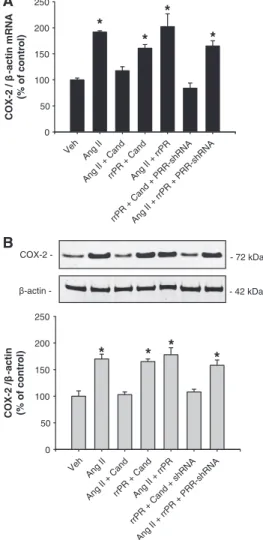
(6) 448 Hypertension February 2013 A COX-2 / β -actin mRNA (% of control). 250. *. 200. cells using transgenic models that overexpress PRR; however, the present study supports the notion that in the renal inner medulla the activation of PRR contributes to the stimulation of COX-2 via ERK1/2. These findings are of great relevance in the light of recent in vivo evidence demonstrating that during AngII-dependent hypertension there is stimulation of renin and prorenin synthesis and secretion by the CD cells17,18 and upregulation of PRR transcript. Clearly, more studies are needed to carefully test whether during AngII-dependent hypertension, the activation of PRR in intercalated and interstitial cells by its natural agonists, contribute to buffer the local effects of AngII in the renal medulla by stimulating COX-2 and promoting the synthesis of vasodilator and natriuretic prostanoids.. * *. *. 150 100 50. II II + rrP Ca nd R + + C An C An an an g g d d II II + + + P r rrP rP R R R -s R + PR hR R NA -s hR N A. g. rrP R. An. g. An. Ve. h. 0. B. COX-2 -. - 72 kDa. β-actin -. - 42 kDa. COX-2 /β -actin (% of control). 250 200. *. *. *. *. 150. Sources of Funding M.C.P. received funds from the National Institutes of Health (NIH) through the Institutional Developmental Award Program of the National Center for Research Resources (P20RR-017659), HL26371, American Heart Association (AHA; 09BGIA2280440), and Eunice Kennedy Shriver National Institute of Child Health and Human Development (K12HD043451). C.P.V. is supported by PFB 12–2007, FONDECYT 1080590, Chile.. Disclosures. 100. None.. 50. References A. A. hR. N. N. +. R PR. d. +. an. R. C + An. g. II. +. rrP. R rrP. -s. sh. + II. R. rrP. R. d. d. an C. + R. An. II. + rrP. g An. g. II. an. C. g An. Ve. h. 0. Figure 4. Prorenin receptor (PRR) knockdown suppressed the PRR-mediated upregulation of cyclooxygenase-2 (COX-2) in inner medullary (IM) cells. COX-2 mRNA (A) and protein (B) levels were augmented by angiotensin II (AngII, 100 nmol/L), and this effect was suppressed by AT1R blockade with candesartan (Cand; 1 μmol/L). Rat recombinant prorenin (rrPR; 100 nmol/L) plus Cand also upregulates COX-2, demonstrating an independent effect. PRR-mediated upregulation of COX-2 was completely blunted in IM cells previously transfected with PRRshRNA. *P<0.05 vs vehicle; n=6. Veh indicates vehicle.. PRR protein expression prevented the upregulation of COX-2 mRNA and protein, supporting our hypothesis of an AngIIindependent pathway for COX-2 regulation. In summary, type-A CD intercalated cells and interstitial cells coexpress COX-2, PRR, and AT1R. COX-2 expression is upregulated in rat renal IM cells through the independent activation of both PRR and AT1R. These findings provide basis for the critical contributions of PRR and AT1R in the regulation of COX-2 in the renal medulla.. Perspectives Although previous studies in vivo have shown that AT1R activation in the renal medulla led to COX-2-dependent PGE2 synthesis, our data demonstrate that COX-2 can also be upregulated by PRR activation independently of AngII in rat IM cells. Most of the pathophysiological effects of PRR have been reported in renal cortical tissues, particularly in mesangial. 1. Ludwig J, Kerscher S, Brandt U, Pfeiffer K, Getlawi F, Apps DK, Schägger H. Identification and characterization of a novel 9.2-kDa membrane sector-associated protein of vacuolar proton-ATPase from chromaffin granules. J Biol Chem. 1998;273:10939–10947. 2. Advani A, Kelly DJ, Cox AJ, White KE, Advani SL, Thai K, Connelly KA, Yuen D, Trogadis J, Herzenberg AM, Kuliszewski MA, Leong-Poi H, Gilbert RE. The (Pro)renin receptor: site-specific and functional linkage to the vacuolar H+-ATPase in the kidney. Hypertension. 2009;54:261–269. 3. Nguyen G, Delarue F, Berrou J, Rondeau E, Sraer JD. Specific receptor binding of renin on human mesangial cells in culture increases plasminogen activator inhibitor-1 antigen. Kidney Int. 1996;50:1897–1903. 4. Nguyen G. Renin/prorenin receptors. Kidney Int. 2006;69:1503–1506. 5. Nguyen G, Contrepas A. Physiology and pharmacology of the (pro)renin receptor. Curr Opin Pharmacol. 2008;8:127–132. 6. Ichihara A, Sakoda M, Kurauchi-Mito A, Kaneshiro Y, Itoh H. Involvement of (pro)renin receptor in the glomerular filtration barrier. J Mol Med. 2008;86:629–635. 7. Gonzalez AA, Lara LS, Luffman C, Seth DM, Prieto MC. Soluble form of the (pro)renin receptor is augmented in the collecting duct and urine of chronic angiotensin II-dependent hypertensive rats. Hypertension. 2011;57:859–864. 8. Nabi AH, Kageshima A, Uddin MN, Nakagawa T, Park EY, Suzuki F. Binding properties of rat prorenin and renin to the recombinant rat renin/ prorenin receptor prepared by a baculovirus expression system. Int J Mol Med. 2006;18:483–488. 9. Nguyen G, Burcklé CA, Sraer JD. Renin/prorenin-receptor biochemistry and functional significance. Curr Hypertens Rep. 2004;6:129–132. 10. Adderley SR, Fitzgerald DJ. Oxidative damage of cardiomyocytes is limited by extracellular regulated kinases ½-mediated induction of cyclooxygenase-2. J Biol Chem. 1999;274:5038–5046. 11. Rodríguez-Barbero A, Dorado F, Velasco S, Pandiella A, Banas B, LópezNovoa JM. TGF-beta1 induces COX-2 expression and PGE2 synthesis through MAPK and PI3K pathways in human mesangial cells. Kidney Int. 2006;70:901–909. 12. Kaneshiro Y, Ichihara A, Takemitsu T, Sakoda M, Suzuki F, Nakagawa T, Hayashi M, Inagami T. Increased expression of cyclooxygenase-2 in the renal cortex of human prorenin receptor gene-transgenic rats. Kidney Int. 2006;70:641–646. 13. Jaimes EA, Tian RX, Pearse D, Raij L. Up-regulation of glomerular COX-2 by angiotensin II: role of reactive oxygen species. Kidney Int. 2005;68:2143–2153..
(7) Gonzalez et al PRR Stimulates COX-2 in the Inner Medulla 449 14. Kaneshiro Y, Ichihara A, Sakoda M, Takemitsu T, Nabi AH, Uddin MN, Nakagawa T, Nishiyama A, Suzuki F, Inagami T, Itoh H. Slowly progressive, angiotensin II-independent glomerulosclerosis in human (pro)renin receptor-transgenic rats. J Am Soc Nephrol. 2007;18:1789–1795. 15. Qi Z, Hao CM, Langenbach RI, Breyer RM, Redha R, Morrow JD, Breyer MD. Opposite effects of cyclooxygenase-1 and -2 activity on the pressor response to angiotensin II. J Clin Invest. 2002;110:61–69. 16. Prieto-Carrasquero MC, Harrison-Bernard LM, Kobori H, Ozawa Y, HeringSmith KS, Hamm LL, Navar LG. Enhancement of collecting duct renin in angiotensin II-dependent hypertensive rats. Hypertension. 2004;44:223–229. 17. Gonzalez AA, Liu L, Lara LS, Seth DM, Navar LG, Prieto MC. Angiotensin II stimulates renin in inner medullary collecting duct cells via protein kinase C and independent of epithelial sodium channel and mineralocorticoid receptor activity. Hypertension. 2011;57:594–599. 18. Liu L, Gonzalez AA, McCormack M, Seth DM, Kobori H, Navar LG, Prieto MC. Increased renin excretion is associated with augmented urinary angiotensin II levels in chronic angiotensin II-infused hypertensive rats. Am J Physiol Renal Physiol. 2011;301:F1195–F1201. 19. Zhuo J, Alcorn D, McCausland J, Mendelsohn FA. Localization and regulation of angiotensin II receptors in renomedullary interstitial cells. Kidney Int. 1994;46:1483–1485. 20. Zhuo JL. Renomedullary interstitial cells: a target for endocrine and paracrine actions of vasoactive peptides in the renal medulla. Clin Exp Pharmacol Physiol. 2000;27:465–473. 21. Ferguson S, Hébert RL, Laneuville O. NS-398 upregulates constitutive cyclooxygenase-2 expression in the M-1 cortical collecting duct cell line. J Am Soc Nephrol. 1999;10:2261–2271. 22. Câmpean V, Theilig F, Paliege A, Breyer M, Bachmann S. Key enzymes for renal prostaglandin synthesis: site-specific expression in rodent kidney (rat, mouse). Am J Physiol Renal Physiol. 2003;285:F19–F32. 23. Gonzalez-Villalobos RA, Satou R, Ohashi N, Semprun-Prieto LC, Katsurada A, Kim C, Upchurch GM, Prieto MC, Kobori H, Navar LG. Intrarenal mouse renin-angiotensin system during ANG II-induced hypertension and ACE inhibition. Am J Physiol Renal Physiol. 2010;298:F150–F157. 24. Prieto-Carrasquero MC, Botros FT, Pagan J, Kobori H, Seth DM, Casarini DE, Navar LG. Collecting duct renin is upregulated in both kidneys of 2-kidney, 1-clip goldblatt hypertensive rats. Hypertension. 2008;51:1590–1596. 25. Kang JJ, Toma I, Sipos A, Meer EJ, Vargas SL, Peti-Peterdi J. The collecting duct is the major source of prorenin in diabetes. Hypertension. 2008;51:1597–1604. 26. Cruciat CM, Ohkawara B, Acebron SP, Karaulanov E, Reinhard C, Ingelfinger D, Boutros M, Niehrs C. Requirement of prorenin receptor and vacuolar H+-ATPase-mediated acidification for Wnt signaling. Science. 2010;327:459–463. 27. Cousin C, Bracquart D, Contrepas A, Corvol P, Muller L, Nguyen G. Soluble form of the (pro)renin receptor generated by intracellular cleavage by furin is secreted in plasma. Hypertension. 2009;53:1077–1082.. 28. Nguyen G. Increased cyclooxygenase-2, hyperfiltration, glomerulosclerosis, and diabetic nephropathy: put the blame on the (pro)renin receptor? Kidney Int. 2006;70:618–620. 29. Huang J, Matavelli LC, Siragy HM. Renal (pro)renin receptor contributes to development of diabetic kidney disease through transforming growth factor-β1-connective tissue growth factor signalling cascade. Clin Exp Pharmacol Physiol. 2011;38:215–221. 30. Burcklé CA, Jan Danser AH, Müller DN, Garrelds IM, Gasc JM, Popova E, Plehm R, Peters J, Bader M, Nguyen G. Elevated blood pressure and heart rate in human renin receptor transgenic rats. Hypertension. 2006;47:552–556. 31. Ichihara A, Itoh H, Inagami T. Critical roles of (pro)renin receptor-bound prorenin in diabetes and hypertension: sallies into therapeutic approach. J Am Soc Hypertens. 2008;2:15–19. 32. Hao CM, Kömhoff M, Guan Y, Redha R, Breyer MD. Selective targeting of cyclooxygenase-2 reveals its role in renal medullary interstitial cell survival. Am J Physiol. 1999;277(3 pt 2):F352–F359. 33. Harris RC, McKanna JA, Akai Y, Jacobson HR, Dubois RN, Breyer MD. Cyclooxygenase-2 is associated with the macula densa of rat kidney and increases with salt restriction. J Clin Invest. 1994;94:2504–2510. 34. Vio CP, Cespedes C, Gallardo P, Masferrer JL. Renal identification of cyclooxygenase-2 in a subset of thick ascending limb cells. Hypertension. 1997;30(3 pt 2):687–692. 35. Yang T, Zhang A, Pasumarthy A, Zhang L, Warnock Z, Schnermann JB. Nitric oxide stimulates COX-2 expression in cultured collecting duct cells through MAP kinases and superoxide but not cGMP. Am J Physiol Renal Physiol. 2006;291:F891–F895. 36. Harris RC. An update on cyclooxygenase-2 expression and metabolites in the kidney. Curr Opin Nephrol Hypertens. 2008;17:64–69. 37. Green T, Rodriguez J, Navar LG. Augmented cyclooxygenase-2 effects on renal function during varying states of angiotensin II. Am J Physiol Renal Physiol. 2010;299:F954–F962. 38. Ferreri NR, An SJ, McGiff JC. Cyclooxygenase-2 expression and function in the medullary thick ascending limb. Am J Physiol. 1999;277 (3 pt 2):F360–F368. 39. Kammerl MC, Nüsing RM, Schweda F, Endemann D, Stubanus M, Kees F, Lackner KJ, Fischereder M, Krämer BK. Low sodium and furosemideinduced stimulation of the renin system in man is mediated by cyclooxygenase-2. Clin Pharmacol Ther. 2001;70:468–474. 40. Yang T, Singh I, Pham H, Sun D, Smart A, Schnermann JB, Briggs JP. Regulation of cyclooxygenase expression in the kidney by dietary salt intake. Am J Physiol. 1998;274(3 pt 2):F481–F489. 41. Zhang MZ, Hao CM, Breyer MD, Harris RC, McKanna JA. Mineralocorticoid regulation of cyclooxygenase-2 expression in rat renal medulla. Am J Physiol Renal Physiol. 2002;283:F509–F516. 42. Huang J, Siragy HM. Sodium depletion enhances renal expression of (pro)renin receptor via cyclic GMP-protein kinase G signaling pathway. Hypertension. 2012;59:317–323.. Novelty and Significance What Is New? • This study provides evidence for a new role of the prorenin receptor (PRR) in the regulation of cyclooxygenase-2 (COX-2) in the rat renal medulla via mitogen-activated protein kinase/extracellular regulated kinases. In addition, we provide evidence that the PRR and COX-2 are colocalized in the intercalated cells of the collecting duct and in the interstitial cells, which further supports our hypothesis. What Is Relevant? • Our findings are of critical importance because they support the notion that activation of PRR by upregulating COX-2 via extracellular regulated kinases 1/2 in the interstitial and intercalated cells may. increase prostaglandins synthesis, thus contributing to buffer local vasoconstrictor and antinatriuretic effects of angiotensin II.. Summary PRR and COX-2 are coexpressed in interstitial cells and intercalated collecting duct cells. Activation of PRR by recombinant prorenin upregulated COX-2 even in the presence of AT1 receptor blockade in rat primary cultured renal inner medullary cells. Upregulation of COX-2 by angiotensin II or prorenin was extracellular regulated kinases 1/2 signaling–dependent. PRR knockdown prevented COX-2 upregulation mediated by prorenin treatment in rat inner medullary cells. Upregulation of COX-2 in inner medullary cells is mediated by angiotensin II and by the angiotensin II–independent activation of PRR..
(8)
Figure
Documento similar
Nijkamp has placed special emphasis on spatial-economic competition and ascribes to it a central function. While we recognise the centrality of spatial- economic
Pero, al fin y al cabo, lo que debe privar e interesar al sistema, es la protección jurisdiccional contra las ilegalidades de la Administración,221 dentro de las que se contemplan,
Dado un espazo topol´ oxico, denominado base, e dado un espazo vec- torial para cada punto de dito espazo base, chamaremos fibrado vectorial ´ a uni´ on de todos estes
Este estimador é asintoticamente insesgado para a densidade espectral, pero non é consistente dado que a súa varianza é proporcional ao valor da densidade espectral en cada frecuencia
La solución que se ha planteado, es que el paso o bien se hiciese exclusivamente por el adarve de la muralla, o que una escalera diese acceso por la RM evitando la estancia (De
Dentro de esta franja, diversos prospectos de mineralización epitermal de minerales preciosos (Au) están asociados a zonas de cizallamiento compresivo, los cuales tienen como
De esta manera, ocupar, resistir y subvertir puede oponerse al afrojuvenicidio, que impregna, sobre todo, los barrios más vulnerables, co-construir afrojuvenicidio, la apuesta
after purification of myeloid cells from heart tissue, we found that only a particular subset expressed COX-2, being the levels of COX-2 expression higher in C57BL/6 than BALB/c
