Toxoplasma gondii infection in wild mustelids
and cats across an urban-rural gradient
Macarena Barros1, Oscar Cabezo´ n2,3, Jitender P. Dubey4, Sonia Almerı´a2,5, Marı´a P. Ribas2,3, Luis E. Escobar6, Barbara Ramos1, Gonzalo Medina-Vogel1*
1 Centro de Investigacion para la Sustentabilidad, Universidad Andres Bello, Repu´blica, Santiago, Chile, 2 UAB, Centre de Recerca en Sanitat Animal (CReSA, IRTA-UAB), Campus de la Universitat Autònoma de Barcelona, Bellaterra, Spain, 3 Servei d’Ecopatologia de Fauna Salvatge, Departament de Medicina I Cirugia Animals, Universitat Autònoma de Barcelona, Bellaterra, Spain, 4 Animal Parasitic Diseases Laboratory, Beltsville Agricultural Research Center, Agricultural Research Service, United States Department of Agriculture, Beltsville, Maryland, United States of America, 5 Departament de Sanitat I d´Anatomia Animals, Universitat Autònoma de Barcelona, Bellaterra, Spain, 6 Department of Fish and Wildlife Conservation, Virginia Tech., Blacksburg, Virginia, United States of America
Abstract
The increase in human population and domestic pets, such as cats, are generating impor-tant consequences in terms of habitat loss and pathogen pollution of coastal ecosystems with potential to generate negative impacts in marine biodiversity. Toxoplasma gondii is the etiological agent of zoonotic disease toxoplasmosis, and is associated with cat abundance and anthropogenic disturbance. The presence of T. gondii oocysts in the ocean has nega-tively affected the health status of the threatened Southern sea otter (Enhydra lutris nereis) populations. The present study analyzed seroprevalence and presence of T. gondii DNA in American mink (Neovison vison), Southern river otters (Lontra provocax) and domestic cats (Felis silvestris catus) in four different areas in Southern Chile comprising studies in rivers and lakes in Andean foothills and mountains, marine habitat and island coastal ecosystems. Mean seroprevalence of T. gondii in the study was 64% of 151 total animals sampled: 59% of 73 American mink, 77% of 13 Southern river otters, 68% of 65 domestic cats and in two of two kodkods (Leopardus guigna). Toxoplasma gondii DNA was detected in tissues from one American mink and one Southern river otter. The present study confirms the widespread distribution of T. gondii in Southern Chile, and shows a high exposure of semiaquatic muste-lids and domestic cats to the parasite. Cats and anthropogenic disturbance have a role in the maintenance of T. gondii infection in ecosystems of southern Chile.
Introduction
Contamination of the aquatic environment derived from human activities is a concern world-wide, and the study of the biological effects of these activities in the marine ecosystem has been declared a goal for future research by the scientific community [1]. The effect of human settle-ment in coastal areas causes significant negative impacts on coastal marine habitats and their wildlife species, including the flow of human/terrestrial pathogens [2,3,4].Toxoplasma gondii, a1111111111
Citation: Barros M, Cabezo´n O, Dubey JP, Almerı´a
S, Ribas MP, Escobar LE, et al. (2018) Toxoplasma
gondii infection in wild mustelids and cats across
an urban-rural gradient. PLoS ONE 13(6): e0199085.https://doi.org/10.1371/journal. pone.0199085
Editor: Michael E. Grigg, NIH, UNITED STATES
Received: May 12, 2017
Accepted: May 31, 2018
Published: June 20, 2018
Copyright:©2018 Barros et al. This is an open access article distributed under the terms of the
Creative Commons Attribution License, which permits unrestricted use, distribution, and reproduction in any medium, provided the original author and source are credited.
Data Availability Statement: All relevant data are
within the paper and its Supporting Information files.
Funding: This study was supported by FONDECYT
a worldwide distributed zoonotic protozoan parasite, presents an indirect cycle with domestic and wild felines as definitive hosts. Oocysts are eliminated to the environment through feces where they can remain infective for months or years. Warm-blooded species can become infected by ingesting the sporulated oocysts and infection can persist in infected hosts for the life of the host [5]. Although better studied in terrestrial landscapes,T.gondiihas also emerged as a significant aquatic pathogen linked to marine mammal infection and water-borne out-breaks of disease in humans worldwide [6]. The presence ofT.gondiiin the marine and fresh-water ecosystems has been confirmed by the exposure of several aquatic species to this parasite [7,8,9,10], with clinical disease observed in seals, dolphins, whales, sea otters, and manatees [7,11,12]. Animals living in freshwater systems are also at risk. As many as 85% (82 of 95) of free-ranging Amazon river dolphins had antibodies toT.gondii[13]. In this respect,T.gondii
has been used as a model for the study of land-to-sea pathogen contamination [14,15,16]. Higher flow ofT.gondiioocysts to the marine ecosystem has been related to the high coastal human densities and estuarine wetland degradation due to human activities [14,17]. The effect of this higherT.gondiipresence in the ocean has negatively affected the health status of the threatened Southern sea otter (Enhydra lutris nereis) populations, increasing its mortality related to the parasite [18,19], and supporting the hypothesis of land to sea transmission of this parasite through contaminated freshwater runoff [20]. WidespreadT.gondiiinfection in aquatic mammals suggests that contamination of terrestrial watersheds withT.gondiiis preva-lent, and that sufficient numbers of oocysts are distributed in freshwater and marine ecosys-tems to infect and cause disease in both near-shore and pelagic mammals [10].
Chile has several aquatic wild carnivore endemic species classified as endangered by the International Union for the Conservation of Nature (IUCN) [21]. One of the most important species is the Southern river otter (Lontra provocax, referred here as otter), a semi-aquatic spe-cies that is a top predator of river and marine fauna [22] and is considered to be the otter with the smallest distribution in the world [23,24]. The distribution of this otter has declined drasti-cally due to combined pressures from the destruction of habitat, removal of vegetation, river and stream canalization, and extensive dredging [24,25]. This species is considered “endan-gered” by the IUCN Red List of Threatened Species [21]. This otter shares its ecosystem with the American mink (Neovison vison), a widely distributed exotic invasive mustelid present worldwide [26,27]. American mink can negatively impact otter populations in different ways, including competition for resources and disease transmission. In this way, the presence of other species, such as the mink, can serve as reservoirs of diseases for the otters. For example, the exposure of the otter toT.gondiiwill be influenced by the presence of felids in the field. In this respect, the domestic and feral cats represent the most important epidemiological factor in the dispersion of the parasite [28] and its distribution and density is directly influenced by human presence. In Southern Chile, there may be other potential felid definitiveT.gondii
hosts such as the kodkod (Leopardus guigna). The role of this species in the epidemiology ofT.
gondiihas not been evaluated. Kodkod is the smallest wild felid in the Americas. It lives pri-marily in central and southern Chile and marginally in adjoining areas of Argentina. Its area of distribution is small compared to the other South American felids. Since 2002, it has been listed as Vulnerable on the IUCN Red List as the total effective population may comprise less than 10,000 mature individuals, and is threatened due to persecution and loss of habitat and prey base [21].
The epidemiology ofT.gondiiis understudied in Chile. Although some studies have reported its circulation in human beings, domestic animals, and wildlife, these studies have been undertaken in few species and geographic areas [29,30–34]. Therefore, the objective of the present study was to analyzeT.gondiidistribution in the diverse terrestrial/aquatic inter-faces in several areas of Southern Chile, particularly in relation to their proximity to human manuscript and there was no additional external
funding received for this study.
Competing interests: The authors have declared
settlements and the presence of domestic cats, assaying DNA and antibodies against this para-site in semi-aquatic carnivores.
Materials and methods
Study areas
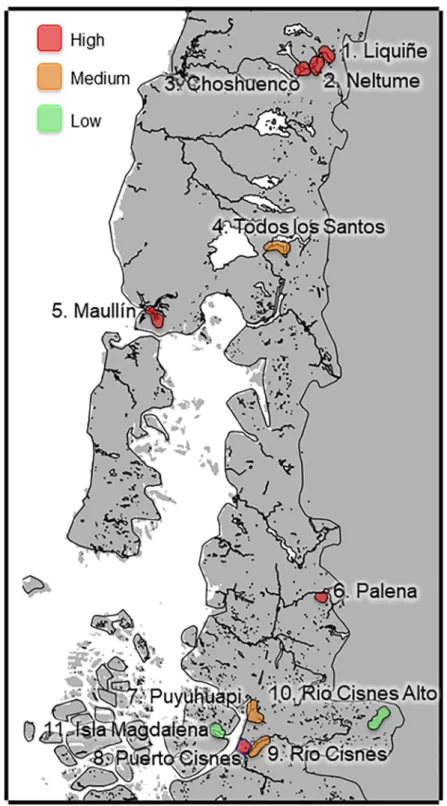
Eleven study sites were chosen according to human, cat, mink and otter presence and then grouped into four study areas according to landscape characteristics in Southern Chile between 39˚-45˚ S latitudes (Fig 1).
Area 1 (39.3˚ - 41.3˚S; 200–300 m.a.s.l; sites 1 to 4;Fig 1) represents an Andean foothill eco-system and comprised the Liquiñe river, Neltume lake, Choshuenco lake and Todos-los-Santos lake. This area has a cold temperate rainy Mediterranean climate. The annual average tempera-ture is low (11˚C) and falls toward the south, with strong daily thermal oscillation and high rainfall throughout the year. In winter, the precipitation increases and there are almost no dry months. The vegetation corresponds to Andean rainforest of deciduous and coniferous trees, comprisingNothofagus dombeyi,Podocarpus nubigenus,Aextoxicon punctatum, andDrymis winteri[35]. The four sites sampled had little slope. The landscape has residential, tourist and agricultural use with pervious surfaces.
Area 2 (41.5˚ - 44.8˚S; 20–80 m.a.s.l; sites 5, 7, 8 and 9;Fig 1) represents a marine coastal ecosystem and comprises the Puerto-Cisnes and Puyuhuapi fiords, the Maullı´n, and Rio-Cisnes rivers. This area has a rainy temperate maritime climate without a dry season. The area is characterized by low temperatures, with an average annual temperature of 10.2˚C. Rainfall is abundant and present throughout the year. The vegetation present in Maullı´n is the same as Area 1, while in Puerto-Cisnes is the Patagonian steppe [35]. The Maullı´n, Puyuhuapi and Rio-Cisnes areas all have residential, tourist, and agricultural use with pervious surfaces. Puerto-Cisnes is a residential area with primarily impervious surface.
Area 3 (43.3˚ - 44.6˚S; 250–750 m.a.s.l; sites 6 and 10;Fig 1) represents Southern Andean mountain valleys and is comprised of the Palena and Rio-Cisnes-Alto rivers. The Palena river has a microclimate with average annual temperatures of 9.5˚C, with average rainfall of 1700mm. The Rio-Cisnes-Alto river has a cold steppe climate affected by the eastern slope of the Andes. The average annual temperature is 7.1˚C. Rainfall decreases during autumn, and in winter snow precipitation occurs. The vegetation type is tundra [35]. This area has little slope. The landscape has residential, tourist and agricultural use with pervious surfaces.
Fig 1. Study area. Eleven study sites, grouped into four study areas in Southern Chile, between latitude 39˚-45˚ S. Area
1, represents an Andean foothill ecosystem (1:Liquiñe, 2:Neltume, 3:Choshuenco, 4:Todos los Santos); area 2, represents a marine coastal ecosystem (5:Maullı´n, 7:Puyuhuapi, 8:Puerto Cisnes, 9:Rio-Cisnes); area 3, represents Southern Andean mountain valleys (6:Palena, 10:Rio-Cisnes-Alto); area 4, represents an island (11:Magdalena Island). Red: High degree of presence human-domestic cat. Orange: Medium degree of presence human-domestic cat. Green: Low degree of presence human-domestic cat.
Animals and samples
From 2009 to 2013, American mink (n = 73) were captured using modified Tomahawk traps with double doors [38]. Once captured, animals were immobilized mechanically with a plastic mesh and then chemically with an intramuscular (IM) administration of 10 mg/kg of Keta-mine (Ketamina 1001, Chemie) and 0.025 mg/kg of Dexmedetomidine (Dexdomitor1, Pfi-zer). Blood samples were obtained though intracardiac puncture. After blood sampling, all mink were euthanized with thiopental (Tiopental So´dico1, Chemie). All animals were nec-ropsied and tissues samples from the brain, lung, liver, and kidney were obtained. In total, tis-sues samples from 55 animals were collected. Otters (n = 13) were captured in leg hold soft-catch traps [39,40]. After manual immobilization, a combination of Ketamine:dexmedetomi-dine 5:0.025 mg/kg IM was injected. Blood samples were obtained from a jugular vein. Once the individuals were microchipped and fully recovered from anesthesia they were released at the site of capture. Cats (n = 65) were sampled near the area where mink and otters were trapped. With the owner’s consent, each cat was manually immobilized to obtain a blood sam-ple from a jugular vein. During the field work, two kodkod were accidentally captured and thus also sampled, one in Neltume and one in Puyuhuapi. In addition, a dead otter from Valdi-via was also examined. All blood samples were obtained using sterile syringes and collected in 2 ml tubes with and without anticoagulant. To obtain serum, the blood without anticoagulant was centrifuged at 1200 g for 10 minutes. All samples were stored in liquid nitrogen until anal-ysis. The tissue samples were preserved in alcohol 70% to preserve the tissues and ensure read-ability. Capture and sampling methods was carried out in strict accordance with the
recommendations by Bioethics Committee for Animal Research of the Universidad Andres Bello and National Commission for Science and Technology. The field permit was granted by Subsecretarı´a de Pesca (Subpesca) for otters. Mink euthanasia was ordered by Servicio Agrı´c-ola y Ganadero (SAG). Sampling procedures in the field were reviewed and approved by veter-inarians. All efforts were made to minimize suffering. Animal research ethics committee prospectively approved this research.
Serological analysis
Sera from American mink, otter, domestic cat, and kodkod were evaluated for the detection of IgG antibodies againstT.gondiiusing the modified agglutination test (MAT) [41]. MAT is one of the most evaluated and widely used tests both in clinical and epidemiological surveys for the detection ofT.gondiiin several animal species, including humans [5]. Sera were tested at 1:25, 1:50, 1:100, and 1:500 dilutions. Titers25 were considered positive and those with doubtful results were re-tested. Positive and negative controls were included in this test. Additionally, we used Toxo-Screen DA fast kit [42] to as a complementary test of MAT in 40 samples of wild animals (29 American mink and 11 otter, were also included in MAT). Toxo-Screen DA fast kit, allows detection of IgG by direct agglutination. Sera were tested at 1:40 and 1:4000 dilutions. Titers40 were considered positive [42]. The MAT and Toxo-Screen DA kit use same procedures and reagent [5].
Molecular analysis
GT—BHQ1 3') using a commercial kit (TaqMan PCR Master Mix, Applied Biosystems, Carls-bad, CA, USA).
Domestic cat presence
Cat and human presence was determined via field trapping, human surveys, and spatial analy-sis using Quantum GIS version 2.0.1 (http://www.qgis.org/es/site/). To establish our study sites, we made a buffer area of 4 km around each American mink captured, individual buffers were then merged to produce a wider area, creating 11 study sites with a similar size. Buffered areas were divided in 1 km2cells, which were then categorized as containing or not containing human presence. Google Earth (https://www.google.com/earth/) imagery was used to deter-mine the presence of houses via visual identification of roofs, and cells with one or more houses were designated as having human presence and cells without houses were designated as not having human presence. Then, we counted the number of roofs per cell to have an approx-imate number of houses per site as a proxy of human presence or anthropogenic disturbance. Additionally, we conducted an on-ground survey for home owners to estimate the number of cats per house detected in Google Earth. Data of human presence and cats per house were used in posterior analyses as raw-continuous and categorical variables. Estimated number of cats per cell were divided in three categories to denote areas of low (0.2 cats per km2), medium (0.21–0.6 cats per km2), and high (0.61–5.5 cats per km2) degree of cat density.
Land cover change
We explored the potential effects of anthropogenic land use change on the occurrence ofT.
gondiiin the wild species used as sentinels. Land cover change was estimated via comparing the vegetation change in a ten-year period. MODIS imagery from the Terra satellite at 500 m. spatial resolution (MOD13A1.005) was used to obtain information of the landscape configura-tion. Specifically, employing 16-day composites Enhanced Vegetation Index (EVI) variables for February 2002 and 2012, the last representing the sampling period. The open access vari-ables [44] were first processed using the MODIS Reprojection Tool version 4.1 to obtain raster files for posterior analyses [44]. Rasters were then analyzed using a principal components anal-ysis in ArcGIS 10.3 [45]. The second component was used to identify areas of vegetation loss and gain. The second principal component from original vegetation indices is an excellent proxy of landscape variation [46]. This component was visually inspected to corroborate areas with loss and gain of vegetation.
Statistical analysis
package in R [50], following protocols described elsewhere [51]. To compare the two serologi-cal tests we used Cohen’s kappa analysis [52]. Finally, to assess the effect of Land Cover Change we used the locations of otter and mink captured,T.gondiipositive and negative, to assess binary association ofT.gondiiin wildlife with continuous values landscape variation (i.e., prin-cipal component two of EVI data). This analysis was done separately from the previous ones using a logistic regression in R [53].
Results
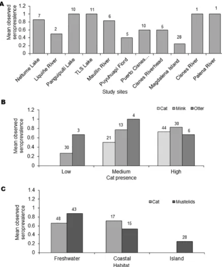
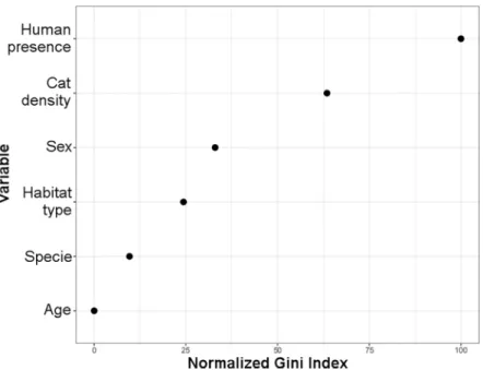
The overallT.gondiiseroprevalence, according to the MAT analysis, was 64% (97/151), with 68% (44/65) in domestic cats, 59% (43/73) American mink, and 77% (10/13) in otter (Table 1). The two kodkods were also seropositive. With GLM and Kruskal-Wallis test, seroprevalence forT.gondiiamong study sites showed no statistical differences (Fig 2A). However, there were differences between study areas (F3,147= 9.7; P<0.01) because the significantly lower preva-lence in area 4 (Magdalena Island), there were not significantly difference between the other areas (F2,120= 1.12; P = 0.33). Analysis by species showed a consistent prevalence in cats among all of the analyzed study sites, areas and habitats, but not in Magdalena island. No dif-ferences were found betweenT.gondiiseroprevalences of mink and otter, or between animal sex or age, although a tendency towards higher prevalence was observed in older animals. Mink had significantly higher seroprevalences in the study sites associated to medium and high presence of domestic cats (H2= 22.0; P<0.01) (Fig 2B), and significantly less observed seroprevalences in marine habitats (H2= 28.9; P<0.01) (Fig 2C). In fact, the only study site with significantly less observed seroprevalence was Magdalena Island (Fig 2A). Otter seroposi-tivity had no relationship with cat presence, but was also significantly lower in marine habitats (H1= 31.5; P = 0.04) (Fig 2C). Indeed, the differences of seroprevalences between study sites for otters were close to significant (F5,7= 3.5; P = 0.07). Our random forest analysis revealed that seropositivity toToxoplasma gondiiwas mainly explained by presence of humans and den-sity of cats (Fig 3).Toxoplasma gondiiDNA was detected in the liver of one American mink in
Choshuenco(Area 1), and in the lung of the otter found dead in Valdivia (outside of the study
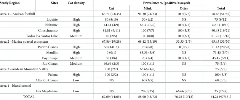
Table 1. Seroprevalence ofToxoplasma gondii. Data from 11 study sites with varying degrees of domestic cat presence, (NS = Not samples).
Study Region Sites Cat density Prevalence % (positive/assayed)
Cat Mink Otter Total
Area 1—Andean foothill 65.71 (23/35) 91.30 (21/23) 100 (7/7) 78.46 (51/65) Liquiñe High 80 (8/10) 50 (1/2) NS 75 (9/12) Neltume High 44.44 (4/9) 83.33 (5/6) 100 (1/1) 62.5 (10/16) Choschuenco High 81.81 (9/11) 100 (7/7) 100 (3/3) 90.48 (19/21) Todos los Santos Lake Medium 40 (2/5) 100 (8/8) 100 (3/3) 81.25 (13/16) Area 2 –Marine coastal ecosystem 67.86 (19/28) 68.42 (13/19) 33.33 (1/3) 65.45 (33/50) Puerto Cisnes High 50 (14/18) 75 (6/8) 0 (0/2) 71.43 (20/28) Maullı´n High 0 (0/1) 83.33 (5/6) NS 71.43 (5/7) Puyuhuapi Medium 50 (3/6) 25 (1/4) 100 (1/1) 45.45 (5/11) Rio Cisnes Medium 66.66 (2/3) 100 (1/1) NS 75 (3/4) Area 3 –Andean Mountain Valley 100 (2/2) 66.66 (4/6) 75 (6/8) Palena High 100 (2/2) 100 (1/1) NS 100 (3/3) Alto Rio Cisnes Low NS 60 (3/5) NS 60 (3/5) Area 4 –Island coastal
Isla Magdalena Low NS 20 (5/25) 66.66 (2/3) 25 (7/28) TOTAL 67.69 (44/65) 58.90 (43/73) 76.92 (10/13) 64.24 (97/151)
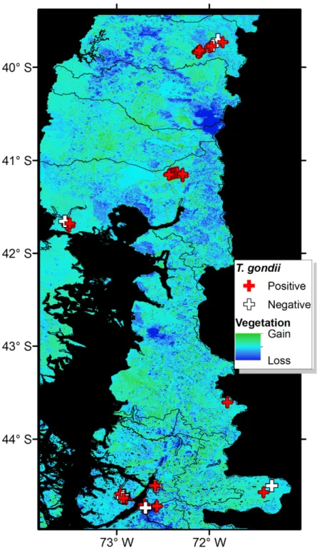
areas). The analysis of the vegetation structure revealed considerable variation in the vegeta-tion biomass in the ten-year period assessed (Fig 4). At province level, areas of dramatic vege-tation loss were identified in central western Valdivia, eastern Ranco, dispersed areas across Llanquihue, central west Palena, and across the southern sampled areas in Aysen. A significant association was found between theT.gondiiseropositivity and the level of vegetation loss of the study sites (COR = 0.65; df = 85;P<0.001) with habitat loss being associated with increase ofT.gondiiseroprevalence. Results obtained by Toxo-Screen DA fast kit were similar to those obtained with MAT. Of the 40 samples, 27 were equally positive and 9 equally negative, with 2 “false positives” and 2 “false negatives”. The Cohen’s kappa analysis shows a good agreement between the two tests (Kappa = 0.75; 95% confidence interval = 0.52–0.98;P<0.001).
Discussion
The present study confirms the widespread distribution ofT.gondiiin Southern Chile, and shows a high exposure of semiaquatic mustelids and domestic cats to the parasite. The high explanatory power of human presence followed of cat density in our machine learning analysis (Fig 3), suggests the role of domestic cats as a form of anthropogenic disturbance for the main-tenance ofT.gondiiinfections in Southern Chilean ecosystems. The mean prevalences
Fig 2. Effects of landscape variables on seroprevalence. A) Observed seroprevalence in mink and otter by study site.
B) Observed seroprevalence by degree of presence of domestic cat. C) Observed seroprevalence by habitat.Number
above bars indicate sample size. Y axis show mean seroprevalence.
observed in the study, 59% in mink, 77% in otters, and 68% in domestic cats are consistent with those observed in previous studies. A 70% prevalence ofT.gondiiin American mink has been reported in the Maullı´n River [32]. We observed similarly high seroprevalence level at the same site in mink in the current study (83%). Our findings also agree with recent reports ofT.
gondiiseroprevalences of 77% in American mink in the USA [54]. Seroprevalence ofT.gondii
revealed the lowest values (25%) in Magdalena Island. These results were expected considering that there are no reports of the presence of domestic cats in this island due to its geographic isolation (five km distant from the continent). Our results also agree with another study in USA which suggests that prevalence ofT.gondiiantibodies below 40% indicates low exposure to the parasite [55]. However, the prevalence on this island may support the idea of horizontal transmission ofT.gondii. Mink and otters from Magdalena Island may have become infected while feeding on other intermediate hosts.
Studies of the mechanisms ofT.gondiitransmission in islands are of great interest since iso-lated environments can affect the epidemiology of different pathogens. In this respect, several studies have reported positive relationship between prevalences ofT.gondiiin intermediate hosts and the level of the presence of cats. However, the presence of the parasite in islands without felids has been reported in Arctic foxes (Vulpes lagopus) from Svalbard Island [42].
Toxoplasma gondiiseroposivity of seagull chicks in an island without cats has also been reported, confirming the capability of seagulls to introduce pathogens in naïve territories and expose other susceptible hosts [16]. Sites with moderateT.gondiiprevalence (i.e., 40–60%) [55] were Puyuhuapi Fiord (45.4%) which is about 20 km from the nearest village, and Alto-Rio-Cisnes (60%). All other sites showed high seroprevalences (>60%;Fig 2A).
As reported previously, we observed lower seroprevalence in wildlife from study sites with low degree of housing and therefore low human presence. However, this result was not observed in cats. Domestic cats seroprevalence did not show differences based on cat density (Fig 2B,Table 1). This may be explained by the role of the felids in the epidemiological cycle of
Fig 3. Variable importance analysis performed using random forest. The set of four categorical variables (age,
species, habitat type, sex) and two continuous variables (human presence and cat density) used for classification the seropositivity toToxoplasma gondii. Variables are ordered by their importance from top to bottom as estimated by the random forest model and denoted using an Gini index ranging from 0 to 100.
T.gondii. The domestic cat is the key species for the maintenance ofT.gondiioocyts in the environment, excreting millions of oocysts in a short period of time [56]. It has been estimated that between the 1% and 2% of all domestic cats excreteT.gondiioocysts at any given time [57,
58,59].
Fig 4. Land cover change andToxoplasma gondiireports. Positive (red crosses) and negative (white crosses) reports
ofT.gondiiin wildlife compared with vegetation change represented in the second principal component of the EVI 2002 and 2012. Change was denoted as areas of vegetation gain (green) and stability (light blue), and considerable vegetation loss (dark blue).
In Chile, feral cat populations are scattered and found mainly in Central-South regions; thus, cats have sympatric distribution with the otter and the American mink [60]. In rural areas of Central Chile, up to 54.6% seropositivity againstT.gondiihas been recorded in domes-tic cats [61] while in the Southern areas, i.e. in the city of Valdivia, 33% [62]. Recently, in a smaller city, the city of San Carlos, north of Valdivia, a seroprevalence of 48.3% was reported in domestic cats, suggesting a consistent range ofT.gondiiprevalence in the area [63]. Thus, the numbers of oocysts released to the environment is not dependent on the cat population density (cats per area), but on their abundance (total number of cats). Therefore, we did not expect to find significant differences in prevalence among domestic cat populations along a certain territory. Contrary, if there is an important participation of the cat’s population in a process of pathogen pollution, we expect higher levels ofT.gondiiin the environment where there are larger populations of cats [64].
As an example of environmental contamination, in North Carolina, USA, higher seropreva-lences ofT.gondiiwere found in feral cats than in pet cats, and higher seroprevalences in pet cats that had access to outdoors than those that did not [65]. Nevertheless research onT.gondii
in native wild felids has been limited even considering their crucial role in the ecology and epi-demiology of toxoplasmosis [66,67]. In southern Brazil,T.gondiiseroprevalences ranged between 14% to 100% in six free ranging wild small neotropical feline species [68]. The present study only tested two kodkods and both were seropositive.
We recorded higher seroprevalence in native and invasive wild species (semiaquatic carni-vores) in three areas than that of domestic cats (Fig 2B). Higher levels of infection ofT.gondii
in semiaquatic than terrestrial mammals have been reported recently elsewhere [54]. These results support the hypothesis that American mink and otter may be consuming oocysts directly from contaminated water or filter feeders such as mussels as previously reported [69,
70]. The spatial patterns of prevalence in the American mink offer a proxy for the oocyst load in coastal waters [55]. Nevertheless, mink also predate on mice and rats [71,72]. Thus, mink may be competing with domestic cats and wild cats for prey. Furthermore, the association found between the occurrence ofT.gondiiand landscape change suggests that habitat loss may also affect the ecology ofT.gondii, for example, altering small mammal communities [73,74] and also altering runoff and flooding. Under this scenario, the effects of native forest loss, increase farming, together with the invasion of mink on small mammal communities should be studied in Patagonia.
Toxoplasma gondiiDNA was detected in liver tissue from an American mink and in lung tissue from an otter. Interestingly, the presence of the parasite in these tissues may indicate an acute disseminated disease in these animals. Protozoan parasites such asT.gondiihave been proven to compromise the viability of some aquatic mustelid populations in other geographic areas [18,19]. In this respect, it is important to evaluate the effect ofT.gondiiexposure of wild endangered species populations from Chile.
Supporting information
S1 Protocol. (PDF)
S1 File. (XLSX)
S2 File. (7Z)
Acknowledgments
Author thank the Bioethics Committee at University Andres Bello, the National Commission for Science and Technology, and the Subsecretarı´a de Pesca (Subpesca) for permissions pro-vided to develop this study. The authors also thank Federico Villatoro, Daniela Poo-Muñoz, Rene Monsalve, Rodolfo Tardone, and Sergio Navarrete for their help. Authors thank Emily Geary for her input in the manuscript and especial thank to Dra. Liz Chadwick for important comments in the final version of the manuscript.
Author Contributions
Conceptualization: Gonzalo Medina-Vogel.
Data curation: Macarena Barros, Barbara Ramos.
Formal analysis: Macarena Barros, Oscar Cabezo´n, Jitender P. Dubey, Sonia Almerı´a, Marı´a P. Ribas, Luis E. Escobar, Barbara Ramos, Gonzalo Medina-Vogel.
Funding acquisition: Gonzalo Medina-Vogel.
Investigation: Macarena Barros, Oscar Cabezo´n, Jitender P. Dubey, Sonia Almerı´a, Marı´a P. Ribas, Luis E. Escobar, Gonzalo Medina-Vogel.
Methodology: Macarena Barros, Gonzalo Medina-Vogel.
Project administration: Gonzalo Medina-Vogel.
Resources: Gonzalo Medina-Vogel.
Software: Luis E. Escobar, Gonzalo Medina-Vogel.
Supervision: Gonzalo Medina-Vogel.
Validation: Gonzalo Medina-Vogel.
Visualization: Macarena Barros, Oscar Cabezo´n, Jitender P. Dubey, Sonia Almerı´a, Marı´a P. Ribas, Luis E. Escobar.
Writing – original draft: Macarena Barros.
Writing – review & editing: Oscar Cabezo´n, Jitender P. Dubey, Sonia Almerı´a, Marı´a P. Ribas, Luis E. Escobar, Barbara Ramos, Gonzalo Medina-Vogel.
References
1. Yoder J, Doney S, Siegel D, Wilson C. Study of marine ecosystems and biogeochemistry now and in the future: Examples of the unique contributions from space. Oceanography. 2010; 23(4): 104–117. 2. Daszak P, Cunningham AA, Hyatt AD. Anthropogenic environmental change and the emergence of
3. Miller MA, Byrne BA, Jang SS, Dodd EM, Dorfmeier E, Harris MD, et al. Enteric bacterial pathogen detection in southern sea otters (Enhydra lutris nereis) is associated with coastal urbanization and freshwater runoff. Veterinary Research. 2010; 41(1): 1–13.https://doi.org/10.1051/vetres/2009049
PMID:19720009
4. Shapiro K. Climate and coastal habitat change: A recipe for a dirtier ocean. Marine Pollution Bulletin. 2012; 64(6): 1079–1080.https://doi.org/10.1016/j.marpolbul.2012.01.040PMID:22341407
5. Dubey JP. Toxoplasmosis in Animals and Humans. 2nd ed. CRC Press. Boca Raton, FL; 2010. 6. Jones JL, Dubey JP. Waterborne toxoplasmosis- recent developments. Experimental Parasitology.
2010; 124(1): 10–25.https://doi.org/10.1016/j.exppara.2009.03.013PMID:19324041
7. Dubey JP, Zarnke R, Thomas NJ, Wong SK, Van Bonn W, Briggs M, et al. Toxoplasma gondii, Neos-pora caninum, Sarcocystis neurona, and Sarcocystis canis-like infections in marine mammals. Veteri-nary Parasitology. 2003; 116(4): 275–296. PMID:14580799
8. Cabezo´n O, Resendes AR, Domingo M, Raga JA, Agustı´ C, Alegre F, et al. Seroprevalence of Toxo-plasma gondii antibodies in wild dolphins from the Spanish Mediterranean coast. Journal of Parasitol-ogy. 2004; 90(3): 643–644.https://doi.org/10.1645/GE-257RPMID:15270114
9. Cabezo´n O, Hall AJ, Vincent C, Pabo´n M, Garcı´a-Bocanegra I, Dubey JP, et al. Seroprevalence of Toxoplasma gondii in North-eastern Atlantic harbor seal (Phoca vitulina vitulina) and grey seal (Hali-choerus grypus). Veterinay Parasitology. 2011; 179(1): 253–256.
10. VanWormer E, Fritz H, Shapiro K, Mazet JA, Conrad PA. Molecules to modeling: Toxoplasma gondii oocysts at the human-animal-environment interface. Comparative Immunology, Microbiology and Infec-tious Diseases. 2013a; 36(3): 217–31.https://doi.org/10.1016/j.cimid.2012.10.006PMID:23218130
11. VanWormer E, Conrad PA, Miller MA, Melli AC, Carpenter TE, Mazet JA. Toxoplasma gondii, source to sea: higher contribution of domestic felids to terrestrial parasite loading despite lower infection preva-lence. EcoHealth. 2013b; 10(3): 277–289.https://doi.org/10.1007/s10393-013-0859-xPMID:
24048652
12. Resendes AR, Almerı´a S, Dubey JP, Obo´n E, Juan-Salle´s C, Degollada E, et al. Disseminated toxo-plasmosis in a Mediterranean pregnant Risso’s dolphin (Grampus griseus) with transplacental fetal infection. Journal of Parasitology. 2002; 88(5): 1029–1032.https://doi.org/10.1645/0022-3395(2002) 088[1029:DTIAMP]2.0.CO;2PMID:12435153
13. Santos PS, Albuquerque GR, da Silva VM, Martin AR, Marvulo MF, Souza SL, et al. Seroprevalence of Toxoplasma gondii in free-living Amazon River dolphins (Inia geoffrensis) from central Amazon, Brazil. Veterinary Parasitology. 2011; 183(1): 171–3.
14. Shapiro K, Conrad PA, Mazet JA, Wallender WW, Miller WA, Largier JL. Effect of estuarine wetland degradation on transport of Toxoplasma gondii surrogates from land to sea. Applied Environmental Microbiology. 2010; 76(20): 6821–6828.https://doi.org/10.1128/AEM.01435-10PMID:20802072
15. Shapiro K, Krusor C, Mazzillo FF, Conrad PA, Largier JL, Mazet JA, et al. Aquatic polymers can drive pathogen transmission in coastal ecosystems. Proceedings of the Royal Society of London: Biological Sciences. 2014; 281(1795): 20141287.https://doi.org/10.1098/rspb.2014.1287PMID:25297861
16. Cabezo´n O, Cerdà-Cue´ llar M, Morera V, Garcı´a-Bocanegra I, Gonza´lez-Solı´s J, Napp S, et al. Toxo-plasma gondii infection in seagull chicks is related to the consumption of freshwater food resources. PLoS One. 2016; 11(3): e0150249https://doi.org/10.1371/journal.pone.0150249PMID:26974667
17. Fayer R, Dubey JP, Lindsay DS. Zoonotic protozoa: from land to sea. Trends in Parasitology. 2004; 20 (11): 531–536.https://doi.org/10.1016/j.pt.2004.08.008PMID:15471705
18. Tenter AM, Heckeroth AR, Weiss LM. Toxoplasma gondii: from animals to humans. International Jour-nal for Parasitology. 2000; 30(12): 1217–1258.
19. Kreuder C, Miller MA, Jessup DA, Lowenstine LJ, Harris MD, Ames JA, et al. Patterns of mortality in southern sea otters (Enhydra lutris nereis) from 1998–2001. Journal of Wildlife Diseases. 2003; 39(3): 495–509.https://doi.org/10.7589/0090-3558-39.3.495PMID:14567210
20. Miller MA, Gardner IA, Kreuder C, Paradies DM, Worcester KR, Jessup DA, et al. Coastal freshwater runoff is a risk factor for Toxoplasma gondii infection of southern sea otters (Enhydra lutris nereis). Inter-national Journal for Parasitology. 2002; 32(8): 997–1006. PMID:12076629
21. International Union for Conservation of Nature and Natural Resources-IUCN; 2015. Available:http:// www.iucnredlist.org.
22. Vianna JA, Medina-Vogel G, Chehe´bar C, Sielfeld W, Olavarrı´a C, Faugeron S. Phylogeography of the Patagonian otter Lontra provocax: adaptive divergence to marine habitat or signature of southern glacial refugia?. BMC Evolutionary biology, 2011; 11(1), 1.
23. Chehe´bar C. “The Huillı´n in Argentina”. IUCN, Otter Specialist Group Bulletin. 1986; 1: 17–19. 24. Medina G. “Conservation status of Lutra provocax in Chile”. Pacific Conservation Biology. 1996; 2(4):
25. Medina-Vogel G, Kaufman VS, Monsalve R, Gomez V. The influence of riparian vegetation, woody debris, stream morphology and human activity on the use of rivers by southern river otters in Lontra pro-vocax in Chile. 2003; Oryx, 37(4), 422–430.
26. Larivière S. “Mustela vison”. Mammalian Species Archives. 1999; 608:1–9.
27. Rozzi R, Sherriffs M. “El viso´n (Mustela vison schreber, carnı´vora: Mustelidae), un nuevo mamı´fero exo´tico para la isla Navarino”. Anales del Instituto de la Patagonia. 2003; 31: 97–104.
28. Milla´n J, Cabezo´n O, Pabo´n M, Dubey JP, Almerı´a S. Seroprevalence of Toxoplasma gondii and Neos-pora caninum in feral cats (Felis silvestris catus) in Majorca, Balearic Islands, Spain. Veterinary Parasi-tology. 2009; 165(3): 323–326.
29. Contreras M, Schenone H, Salinas P, Sandoval L, Rojas A, Villarroel F. Seroepidemiology of human toxoplasmosis in Chile. Revista do Instituto de Medicina Tropical de Sao Paulo. 1996; 38(6): 431–435. PMID:9293090
30. Gorman T, Arancibia JP, Lorca M, Hird D, Alcaino H. Seroprevalence of Toxoplasma gondii infection in sheep and alpacas (Llama pacos) in Chile. Preventive Veterinary Medicine. 1999; 40(3): 143–149. 31. Dubey JP, Patitucci AN, Su C, Sundar N, Kwok OC, Shen SK. Characterization of Toxoplasma gondii
isolates in free-range chickens from Chile, South America. Veterinary Parasitology. 2006; 140(1): 76– 82.
32. Sepu´lveda MA, Muñoz-Zanzi C, Rosenfeld C, Jara R, Pelican KM, Hill D. Toxoplasma gondii in feral American mink at the Maullı´n River, Chile. Veterinary Parasitology. 2011; 175(1): 60–65.
33. Munoz-Zanzi CA, Fry P, Lesina B, Hill D. Toxoplasma gondii oocyst-specific antibodies and source of infection. Emerging Infectious Diseases. 2010; 16(10): 1591–1593.https://doi.org/10.3201/eid1610. 091674PMID:20875286
34. Munoz-Zanzi CA, Campbell C, Berg S. Seroepidemiology of toxoplasmosis in rural and urban communi-ties from Los Rios Region, Chile. Infection Ecology and Epidemiology. 2016; 6,https://doi.org/10.3402/ iee.v6.30597PMID:26968154
35. Instituto Nacional de Estadı´sticas (INE). 2010.www.ine.cl.
36. Veblen T, Schlegel F. “Reseña ecolo´gica de los bosques del sur de Chile”. Bosques. 1982; 4(2): 73– 115.
37. Toledo OX, Zapater AE. Geografı´a General y Regional de Chile. Editorial Universitaria, Santiago de Chile. 1989.
38. Powell R, Proulx G. “Trapping and Marking Terrestrial Mammals for Research: Integrating Ethics, Per-fomance Criteria, Techniques and Common Sense”. ILAR journal. 2003; 44(4): 259–276. PMID:
13130157
39. Blundell G, Kern J, Bowyer T, Duffy L. “Capturing river otters: a comparison of Hancock and leg-hold traps”. Wildlife Society Bulletin. 1999; 27(1): 184–192.
40. Soto-azat C, Reyes R, Gomez V, Medina-Vogel G. “Anestesia reversible a base de ketamina y medeto-midina en huillines silvestres”. En: Cassini M; Sepu´lveda M. El Huillı´n Lontra provocax: Investigaciones sobre una nutria patago´ nica en peligro de extincio´n. 1a ed. Buenos Aires: Publicacio´n de la Organiza-cio´n PROFAUNA, 2006; p.153–160.
41. Dubey JP, Desmonts G. Serological responses of equids fed Toxoplasma gondii oocysts. Equine Veter-inary Journal. 1987; 19(4): 337–339. PMID:3622463
42. Prestrud KW,Åsbakk K, Fuglei E, Mørk T, Stien A, Ropstad E, et al. Serosurvey for Toxoplasma gondii in arctic foxes and possible sources of infection in the high Arctic of Svalbard. Veterinary Parasitology. 2007; 150(1): 6–12.
43. Homan WL, Vercammen M, De Braekeleer J, Verschueren H. Identification of a 200- to 300-fold repeti-tive 529 bp DNA fragment in Toxoplasma gondii, and its use for diagnostic and quantitarepeti-tive PCR. Inter-national Journal for Parasitology. 2000; 30(1): 69–75. PMID:10675747
44. NASA, USGS. Data Pool.https://lpdaac.usgs.gov/data_access/data_pool. Accessed 17 Sep 2016. 45. ESRI. ArcGIS Desktop: Release 10.3. Environmental Systems Research Institute, Redlands, CA.
2016.
46. Horning N, Robinson J, Sterling E, Turner W, Spector S. Remote Sensing for Ecology and Conserva-tion. Oxford University Press, New York. 2010.
47. Chadwick EA, Cable J, Chinchen A, Francis J, Guy E, Kean EF, et al. Seroprevalence of Toxoplasma gondii in the Eurasian otter (Lutra lutra) in England and Wales. Parasites & vectors, 2013; 6(1): 75.
48. Wilkinson L, Blank G, Gruber C. Desktop data analysis with SYSTAT. Prentice Hall, New Jersey. 1996; Pp: 798.
50. Kuhn M. caretClassification and Regression Training.http://cran.r-project.org/web/packages/caret/ index.html. R package version 5.15–052, Accessed 2018.
51. Machado G, Mendoza MR, Corbellini LG. What variables are important in predicting bovine viral diar-rhea virus? A random forest approach. Veterinary Research, 2015; 46: 1–15.https://doi.org/10.1186/ s13567-014-0124-5
52. Meireles L. R., Galisteo A. J., Pompeu E., & Andrade H. F. Toxoplasma gondii spreading in an urban area evaluated by seroprevalence in free-living cats and dogs. Tropical medicine & international health, 2004; 9(8), 876–881.
53. R Core Team. R: A language and environment for statistical computing. R Foundation for Statistical Computing, Vienna, Austria. 2016.
54. Ahlers A, Mitchell M, Dubey J, Schooley R, Heske E. Risk factors for Toxoplasma gondii exposure in semiaquatic mammals in a freshwater ecosystem. Journal of Wildlife Diseases. 2015; 51(2): 488–492.
https://doi.org/10.7589/2014-03-071PMID:25574808
55. VanWormer E, Caroenter TE, Singh P, Shapiro K, Wallender WW, Conrad PA, et al. Coastal develop-ment and precipitation drive pathogen flow from land to sea: evidence from a Toxoplasma gondii and felid host system. Scientific Reports. 2016; 6.https://doi.org/10.1038/srep29252PMID:27456911
56. Dubey JP. Toxoplasmosis in Animals and Humans. 2nd ed. CRC Press. Boca Raton, FL. 2010. 57. Dubey JP. Toxoplasmosis–a waterborne zoonosis. Veterinary Parasitology. 2004; 126(1): 57–72. 58. Dubey JP, Jones JL. Toxoplasma gondii infection in humans and animals in the United States.
Interna-tional Journal for Parasitology. 2008; 38(11): 1257–1278.https://doi.org/10.1016/j.ijpara.2008.03.007
PMID:18508057
59. Elmore SA, Jones JL, Conrad PA, Patton S, Lindsay DS, Dubey JP. Toxoplasma gondii: epidemiology, feline clinical aspects, and prevention. Trends in Parasitology. 2010; 26(4): 190–196.https://doi.org/10. 1016/j.pt.2010.01.009PMID:20202907
60. Iriarte A. “Mustela vison”. En: Iriarte. Mamı´feros de Chile. Barcelona: Lynx. 2008; p. 361–362. ISBN: 978-84-96553-31-6.
61. Schenone H, Contreras MDC, Salinas P, Tello P, Sandoval L, Peña A, et al. Epidemiologı´a de la toxo-plasmosis en Chile: III Prevalencia de la infeccio´n humana, estudiada mediante la reaccio´n de hema-glutinacio´n indirecta, en la Regio´n Metropolitana de Santiago, 1982–1987. Boletı´n chileno de parasitologı´a. 1987; 42(1/2), 28–32.
62. Ovalle F, Garcı´a A, Thibauth J, Lorca M. Frecuencia de anticuerpos anti Toxoplasma gondii en gatos de la ciudad de Valdivia, Chile. Boletı´n Chileno de Parasitologı´a. 2000; 55(3–4), 94–99. PMID:
11338982
63. Troncoso Toro IE, Uribe Henrı´quez PA, Arrue´ Brenet KC, Valenzuela Contreras AA Fischer Wiethuch-ter C. Seroprevalence of Toxoplasma gondii in cats (Felis catus, Linnaeus 1758) living in San Carlos (Chile). Revista de Medicina Veterinaria. 2015; (29): 23–31.
64. Botzler RG, Brown RN. Foundations of Wildlife Diseases. University of California Press. Oakland, Cali-fornia. 2014; USA.449 Pp.Thrusfield M. Veterinary epidemiology. Elsevier, 2013.
65. Nutter FB. Seroprevalences of antibodies against Bartonella henselae and Toxoplasma gondii and fecal shedding of Cryptosporidium spp, Giardia spp, and Toxocara cati in feral and pe´t domestic cats. Journal of the American Veterinary Medical Association. 2004; 229(9): 1394–1398.
66. Carlin JB, Hocking J. Design of cross-sectional surveys using cluster sampling: an overview with Aus-tralian case studies. AusAus-tralian and New Zealand Journal of Public Health. 1999; 23(5): 546–551. PMID:10575783
67. Dubey JP, Beattie CP. Toxoplasmosis of Animals and Man. CRC Press. 1988.
68. Jittapalapong S, Sarataphan N, Maruyama S, Hugot JP, Morand S, Herbreteau V. Toxoplasmosis in rodents: ecological survey and first evidences in Thailand. Vector-Borne and Zoonotic Diseases. 2011; 11(3): 231–237.https://doi.org/10.1089/vbz.2009.0238PMID:20645868
69. Cañon-Franco WA., Arau´jo FAPD, Gennari SM. Toxoplasma gondii in small neotropical wild felids. Bra-zilian Journal of Veterinary Research Animal Science. 2013; 50(1): 55–67.
70. Cole RA, Lindsay DS, Howe DK, Roderick CL, Dubey JP, Thomas NJ, et al. Biological and molecular characterizations of Toxoplasma gondii strains obtained from southern sea otters (Enhydra lutris
ner-eis). Journal of Parasitology. 2000; 86(3): 526–530.https://doi.org/10.1645/0022-3395(2000)086
[0526:BAMCOT]2.0.CO;2PMID:10864250
71. Riedman ML JA Estes. The Sea Otter (Enhydra Lutris): Behavior, Ecology, and Natural History. United States Fish and Wildlife Service, Biological Report. 1990; 90(14): 1–126.
73. Medina-Vogel G, Barros M, Organ JF, Bonesi L. Coexistence between the southern river otter and the alien invasive North American mink in marine habitats of southern Chile. Journal of Zoology. 2013; 290 (1): 27–34.