B amyloid causes depletion of synaptic vesicles leading to neurotransmission failure
10
0
0
Texto completo
(2) Synaptic Depletion by A Peptide. JANUARY 22, 2010 • VOLUME 285 • NUMBER 4. JOURNAL OF BIOLOGICAL CHEMISTRY. 2507. Downloaded from http://www.jbc.org/ at PONTIFICIA UNIVERSIDAD on May 16, 2016. tion contained 150 mM NaCl, 5.4 mM KCl, 2.0 mM CaCl2, 1.0 mM MgCl2, 10 mM glucose, and 10 mM HEPES (pH 7.4, 330 mOsmol). The standard internal solution in the patch pipette contained 120 mM KCl, 4.0 mM MgCl2, 10 mM BAPTA, and 2.0 mM Na2-ATP (pH 7.4, 310 mOsmol). Some studies involved the use of external solutions without calcium (10 M by atomic absorption) or Na7 (100 M). For longer incubations, A was diluted in the culture medium to a final working concentration in the absence or presence of 1 mM EGTA (for low calcium solution) or with Na7, Na13, or Na15 (100 M; AnaSpec, Fremon, CA). Evoked membrane currents were obtained by local application of 100 M AMPA using a series of glass flow pipettes fed by gravity. The data are presented normalFIGURE 1. Inhibition of synaptic transmission by nanomolar A. A and B, hippocampal neurons were exposed to different concentrations of A for 24 h. Soluble fibrils and reverse (1 M) peptides were also ized to cell capacitance. Analysis examined on frequency and amplitude of miniature currents. The bars are mean ⫾ S.E. from 16 different of cell capacitance was done with neurons (*, p ⬍ 0.05). DMSO, dimethyl sulfoxide. C, typical miniature synaptic currents obtained under different aggregation states. D, electron micrographs showing three states of A aggregation (all at 80 M): monomers ClampFit software using a 5-mV (fresh solution) (a), mixture of aggregates (obtained after 120 min of aggregation) (b), and fibrils (obtained after depolarizing pulse. Spontaneous 72 h of aggregation) (c). Scale bars, 50 nm. postsynaptic currents were recorded in 2-min segments. GABAA neuroEXPERIMENTAL PROCEDURES transmission was isolated pharmacologically in the presence of Hippocampal Cultures—Hippocampal neurons were ob- 2 M CNQX. The currents were blocked by 4 M bicuculline. tained from 18-day-old mice embryos as described previously To isolate AMPA currents pharmacologically, the external (16) in accordance with National Institutes of Health recom- solution contained 4 M bicuculline and 2 mM Mg2⫹. The mendations. The neuronal feeding medium consisted of 90% release of the readily releasable pool (RRP) was achieved using a minimal essential medium (Invitrogen), 5% heat-inactivated hyperosmotic external solution (500 mOsm/liter sucrose). The horse serum, 5% fetal bovine serum, and a mixture of nutrient data were analyzed with the MiniAnalysis 8.0 program (Synapsupplements. All animals were handled in strict accordance tosoft, Inc., Leonia, NY) to obtain mean averages of peak ampliwith National Institutes of Health recommendations, and all tude, frequency, and decay time constant (between 90 and 10% animal work was approved by the appropriate committee at the of decay). Amplitude histograms were plotted with a bin width University of Concepción. of 2–5 pA. From the resulting data, cumulative or frequency A Aggregation—Both human synthetic A1-40 (wild-type) histograms were generated. Western Blotting—Standard Western blotting procedures and A40-1 (reverse sequence) peptides (Tocris Bioscience, Ellisville, MO) were dissolved in dimethyl sulfoxide at a concen- were used (13). Equal amounts of protein were separated on tration of 10 mg/ml and immediately stored in aliquots at 10 –12% SDS-polyacrylamide gels. Protein bands were trans⫺20 °C. 25 l of stock solution was diluted to a final concentra- ferred onto nitrocellulose membranes, blocked with 5% tion of 80 M in 725 l of phosphate-buffered saline (GIBCO) nonfat milk, and incubated with primary antibodies using and stirred continuously (200 rpm) at 37 °C. The formation of the following concentrations: 1:500 anti-dynamin-1 (Chemiaggregates was monitored by turbidity measurements (A405 nm) con, Temicula, CA), 1:500 anti-synapsin-1 (Chemicon), 1:500 anti-synaptophysin (Zymed Laboratories), 1:1000 anti-SV2 and stored at 4 °C until use. Patch Clamp Recordings—Patch pipettes (1–3 M⍀) were (Developmental Studies Hybridoma Bank, Iowa City, IA), prepared from filament-containing borosilicate micropipettes. 1:2000 anti-␣-tubulin (Sigma), 1:500 anti-syntaxin-1 (ChemiAcute applications of A were done with a mobile series of con), 1:500 anti-GluR1 (Chemicon), 1:500 anti-NR1 (Chemipipettes (approximately 200 m in diameter). Currents were con), 1:1000 anti-PSD-95 (Affinity Bioreagents), and 1:500 antimeasured with the whole cell patch clamp technique at a hold- ␣1-RGABAA (Santa Cruz Biotechnology, Santa Cruz, CA). ing potential of ⫺60 mV using an Axopatch 200B amplifier Bands were visualized with the ECL Plus Western blotting (Axon Instruments). The data were displayed and stored using detection system (PerkinElmer). a 1322A Digidata acquisition board and analyzed with electroCalcium Imaging—Neurons were loaded with Fluo-3 AM (1 physiological software (Axon Instruments). The external solu- M in pluronic acid/dimethyl sulfoxide; Molecular Probes).
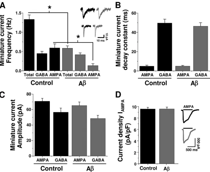
(3) Synaptic Depletion by A Peptide. for 30 min at 36 °C. The neurons were then washed twice with external solution and incubated for 30 min at 36 °C. The cells were mounted in a perfusion chamber that was placed on the stage of an inverted fluorescent microscope (Eclipse TE; Nikon) equipped with a xenon lamp and a ⫻40 objective (22–24 °C). The cells were briefly illuminated (200 ms) using a computer-controlled Lambda 10-2 filter wheel (Sutter Instruments). Regions of interest were simultaneously selected on neuronal somata containing Fluo-3 fluorescence (excitation 480 nm, emission 510 nm) in an optical field having usually more than 10 cells. Images were collected at 2–5-s intervals during a continuous 5-min period. The imaging was carried out with a 12-bit cooled SensiCam camera (PCO, Kelheim, DE). FM1-43 Loading and Unloading—Presynaptic vesicles were labeled by exposure to styryl dye FM1-43 (15 M; Molecular Probes) during a high K⫹ depolarization for 5 min and washed immediately. Coverslips were mounted on a rapid switching flow perfusion chamber with an epifluorescence microscope (Nikon Eclipse 3000). Depolarization-dependent destaining. 2508 JOURNAL OF BIOLOGICAL CHEMISTRY. was induced by bath perfusion with 30 mM K⫹ (equiosmolar replacement of Na⫹). To measure the effect of A on poststimulus endocytosis, control and treated neurons were stimulated for 5 min in 30 mM K⫹ and exposed to FM1-43 for 5 min after a variable delay (⌬t) from the onset of the stimulus. As the ⌬t increased, the endocytosis cycle was completed, reducing actual FM1-43 uptake (17). The reduced endocytosis was expressed relative to that of ⌬t ⫽ 0. Immunofluorescence—Presynaptic terminals were loaded with AM1-43 (15 M; Biotium, Inc., Hayward, CA) during high K⫹ depolarization for 5 min and immediately washed in dye-free solution with nominal Ca2⫹ to minimize spontaneous dye loss, fixed for 30 min with 4% paraformaldehyde, and permeabilized with 0.1% Triton X-100 in phosphate-buffered saline. Nonspecific immunoreactivity was blocked with 10% horse serum for 1 h at room temperature. Monoclonal synapsin-1 antibody (1:100; Santa Cruz Biotechnology) was incubated overnight followed by incubation with an antigoat secondary antibody conjugated with fluorescein isothiocyanate (1:500; Jackson ImmunoResearch Laboratories). VOLUME 285 • NUMBER 4 • JANUARY 22, 2010. Downloaded from http://www.jbc.org/ at PONTIFICIA UNIVERSIDAD on May 16, 2016. FIGURE 2. Effect of A on glutamatergic and GABAergic transmissions. A, A (500 nM, 24 h) on frequency of total and isolated AMPA and GABAA miniature currents. Insets, characteristic GABAA (upper) and AMPA (lower) currents. B and C, effects of A on time course and peak amplitude of postsynaptic currents. D, current density for evoked AMPA responses in control and A-treated neurons. The symbols are mean ⫾ S.E. from at least five neurons (*, p ⬍ 0.05)..
(4) Synaptic Depletion by A Peptide. JANUARY 22, 2010 • VOLUME 285 • NUMBER 4. JOURNAL OF BIOLOGICAL CHEMISTRY. 2509. Downloaded from http://www.jbc.org/ at PONTIFICIA UNIVERSIDAD on May 16, 2016. processes (50 m) were counted for control and treated neurons. Transmission Electronic Microscopy—Samples of 20 l of A, at a concentration of 50 M, were applied to carbon-coated Formvar grids (ORIGEN) pretreated with glutaraldehyde solution. Amyloid fiber or aggregates were stained with 20 l of 2% (w/v) uranyl acetate solution, and the grid was air-dried. Samples were examined using a JEOL 1200 EX II electronic microscope. In other experiments, neurons plated directly onto the bottom of 35-mm plastic tissue culture plates were used. Cultures of 12 days in vitro neurons were incubated with A for 24 h, subsequently washed, and then fixed for 30 min with 2% glutaraldehyde. The samples were placed in a plastic resin, sectioned, and mounted on a grid. The samples were visualized on a Phillips electronic microscope (model EM-300) operated between 60 and 120 kV. Using a double-blind protocol, synaptic boutons from control and A-treated neurons were counted. Here, all sections were analyzed in codified samples. Only at the end of the experiment were the sections identified as part of an experimental condition. The number of ⫹ FIGURE 3. A reduced synaptic vesicle recycling. A, K -induced destaining of FM1-43 in control and synaptic vesicles was counted in the A-treated neurons. B, effect of A (500 nM, 24 h) on fractional and time constant of FM1-43 release. area that included the high electron C, ratio of endocytosis of FM1-43 following a variable time delay (0 –300 s) from an initial depolarizing K⫹ dense postsynaptic zone. pulse in control and treated neurons. D, protocol used to load FM after variable times of exocytosis. E, time Data Analysis—Nonlinear analycourse of FM1-43 fluorescence constructed from the data in C for control and treated neurons. The lines are the best single-exponential fit to the data. The symbols are mean ⫾ S.E. from 12 independent record- sis was performed using Origin ings (*, p ⬍ 0.05). (Microcal). The values are expressed as mean ⫾ S.E. Statistical The samples were mounted in fluorescent mounting me- differences were determined using Student’s t test or analysis of variance. The experiments were performed in triplicate. dium (DAKO) for confocal analysis. Fluorescence Measurements and Confocal Microscopy— FM1-43 immunofluorescence was acquired using an epifluo- RESULTS rescence Nikon microscope (Eclipse TE; Nikon) equipped with Effects of Chronic A on Synaptic Transmission in Hippocama ⫻100 objective (Neofluor, oil immersion, NA 1.0). FM1-43 pal Neurons—Previous studies have shown that A inhibits fluorescence was excited at 540 nm and collected with 620-nm synaptic transmission in brain neurons (6, 18, 19). Those studlong pass filters. Fluorescence intensity was measured using a ies suggested that the inhibition was due to a postsynaptic 2 ⫻ 2 binning with a CCD camera (SensiCam; PCO). Images action because A reduced long term potentiation and excitwere digitized and processed with Imaging Axon Workbench atory postsynaptic current amplitudes. However, only a single 2.2 (Axon Instruments). AM1-43 and fluorescein isothiocya- concentration of A (usually 1 M) and one time point were nate immunofluorescence were visualized using a confocal analyzed, making it difficult to determine the mechanisms and Nikon C1 TE2000U microscope (⫻60, water immersion, NA kinetics of the inhibition. To gain further insights on potential 1.4) with lasers of argon (488 nm) and He-Ne (543 nm) for differences at a range of concentrations, hippocampal neurons fluorescein isothiocyanate and AM1-43, respectively. After were exposed for 24 h to several concentrations of A (5 nM–10 acquisition, images were processed with ImageJ (National Insti- M). A low concentration of A (5 nM) was without effect on tutes of Health). Co-localized punctas in the soma and primary synaptic transmission parameters. In contrast, higher con-.
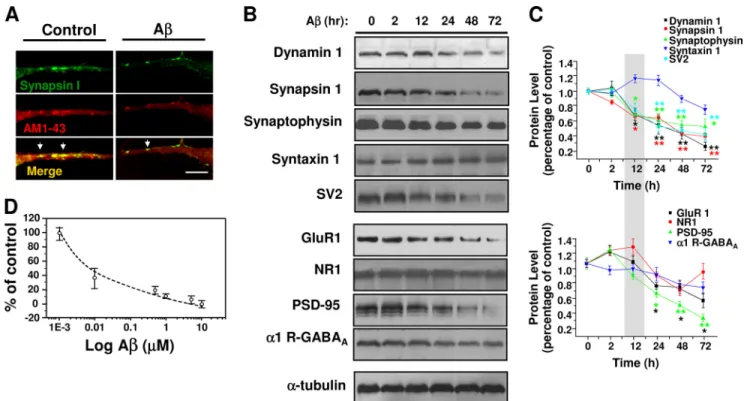
(5) Synaptic Depletion by A Peptide. centrations (50 –500 nM) reduced the frequency of miniature currents, without manifesting changes in amplitude (Fig. 1, A and B). A decreased quantal current amplitude at concentrations of 1 M and above (Fig. 1B). On the other hand, neurons exposed to A monomers, fibrils, or reverse A40-1 (all at 500 nM) displayed unchanged neurotransmission (Fig. 1C). The ultrastructural forms (oligomers and short fibrils) that appeared after 120 –150 min of agitation, corresponding to the neuroactive A forms, were structurally (Fig. 1Db) and functionally similar to a heterogeneous array of forms defined as oligomers, protofibrils, and A-derived diffusible ligands, known to enhance neuronal spiking and intracellular calcium (14, 20, 21). These features were not observed in either nonaggregated peptide (monomers) (Fig. 1Da) or in aggregated peptides after 3 days of incubation, where long mature fibrils were mainly observed (Fig. 1Dc). Therefore, the neuroactive forms of A able to depress synaptic transmission corresponded to intermediate aggregates. Under the present experimental conditions, the predominant glutamatergic transmission was AMPAergic, whereas the inhibitory transmission was GABAergic. Few low amplitude synaptic NMDA currents were detected in our recordings, and because of conflicting effects of A on this receptor (4, 22), we decided to focus the present study mainly on pharmacologically isolated AMPA and GABAA-mediated synaptic currents. The inhibitory effect of chronic A was highly selective for glutamatergic transmission because the miniature GABAergic currents were not significantly altered (Fig. 2A). On the other hand, analyses of the peak current amplitude and time constant of decay, parameters related to the postsynaptic responsiveness to neurotransmitters, showed that AMPA and GABAA receptors. 2510 JOURNAL OF BIOLOGICAL CHEMISTRY. were affected to a lower extent (Fig. 2, B and C). Furthermore, direct application of 100 M AMPA to the postsynaptic membrane (Fig. 2D) and analysis of cumulative probability of AMPA synaptic currents in control and treated neurons (not shown) showed that the current density was unchanged by 24 h of A incubation suggesting that membrane levels of AMPA receptors were not affected. Effects of A on Vesicular Recycling in Hippocampal Neurons— To monitor directly whether the decrease in miniature excitatory postsynaptic current frequency was related to changes in exocytosis-endocytosis vesicular cycles, we performed cellular imaging with FM1-43 (23, 24, 17). Application of 30 mM external K⫹ indicated that the destaining (exocytosis) of FM1-43 was significantly reduced by chronic application (24 h) of 500 nM A (Fig. 3A). Additional experiments using AM1-43, a fixable form of FM1-43, showed that the vesicular uptake was reduced by more than 40% with chronic A (95 ⫾ 5 versus 55 ⫾ 5 arbitrary fluorescent units, n ⫽ 3). Moreover, although the level of released FM1-43 was reduced by A, the time constant of exocytosis was similar (Fig. 3B). Next, we examined the capacity of the stimulated vesicles to reuptake the fluorescent dye to determine whether endocytosis was being altered by A. Using a modified FM1-43 reuptake protocol (17), we found that control vesicles completed ⬃80% of the dye endocytosis within 60 s of the depolarizing stimuli (Fig. 3, C and D), with a time constant for endocytosis of 35 ⫾ 5 s (Fig. 3E), which is in agreement with previous studies (17). A caused a lengthening of the time constant of this process (66 ⫾ 6 s). Therefore, the data showed that a low concentration of A reduced the magnitude of exocytosis and that the remaining synaptic vesicles displayed a much slower speed of endocytosis. VOLUME 285 • NUMBER 4 • JANUARY 22, 2010. Downloaded from http://www.jbc.org/ at PONTIFICIA UNIVERSIDAD on May 16, 2016. FIGURE 4. A reduced several synaptic vesicle proteins. A, confocal micrographs showing the effect of chronic A (500 nM, 24 h) on active synaptic proteins visualized by co-localization of synapsin-1 and AM1-43 (yellow and arrows). B, time course of A effects on the level of several pre- and postsynaptic proteins. C, graphs showing the quantification of signal intensity obtained at different times of treatment. The faster effect of A at 12 h was on presynaptic proteins. The bars are mean ⫾ S.E. from three independent experiments (*, p ⬍ 0.05; **, p ⬍ 0.01)..
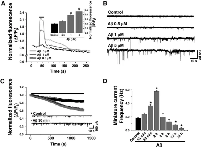
(6) Synaptic Depletion by A Peptide. Early Effects of A on Presynaptic Proteins—Confocal microscopy showed a reduction in active synaptic vesicles in A-treated neurons. This was shown as a diminished co-localization of synapsin-1 and the FM1-43 fixable analog AM1-43, expressed by the number of presynaptic puncta/10-m length (Fig. 4, A and D). In addition, quantitative analyses of several presynaptic proteins showed that with the exception of syntaxin-1 they were all reduced with 12 h of treatment with A, partly explaining the synaptic failure. On the other hand, among the postsynaptic proteins, only PSD-95 and GluR1 levels were significantly altered at 24 h with A (Fig. 4B). These data indicate that the low A concentration induced an earlier presynaptic deficit, which was followed by a postsynaptic alteration. The lack of functional differences (Fig. 2D) suggests that under this condition (time and concentration) active AMPA membrane receptors were still basically normal. Previous experiments suggested that A reduced several presynaptic proteins (12, 25, 26). Therefore, we decided to examine the ultrastructural features of the synapses using electron microscopy in double-blind experiments. The data showed that neurons cultured with A displayed a reduced number of synaptic vesicles, especially those near the presynaptic active zones (Fig. 5A). Quantitative analysis showed that the number of synaptic vesicles was reduced from 50 ⫾ 4 vesicles/m2 in control to 18 ⫾ 3 vesicles/m2 in A-treated neurons (Fig. 5B). These data suggest that chronic application of A might affect the JANUARY 22, 2010 • VOLUME 285 • NUMBER 4. RRP of synaptic vesicles. To test this possibility further, we studied the synaptic transmission induced by a hyperosmotic sucrose solution, known to cause the release of RRP (27, 28). These experiments showed that unlike control neurons (top trace, Fig. 5C), which had a 4-fold increase in synaptic currents in the hypertonic solution (Fig. 5D), the A-treated neurons had few miniature synaptic currents (Fig. 5D), and their frequency was less affected by the hypertonic solution. Acute A Increased Intracellular Calcium, Vesicle Release, and Synaptic Transmission—A was shown to increase intracellular calcium in non-neuronal cells (21, 22). In agreement, short (60-s) applications of A (500 nM–5 M) with a puffer pipette induced a rapid and reversible increase in intracellular calcium in hippocampal neurons (Fig. 6A). It seemed feasible that this increase in intracellular calcium could affect the discharge of synaptic vesicles (29). We found that A perfusions (500 nM–5 M, 30 min) produced a concentration-dependent increase in miniature current frequency (Fig. 6B). Parallel analysis of FM1-43 destaining in the presence of A (500 nM) showed enhancement in the slope of fluorescent decay (⫺1.2 ⫻ 10⫺4 ⫾ 3.0 ⫻ 10⫺6 versus ⫺3.3 ⫻ 10⫺4 ⫾ 3.2 ⫻ 10⫺6 URF/s; p ⬍ 0.0001) after 10 min of application (Fig. 6C). The above mentioned results showed that a short application (30 min) of A increased the frequency of miniature synaptic currents in the neurons, whereas longer exposures (24 h) reduced it (Figs. 1A and 6C). Therefore, we decided to examine JOURNAL OF BIOLOGICAL CHEMISTRY. 2511. Downloaded from http://www.jbc.org/ at PONTIFICIA UNIVERSIDAD on May 16, 2016. FIGURE 5. A reduced the synaptic vesicular pool. A, electron micrographs show synapses from control (left) and 500 nM A-treated (right) neurons. Rectangles illustrate the areas for quantification. Scale bars, 100 nm. B, columns showing the number of vesicles in both conditions from 12 selected active zones using a double-blind protocol (*, p ⬍ 0.05). C and D, effect of hypertonic sucrose solution (500 mOs) on miniature current frequencies in control and A (500 nM)-treated neurons. Bars are mean ⫾ S.E. from 18 neurons (***, p ⬍ 0.001; *, p ⬍ 0.05)..
(7) Synaptic Depletion by A Peptide. the effects of A on synaptic transmission after exposing the neurons to A for various times (15 min up to 24 h). The data showed that the frequency of miniature currents was greatly increased during the first 2 h, and thereafter the frequency decreased progressively to well below the control level (Fig. 6D). The results mentioned above suggest that the synaptic failure produced by the prolonged action of A on the synapse may be due to the strong early enhancement of quantal transmitter release leading to neurotransmitter vesicle depletion. Before testing this hypothesis, it was relevant to determine the mechanism by which A enhanced intracellular calcium and synaptic release. Given that the increase in intracellular calcium could be mediated by voltage-dependent calcium channels (30), glutamatergic receptors (NMDA and AMPA) (8, 31), or amyloid pores (32), we studied the action of several inhibitors. Analysis of A-induced increase in intracellular calcium, monitored with Fluo-3, showed that it was not blocked by a mixture containing calcium channel antagonists (-conotoxin, agatoxin-VI, and nifedipine) or CNQX and AP5 ((2R)-amino-5-phosphonopentanoate), antag-. 2512 JOURNAL OF BIOLOGICAL CHEMISTRY. onists of AMPA and NMDA receptors, respectively. Next, we used a small seven-amino acid peptide (Na7) that has been shown to block the ion current induced by A amyloid pores in lipid membranes, as well as A cellular toxicity (33, 34). Interestingly, this peptide blocked most of the increase in intracellular calcium induced by A (Fig. 7A), suggesting that the calcium increase was mediated by the formation of amyloid pores in the neuronal membrane. In agreement with the involvement of a calcium-dependent mechanism on the acute and long term effects of A on synaptic transmission, we found that its early and chronic synaptic effects were blocked by reducing extracellular calcium (from 2 to 0.01 mM) or by adding Na7 (Fig. 7B). Na7 also antagonized the A-induced decrease in SV2 (Fig. 7C). Two analogs (Na13 and Na15) previously reported to be inactive blocking the ion permeation through the A pore were not able to antagonize A actions on a presynaptic marker (Fig. 7, C and D), emphasizing the role of calcium influx through the amyloid pore in synaptic failure. Additionally, these data support the critical role proposed for the histidines in Na7 (33). VOLUME 285 • NUMBER 4 • JANUARY 22, 2010. Downloaded from http://www.jbc.org/ at PONTIFICIA UNIVERSIDAD on May 16, 2016. FIGURE 6. Acute A increased intracellular calcium and the release of synaptic vesicles. A, Fluo-3-associated fluorometric recordings showing increases in intracellular calcium produced by short applications of A (500 nM–5 M). Inset, concentration-dependent effects of A on intracellular calcium from three independent experiments (p ⬍ 0.05). B, traces showing the rapid increase in miniature synaptic current frequency with different concentrations of A. C, FM1-43 destaining under control (filled symbols) and A containing solutions (open symbols). The current traces were obtained after 30 min of exposure to control and 500 nM A. D, effect of different A incubation times on the frequency of miniature synaptic currents. Bars are mean ⫾ S.E. from at least six neurons (*, p ⬍ 0.05)..
(8) Synaptic Depletion by A Peptide. JANUARY 22, 2010 • VOLUME 285 • NUMBER 4. JOURNAL OF BIOLOGICAL CHEMISTRY. 2513. Downloaded from http://www.jbc.org/ at PONTIFICIA UNIVERSIDAD on May 16, 2016. on presynaptic release. Nevertheless, higher concentrations and longer exposure times with A affected postsynaptic properties such as current amplitude and protein expression levels (Figs. 1 and 4). The effect of A on cultured hippocampal neurons was clearly evident on AMPAergic transmission in agreement with other studies in hippocampal slices (38). Clear postsynaptic effects of A were not evident and might be complicated by the concentration and the type of active species. Although the present study was done with a mixture of amyloid aggregates, most likely present in the aging brain, previous studies utilized an oligomer-rich preparation (8, 38). Mechanisms of A Action—Presynaptic vesicle fusion to the active zones is strongly dependent on the entry of extracellular calcium and subsequent activation of intracellular signaling and cytoskeleton reFIGURE 7. Blockade of A induced increases in intracellular calcium and synaptic transmission by a small modeling (39). Consequently, the peptide. A, effect of several calcium channel blockers on the increase in intracellular calcium, measured with early increase in synaptic transmitFluo-3, induced by the application of 500 nM A with a puffer pipette (indicated by a horizontal line in Fig. 6A). ter release by A was associated The concentrations of the blockers were 1 M CNQX, 50 M D-AP5, 1 M conotoxin, 1 M agatoxin, 3 M nifedipine, and 100 M Na7. The effects were normalized with respect to control (p ⬍ 0.05). B, result of lowering with the rise in intracellular calcalcium influx using a nominally Ca2⫹ free solution or 1 mM EGTA on the early and chronic effects of A on cium, in agreement with previous miniature current frequency from three independent experiments (p ⬍ 0.05). For these experiments, the neurons were exposed to A alone or in the presence of 100 M Na7. C and D, effect of A-induced reduction studies on liposomes and clonal cell on SV2 in the presence and absence of Na7, Na13, and Na15. The bars are mean ⫾ S.E. from nine different lines (21, 22). This increase might neurons (p ⬍ 0.05). depend on voltage-gated calcium channels (40), AMPA receptors (38), DISCUSSION NMDA receptors (8), or formation of calcium-permeable Effects of Low A Concentrations on Synaptic Transmission— membrane pores (21, 22, 32, 41). Our data showed that antagOnly recent studies have dealt with the action of A at concen- onists for calcium channels, NMDA and AMPA receptors were trations without overt neurotoxicity on synaptic properties unable to block the A-induced increase in intracellular cal(35). Although some debate still exists on whether A can cium, although it was blocked by Na7, a small peptide reported down-regulate specific components of synaptic transmission, to block calcium influx, through the amyloid pore (34, 42). several studies in rodent hippocampus showed that A affected Noteworthy, the early and delayed effects of A on synaptic long term potentiation, NMDA- and AMPA-evoked currents transmission were also blocked by Na7, suggesting that these (8, 36, 37). Furthermore, studies in transgenic animals revealed synaptic effects resulted as a consequence of pore formation that overexpression of amyloid precursor protein reduced pre- and subsequent calcium influx. Interestingly, the experimental synaptic proteins (36) together with complex brain functions. procedures used to block the early synaptic potentiation also In addition, it was shown that micromolar concentrations of inhibited the delayed synaptic failure, suggesting that they are A inhibited long term potentiation and NMDA-evoked cur- linked. Finally, a significant dysfunction in calcium homeostasis rents (8). Based on all this evidence, it is now accepted that with A is in good agreement with several studies that related minute synaptic dysfunctions may represent the earliest signs AD to ion alterations, with calcium being central to these mechof AD (35). anisms (43). However, the contribution of other ions such as The present study, with low concentrations of A, demon- copper, iron, or zinc to the mechanism behind A synaptotoxstrates a largely unrecognized action of A as an inhibitor of icity cannot be ruled out (44). In fact, histidine 13 and 14 of the presynaptic function. For instance, release of synaptic vesi- A sequence, corresponding to key amino acids for the metalcles, as measured by real time imaging with FM1-43, was binding site of this peptide, are also determinants for the A increased. A also enhanced the frequency of miniature syn- pore permeability (45). In terms of a mechanism explaining the aptic currents, without changes in the amplitude and kinetics synaptic failure, we found that chronic A reduced the number of the postsynaptic currents, supporting a primary effect of A of total and docked synaptic vesicles in nerve terminals and.
(9) Synaptic Depletion by A Peptide. Acknowledgments—We thank Dr. Ricardo Miledi for critically reading the manuscript and Lauren Aguayo for technical and editing assistance. REFERENCES 1. Selkoe, D. J. (2002) Science 298, 789 –791 2. Mattson, M. P. (1997) Physiol. Rev. 77, 1081–1132 3. McLean, C. A., Cherny, R. A., Fraser, F. W., Fuller, S. J., Smith, M. J., Beyreuther, K., Bush, A. I., and Masters, C. L. (1999) Ann. Neurol. 46, 860 – 866 4. Shankar, G. M., Bloodgood, B. L., Townsend, M., Walsh, D. M., Selkoe, D. J., and Sabatini, B. L. (2007) J. Neurosci. 27, 2866 –2875 5. Goodman, L. S., Hardman, J. G., Limbird, L. E., and Gilman, A. G. (2001) Goodman and Gilman’s the Pharmacological Basis of Therapeutics, pp. 203–220, 10th Ed., McGraw-Hill, New York 6. Oddo, S., Caccamo, A., Shepherd, J. D., Murphy, M. P., Golde, T. E., Kayed, R., Metherate, R., Mattson, M. P., Akbari, Y., and LaFerla, F. M. (2003) Neuron 39, 409 – 421 7. Shankar, G. M., Li, S., Mehta, T. H., Garcia-Munoz, A., Shepardson, N. E., Smith, I., Brett, F. M., Farrell, M. A., Rowan, M. J., Lemere, C. A., Regan, C. M., Walsh, D. M., Sabatini, B. L., and Selkoe, D. J. (2008) Nat. Med. 14, 837– 842 8. Snyder, E. M., Nong, Y., Almeida, C. G., Paul, S., Moran, T., Choi, E. Y., Nairn, A. C., Salter, M. W., Lombroso, P. J., Gouras, G. K., and Greengard, P. (2005) Nat. Neurosci. 8, 1051–1058 9. Nomura, I., Kato, N., Kita, T., and Takechi, H. (2005) Neurosci. Lett. 391, 1– 6 10. Chauhan, N. B., and Siegel, G. J. (2002) J. Neurosci. Res. 69, 10 –23 11. Reddy, P. H., Mani, G., Park, B. S., Jacques, J., Murdoch, G., Whetsell, W., Jr., Kaye, J., and Manczak, M. (2005) J. Alzheimers Dis. 7, 103–117 12. Kelly, B. L., Vassar, R., and Ferreira, A. (2005) J. Biol. Chem. 280, 31746 –31753 13. Kelly, B. L., and Ferreira, A. (2006) J. Biol. Chem. 281, 28079 –28089 14. Ye, C., Walsh, D. M., Selkoe, D. J., and Hartley, D. M. (2004) Neurosci. Lett. 366, 320 –325. 2514 JOURNAL OF BIOLOGICAL CHEMISTRY. 15. Glabe, C. G., and Kayed, R. (2006) Neurology 66, S74 –S78 16. Tapia, J. C., Mentis, G., Navarrete, R., Nualart, F., Figueroa, E., Sánchez, A., and Aguayo, L. G. (2001) Neuroscience 108, 493–506 17. Ryan, T. A., Smith, S. J., and Reuter, H. (1996) Proc. Natl. Acad. Sci. U.S.A. 93, 5567–5571 18. Walsh, D. M., and Selkoe, D. J. (2004) Protein Pept. Lett. 11, 213–228 19. Wang, Q., Walsh, D. M., Rowan, M. J., Selkoe, D. J., and Anwyl, R. (2004) J. Neurosci. 24, 3370 –3378 20. Rhee, J. S., Ebihara, S., and Akaike, N. (1994) J. Neurophysiol. 72, 1103–1108 21. Alarcón, J. M., Brito, J. A., Hermosilla, T., Atwater, I., Mears, D., and Rojas, E. (2006) Peptides 27, 95–104 22. Demuro, A., Mina, E., Kayed, R., Milton, S. C., Parker, I., and Glabe, C. G. (2005) J. Biol. Chem. 280, 17294 –17300 23. Ryan, T. A., Reuter, H., Wendland, B., Schweizer, F. E., Tsien, R. W., and Smith, S. J. (1993) Neuron 11, 713–724 24. Ryan, T. A., and Smith, S. J. (1995) Neuron 14, 983–989 25. Yao, P. J., and Coleman, P. D. (1998) Neurosci. Lett. 252, 33–36 26. Grace, E. A., Rabiner, C. A., and Busciglio, J. (2002) Neuroscience 114, 265–273 27. Kriebel, M. E., and Pappas, G. D. (1987) Neuroscience 23, 745–756 28. Lonart, G., and Simsek-Duran, F. (2006) Brain Res. 1107, 42–51 29. Cousin, M. A., and Robinson, P. J. (2000) J. Neurosci. 20, 949 –957 30. Bobich, J. A., Zheng, Q., and Campbell, A. (2004) J. Alzheimers Dis. 6, 243–255 31. Blanchard, B. J., Konopka, G., Russell, M., and Ingram, V. M. (1997) Brain Res. 776, 40 –50 32. Arispe, N., Pollard, H. B., and Rojas, E. (1994) Ann. N.Y. Acad. Sci. 747, 256 –266 33. Arispe, N. (2004) J. Membr. Biol. 197, 33– 48 34. Arispe, N., Diaz, J. C., and Simakova, O. (2007) Biochim. Biophys. Acta 1768, 1952–1965 35. Haass, C., and Selkoe, D. J. (2007) Nat. Rev. Mol. Cell Biol. 8, 101–112 36. Hsia, A. Y., Masliah, E., McConlogue, L., Yu, G. Q., Tatsuno, G., Hu, K., Kholodenko, D., Malenka, R. C., Nicoll, R. A., and Mucke, L. (1999) Proc. Natl. Acad. Sci. U.S.A. 96, 3228 –3233 37. Stéphan, A., Laroche, S., and Davis, S. (2001) J. Neurosci. 21, 5703–5714 38. Hsieh, H., Boehm, J., Sato, C., Iwatsubo, T., Tomita, T., Sisodia, S., and Malinow, R. (2006) Neuron 52, 831– 843 39. Schneggenburger, R., and Neher, E. (2005) Curr. Opin. Neurobiol. 15, 266 –274 40. Ramsden, M., Plant, L. D., Webster, N. J., Vaughan, P. F., Henderson, Z., and Pearson, H. A. (2001) J. Neurochem. 79, 699 –712 41. Quist, A., Doudevski, I., Lin, H., Azimova, R., Ng, D., Frangione, B., Kagan, B., Ghiso, J., and Lal, R. (2005) Proc. Natl. Acad. Sci. U.S.A. 102, 10427–10432 42. Qi, J. S., and Qiao, J. T. (2001) Neuroscience 105, 845– 852 43. Mattson, M. P., and Chan, S. L. (2003) Cell Calcium 34, 385–397 44. Opazo, C., Huang, X., Cherny, R. A., Moir, R. D., Roher, A. E., White, A. R., Cappai, R., Masters, C. L., Tanzi, R. E., Inestrosa, N. C., and Bush, A. I. (2002) J. Biol. Chem. 277, 40302– 40308 45. Díaz, J. C., Linnehan, J., Pollard, H., and Arispe, N. (2006) Biol. Res. 39, 447– 460 46. Khvotchev, M., Lonart, G., and Südhof, T. C. (2000) Neuroscience 101, 793– 802 47. Rosenmund, C., and Stevens, C. F. (1996) Neuron 16, 1197–1207 48. Rizzoli, S. O., and Betz, W. J. (2005) Nat. Rev. Neurosci. 6, 57– 69 49. Ashton, A. C., Volynski, K. E., Lelianova, V. G., Orlova, E. V., Van Renterghem, C., Canepari, M., Seagar, M., and Ushkaryov, Y. A. (2001) J. Biol. Chem. 276, 44695– 44703 50. Kourie, J. I., Henry, C. L., and Farrelly, P. (2001) Cell. Mol. Neurobiol. 21, 255–284. VOLUME 285 • NUMBER 4 • JANUARY 22, 2010. Downloaded from http://www.jbc.org/ at PONTIFICIA UNIVERSIDAD on May 16, 2016. reduced the RRP demonstrated by the hyperosmotic sucrose solution (46, 47). Structurally, synaptic vesicles are found in three pools: (i) RRP that correspond to docked vesicles in the active site ready to be released (although see Ref. 48), (ii) the recycling pool sensitive only to strong stimulations (24), and (iii) the reserve pool located at a distance from the active zone. Therefore, these new results strongly support the idea that A affects RRP and recycling pools, directly apposed to the active zone. Overall, we postulate that the earliest effects of low A concentrations are mainly presynaptic and reflect a combination of disruption of RRP and recycling pools. Interestingly, these novel actions of A show strong similarities, although with lower potency, to the effect of ␣-latrotoxin on neurotransmission. For example, after a strong enhancement of synaptic transmission, ␣-latrotoxin induced vesicle depletion and diminution in miniature potentials by a pore-forming mechanism (49), having conductance and kinetic properties very similar to those of pores formed by A in lipid bilayers (50). The characterization of A-induced calcium-permeable pores, displaying properties similar to those of ␣-latrotoxin, is currently under study in our laboratory..
(10) β-Amyloid Causes Depletion of Synaptic Vesicles Leading to Neurotransmission Failure Jorge Parodi, Fernando J. Sepúlveda, Jorge Roa, Carlos Opazo, Nibaldo C. Inestrosa and Luis G. Aguayo J. Biol. Chem. 2010, 285:2506-2514. doi: 10.1074/jbc.M109.030023 originally published online November 13, 2009. Access the most updated version of this article at doi: 10.1074/jbc.M109.030023. Click here to choose from all of JBC's e-mail alerts This article cites 49 references, 16 of which can be accessed free at http://www.jbc.org/content/285/4/2506.full.html#ref-list-1. Downloaded from http://www.jbc.org/ at PONTIFICIA UNIVERSIDAD on May 16, 2016. Alerts: • When this article is cited • When a correction for this article is posted.
(11)
Figure
Documento similar
In the preparation of this report, the Venice Commission has relied on the comments of its rapporteurs; its recently adopted Report on Respect for Democracy, Human Rights and the Rule
On the other hand, the larvae from 18 ovipostions that were kept in the laboratory at ambient temperatu- re emerged between two and four days after they were collected in the
In conclu- sion, the results of the study showed that of the variables of executive function analyzed, it is deficits in inhibition that relate to the greatest
The Dome of the Rock does attest the existence, at the end of the seventh century, of materials immediately recognisable as Koranic in a text that not infrequently
Parameters of linear regression of turbulent energy fluxes (i.e. the sum of latent and sensible heat flux against available energy).. Scatter diagrams and regression lines
It is generally believed the recitation of the seven or the ten reciters of the first, second and third century of Islam are valid and the Muslims are allowed to adopt either of
The purpose of the research project presented below is to analyze the financial management of a small municipality in the province of Teruel. The data under study has
Quantitative comparisons of structural synaptic parameters To substantiate the above observations, structural parameters of Po axon synapses were statistically compared between in
