Edited by:
Renato Grillo, Universidade Federal do ABC, Brazil
Reviewed by:
Benjaram M. Reddy, CSIR—Indian Institute of Chemical Technology, India Eric D. Van Hullebusch, UNESCO-IHE Institute for Water Education, Netherlands Monique C. P. Mendonça, University of Campinas, Brazil
*Correspondence:
Bryan Calderón Jiménez [email protected] José R. Vega Baudrit [email protected]
Specialty section:
This article was submitted to Green and Environmental Chemistry, a section of the journal Frontiers in Chemistry
Received:21 December 2016
Accepted:06 February 2017
Published:21 February 2017
Citation:
Calderón-Jiménez B, Johnson ME, Montoro Bustos AR, Murphy KE, Winchester MR and Vega Baudrit JR (2017) Silver Nanoparticles: Technological Advances, Societal Impacts, and Metrological Challenges. Front. Chem. 5:6. doi: 10.3389/fchem.2017.00006
Silver Nanoparticles: Technological
Advances, Societal Impacts, and
Metrological Challenges
Bryan Calderón-Jiménez1, 2*, Monique E. Johnson1, Antonio R. Montoro Bustos1, Karen E. Murphy1, Michael R. Winchester1and José R. Vega Baudrit3*
1Material Measurement Laboratory, Chemical Sciences Division, National Institute of Standards and Technology,
Gaithersburg, MD, USA,2Chemical Metrology Division, National Laboratory of Metrology, San Jose, Costa Rica,3National
Laboratory of Nanotechnology, National Center of High Technology, San Jose, Costa Rica
Silver nanoparticles (AgNPs) show different physical and chemical properties compared to their macroscale analogs. This is primarily due to their small size and, consequently, the exceptional surface area of these materials. Presently, advances in the synthesis, stabilization, and production of AgNPs have fostered a new generation of commercial products and intensified scientific investigation within the nanotechnology field. The use of AgNPs in commercial products is increasing and impacts on the environment and human health are largely unknown. This article discusses advances in AgNP production and presents an overview of the commercial, societal, and environmental impacts of this emerging nanoparticle (NP), and nanomaterials in general. Finally, we examine the challenges associated with AgNP characterization, discuss the importance of the development of NP reference materials (RMs) and explore their role as a metrological mechanism to improve the quality and comparability of NP measurements.
Keywords: silver nanoparticles, synthesis, characterization, environment health and safety, metrology, reference materials
DEFINING NANOMATERIALS AND NANOPARTICLES: THEIR
IMPORTANCE IN NANOSCIENCE, AND NANOTECHNOLOGY
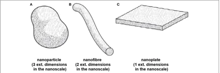
is described as a “discrete piece of material with one, two or three external dimensions in the nanoscale” (ISO/TS 80004-1, 2015) and a nanostructured material is a “material having internal structure or surface structure in the nanoscale” (ISO/TS 80004-4, 2011). Nano-objects, can be classified into three categories (seeFigure 1) depending on their size and shape characteristics (ISO/TS 80004-1, 2015):
1. Nanoparticle (NP): “Nano-object with all external dimensions at the nanoscale where the lengths of the longest and shortest axes of the nano-object do not differ significantly”,
2. Nanofiber: “Nano-objects with two external dimensions at the nanoscale and the third dimension significantly larger”, 3. Nanoplate: “Nano-objects with one external dimension in the
nanoscale and the other two dimensions significantly larger”, ISO also provides a simple and general definition for engineered nanomaterials indicating that they are “nanomaterials designed for specific purposes or functions” (ISO/TS 80004-1, 2015).
Other organizations, federal agencies, and government bodies have developed their own approach to categorizing nanomaterials with the goal of assessing and controlling risk. The United States Environmental Protection Agency (U.S. EPA) has developed reporting and recordkeeping requirements for companies that manufacture or process nanoscale chemical substances. The entity describes “nanoscale chemical substances” as “chemical substances containing primary particles, aggregates, or agglomerates in the size range of 1 to 100 nm in at least one dimension” (EPA, 2015). The U.S. Food and Drug Administration (U.S. FDA) has issued a series of guidance documents with respect to the use of nanotechnology in FDA-regulated products (Hamburg, 2012; U.S. FDA, 2015). For example, when considering whether an U.S. FDA-regulated product involves nanotechnology, the U.S. FDA offers “Points to Consider” such as whether a material or product is engineered to have at least one dimension in the nanoscale range, or whether it exhibits chemical or physical properties or biological effects attributable to its dimensions (Croce, 2014; U.S. FDA, 2014). In recent years, the European Union (EU) has been engaged in a number of efforts to define “nanomaterials” and “engineered nanomaterial.” Particularly, the European Commission recommended the following definition for a nanomaterial: “Nanomaterial means a natural, incidental, or manufactured material containing particles, in an unbound state or as an aggregate or as agglomerate and where, for 50% or more of the particles in the number size distribution, one or more external dimensions in the size range 1–100 nm” (Commission Recommendation, 2011). The definitions cited in this directive are generally based on the ISO definition; however, they have been adapted with the goal of incorporating other technical concepts such as aggregation/agglomeration, particle size distribution, and particle number concentration (Commission Recommendation, 2011). Additionally, the EU has issued a series of directives in the fields of cosmetics (Regulation (EC) No 1223/2009), biocides (Regulation (EU) No 528/2012), food (Regulation (EU) No 1363/2013, Regulation (EU) No 1363/2013), and any food that was not used for human
consumption to a significant degree, commonly denominate “novel food”. (Regulation (EU) 2015/2283). Recently, extensive technical work has begun to focus on the goal of providing recommendations on the possible use and limitations of some measurement techniques (MTs) with respect to the application of the EU definition (Babick et al., 2016).
Efforts to adapt and/or recast existing regulations to define fundamental concepts and applications of nanomaterials in consumer products are taking place in France (Decree No 2012-232) Belgium (Decree No 2014/24329), Denmark (Decree No 644 of 13/06/2014), and Canada (Health Canada, 2011). These countries have recently enacted their own policies to study the potential risks associated with the commercialization of nanomaterials by collecting information and establishing inventories. For instance, with the goal of identifying and assessing potential risks and benefits, Canadian regulatory agencies request information from manufacturers and other stakeholders on physical-chemical properties such as composition, purity, morphology, particle size/size distribution, chemical reactivity, agglomeration/aggregation state, as well as information on the methods used to assign these properties (Health Canada, 2011).
Despite efforts in recent years to properly define nanotechnology-related terms, more work needs to be done with respect to the harmonization and standardization of the terminology used in this field. For example, the term “nanoparticle” is defined differently by ISO (ISO/TS 80004-2, 2015), ASTM (ASTM E2456-06, 2012), and IUPAC (Alemán et al., 2007) with regard to the number of dimensions and shapes that can be attributed to NPs. This however, does not imply that one definition is accurate while another is not; rather it demonstrates that definitions and terms in the nanotechnology field are still evolving and highlights the importance of generating robust descriptors for these emerging materials to satisfy the variety of angles where the terminology would be applied.
IMPACTS OF THE NANOPARTICLES AND
SILVER NANOPARTICLES (AgNP
S) ON
COMMERCE, TECHNOLOGY AND
SOCIETY
FIGURE 1 | Schematic diagrams displaying shape designations for nano-objects (A)Nanoparticle,(B)Nanofiber(C)Nanoplate©ISO. This material is excerpted from ISO 80004-2:20015 with permission from the American National Standard Institute (ANSI) on behalf of ISO. All rights reserved.
FIGURE 2 | (A)Number of nanotechnology patents published by the United State Patent and Trademark Office (USPTO), the European Patent Office (EPO), and the Japan Patent Office (JPO) between 1976 and 2005, demonstrating the exponential growth of this emerging technology. The drop in the number of USPTO patents in 2005 is due to the USPTO enforcing a stricter definition of the term “nanotechnology.” The decline in the number of JPO patents for 2005 and 2006 is due to the delay between the publication and granting of patents at the JPO (Chen et al., 2008a). Reprinted by permission from Macmillan Publishers Ltd: [Nature Nanotechnology], copyright (2008)(B)Number of nanoscience papers indexed in Scopus®Elsevier between 2000 and 2014 byShin et al. (2015)CC BY 2.0. This figure demonstrates the quick and substantial advances in the investigation in the nanoscience field.
(Parisi et al., 2015; Phogat et al., 2016), and energy (Lohse and Murphy, 2012).
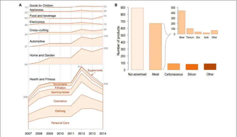
All of this escalation in the research and development of new NP applications will have a direct impact on commerce and society. In 2011, it was estimated that US$ 65 billion had been invested into the nanotechnology field (Miller and Wickson, 2015). Moreover, it was projected that a cumulative investment of US$150 billion would be made by the private sector into the field by 2015 (Cientifica, 2011). It was further predicted that nanotechnology in the form of NPs would impact different fields such as electronics, information technology and manufactured goods in health care and life sciences (Lux Research, 2008; Fiorino, 2010; Sargent, 2016). These projections are reflected in the growth of the numbers of consumer products incorporating NPs into their formulations. These numbers have grown from a total of 54 products identified in 2005 to over 1,800
nanomaterial-and NP-containing consumer products in 2014 produced by 622 companies in 32 countries (Vance et al., 2015). The variety of products ranged from goods for children to personal care products (Figure 3), with metals and metal oxides being the most commonly used NPs in commercial products. Although, silicon dioxide NPs (SiO2-NPs), titanium oxide NPs (TiO2-NPs),
and zinc oxide NPs (ZnO-NPs), are produced in the greatest quantities worldwide, with a global production of 5,500 t per year, 3,000 t per year and 550 t per year, respectively (Piccinno et al., 2012; Keller et al., 2013).
FIGURE 3 | (A)Number of available nanomaterial-containing consumer products over time (since 2007) by category (black print) and sub category (red print).(B)
Claimed composition of nanomaterials listed in the Nanotechnology Consumer Product Inventory, grouped into five major categories: Not advertised, metal (including metals and metal oxides), carbonaceous nanomaterials (carbon black, carbon nanotubes, fullerenes, graphene), silicon-based nanomaterials (silicon and silica), and other (organics, polymers, ceramics, etc.). The insert in 3b shows the claimed elemental composition of nanomaterials listed in the metals category: silver, titanium, zinc, gold, and other metals (magnesium, aluminum oxide, copper, platinum, iron, and iron oxides, etc.). Adapted fromVance et al. (2015)with the permission of Beilstein-Institut. CC BY 2.0.
would potentially be produced per yearHendren et al. (2011). It is projected that the global nanotechnology industry will continue to grow significantly. Specifically, the production of AgNPs is expected to reach approximately 800 t by 2025 ( Pulit-Prociak and Banach, 2016). Vance et al. (2015) showed that AgNPs have greater marketing value than other NPs and their presence in consumer products are more widely advertised. This noted popularity can be attributed to the well-documented antimicrobial properties of ionic silver (Le Ouay and Stellacci, 2015). It should be clear that AgNPs by themselves have no antibacterial or antifungal properties, but it is the release of silver ions due to the destabilization of the AgNPs which confers such properties. Other distinctive physico-chemical properties of AgNPs such as high electrical and thermal conductivity (Alshehri et al., 2012), surface-enhanced Raman scattering (Nie and Emory, 1997), catalytic activity (Xu et al., 2006), and non-linear optical properties (Kelly et al., 2003), have led to a variety of new products and scientific applications (Tran et al., 2013).
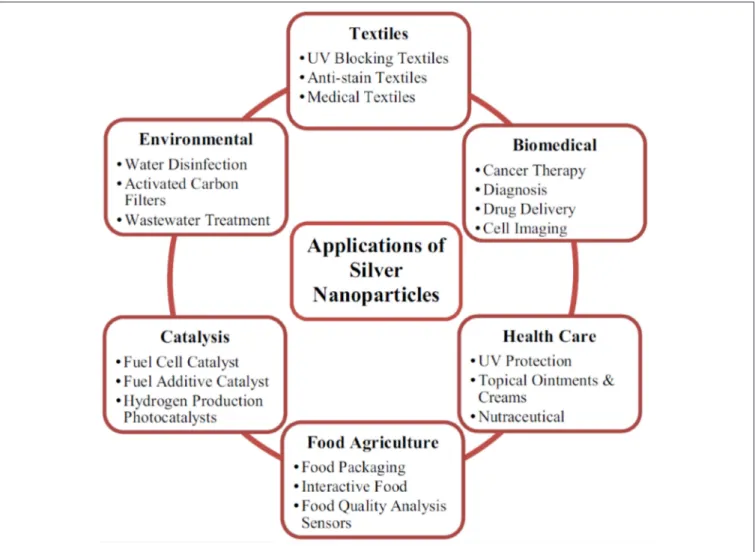
The physico-chemical properties mentioned above offer AgNPs the capability of being used in a plethora of new commercial and technological applications, including as antiseptic agents in the medical field, cosmetic, food packaging, bioengineering, electrochemistry, and catalysis industries (Keat
et al., 2015). As displayed in Figure 4, the antibacterial and antimicrobial activity of AgNPs are among the main reasons for their use in the formulation of surface cleaners, toys, textiles, air and water disinfection, antimicrobial catheters, antimicrobial gels, antimicrobial paints, food packaging supplies, clinical clothing, and food preservation etc. (Wijnhoven et al., 2009; Tolaymat et al., 2010; Tran et al., 2013).
FIGURE 4 | Applications of AgNPs.Reproduced fromKeat et al. (2015)with permission of Bioresources and Bioprocessing. CC BY 4.0
obtained were used to generate a novel third generation biosensor capable of measuring lactose in a large linear range, with high sensitivity and long-term stability.
Despite the promising economic benefit of the use of AgNPs and NPs in general, there are societal concerns associated with their use. For example, Miller and Wickson (2015) and Patenaude et al. (2015) discussed some barriers to accurate risk assessment and management of NPs and nanomaterials in general. These barriers include the lack of specific regulations for different types of NPs, the discrepancy between definitions, the lack of validated analytical methods and test protocols, the scarcity of reliable information about commercial use, and the lack of reliable exposure and toxicity data. Similarly, Hofmann et al. (2015)discussed the need for analytical methodology to accurately characterize NP morphology as well as the need for relevant toxicity assays in order to aid the development of regulations concerning inorganic NPs in the biomedical field. All of these developments, capital investment, research and development, legislative directives, and debate over regulatory approaches demonstrate the emergent role of NPs in technology, commerce, and society and show the importance of thoroughly
evaluating environment, health and safety aspects associated with their use.
SILVER NANOPARTICLES (AgNPs):
POSSIBLE IMPACTS ON ENVIRONMENT,
HEALTH AND SAFETY (EHS)
Potential Release of Ag and AgNPs in the
Environment
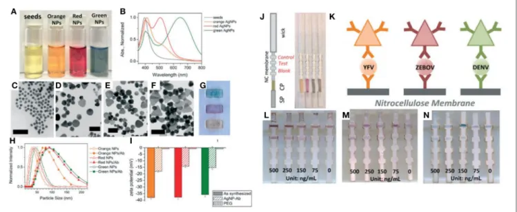
FIGURE 5 | An example of the use of AgNPs for multiplexed detection. (A)Vials of AgNPs during stepwise growth and(B)their corresponding absorption spectra. TEM images of(C)Ag seeds,(D)orange AgNPs,(E)red AgNPs, and(F)green AgNPs. Scale bars: 50 nm.(G)Green, red, and orange (top to bottom) AgNPs on nitrocellulose paper.(H)DLS and(I)zeta-potential of AgNPs before and after antibody conjugation.(J–N)Illustration of the flow strips conjugate with the different antibodies and limit of detections of each biomarker. Yellow Fever (YFV), Zaire Ebola virus (ZEBOV), Dengue virus (DENV). Adapted fromYen et al. (2015)with permission of the Royal Society of Chemistry.
The cache of studies related to the effects of AgNP production and use on the environment are still developing, however there is general agreement that AgNPs may be released into the environment during several routes and processes: Synthesis, during the manufacturing process and incorporation into products, recycling, and disposal (seeFigure 6;Gottschalk and Nowack, 2011). One such study was conducted by researchers at the United States Consumer Product Safety Commission (Quadros et al., 2013), where the potential child AgNP exposure from a variety of consumer products (i.e., toys, fabric products, human milk storage bags, humidifiers, and accessories, etc.) was assessed by measuring the release of Ag+
and AgNPs into water, air, dermal wipes, orange juice, milk formula, and synthetic saliva, sweat, and urine. They were able to rank the products and categories on the basis of their potential for Ag bioavailability, from most likely to least likely to be a source of bioavailable Ag. Almost all the Ag released from fabric and toy samples was in the ionic form. They found that sweat and urine yielded the highest Ag+ release, while tap water had the lowest yield.
While there are currently no guidelines for Ag in consumer products, their findings were significant as a proxy for release of Ag as AgNPs incorporated into various textiles, fabric, and cleaning products for antibacterial and purposes. Later,Mitrano et al. (2014)utilized a laboratory washing machine to simulate household laundering of textiles known to have undergone Ag and AgNP treatments to characterize and quantify total Ag release. Interestingly, conventional Ag treated fabrics yielded more total Ag and more nanoparticulate-sized Ag during fabric washing than the AgNP-treated fabrics. This was evidence that conventional forms of Ag precipitate to form nanosized Ag (complexes) and warrant careful considerations for regulatory
action of nano-Ag as compared to conventional Ag forms. In fact, several other studies have focused on assessments and quantification of the release of Ag from AgNP-containing consumer products (Benn and Westerhoff, 2008; Kulthong et al., 2010; Von Goetz et al., 2013). Studies such as these allow researchers to understand the behavior of AgNPs in real-world scenarios as well as to aid risk assessments.
Interaction of AgNPs and Soil-Plant
Systems
FIGURE 6 | Synthesis, application, routes of exposure, factors governing toxicology, and paradigm changes related to the AgNP production and use.
Reproduced fromLeón-Silva et al. (2016)with permission of Springer.
Ag and AgNP composites have found use in the control of various phytopathogens as well as for plant disease management (Liu et al., 2002; Park et al., 2006; Jo et al., 2009). Over the course of several studies, it was demonstrated that AgNPs were effective against plant fungus, providing evidence that AgNPs could serve a great purpose for controlling spore-producing fungal plant pathogens (Kim et al., 2009b; Jung et al., 2010; Lamsal et al., 2011a,b). While the latter studies demonstrate the benefit of AgNP soil treatment, AgNPs have also been found to have a deleterious effect, resulting in a drop in the metabolic abilities and diversity of necessary soil microbial populations (Jo et al., 2009). Hänsch and Emmerling (2010)identified that exposure to AgNPs of increasing concentration resulted in a significant decrease in microbial mass. A study byZhai et al. (2016)demonstrated the potential for AgNPs of different shapes to disrupt the metabolic processes of natural soil microbial communities and also that soil microbes were more vulnerable to AgNPs on the smaller size spectrum.
Interactions of AgNPs with Biological
Media
The state of AgNPs is highly dependent upon their interaction with surrounding medium (Stebounova et al., 2011). Studies have
immersion in different media over 4,000 h. In their study, they hypothesized that the release of silver ions led directly to silver toxicity and confirmed this via cell culture-, microbiological-, and reactive oxygen species experiments. Researchers have also demonstrated that AgNPs in blood readily interact with surrounding biomolecules such as proteins and lipids, leading to the formation of protein coronas on the NP surface (Walczyk et al., 2010; Mahmoudi et al., 2013). On the other hand, it has been shown that the release of silver ions can be potentially suppressed by the addition of humic and fulvic acids, dissolved oxygen, natural and low salt sea water, and other organic matter (Liu and Hurt, 2010).
In Vitro
and
In Vivo
AgNP Studies
In the past two decades, a large research effort has been devoted to the aspects of the toxicity of AgNPs, covering investigations of environmental fate, and including a plethora of in vivoand
in vitrostudies (Marambio-Jones and Hoek, 2010; Fabrega et al., 2011; Zhang et al., 2014). Comprehensive reviews have been compiled that detail the synthesis, application, subsequent routes of exposure, and toxicological mechanisms related to AgNP production and use (seeFigure 6;León-Silva et al., 2016; Wen et al., 2016). Published cytotoxicity tests and in vivo assays lend limited evidence to claims that silver is carcinogenic in any tissue (U.S. Department of Health and Human Resources, 2010). However, a plethora of in vitro studies have provided evidence that AgNPs are not only transported into cells and internalized, but target endosomes and lysosomes (Asharani et al., 2009a; Luther et al., 2011), induce lung fibroblasts, impair the cellular membrane, cause DNA damage and genotoxicity, chromosome aberration, and apoptosis (Almofti et al., 2003; Asharani et al., 2009b; Yang et al., 2012; Jiang et al., 2013). Exposing A549 cells (human alveolar basal epithelial cells) to AgNPs resulted in not only reactive oxygen species generation, but reductions in cell viability and mitochondrial membrane potential (Chairuangkitti et al., 2013). Conversely, exposure to AgNPs at high concentrations (up to 6.25µg/mL) caused not
only apoptosis and oxidative stress but morphology changes in HT 1080 (human fibrosarcoma) and A431 cells (human skin/carcinoma) cells which became less polyhedral, more fusiform, shrunken, and rounded (Arora et al., 2008).
While there is evidence that AgNPs are toxic (Maurer and Meyer, 2016), the full mechanisms of toxicity are still not well-understood and research efforts should be devoted to gaining more clarity. The main drawbacks to establishing a systematic comparison of the current published studies are the lack of uniformity (in terms of size and shape) in the synthesis and the purification procedures of AgNPs, varying size distributions, coatings, and precursors, a lack of particle characterization, and the lack of implementation of validation with reference materials (Gliga et al., 2014; Gorham et al., 2014). Nonetheless, increased oxidative stress, apoptosis, and genotoxicity have been highlighted as the main in vitro outcomes of AgNP exposure (Kim and Ryu, 2013). These confounding differences in methodology have often lead to contradictory findings inin vitrostudies. Studies that compare AgNPs of varying sizes show a greater toxic effect for particles of smaller diameter (Carlson
et al., 2008; Braydich-Stolle et al., 2010). Oxidative stress has been the main link to the toxicity of AgNPs themselves (Kim et al., 2009a), but far more frequently, it is the dissolution of AgNPs that leads to toxic effects which makes an understanding of the ion release kinetics for AgNPs paramount (Foldbjerg et al., 2015).Burrell (2003)found that although inert in the presence of human tissues, metallic silver ionizes in the presence of bodily fluids and secretions, to release the biologically active Ag+which
has a high affinity to sulfydryl groups and other anionic ligands of proteins, cell membranes, and tissue debris (Burrell, 2003). Although Ag ion release has often been highlighted as the main cause of cytotoxicity and toxic effects, researchers find difficulty in determining the extent of the toxicity of AgNPs when Ag ions are also present in solution (the Ag ion induced effects often mask the effect of AgNPs at high metal ion concentrations). Foldbjerg et al. (2015)assert that research studies are still rife with confounding results the make ascertaining the cause of toxicity difficult to decipher. To date, the weight researchers must place on ion release when discussing AgNP toxicity is still a difficult concept to discern.
While AgNPs have been shown to be toxic to bacteria, hence their main use in the formulation of antibacterial products, significant evidence is present to support the toxicity of AgNPs to other organisms.Marambio-Jones and Hoek (2010)provide comprehensive evidence that AgNPs cause inactivity not only in bacterial cells, but also fungi, virii, and algae. AgNPs have also been found to be toxic to models such as zebrafish (Yeo and Yoon, 2009),Drosophila melanogaster(Ahamed et al., 2010),
Daphnia magna(Scanlan et al., 2013), andCaenorhabditis elegans
(Meyer et al., 2010 and Yang et al., 2014). Yeo and Yoon (2009) found that nano-silver ions penetrated the skin and blood tube of zebrafish larvae in the form of aggregates, while Ahamed et al. (2010) found that AgNPs induced heat shock, oxidative stress, DNA damage, and apoptosis in Drosophila melanogaster. Further, silver nanowires were not only toxic to
Daphnia magna, butScanlan et al. (2013)found that the surface coating of silver nanowires (AgNWs) was dramatically modified (as compared to pristine AgNWs) when extracted from the organism’s hemolymph. In correlation with the effect that AgNPs have on soil and soil ecosystems, toxic effects have also been reported on a diverse range of soil invertebrates which include
Eisenia fetida, Enchytraeus albidus, Eisenia andrei, Porcellio scaber, andFolsomia candida(Tkalec et al., 2011; Hayashi et al., 2012, 2013; Gomes et al., 2013; Schlich et al., 2013; Waalewijn-Kool et al., 2014; Gomes et al., 2015).
Effects of Ag, AgNPs, and Ag Constituents
on Human Health
regarded as toxic to the immune, cardiovascular, nervous, or reproductive systems (ATSDR, 1990; Lansdown, 2010). Even though the benefits of the Ag on human health are yet to be proven, colloidal silver suspensions are being incorporated into health supplements (Fabrega et al., 2011). Occupational health studies have found that long-term exposures to Ag have led to irreversible conditions such as argyria, wherein the skin turns bluish in color as a response to the accumulation of Ag in body tissues (Hill, 1941; Wadhera and Fung, 2005). It is worthy to note that the critical oral dosage that elicits this effect is not known and may vary from individual to individual. Silver and nanosilver accumulation in the skin, liver, kidneys, corneas, gingiva, mucous membranes, nails, and spleen are also possible (Rosenman et al., 1979; DiVincenzo et al., 1985; Hollinger, 1996; Sue et al., 2001; Wan et al., 1991). An extensive review of the exposure-related health effects of silver and silver related compounds was conducted by Drake and Hazelwood in 2005 and later by Lansdown in 2010 (Drake and Hazelwood, 2005; Lansdown, 2010). Studies have listed the liver as the primary organ for silver accumulation and elimination. Even though the majority of Ag-containing consumer products are designated for topical application, the risk of percutaneous absorption of silver is very low as the human epidermis is a relatively impenetrable barrier (the exception being dermal abrasions, wounds, and cuts). Lansdown (2006)also reasons that although there is an increasing use of Ag in silver thread and textile fibers, there has been no evidence of increased blood silver or accumulation of silver precipitates in the skin in chronic exposure and the risks of argyria in these cases have been deemed negligible. In the same vein, the toxic risks associated with silver ingestion are low, as most products releasing Ag ions for oral or gastrointestinal hygiene were removed from pharmacopeias and permitted lists in most countries, in light of the risks of argyria (Lansdown, 2010). More comprehensive studies and research efforts are necessary to clearly aid risk assessment, identify the toxic mechanisms of AgNPs and their toxicological effects where areas of human health are concerned.
SYNTHESIS AND STABILIZATION OF
SILVER NANOPARTICLES (AgNPs) IN
LIQUID PHASE
Generally, the synthesis of NPs can be classified in two main categories: Top-down, where the procedure involves the use of bulk materials, such as metallic silver, that are reduced to form NPs using physical, chemical, or mechanical processes; or bottom-up, where the procedure requires starting from molecules, atoms, or ions to obtain NPs (Hornyak et al., 2008). Most NP synthesis approaches focus on bottom-up procedures, particularly in liquid phase media (Klabunde, 2001; Cunningham and Bürgi, 2013; Cushing et al., 2014; Majdalawieh et al., 2014) and nucleation theories and mechanisms have been extensively described byCushing et al. (2014),Viswanatha and Sarma (2007), Finney and Finke (2008), Thanh et al. (2014), and Kettemann et al. (2016).
In recent years, the development of methods for the synthesis of AgNPs has been the subject of significant interest (Tran et al., 2013). Generally, AgNPs are synthetized in liquid phase using chemical methods such as: Classical reduction with citrate (Turkevich et al., 1951), reduction with NaBH4 (Lee and
Meisel, 1982), reduction with gallic acid (Park et al., 2016), polyol synthesis (Kim et al., 2006), synthesis with organic solvents (Pastoriza-Santos and Liz-Marzán, 1999), as well as photochemical (Sun and Xia, 2002), electrochemical ( Rodrıguez-Sanchez et al., 2000), and sonochemical methods (Jiang et al., 2004). However, despite the myriad of AgNP synthesis methods, few offer the capability to achieve shape and size control. The main impediments to the production of monodisperse, uniformly spherical AgNPs are the formation of secondary products (smaller and/or larger sizes) or undesirable shapes, such as nanorods, nanocubes, nanotriangles, nanodipyramids, and nanooctahedra (Shirtcliffe et al., 1999; Yang et al., 2011). Therefore, it is necessary to control and establish reaction conditions that facilitate reproducible synthesis of spherical NPs with uniform size distributions. In this context, some of the variables that can be tuned in the chemical synthesis process to control the size and shape of AgNPs are:
i) the type and concentration of reducing agent (Dadosh, 2009) or stabilizing agent (Zhao et al., 2010);
ii) the addition of complexing agents (i.e., NH3) for removing
precursor agents and decreasing particle size (Zhao et al., 2010);
iii) the addition of alkaline co-reducers using strong and/or weak reducing agents (Agnihotri et al., 2014).
Alternatively, other synthesis routes employ seed methods, where small NPs serve as seed or nucleation centers that allow control of the shape and particle size of the AgNPs (Jana et al., 2001; Pyatenko et al., 2007; Qu and Ma, 2012; Wan et al., 2013). The most common methods used for the synthesis of uniform and spherical AgNPs are summarized inTable 1.
TABLE 1 | Chemical methods for the synthesis of monodisperse and quasi-spherical AgNPs in liquid phase.
Precursor agent Reduction agent Capping agent Some experimental conditions/results References
AgNO3 ascorbic acid Glycerol/PVP d≈(20 to 100) nm; temp≈90◦C Steinigeweg and Schlücker, 2012
AgNO3 Na3Cit Na3Cit/ TA d≈(10 to 100) nm; temp≈90◦C Bastús et al., 2014
AgNO3 EG PVP/EG d≈(10 to 80) nm; temp≈160◦C; t≈4 h Zhao et al., 2010
AgNO3 Na3Cit Na3Cit d≈(10 to 80) nm; temp≈ b.p Pyatenko et al., 2007
AgNO3 Na3Cit Na3Cit d≈(30 to 96) nm; temp≈b.p; pH≈5.7 to 11.1 Dong et al., 2009
AgNO3 ascorbic acid Daxad 19 d≈(15 to 26) nm; temp≈b.p Sondi et al., 2003
AgNO3 NaBH4or Na3Cit Na3Cit d≈(28 to 73) nm; temp≈b.p Wan et al., 2013
AgNO3 Alanine/NaOH DBSA d≈8.9 nm; temp≈90◦C, t≈60 min Yang et al., 2011
AgNO3 Na3Cit Na3Cit/ TA d≈(18 to 30) nm; temp≈60◦C to b.p; t≈20 min Dadosh, 2009
AgNO3 NaBH4/ Na3Cit Na3Cit d≈(5 to 100) nm; temp≈90◦C; pH: 10.5; t≈20 min Agnihotri et al., 2014
AgNO3 Oleic Acid sodium oleate d≈(5 to 100); temp≈(100 to 160)◦C; t≈(15 to 120) min Xu and Hu, 2012
Na3Cit, Trisodium citrate; PVP, (C6H9NO)nPolyvinylpyrrolidone; TA, (C76H52O46) Tannic acid; DBSA, (C18H29NaO3S) dodecylbenzenesulfonic acid; Daxad 19, Sulfonated Naphthalene
Condensate (surfactant); b.p, boiling point.
NPs are mostly through chemisorption processes, electrostatic attractions, or hydrophobic interactions (Kraynov and Müller, 2011; Manojkumar et al., 2016).Figure 7provides an illustration of functional groups with strong surface interactions with AgNPs (-SH, -NH, -COOH, -C=O) that allow for functionalization and
further stabilization (Sperling and Parak, 2010).
Depending on the type of NP (i.e., the core material) and the dispersant solvent, the choice of a specific ligand can provide either higher or lower stability. Molecules with low molar mass have been used as stabilizing agents (Warner et al., 2000; Nath et al., 2010), however these types of molecules exhibit several limitations, including the easy desorption of ligands and the promotion of agglomeration and aggregation. (Van Hyning and Zukoski, 1998). Alternatively, synthetic polymers can be used for the stabilization of NPs. In this context, amphiphilic polymers have been employed to stabilize NPs (Mayer, 2001). Polymeric ligands tend to generate more contact points with the NP surface, creating better interaction ligand/surface interactions (adsorption) (Toshima and Yonezawa, 1998). On the other hand, hydrophilic polymer chain interactions generate external loops which can interact with the solvent and sterically stabilize NPs, (seeFigure 8).
Stabilization will directly impact the physical and chemical properties of AgNPs, and subsequently may limit their applications. For example, studies have shown that AgNPs coated with polyvinylpyrrolidone (PVP) and polyethyleneglycol (PEG) have greater stability under environmental conditions than AgNPs stabilized using citrate (Lead et al., 2014). However, besides capping agents, storage temperatures are also critical to the stability of these materials. It has been shown that different storage temperatures can produce oxidation processes promoting unwanted shapes such as nanorods and nanoprisms or AgNP aggregation and/or agglomeration (Pinto et al., 2010). These processes are unintended in the synthesis of spherical and uniform size distributions of AgNPs; therefore, it is key to control the temperature of these colloidal systems to avoid (thermodynamically) the formation of such structures. Also, AgNPs can be modified and destabilized by photochemical
reactions.Gorham et al. (2012)showed that AgNPs coated with citrate can be destabilized with UV radiation exposure. Other factors to take into account with regard to the destabilization of AgNPs are post-synthesis residues and incorrect purification procedures. For examples, high concentrations of remnant precursors and/or reducing agents in the liquid phase promote the transformation of AgNPs into new shapes such as nanorods, nanocubes, and nanotriangles (Murphy et al., 2001; Dadosh, 2009; Pinto et al., 2010). Additionally, pH plays an important role which is shown where amino acid-coated AgNPs have improved stability under acidic conditions (pH ≈ 3), eliminating the
formation of agglomerates due to suppression of intermolecular interactions between solvent and ligand (Bayram et al., 2015).
Recently, it has been discovered that the use of biopolymers as a capping agents foster biocompatibility and safety from toxicological points of view (Jena et al., 2012). Specifically, different carbohydrates and their derivatives such as a guar gum (Vanamudan and Sudhakar, 2016), carboxymethyl cellulose (CMC) (Velusamy et al., 2016), dextran (Caki´c et al., 2016), kappa-carrageen (Elsupikhe et al., 2015), sodium alginate (Chunfa et al., 2016), chitosan (Shanmugaraj and Ilanchelian, 2016), heparin (Kemp et al., 2009), and hyaluronic acid (Yahyaei et al., 2016) have been employed to stabilize AgNPs. Proteins have also been employed for the stabilization of AgNPs.Darroudi et al. (2011)provided a procedure for the sonochemical synthesis of AgNPs using gelatin as both a reducing and coating agent, obtaining very promising results in terms of sphericity and distribution of particles in the sub-10 nm range.
Furthermore, some studies demonstrate the good stability of capped biopolymer-AgNPs. Chen et al. (2008b) obtained highly stable AgNPs-CMC that showed no apparent change in their optical spectrum extinction when stored at 25◦C
FIGURE 7 | An illustration of some selected surface chemistries and conjugation strategies that are applied to NPs.Reproduced fromSperling and Parak (2010)with permission of the Royal Society.
FIGURE 8 | Steric stabilization of AgNPs. Reproduced fromZamiri et al. (2010)with permission of MDPI, Basel, Switzerland. CC BY 3.0.
biopolymers can be used as capping agents and provide evidence for employing these macromolecules as stabilizing agents for NPs in liquid phase. Overall, the stabilization of AgNPs and other NPs in liquid phase is still considered a
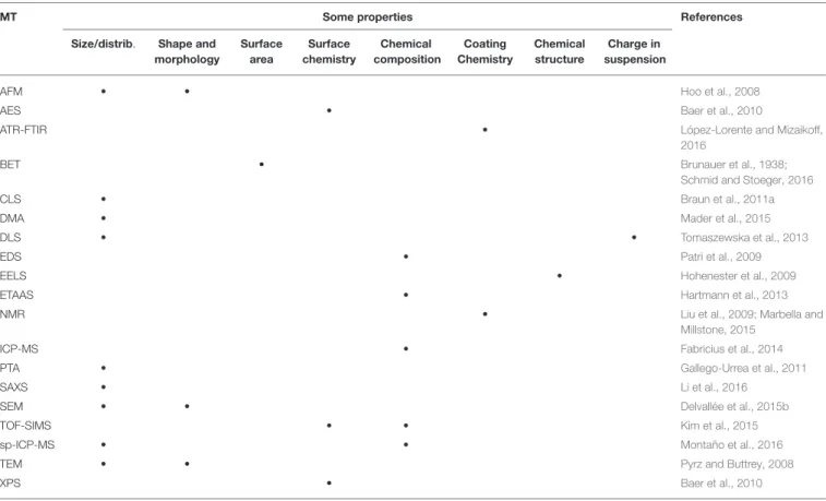
TABLE 2 | Common measurement techniques (MT) used for the characterization of NPs.
MT Some properties References
Size/distrib. Shape and morphology
Surface area
Surface chemistry
Chemical composition
Coating Chemistry
Chemical structure
Charge in suspension
AFM • • Hoo et al., 2008
AES • Baer et al., 2010
ATR-FTIR • López-Lorente and Mizaikoff, 2016
BET • Brunauer et al., 1938;
Schmid and Stoeger, 2016
CLS • Braun et al., 2011a
DMA • Mader et al., 2015
DLS • • Tomaszewska et al., 2013
EDS • Patri et al., 2009
EELS • Hohenester et al., 2009
ETAAS • Hartmann et al., 2013
NMR • Liu et al., 2009; Marbella and
Millstone, 2015
ICP-MS • Fabricius et al., 2014
PTA • Gallego-Urrea et al., 2011
SAXS • Li et al., 2016
SEM • • Delvallée et al., 2015b
TOF-SIMS • • Kim et al., 2015
sp-ICP-MS • • Montaño et al., 2016
TEM • • Pyrz and Buttrey, 2008
XPS • Baer et al., 2010
AFM, Atomic Force Microscopy; AES, Auger Electron Spectroscopy; BET, Brunauer-Emmett-Teller method; ATR-FITR, Attenuated Total Reflectance Fourier Transfom-Infrared Spectroscopy; CLS, Centrifugal Liquid Sedimentation; DMAS, Differential Mobility Analysis; DLS, Dynamic Light Scattering; ET-AAS, Electrothermal Atomic Absorption; EELS, Electron Energy Loss Spectroscopy; EDS, Energy Disperse X-Ray Spectroscopy; ICP/MS, Inductively Couple Plasma Mass Spectrometry; NMR, Nuclear Magnetic Resonance; PTA, Particle Tracking Analysis; SAXS, Small-Angle X-Ray Scattering; SEM, Scanning Electron Microscopy; TEM, Transmission Electron Microscopy; SIMS, Secondary Ion Mass Spectrometry; TOF-SIMS, Time of Flight Secondary Ion Mass Spectrometry; spICP-MS, Single Particle Inductively Coupled Plasma Mass Spectrometry; XPS, X-Ray Photoelectron Spectroscopy.
processes that AgNP experience which can reduce their entropy.
MEASUREMENT AND
CHARACTERIZATION OF SILVER
NANOPARTICLES (AgNPs): A
METROLOGICAL APPROACH
As previously mentioned, NPs constitute a focus of interest in nanoscience and nanotechnology (Kang and Haider, 2015; Sharma et al., 2015). Particularly, there is an interest in establishing controlled chemical (e.g., chemical composition of the core, surface chemistry, bulk element composition, internal/external chemistry of mixing state, and oxidation state) and physical properties (e.g., size, shape, number and mass concentration, surface area, total mass, crystallinity, morphology, and optical properties) of these nanoobjects. Moreover, due to advancements in the production and applications of nanomaterials, scientists are developing new, and adapting classic, analytical techniques for the detection, characterization, and quantification of NPs. An extensive discussion of the fundamentals and analytical capabilities
of the most common techniques for the characterization of NPs (specifically metal, metal oxide and metalloid) has been thoroughly reviewed (Gunsolus and Haynes, 2015; Costa-Fernandez et al., 2016; Laborda et al., 2016; Majedy and Lee, 2016) and can be used for further consultation. The main current measurement techniques (MTs) for the characterization of NPs in general, and AgNPs in particular, and the requisite information they provide are listed in Table 2.
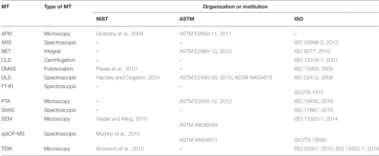
TABLE 3 | Representative standards, guides, and protocols developed in the recent years for the characterization of NPs.
MT Type of MT Organization or institution
NIST ASTM ISO
AFM Microscopy Grobelny et al., 2009 ASTM E2859-11, 2011 –
ARS Spectroscopic – – ISO 20998-2, 2013
BET Integral – ASTM E2864-13, 2013 ISO 9277, 2010
CLS Centrifugation – – ISO 13318-1, 2001
DMAS Fractionation Pease et al., 2010 – ISO 15900, 2009
DLS Spectroscopic Hackley and Clogston, 2015 ASTM E2490-09, 2015;ASTM WK54872 ISO 22412, 2008
FT-IR Spectroscopic – –
ISO/TS 1410
PTA Microscopy – ASTM E2834-12, 2012 ISO 19430, 2016
SAXS Spectroscopic – – ISO 17867, 2015
SEM Microscopy Vladár and Ming, 2010
ASTM WK39049
ISO 13322-1, 2014
spICP-MS Spectroscopic Murphy et al., 2015
ASTM WK54613 ISO/TS 19590
TEM Microscopy Bonevich et al., 2010 – ISO 29301, 2010; ISO 13322-1, 2014
AFM, Atomic Force Microscopy; ARS, Acoustic Resonance Spectroscopy; BET, Brunauer-Emmett-Teller method; CLS, Centrifugal Liquid Sedimentation; DMAS, Differential Mobility Analysis system; DLS, Dynamic Light Scattering; FITR, Fourier Transfom-Infrared Spectroscopy; PTA, Particle Tracking Analysis; SAXS, Small-Angle X-Ray Scattering; SEM, Scanning Electron Microscopy; spICP-MS, Single Particle Inductively Coupled Plasma Mass Spectrometry; TEM, Transmission Electron Microscopy.
procedures for the correct measurement and characterization of NPs (seeTable 3).
While an extensive discussion of the measurement techniques and their analytical capabilities are beyond the scope of this review, we will focus on the metrological aspects of some of the most important measurement techniques for the characterization of AgNPs. In this context, microscopy techniques are extremely powerful analytical tools for the characterization of AgNPs. For example,MacCuspie (2011)used Atomic Force Microscopy (AFM) with the goal of exploring the stability of AgNPs capped with citrate and bovin serum albumin (BSA) in solvents with different electrolyte concentrations and pH conditions. Their AFM results, accompanied with measurements of ultraviolet-visible spectroscopy (UV-Vis) and dynamic light scattering (DLS), showed how the stability of the AgNPs are highly affected by different factors (pH, electrolytes concentrations, and capping agent). Also, they demonstrated how different MTs such as AFM, UV-Vis, and DLS, can be used for evaluating the stability and characterizing these colloidal systems. Transmission electron microscopy (TEM) and scanning electron microscopy (SEM) are other microscopy techniques widely used in the characterization of AgNPs in the metrological field. Klein et al. (2011b) used SEM and TEM in order to characterize and establish the particle size and size distribution of its representative test material, NM-300 (see definition of representative test material, RTM, in the next section of this document). The use of complementary MTs such a UV-Vis, graphite furnace atomic absorption (GFAAS), and inductively coupled plasma optical emission spectrometry (ICP-OES) were used to study stability, the release of ionic silver, and to quantify the total silver mass of this RTM, respectively. Recently, Verleysen et al. (2015) used TEM for the measurement and the validation of 23 dimensional and morphological parameters (diameter, perimeter, central distance, shape factor among
others) of AgNPs, providing the measurement uncertainty of these parameters. In the same context,Dudkiewicz et al. (2015) reported the use of electron microscopies (SEM and TEM) for the characterization of AgNPs spiked into two different food matrices (chicken paste and tomato soup). Their study has generated a key metrological input in the determination of particle size in a complex matrix (food) by electron microscopy techniques, because they assessed the impact of different sources of uncertainty such as sampling, sample preparation prior to imaging, and image analysis in the total uncertainty of the particle size determination.
different varieties of NPs (metal, metal oxide, and metalloid NPs). A good example is the development of RMs in the nanoscale, where the combination of multiple methods is necessary to assign and characterize the different properties of these materials.
In the specific case of AgNPs, many of the MTs described above can be used for the characterization of their chemical and physical properties. For example,MacCuspie et al. (2011), report the use of multiple MTs such AFM, TEM, DLS, NTA, and ultrasmall angle X-ray scattering (USAXS) for the physico-chemical characterization of AgNPs. The same group also discuss the different results obtained by these MTs in the determination of the size, size distribution and agglomeration of the AgNPs. Martin et al. (2014) used USAXS and TEM to understand and study the dissolution, agglomeration, morphology, and stability of AgNPs exposed under different acid concentration (HNO3). Moreover, they used UV-Vis and DLS
to investigate the stability of AgNPs in strong acid media and evaluated the morphology the AgNPs coated with BSA.Murphy et al. (2015) established a protocol for the determination of mean nanoparticle size (equivalent spherical particle diameter), number based size distribution, particle number concentration, and mass concentration of ions in an aqueous suspension of AgNPs using single particle inductively coupled plasma mass spectrometry (spICP-MS). These are just some examples of the different MTs that can be used for the characterization of AgNPs. Finally, all these techniques can be employed in concert toward one of the most important task in the chemical metrology field: The development of reference materials (seeTable 4). A good example of this is the multimethod approach used by NIST in the development of the NIST RM 8017 PVP-coated AgNPs (NIST, 2015d). In their investigation report, AFM, TEM, USAXS, and DLS was used by NIST researchers to determinate the particle size of this nano-object. It is important to mention that the determination of the particle size of NPs is method dependent, and as a result of this, NIST attempted to characterize its RM using different MTs. Other MTs such as isotopic dilution mass spectroscopy (IDMS), asymmetric-flow field-flow fractionation (AF4), ICP-MS, UV-Vis, and spICP-MS have been used to characterize important properties in the RM including the silver mass content, elemental impurities, absorbance spectrum and others.
Despite this, further advancements are necessary to work toward improving the measurement and characterization of AgNPs and NPs in general, as many analytical techniques are still hampered with limitations (especially at the small end of the nanoscale range, i.e., sub-10 nm). In the specific case of AgNPs, the simultaneous determination of ionic silver and AgNPs in colloid suspensions still present an analytical challenge for most of the MTs. This aspect is solved partially by techniques like spICP-MS, however limitations such as limit of detection (LOD) and the overlap of ionic silver and AgNPs signals still obstruct the characterization by this technique in some cases. On the other hand, a large number of nanotoxicological and environmental studies lack a metrological approach, leaving out important metrological tools that enable the comparability and reproducibility of results. Such tools include standardized/validated methods, use of reference
materials, and the estimation of the measurement uncertainty in the nanoscale. The studies described above reflect the continued importance of the development of robust, comparable, analytical methodology in order to achieve improvement of measurement in the nanoscale.
DEVELOPMENT OF NANOPARTICLE
REFERENCE MATERIALS (RMs) IN THE
NANOSCALE
TABLE 4 | NPs reference materials and certified reference materials developed in the recent years.
Material Property measured
Form/ quantity
Value and uncertainty
MTs used NMI(id) Proposed uses References
AuNPsa(RM) Particle size LS/5 ml (8.5±0.3) nm AFM NISTf(RM 8011) Instrument calibrations,
evaluation ofin vitroassays (bioassays), interlaboratory comparison
NIST, 2015a
(9.9±0.1) nm SEM (8.9±0.1) nm TEM (11.3±0.1) nm DMA (8.5±1.8) nm SAXS
AuNPsa(RM) Particle size LS/5 ml (24.9±1.1) nm AFM NISTf(RM 8012) Instrument calibrations, evaluation ofin vitroassays (bioassays), interlaboratory comparison
NIST, 2015b
(26.9±0.1) nm SEM (27.6±2.1) nm TEM (28.4±1.1) nm DMA (28.6±0.9) nm DLS (173◦)
(26.5±3.6) nm DLS (90◦)
(24.9±1.2) nm SAXS
AuNPsa(RM) Particle size LS/5 ml (55.4±0.3) nm AFM NISTf(RM 8013) Instrument calibrations, evaluation ofin vitroassays (bioassays), interlaboratory comparison
NIST, 2015c
(54.9±0.4) nm SEM (56.0±1.5) nm TEM (56.3±1.4) nm DMA (56.6±0.9) nm DLS (173◦)
(55.3±3.6) nm DLS (90◦)
(53.2±1.2) nm SAXS
AgNPsb(RM) Particle size DS/≈2 g (70.1±6.0) nm AFM NISTf(RM 8017) Benchmark and evaluation of potential EHS
NIST, 2015d
(74.6±3.8) nm TEM (67.9±0.5) nm USAXS (105.6±4.6) nm DLS Mass value (2.162±0.020)fmg IDMS AgNPsc(CRM) Particle size LS/5 ml d
10(12.0±1.9)dnm
d50(18.5±2.5)dnm
SAXS BAMg(BAM N001) Used as standard material
for measurements and toxicological test
Menzel et al., 2013
d90(18.5±2.5)dnm
d10(6.9±1.9)enm
d50(12.6±2.5)enm d90(19.4±2.5)enm
SiO2-NPs(CRM) Particle size LS/10 mL (19.0±0.6) nm DLS IRMMh(ERM FD100) Evaluated, Instrument and
method performance
Braun et al., 2011b
(20.1±1.3) nm CLS (19.4±1.3) nm TEM (21.8±0.7) nm SAXS
SiO2-NPs (CRM) Particle size LS/9 mL (42.1±0.6) nm DLS IRMMh(ERM FD 304) Evaluated, Instrument and method performance
Franks et al., 2012
(33.0±3.0) nm CLS
TABLE 4 | Continued
Material Property measured
Form/ quantity
Value and uncertainty
MTs used NMI(id) Proposed uses References
PS (CRM) Particle size LS/5 mL (60.39±0.63) nm DMA NISTfSRM 1964 Calibration/validation of
particle sizing instruments
NIST, 2014a
PS (CRM) Particle size LS/5 mL (60.39±0.63) nm DMA NISTf(SRM 1963a) Calibration/validation of
particle sizing instruments
NIST, 2014b
TiO2(CRM) Specific Surface Area
PPS (55.55±0.70) m2g−1 MP-BET NISTf(SRM 1898) Benchmark and evaluation
of potential EHS
NIST, 2012
(53.85±0.78) m2g−1 SP-BET
CRM, Certified Reference Material; DS, Dry Solid, EHS; Environmental, Health, and Safety Risks; RM, Reference Material; LS, Liquid Suspension; PPS, Powder and Porous Solid.
acitrate-stabilized AuNPs in an aqueous suspension.blyophilized polyvinylpyrrolidone (PVP)-coated AgNP,cAgNPs stabilized against aggregation using polyoxyehylene glycerol trioleate,
polyoxiethylene sorbitan monolaurate,dThe d
10, d50,and d90values are specific particle diameters (volume weighted) that correspond to 10,50, and 90% of the total particles in cumulate undersize distribution,eThe d
10, d50,and d90values are specific particle diameters (number-weighted) that correspond to 10,50, and 90% of the total particles in cumulate undersize distribution,fExpanded uncertainties, U, calculated as U=ku
c, where ucis intended to represent, at the level of one standard deviation, the combined standard uncertainty calculated
according to the ISO/JCGM Guide (BIPM et al., 2008). The coverage factor, k, for 95 % expanded uncertainty intervals is based on a t multiplier with the appropriate associated degrees of freedom,gExpanded combined uncertainty consisting of contributions from method repeatability, measurement setup geometry, method bias, possible but undetected
inhomogeneity and instability, and the model used, in particular binning, expanded by a factor or k=2 corresponding to a confidence level of∼95%,hThe certified uncertainty is the
expanded uncertainty with a coverage factor k=2 corresponding to a level of confidence of about 95 % estimated in accordance with ISO/IEC Guide 98-3:2008 (ISO/IEC Guide 98-3, 2008).
FIGURE 9 | Uses of reference materials in the nanoscale.
makes the development of CRMs more challenging because many of the measurands are method-defined making it difficult to establish a clear link to the SI. The measurement of chemical and physical properties of sub-10 nm nano-objects is a challenge for most analytical techniques and, reactivity, aggregation, agglomeration, and interactions between the dispersant medium add more complexity to the measurement system resulting increase in the uncertainty of the measurement.
For all these reasons, in recent years there has been a strong interest in developing NP RMs in the nanoscale, since they can shed new light not only on the impact of nanomaterials with respect to EHS, but also on ways in which the quality of measurements in the nanoscale can be improved or quality-assured (Hansen et al., 2007; Stefaniak et al., 2013).Aitken et al. (2008), established a priority list of candidate materials for the production of nanotoxicology RMs. This list consisted of several nanomaterials such as a carbon black, single and multiwalled
carbon nanotubes (SWCNT/MWCNT), fluorescent polystyrene, combustion-derived NPs, TiO2-NPs, ZnO-NPs, AgNPs. Others
materials such as AuNPs, CeO2-NPs, SiO2-NPs, ceramics, and
nanoclays were identified also as a potential RMs.Stone et al. (2010), evaluated which of these materials were suitable for employment in ecotoxicological studies. They identified TiO2
-NPs, polysterene beads labeled with fluorescent dyes, and Ag-NPs, as materials that would be useful to produce test- or reference materials. A comprehensive approach for the prioritization of materials that can be developed into reference materials was made recently by Stefaniak et al. (2013), where a list of 25 individual nano-objects was generated with scientific interest for the generation of RMs for risk assessment. In particular, they highlighted NPs such as CeO-NPs, SiO2-NPs, TiO2-NPs, ZnO2
establishment of validated methods with a full estimation of the uncertainty sources that have been involved in the measurement (ISO/Guide 34, 2009). This aspect may seriously hinder the development of RMs, because as previously mentioned, only a few standardized procedures for the characterization of properties of NPs are available, the majority of which are focused on the determination of dimensional properties such as particle size and particle size distribution (seeTable 3).
Matrix plays an important role in the production and certification of NP RMs. Grombe et al. (2015), described the feasibility of the development of RMs for the detection of AgNPs in food matrices. Their results indicate significant differences in particle size when the AgNPs are dispersed in meat materials in comparison to water suspensions. They also reported difficulties in the development of efficient methods for the detection of AgNPs, principally due to AgNP reactivity being higher in comparison to more stable NPs (e.g., metal oxides like TiO2). Furthermore, another important factor to
consider in the production of RMs at the nanoscale, is the form of the nanomaterial. Linsinger et al. (2011), discussed in detail two different forms (states of matter) that are conceivable for NP RMs: Suspensions of particles and dry powders. In suspension, NPs have better motion (promoted by Brownian Motion and diffusion process), producing an easier dispersion and homogenization of the material. However, this can promote the interaction with other molecules or even promote interaction between the NPs (aggregation, agglomeration, Ostwaltd ripening, or coarsening). For example,Gorham et al. (2014)demonstrate AgNPs suspensions capped with citrate lose their physical and chemical integrity by oxidation process and oxidation process followed by photoreduction. On the contrary, in powder form, NPs are more stable, essentially because the chemical changes only progress by diffusion, which is a rather slow process in this state of mater. To promote long-term stability, some dry powder RMs are stored in inert atmospheres, preventing the chemical degradation of the materials (Hornyak et al., 2008). MacCuspie et al. (2013), stabilized AgNPs in excess PVP and then lyophilized the formulated AgNPs to produce a cake of NPs that can be reconstituted simply by adding water. This approach resulted in a practical way to eliminate chemical changes of the AgNPs, conserving the particle size within the shelf-life required for a RM and was used in the development of the NIST RM 8017 Polyvinylpyrrolidone Coated Silver Nanoparticles (Nominal Diameter 75 nm) (NIST, 2015d). A drawback of the use of dry powder RMs is the possible need for redispersion protocols (Linsinger et al., 2011) to ensure that a homogeneous suspension is formed. This can be problematic, especially in the case of users with limited experience or expertise in sample preparation procedures and could generate a bias which is not intrinsic to the property certified. On the other hand, NP RMs in suspension (liquid phase) are characterized with respect to homogeneity and are easier to use. However, as was previously discussed that NPs in liquid phase (colloidal suspensions) need to be correctly functionalized in order to prevent their destabilization, which can create issues in ensuring long-term stability. In this aspect,Orts-Gil et al. (2013) pointed out that the development of functionalized, colloidal,
stable RMs, may improve comparability between results across different laboratories, and provide convenience and feasibility in establishing multi-parametric RMs for engineered NPs.
It is important to mention that the development of a NP RM is an arduous process that involves many technical and production requirements (for example, production planning, production control, material storage, material processing, data acquisition, data evaluation, and in the case of CRMs, establishing metrological traceability, etc.) (ISO Guide 34, 2009). Recently, a new term has been proposed: “Representative Test Material (RTM).” RTMs will serve to cover gaps in the availability of NP RMs (Roebben et al., 2013). Specifically, a RTM is defined as “a material from a single batch, which is sufficiently homogeneous and stable with respect to one or more specified properties, and which implicitly is assumed to be fit for its intended use in the development of test methods which target properties other than the properties for which homogeneity and stability have been demonstrated (ISO/TS 16195, 2013).” In the recent years, the Organization for Economic Co-operation and Development (OECD), in conjunction with the European Joint Research Center (JRC), has worked on the development of a wide range of RTMs to support nanomaterial research and development. Some examples of the RTMs developed at this moment are illustrated bySingh et al. (2011),Klein et al. (2011a),Rasmussen et al. (2014), Roebben et al. (2015)and on the website of the JRC (JRC, 2016).
OUTLOOK AND PERSPECTIVES
Concepts, definitions, and terminology in nanoscience and nanotechnology are currently changing in response to increased research efforts and the extraordinary growth that this area has experienced in the last two decades. Several factors (economic, social, and environmental) are promoting the establishment of robust and well-founded terminology that contribute to building sensible legislation and regulation. However, the development and consistent implementation of defined “nano” terms represent a tremendous challenge.
next years. To address these challenging tasks, more studies and results derived from rigorous in vitroandin vivostudies (e.g., bioaccumulation and bioavailability) will be necessary in order to elucidate the true impact of these materials. These studies will provide a scientific and technical basis for building worldwide consensus on regulation. Also, a concerted multidisciplinary effort must be continued to capitalize on initial findings in order to advance the investigation of relevant environmental scenarios.
Other efforts should be made in the area of NP synthesis and stabilization. Nowadays, among the great variety of chemical routes for the synthesis of AgNPs, only few enable control of the production of NPs with sufficient homogeneity of size and shape, both key parameters and highly demanded in the development of new applications. Besides that, the development of new synthesis routes that are much more efficient and use green synthesis approaches present emerging strategies to make the production of AgNPs more sustainable and environmentally friendly. Additionally, the use of biopolymers such as proteins, carbohydrates, and other types of macromolecules as stabilizing and functionalizing agents can improve the long term stability of the AgNPs in liquid phase and increase biocompatibility with environmental and biological systems. So far research into the stabilization of AgNPs using biopolymers is not sufficiently advanced to establish a clear stabilization mechanism using these coating agents. The behavior of this type of functionalized AgNPs under various conditions or factors that can compromise stability such as pH, temperature, UV radiation, etc., has yet to be studied. Given the above concerns, it is necessary to perform in-depth investigations of synthesis routes using biopolymers that control the shape, size, and stability of AgNPs.
With regard to the metrological field, the characterization of NPs is still considered a challenge because some measurement properties are method dependent, which hampers the comparison of values obtained from different measurement techniques. Continuous efforts have been made by the scientific community to standardize measurement protocols. In fact, some protocols, technical standards, and procedures have already been generated by different international organizations (e.g., ISO/TC 229 and ASTM E56) in order to provide more suitable and robust methods. Specifically, these efforts have focused mainly on the dimensional properties (size, shape,
and distribution) of nanomaterials. However, it is necessary to develop/implement analytical techniques to extend the NP characterization capabilities toward the measurement of other important properties such as surface chemistry, chemical structure, and chemical composition. Moreover, the ability to provide traceability to the SI at the nanoscale level has also proven to be quite a challenge. Some of the MTs may be directly or indirectly linked to the S.I., however many of these MTs provide semiquantitative and/or qualitative measurements that are not metrologically traceable. This is a limiting factor in areas such as nanotoxicology, ecotoxicology, and biomedical applications where these properties like surface charge, hydrophobicity, and agglomeration state play critical roles. Finally, the development of NP RMs is crucial to providing sound metrological tools for industry and the scientific community implementation to evaluate their measurement capabilities. However, presently the availability of NP RMs is quite limited because of the technical complexity that is involved the production of these materials. Though more RMs have been developed in recent years, in many cases RMs are not available for relevant measurands and to cover the myriad of scenarios where nanomaterials are being currently applied. It is expected that in the next few years more RMs and RTMs will be released in order to provide comparability and to assure the quality of measurements in the nanoscale.
AUTHOR CONTRIBUTIONS
BC reviewed the literature and wrote the manuscript text. MJ wrote the entire section entitled "Silver nanoparticles (AgNPs): Possible impacts on environment, health, and safety (EHS)". KM, AM, MW, and JV reviewed the manuscript text.
ACKNOWLEDGMENTS
The authors would like to thank Dr. Justin M. Gorham and Dr. Andre M. Striegel (Materials Measurement Science Division, Chemical Sciences Division, respectively, NIST) for their thorough reviews of the manuscript, as well as the Frontiers in Chemistry reviewers for their very useful comments and suggestions.
REFERENCES
Agnihotri, S., Mukherji, S., and Mukherji, S. (2014). Size-controlled silver nanoparticles synthesized over the range 5–100 nm using the same protocol
and their antibacterial efficacy.RSC Adv.4, 3974–3983. doi: 10.1039/c3ra4
4507k
Ahamed, M., Posgai, R., Gorey, T. J., Nielsen, M., Hussain, S. M., and Rowe, J. J. (2010). Silver nanoparticles induced heat shock protein 70, oxidative stress and apoptosis inDrosophila melanogaster.Toxicol. Appl. Pharmacol.242, 263–269. doi: 10.1016/j.taap.2009.10.016
Ahmad, R., Griffete, N., Lamouri, A., Felidj, N., Chehimi, M. M., and Mangeney, C. (2015). Nanocomposites of gold nanoparticles@molecularly imprinted polymers: chemistry, processing, and applications in sensors.Chem. Mater.27,
5464−5478. doi: 10.1021/acs.chemmater.5b00138
Aitken, R. J., Hankin, S. M., Lang Tran, C., Donaldson, K., Stone, V., Cumpson, P., et al. (2008). A multidisciplinary approach to the identification of reference
materials for engineered nanoparticle toxicology.Nanotoxicology2, 71–78.
doi: 10.1080/17435390802109177
Alemán, J. V., Chadwick, A. V., He, J., Hess, M., Horie, K., Jones, R. G., et al. (2007). Definitions of terms relating to the structure and processing of sols, gels, networks, and inorganic-organic hybrid materials (IUPAC Recommendations
2007).Pure Appl. Chem.79, 1801–1829. doi: 10.1351/pac200779101801
Almofti, M. R., Ichikawa, T., Yamashita, K., Terada, H., and Shinohara, Y. (2003). Silver ion induces a cyclosporine a-insensitive permeability transition in rat
liver mitochondria and release of apoptogenic cytochrome C.J. Biochem.134,
43–49. doi: 10.1093/jb/mvg111