pp 264-270 Case RepORt
www.medigraphic.org.mx
Open bite treatment with first molar extraction.
Case report
Tratamiento de mordida abierta con extracciones
de primeros molares. Reporte de caso
Guillermo Pérez Cortez,* Tely Adriana Soto Castro,§ Nancy Ivette Gallardo Alfaro,II Idalia Selene Isais PeñaII
* Clinical consultant.
§ Bibliographic consultant. II Second-year residents.
Orthodontic Specialty of the Autonomous University of Baja California.
This article can be read in its full version in the following page: http://www.medigraphic.com/ortodoncia
ReSUmeN
La extracción de primeros molares permanentes es una alternati-va viable para el tratamiento de discrepancias leves o moderadas de las estructuras maxilares en pacientes con mordida abierta; el objetivo terapéutico es corregir la maloclusión al tiempo que se in-tenta disimular el problema esquelético. Se presenta caso clínico de una paciente tratada con extracciones de primeros molares perma-nentes, para corrección de mordida abierta esquelética moderada, logrando rotación mandibular, una oclusión funcional y resultados estéticos favorables. el objetivo de este artículo es demostrar que después de una adecuada selección del caso, por medio de un buen diagnóstico, las extracciones de primeros molares resultan una buena alternativa de tratamiento en el caso de mordidas abier-tas de origen esqueletal.
Key words: Open bite, first molar extractions, vertical discrepancy.
Palabras clave: mordida abierta, extracción de primeros molares, discrepancia vertical. AbstrAct
The extraction of first permanent molars is a viable option for the treatment of mild to moderate discrepancies of the maxillary structures in patients with open bite. The therapeutic goal is to correct the malocclusion while trying to camouflage the skeletal problem. A case report is presented of a patient who was treated with extractions of first permanent molar in order to correct the moderate skeletal open bite through mandibular rotation thus achieving a functional occlusion and favorable cosmetic results. The aim of this report is to show that after proper case selection and a good diagnosis, first molar extractions are a suitable alternative treatment for skeletal open bites.
IntroductIon
Canut defined open bite as the obvious lack of contact between the upper and lower teeth of dental or skeletal origin; the latter, due to its multifactorial etiology and consequences represents a great challenge for the orthodontist because despite the fact that extensive research has been conducted on this anomaly, there is fear in the etiology identification and in the proper treatment selection that delivers stable functional and aesthetic results.
Currently the orthodontist has a number of treatment options and/or mechanics to treat this anomaly: extra-oral appliances, bite block, intraoral anchorage appliances, mini-implants, orthognathic surgery and bicuspid or first molar extractions.
Orthodontic treatments where extractions of first permanent molars have been suggested are often considered as difficult to handle, with extended treatment time, reserved prognosis and where the final result may be affected. However there are several
case reports in the literature of patients with open bites treated by means of first molar extractions. Arvystas in 1977 reported one of the first cases, followed by Vaden in 1988, and Aras in 2002, among others. These cases were treated under the principle that by eliminating posterior contact points (possible fulcrums) and moving the posterior segment mesially, a mandibular anterior rotation would occur, thus closing the anterior open bite. However, for this treatment alternative to be successful with stable functional results, it is necessary to do a proper case selection.
www.medigraphic.org.mx
Figure 4. Initial panoramic radiograph.
case report Diagnosis
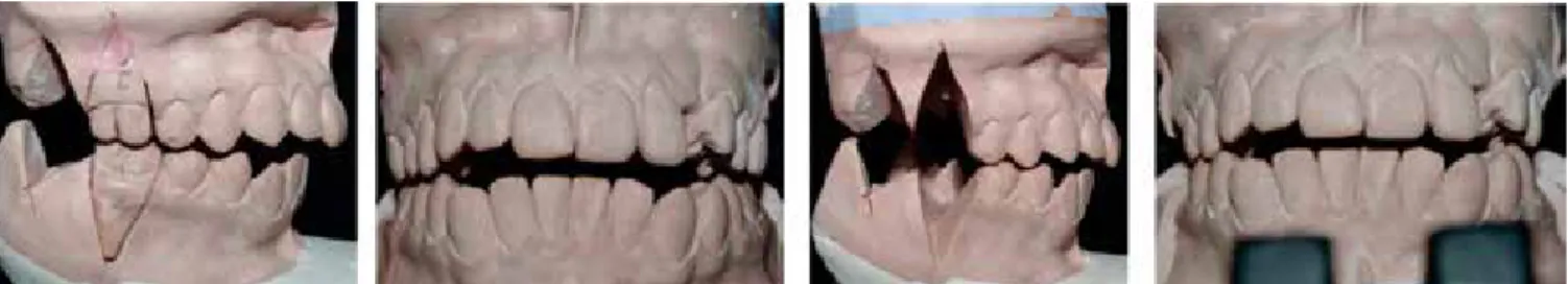
Female patient of 18 years 8 months of age with good overall health attends the Orthodontics Clinic at the Autonomous University of Baja California, mexicali Campus, to seek orthodontic care. On her facial analysis, a slight asymmetry, a convex profile and a retrusivechin were observed (Figure 1). On the intraoral analysis (Figure 2) an ovoid-shaped upper arch, gingival recession of the upper left lateral incisor
Figure 1.
Initial facial analysis.
Figure 2.
Initial intraoral photographs where an anterior open bite from the right premolar area to the first premolar area on the left side may be observed.
Figure 3.
Cephalometric analysis where a hyperdivergent pattern and a considerable upper incisor proclination are noted.
30.1o 33.3o 86.9o 39.4 9.1114.4o 84.6o 48.4o 4.4 13.0 84.3o 124.1o 117.0o 82.3o 117.1o 48.3o 76.2 29.4 48.9 130.6o 80.5o 3.7o 70.1 70.4 52.7o 7.5mm -3mm -13mm 2mm
www.medigraphic.org.mx
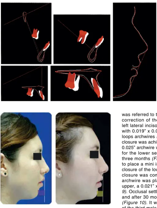
Figure 5. Diagnostic setup. Initially, second molars were eliminated with no success. By extracting the first molars a decrease
in the anterior open bite was obtained.
Figure 6.
Alignment and leveling. Open coil to gain space for the upper left canine.
Figure 7.
Space closure by mesialization of the posterior segments.
www.medigraphic.org.mx
Este documento es elaborado por Medigraphic
Figure 8.
mesialization of the lower second molars.
Figure 9.
www.medigraphic.org.mx
Figure 10.
A functional occlusion with a correct overbite and overjet was obtained. 84.6o 4.4 13.0o 114.4o 9.1 39.4 86.9o 33.3o 30.1o 48.4o 85.3o 32.3o 28.2o49.8 o 1.7 37.2o 3.5 0.9 Figure 11.
Initial and final cephalograms.
www.medigraphic.org.mx
Figure 12.Superimposition areas.
Treatment progress
A multidisciplinary treatment was performed involving orthodontics and periodontics; upper and lower molar extractions were performed.0.022” slot Roth appliances were placed with a 0.014” Nitinol archwire in both dental arches. Additionally, a transpalatalbar with a low button and a lingual arch were fitted to the second molars in order to begin the alignment and leveling (Figure 6). Upon completion of this phase an 0.017” x 0.025” stainless steel archwire was placed and the patient
was referred to the Periodontics Department for the correction of the gingival recession on the upper left lateral incisor. Space closure was then initiated with 0.019” x 0.025” stainless steel double keyhole loops archwires (Figure 7) and after 6 months space closure was achieve don the upper arch. An 0.019” x 0.025” archwire with a mesialization loop was placed for the lower second molars during approximately three months (Figure 8). However it was necessary to place a mini implant as an auxiliary in the space closure of the lower right second molar. Once space closure was completed, a 0.021” x 0.025” Braided archwire was placed on the lower arch and on the upper, a 0.021” x 0.025” upper rigid archwire (Figure 9). Occlusal settlement adjustments were conducted and after 30 months the appliances were removed (Figure 10). It was decided to monitor the eruption of the third molars and continue performing occlusal adjustment so to avoid undesirable contacts.
results
After 30 months of treatment, the facial axis increased 1o, the mandibular axis decreased 1o and
the interincisal angle went from 119o to 137o(Figures 11 and 12).The profile was improved with a good chin projection (Figure 13). Bilateral molar and canine class I, a 2 mm overjet and overbite and good functional guides were obtained. Additionally, root parallelism was achieved and we continued to monitor the eruption of the upper third molar (Figure 14). Occlusal adjustments will be conducted until they finish their eruption.
www.medigraphic.org.mx
Figure 14. Final panoramic radiograph.Figure 15. Retention.
retentIon
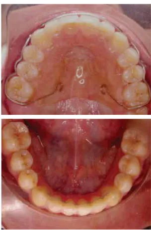
A circumferential removable retainer was placed on the upper arch and in the lower, a fixed retainer from canine to canine (Figure 15).
dIscussIon
Orthodontic treatment with first molar extractions is a therapeutic option that nowadays causes controversy due to the fact that these teeth are considered fundamental keys to the occlusion. However, in the papers by Arvystas in 1977, Vaden in 1988, Sulaiman in 2001 and Aras in 2002 among other authors it has been stated that skeletal open bite treatment with first molar extractions provides functional results with more stability than those obtained in non-extraction treatments.
This treatment option will be successful only if the clinician has the clinical ability and experience to select the case properly and have good control over the biomechanics. It is important to consider that according to Seddon (2004) treatments with first molar extractions may last 7 to 9 months more than conventional treatments.
conclusIons
First molar extractions are a good treatment option that provides stable, functional and esthetic results for skeletal open bite cases. It is important to consider that this therapeutic alternative demands an adequate case selection and a high level of clinical expertise and skill in order not to compromise the treatment results and expectations.
recommended readIngs
1. Aras A. Vertical changes following orthodontic extraction treatment in skeletal open bite subjects. Eur J Orthod. 2002; 24 (4): 407-416.
2. Lopez-Gavito G, Wallen TR, Little Rm, Joondeph DR. Anterior open-bite malocclusion: a longitudinal 10-year postretention
evaluation of orthodontically treated patients. Am J Orthod.
1985; 87 (3): 175-186.
3. english JD. early treatment of skeletal open-bite malocclusions.
Am J Orthod Dentofacial Orthop. 2002; 121 (6): 563-565.
4. Al-emran SeS. extraction of first permanent molars in the management of anterior open bite malocclusion. Saudi Dental Journal. 2001; 13 (3): 155-160.
5. Vaden JL. Nonsurgical treatment of the patient with vertical discrepancy. Am J Orthod Dentofacial Orthop. 1998; 113 (5):
567-582.
6. de Figueiredo mA. early tooth extraction in the treatment of anterior open bite in hyperdivergent patients. World J Orthod.
2007; 8 (3): 249-260.
7. Chui-Shan TNG, Orthodontic treatment of anterior open bite.
International Journal of Peadiatric Dentistry. 2008; 18 (2): 78-83.
8. Sandler PJ, For four sixes. AJO DO. 2000; 117 (4): 418-434.
9. Ortial JP. Vertical dimension and therapeutic choices. Am J Orthod Dentofacial Orthop. 1995; 108 (4): 432-441.
10. Arvystas mG. Treatment of anterior skeletal open-bite deformity.
Am J Orthod. 1977; 72 (2): 147-164.
11. Seddon JL. Extraction of four first molars: a case for a general practitioner? J Orthod. 2004; 31 (2): 80-85.
12. Fränkel R. A functional approach to treatment of skeletal open bite. Am J Orthod. 1983; 84 (1): 54-68.
13. Cangialosi TJ. Skeletal morphologic features of anterior open bite. Am J Orthod. 1984; 85 (1): 28-36
mailing address: Guillermo Pérez cortez e-mail: [email protected]