Processes
A S Raikhel, University of California, Riverside, CA, USA
M R Brown, University of Georgia, Athens, GA, USA X Belles, Institut de Biologia Molecular de
Barcelona (CSIC), Spain
ß2005, Elsevier BV. All Rights Reserved.
3.9.1. Introduction 433
3.9.2. Endocrine Control of Female Reproduction 433
3.9.2.1. Evolution of Endocrine Control of Vitellogenesis and Egg Development 433
3.9.2.2. Yolk Protein Precursors 440
3.9.2.3. Mechanisms of Juvenile Hormone Action in Vitellogenesis 445 3.9.2.4. Molecular Mechanisms of Ecdysteroid Action in Vitellogenesis 449 3.9.2.5. Molecular Endocrinology of Vitellogenesis in the Cyclorrhaphan Diptera 456
3.9.2.6. Hormones and Ovarian Maturation 459
3.9.2.7. Hormone Action in Female Accessory Glands 463
3.9.2.8. Oviposition 464
3.9.2.9. Peptide Hormones Involved in Female Reproduction 464
3.9.3. Hormones and Male Reproduction 469
3.9.3.1. Spermatogenesis 469
3.9.3.2. Male Accessory Gland Function 470
3.9.4. Future Directions 473
3.9.1. Introduction
The years following the 1985 publication of Comprehensive Insect Physiology, Biochemistry and Pharmacology have been marked by stunning developments in insect science. A technological rev- olution in biochemistry, molecular biology, and genetics has swept all areas of biological science, and has profoundly influenced insect science as well.
With this technological revolution, the small size of insects is no longer a barrier to discovering their biochemical make-up, cloning or characterizing genes, or uncovering genetic and hormonal signals governing their functions. Sequencing of insect gen- omes, particularly that ofDrosophila melanogaster, has led to the identification of previously unknown genes and to the development of functional genomic approaches that lead to further elucidation of genetic regulatory networks.
During this period, insect endocrinology has shifted its focus from physiology and biochemistry to molecular biology and genetics. The latter are the subjects of the present volume. This chapter specifi- cally reviews the progress that has been achieved since 1985 in our understanding of the hormonal control of insect reproduction. Particular attention is paid to those areas where significant progress has been made at the molecular and genetic levels.
There are a number of recent reviews on hormonal regulation of yolk protein (yp) genes (Raikhel et al., 2003; Belles, 2004; Bownes, 2004; Wang et al., 2004); vitellogenins and their processing (Sappington et al., 2002; Telfer, 2002;
Tufail et al., 2004); nonvitellogenin yolk pro- teins (Bownes and Pathirana, 2002; Telfer, 2002;
Masuda et al., 2004; Yamahama et al., 2004), and the cell biology of the insect fat body (Giorgiet al., 2004).
3.9.2. Endocrine Control of Female Reproduction
3.9.2.1. Evolution of Endocrine Control of Vitellogenesis and Egg Development
p0020
The insect female reproductive system consists of the ovaries, which contain ovarioles, oviducts, sper- matheca, accessory glands, vagina, and ovipositor (Davey, 1985; Chapman, 1998). Ovarioles are the egg producing units in the ovary. Typically, the insect ovary has four to ten ovarioles; however, some species contain many more ovarioles. Ovar- ioles are tubular and consist of both somatic and germline cells. At the apex of each ovariole, a ger- marium houses the primary germline cells. The fol- licles or egg chambers form within the germarium
and continue to mature along the ovariole tube.
There are three types of ovaries in insects (Bu¨ning, 1994). Ovaries of primitive groups of insects con- tain panoistic ovarioles with egg chambers consist- ing of the oocytes surrounded by the follicular epithelium (panoistic ovarioles). In more advanced insects, where the structure of ovarioles is more complex (meroistic), some germ cells are set aside to form nurse cells that produce massive amounts of numerous products for the developing oocyte. In polytrophic meroistic ovarioles, each egg chamber contains its own group of nurse cells connected to the oocyte. Follicle cells surround the oocyte and nurse cells. In telotrophic meroistic ovaries, each ovariole contains the trophic chamber with large groups of nurse cells. The trophic chamber is connected to the egg chambers by nutritive cords.
Egg chambers of telotrophic ovarioles contain only the oocyte surrounded by the follicle cells.
Endocrine control of female reproduction is governed by different types of hormones: neuropep- tides (see Chapter 3.10), juvenile hormones (JHs;
see Chapter 3.7), and ecdysteroids (see Chapters 3.3 and 3.5). In keeping with common practice among researchers in the field, ecdysteroid is used as the generic term for steroidal insect molting hor- mones, reserving the term ecdysone for the specific chemical compound 2b, 3b, 14a, 22R, 25-pentahy- droxy-5b-cholest-7-en-6-one, originally known as a-ecdysone. The abbreviation 20E will be used to refer to 20-hydroxyecdysone (2b, 3b, 14a, 20R, 22R, 25-pentahydroxy-5b-cholest-7-en-6-one), the ecdysone metabolite believed to serve as the active hormone in most well-characterized responses.
Individually, or in concert, the regulation of par- ticular events during female reproduction by these hormones has evolved along with their effects on molting and metamorphosis. Control of female reproduction in Apterygota remains the most enig- matic. Studies of the firebrat,Thermobia domestica (Zygentoma), have shed some light on the hormonal regulation of vitellogenesis and ovarian develop- ment in primitive apterous insects (Bitsch and Bitsch, 1984; Bitsch et al., 1986). In this insect, molting occurs continually into the adult stage, and oogenesis is coordinated with the ecdysteroid regulated adult molting cycle (Bitsch et al., 1986).
Treatment with the anti-allatal drug precocene blocks ovarian maturation, which indicates its dependence upon juvenile hormone secreted by the corpora allata (CA) (Bitsch and Bitsch, 1984; Bitsch et al., 1986). Further studies are required in order to understand the precise roles of JH and ecdyster- oids in regulating vitellogenesis and oogenesis in apterous insects.
p0030
It is well established that major events of reproduction in all insect orders with incomplete metamorphosis (Hemimetabola) are governed by JH. In the orders with complete metamorphosis (Holometabola), control strategies have evolved dif- ferently. In beetles (Coleoptera), JH remains the major regulatory hormone of reproductive events.
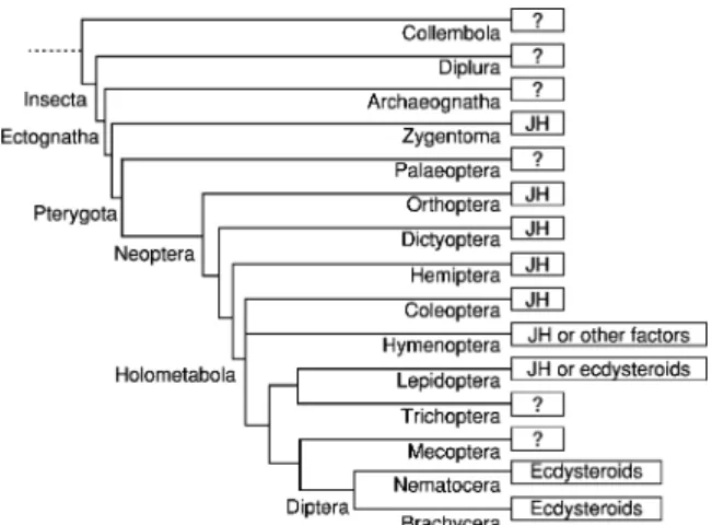
In Hymenoptera, the role of JH is elaborated in eusocial species having a single or a few reproduc- tive females in a colony (seeChapter 3.13). In Lepi- doptera, female reproduction is regulated either by JH or ecdysteroids. In many such species, egg maturation occurs during the pharate adult stage and requires coordination with hormonal signals controlling metamorphosis. In dipteran insects, mosquitos, and flies, ecdysteroids have the leading role as hormonal regulators, but JH has an impor- tant role as a regulator in dipteran females in pre- paring reproductive tissues for ecdysteroid mediated events, such as vitellogenesis. For all insects, neuro- peptides play a key role regulating the production of JH and ecdysteroids. A summary of the primary hormones involved in reproduction according to the phylogeny of insect orders is presented inFigure 1;
for some orders, this is not known.
Figure 1 Utilization of juvenile hormone or ecdysteroids as major regulators of vitellogenesis and reproduction among in- sect orders. Some hemipterans show incomplete dependence on JH. In hymenopterans, JH plays a vitellogenic role in nonso- cial and primitive social groups, but not in advanced groups (seeChapter3.13). In lepidopterans, those species that begin vitellogenesis after adult emergence are JH dependent, and those in which vitellogenesis proceeds between pupal and adult stages are partially dependent on JH, whereas species in which vitellogenesis proceeds within or before the pupal stage are independent of JH. Dipterans, in general, are ecdys- teroid dependent, although JH may play an accessory role in vitellogenesis (Modified from Belles, X.,2004. Vitellogenesis directed by juvenile hormone. In: Raikhel, A.S., Sappington, T.W. (Eds.), Reproductive Biology of Invertebrates, Vol. 12, Part B: Progress in Vitellogenesis. Science Publishers, Enfield, USA/Plymouth, UK, pp. 157–198).
3.9.2.1.1. Juvenile hormone directed female repro- duction In the adult females of Hemimetabola (Dictyoptera to Hemiptera) and Coleoptera, JH is the main regulator and pleiotropically controls most aspects of female reproduction (Figure 2).
The major role of JH in reproduction is to regulate vitellogenin (Vg) gene expression in the fat body, generally, and in ovarian follicular epithelium (Wyatt and Davey, 1996; Engelmann, 1983, 2003;
Belles, 2004). Some gryllid and hemipteran species
show an incomplete dependence upon JH (Wyatt and Davey, 1996; Strambiet al., 1997).
p0040
Cockroaches (Dictyoptera) are the classical model for studies of JH dependent vitellogenesis. In the oviparousPeriplaneta americana(Blattidae),Weaver and Edwards (1990)have shown that allatectomy or treatment with inhibitors of JH synthesis blocks Vg production, oocyte growth, and ootheca formation, whereas JH treatment restores these processes. The regulation of vitellogenesis by JH has been most thoroughly studied inBlattella germanica(Blattelli- dae). In this typical JH dependent species, in which the females carry the ootheca externally until the first inside larvae emerge, JH production (Belles et al., 1987), Vg titers (Martı´n et al., 1995), and Vg mRNA levels (Comaset al., 1999) show cyclical and approximately parallel patterns during the reproductive cycle (Figure 3). The pseudoviviparous cockroach, Leucophaea maderae, has been one of the favorite models for biochemical studies of JH regulation of reproduction. Effective doses to induce vitellogenesis in females in vivorange from 1mg of JH I to 25mg of JH IIIin vivo. Methoprene, a potent JH analog (JHA), induces vitellogenesis in adult males, as well (Don-Wheeler and Engelmann, 1997). In another pseudoviviparous cockroach, Blaberus discoidalis, allatectomy prevents ovarian maturation that can be restored by JH III treatment (Keeley and McKercher, 1985). In decapitated adult females, methoprene or JH III induces Vg protein synthesis (Keeleyet al., 1988) and a 6.5 kb mRNA, presumably corresponding to the Vg transcript (Bradfield et al., 1990). The neuropeptide, hyper- trehalosemic hormone, enhances the vitellogenic
Figure 2 The pleiotropic role of juvenile hormone in insect reproduction. ER, endoplasmic reticulum; Vg, vitellogenin.
(Reproduced with permission from Engelmann, F.,2003. Juve- nile hormone action in insect reproduction. In: Henry, H.L., Norman, A.W. (Eds.), Encyclopedia of Hormones, Vol. 2.
Academic Press, San Diego, pp. 536–539.)
Figure 3 Production rates of juvenile hormone (JH) from the corpora allata, vitellogenin (Vg) from the periovarial fat body, and fat body Vg mRNA during the first reproductive cycle in the cockroachBlattella germanica. (Based on data from Belles, X., Casas, J., Messenguer, A., Piulachs, M.D.,1987.In vitrobiosynthesis of JH III by the corpora allata ofBlattella germanica(L.) adult females.
Insect Biochem. 17, 1007–1010; Martin, D., Piulachs, M.D., Belles, X.,1995. Patterns of hemolymph vitellogenin and ovarian vitellin in the German cockroach, and the role of juvenile hormone.Physiol. Entomol. 20, 59–65; and Martin, D., Comas, D., Piulachs, M.D., Belles, X.,1998. Isolation and sequence of a partial vitellogenin cDNA from the cockroachBlattella germanica(L.) (Dictyoptera, Blattellidae), and characterization of the vitellogenin gene expression.Arch. Insect Biochem. Physiol. 38, 137–146.)
effects of JH (Lee and Keeley, 1994a). InNauphoeta cinerea, in which hemolymph Vg levels show a cyclic pattern during the gonadotropic cycle, JH I, II, and III can induce Vg production (Buschor and Lanzrein, 1983).
In Dermaptera, the CA are required for reproduc- tion in female earwigs,Anisolabis maritima, as first shown by Ozeki (1951; seeRankinet al., 1997), and JH is the main gonadotropic hormone in female Euborellia annulipes, which have vitellogenic cycles (Rankinet al., 1997).Roulandet al. (1981)reported that allatectomized females Labidura riparia do not produce vitellogenic proteins, and 0.1mg of farnesol, a precursor of JH, restores vitellogenesis.
p0050 In Orthoptera, the regulation of vitellogenesis by
JH has been demonstrated in a number of locusts and grasshoppers (Acrididae) through classical experiments involving allatectomy and JH treatment (Wyatt, 1991; Wyatt and Davey, 1996). The most thoroughly studied orthopteran species has been Locusta migratoria, in which Vg synthesis is cyclic (Chinzei and Wyatt, 1985). Induction of Vg syn- thesis in female L. migratoria by JH homologs is only achieved with repeated doses or with high doses coinjected with a JH esterase inhibitor (Wyatt et al., 1987), whereas synthetic JHAs at doses between 2 and 30mg have a much more potent vitel- logenic action (Edwardset al., 1993; Zhanget al., 1993). Neuropeptides may also be involved, since a brain factor enhances vitellogenesis induced by JH in fat body incubated in vitro (Glinka et al., 1995). JH titer, and Vg protein and mRNA levels have been described during the reproductive cycle of the locustSchistocerca gregaria(Mahamatet al., 1997) and the grasshopper Romalea microptera (¼Romalea guttata) (Borstet al., 2000).
p0055 In contrast to other orthopteroids, the phasmid
Carausius morosusis independent of JH for vitello- genesis (Bradleyet al., 1995). Allatectomy does not prevent production of viable eggs in this insect, as first reported by Pflugfelder in 1930, and later confirmed with antibodies against C. morosus Vg that detected its production in allatectomized and intact females (Bradleyet al., 1995). However, it is not clear whether the independence from JH for vitellogenesis applies to all Phasmida.
In crickets (Gryllidae), both JH and ecdyster- oids are involved in the control of vitellogenesis (review:Strambi et al., 1997). In Acheta domesti- cus, the CA are necessary for oocyte growth, and JH restores vitellogenesis in allatectomized or decapitated females (Renucci et al., 1987; Loher et al., 1992). Conversely, female Teleogryllus commodus emerging from allatectomized larvae can still produce eggs (Loher and Giannakakis,
1990). Although allatectomy of Gryllus bimacu- latus reduced the rate of egg production, oocytes in allatectomized females contained a similar amount of yolk protein as oocytes in controls;
JH or JHA restored egg production to a variable extent (Hoffmann and Sorge, 1996). Notably, low doses of ecdysteroids stimulated oocyte growth in females of this species (Chudakovaet al., 1982;
Behrens and Hoffmann, 1983). These results sug- gest that in crickets in which allatectomy does not completely suppress oocyte growth, ecdysteroids may play a vitellogenic role (Hoffmann and Sorge, 1996).
For Hemiptera, the CA were shown to be neces- sary for vitellogenesis inRhodnius prolixusby V.B.
Wigglesworth in the 1930s, and this observation has since been confirmed for several other hemipteran species (Wyatt and Davey, 1996; Davey, 1997;
Belles, 2004). Later studies demonstrated that allatectomy ofR. prolixus does not totally abolish Vg synthesis, but JH treatment does restore normal production (Wang and Davey, 1993). FemaleOnco- peltus fasciatus chemically allatectomized with precocenes produced the Vg precursor, but its con- version to mature Vg, which is incorporated into the oocytes, did not take place, as shown with electro- phoresis of native proteins and immunodiffusion (Kelly and Hunt, 1982). A later study of this species found that the synthesis of two female specific Vg components, 200 and 170 kDa, was inhibited in precocene treated specimens and restored by admin- istration of JH (Martinez and Garcera´, 1987). This discrepancy may be due to differences in the preco- cene treatment. Only one study has demonstrated a role for JH in a homopteran species. In the black bean aphid, Aphis fabae, ovarian development begins prenatally in this parthenogenic species, in such a manner that oocytes differentiate in the em- bryonic germaria, and at emergence each ovariole of a first instar aphid already contains one or two developing embryos. Precocene treatment of aphid nymphs inhibited oocyte development in embryos inside the parental ovaries, whereas JH reversed this inhibition (Hardie, 1987).
p0070
For Coleoptera, JH is the main gonadotropic hor- mone, but surprisingly little is known about the reproductive endocrinology of this holometabolous order with the greatest number of insect species.
A long day regimen for the Colorado potato beetle, Leptinotarsa decemlineata, leads to reproductive ac- tivity and a short day induces diapause. JH or JHA (pyriproxyfen) treatment of short-day females induces Vg synthesis, and the JHA also induces Vg production in last instar larvae (de Kortet al., 1997).
Similarly, females of Coccinella septempunctata
reared on suboptimal artificial diets fail to repro- duce, but Vg synthesis can be induced by treatment with JHA (Zhaiet al., 1984, 1987; Guan, 1989). In the spruce weevil,Pissodes strobi, treatment of pre- vitellogenic females with JH III induces the preco- cious appearance of Vg transcripts, as determined by the identification of Vg mRNA (Leal et al., 1997). This study also showed that Vg mRNA accumulates slower in beetles feeding on Sitka spruce trees (Picea sitchensis), which are resistant to P. strobi attack, thus suggesting that this plant contains anti-juvenoid compounds.
3.9.2.1.2. From nonsocial sawflies to eusocial ants and bees Within the Hymenoptera, JH appears to play a role in female reproduction among nonsocial and eusocial species and through this action may affect caste determination in the eusocial groups (see Chapter 3.13). In the primitive suborder Sym- phyta, onlyAthaliasawflies have received attention.
JH applied to male Athalia rosae induced an in- crease in Vg production, and ovaries implanted into these males incorporated the Vg (Hatakeyama and Oishi, 1990). Later, it was shown that ovaries of Athalia rosae implanted into males of the closely related species,Athalia infumata, incorporated the heterospecific Vg that was induced in the host by JH III treatment (Hatakeyama et al., 1995). Although these studies did not examine vitellogenesis in females, the results suggest a role for JH in females.
The regulation of reproduction by JH varies among the suborder Apocrita, which encompasses a range of species from solitary types to highly social groups like ants and honeybees (for reviews of JH action in social Hymenoptera see Robinson and Vargo, 1997; Hartfelder, 2000; Chapter 3.13). In the primitive eusocial paper wasps (Polistidae), Polistes dominulus(¼Polistes gallicus) andP. metri- cus, JH acts as a gonadotropin. Queens or dominant females of Polistes show high JH titers associated with growing ovaries, whereas subordinate females have low JH titers. In general, allatectomy of domi- nant females leads to a reduction in the dominance status. The bumble bee,Bombus terrestris(Apidae), shows a relatively primitive social structure, and JH titers are high in egg laying queens. A regulatory role for JH is substantiated by the positive cor- relation between ovary development and rate of JH production in queenless workers (Block et al., 2000). The role of JH remains unresolved among the studies of ants and bees with a complex social structure. JH treatment of isolated virgin queens of the fire ant, Solenopsis invicta (Myrmicinae) leads to wing shedding and oviposition, which is pre- vented by allatectomy. Subsequent JH treatment of
allatectomized queens induces these phenomena (see Robinson and Vargo, 1997). Conversely, JH titers are low in reproductive female ants in the genus Diacamma(Ponerine;Sommeret al., 1993).
In the highly eusocial honeybee,Apis mellifera(Api- dae), Vg synthesis in laying queens was only slightly reduced by allatectomy, and JH treatment increased ovary development only weakly (see Robinson and Vargo, 1997). Exogenous JH administered to queen and workers of A. mellifera advances the timing of vitellogenin appearance (Barchuk et al., 2002); together, these results indicate that JH does not play a primary role in honeybee reproduction.
3.9.2.1.3. Butterflies and moths: endocrine com- patibility of metamorphosis and vitellogenesis In Lepidoptera, some species begin vitellogenesis after adult emergence, and studies of Papilionoidea (e.g., Pieris brassicae, Nymphalis antiopa, Poly- gonia caureum, Vanessa cardui, andDanaus plex- ippus) and Noctuoidea (e.g., Heliothis virescens, Helicoverpa zea, andPseudaletia unipuncta) show that JH stimulates vitellogenesis (seeCussonet al., 1994a; Ramaswamy et al., 1997). In P. unipuncta, the release rate of JH from CAin vitrois positively correlated with Vg synthesis (Cussonet al., 1994b).
In this species and H. virescens, vitellogenesis is abolished in decapitated females, and JH treatment restores it (Cusson et al., 1994a; Ramaswamy et al., 1997).
For other groups of Lepidoptera, egg develop- ment proceeds between the pupal and pharate adult stages. Studies of species in the Tortricoidea have shown that decapitation of female Choristo- neura fumiferanaandC. rosaceanareduces egg pro- duction, but Vg remains in the hemolymph (Delisle and Cusson, 1999). Treatment of the decapitated females with methoprene restores egg production.
Similarly, treatment of female codling moths (Cydia pomonella) with fenoxycarb, a JHA, did not affect protein yolk content, but it did stimulate choriona- tion (Webbet al., 1999). In this species, ecdysteroids may have a priming effect on vitellogenesis, since treatment with ecdysteroid agonists (tebufenozide and methoxyfenozide) increased levels of circulat- ing Vg but did not affect its incorporation into the oocytes (Sun et al., 2003). These results suggest that vitellogenesis is not completely dependent on JH and that it plays a primary role in Vg uptake by the oocyte and in chorionation in this family.
In the Sphingid (Bombycoidea) Manduca sexta, vitellogenesis starts 3–4 days before adult emer- gence and proceeds in the absence of the pupal CA; thereafter, JH is necessary to complete oocyte growth and chorionation (Satyanarayana et al.,
1994). Vg was present in prepupae of both sexes at low levels, had disappeared by pupal ecdysis, and reappeared in the late pharate adult females. Meth- oprene treatment induced Vg synthesis and Vg mRNA in prepupae and freshly molted pupae, and simultaneous administration of ecdysteroids abol- ished the vitellogenic action of JH (Satyanarayana et al., 1994). The pyraloid moths studied to date have a similar reproductive physiology. In Plodia interpunctella, the declining ecdysteroid titer trig- gers vitellogenesis in early pupae, and ecdysteroid treatment inhibits vitellogenin uptake by the oocytes (Shirket al., 1992). Similarly, vitellogenesis in Diatrea grandiosellaproceeds in the absence of ecdysteroids, whereas choriogenesis is completed with JH in the pharate adult (Shuet al., 1997).
In non-Sphingid bombycoids, such as Bombyx mori(Bombycidae),Hyalophora cecropia(Saturnii- dae), and Malacosoma pluviale (Lasiocampidae), and in the lymantrid (Noctuoidea) Lymantria dis- par, vitellogenesis proceeds before adult emergence in the absence of JH. In these groups, vitellogenesis seems totally independent of JH, as shown by alla- tectomy and JH treatment, which did not influence oocyte growth (see Wyatt and Davey, 1996 for references). Other studies of L. dispar, in which vitellogenesis starts as early as day 3 of the last larval instar, found that treatment with JHAs on day 2 of this instar selectively prevents the produc- tion of Vg (Fescemyeret al., 1992) and Vg mRNA (Hiremath and Jones, 1992).
3.9.2.1.4. Mosquitos and flies: the shift to ecdys- teroid mediated vitellogenesis For the Diptera, edysteroids have the primary role of regulating reproduction, as demonstrated in detail almost exclusively in mosquitos and flies. JH plays an important priming role in these females by prepar- ing reproductive tissues for ecdysteroid mediated processes. Regulation of Vg production by ecdyster- oids was first demonstrated in the yellow fever mos- quito,Aedes aegypti. Vg synthesis in the mosquito fat body is stimulated by a blood meal and inhibited by removal of ovaries prior to blood feeding (Hagedorn and Fallon, 1973). The ovaries secrete the factor required for Vg production by the fat body, and this was found to be ecdysone (E), which is converted into the active form of the hormone 20-hydroxyecdysone (20E) (Hagedornet al., 1975).
Remarkably, mosquitos and flies are the only insects known to use ecdysteroids as the key regu- lator of reproduction. How or why this shift away from JH- to ecdysteroid-mediated reproduction occurred in Diptera remains an enigma.
The endocrinology of vitellogenesis in mosquitos (suborder Nematocera) has been reviewed in detail for A. aegypti (Hagedorn, 1985; Raikhel, 1992a;
Dhadialla and Raikhel, 1994; Sappington and Raikhel, 1998a; Raikhel et al., 2003; Wang et al., 2004) and investigated in Culex pipiens, Aedes atropalpus, and Anopheles stephensi (Hagedorn, 1985; Klowden, 1997). InA. aegypti, vitellogenesis is separated into four phases: previtellogenic (PV) preparation, arrest, yolk protein (YP) synthesis (vitellogenesis), and termination of vitellogenesis (Raikhel, 1992b; Dhadialla and Raikhel, 1994).
A newly emerged female needs about 3 days to become competent for the physiological demands of intense vitellogenesis. During this phase, the fat body and ovary acquire responsiveness to 20E and become competent for blood meal-activated vitel- logenesis and oogenesis, respectively (Flanagan et al., 1977; Li et al., 2000; Zhu et al., 2003a).
After 3 days of PV preparation, the fat body and ovary enter a state of arrest that persists until a blood meal is taken. During the YP synthesis stage, proteins are produced by the fat body and accu- mulated by developing oocytes (Raikhel, 1992a;
Raikhelet al., 2002). This massive synthesis peaks at around 24 h post-blood meal (PBM), then drops sharply, and terminates by 36–42 h PBM. The fat body undergoes remodeling, and the first batch of eggs completes chorion formation and is oviposited.
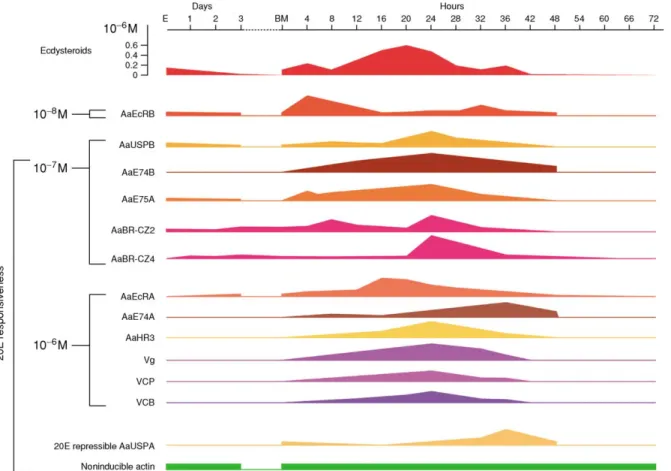
Titers of JH III and ecdysteroids in female A. aegypti are presented in Figure 4. During PV preparation, JH III titer increases and stabilizes during the arrest phase, and the CA remains active (Shapiro et al., 1986). JH is required for the fat body to attain competence for YP synthesis in response to 20E. Following blood feeding, the JH III titer drops as a result of a rapid decrease in CA activity and elevation of JH esterase titer in the hemolymph (Shapiro et al., 1986). Blood feeding triggers the release of the ovarian ecdysteroido- genic hormone (OEH) from the medial neurosecre- tory cells of the brain for up to 12 h postfeeding (Figure 5; Lea, 1972; Brown et al., 1998). In response to OEH, the ovary produces ecdysteroids, and the hemolymph titer of ecdysteroids in female mosquitos is correlated with the rate of YP synthesis in the fat body (Hagedornet al., 1975). The ecdys- teroid titers are only slightly elevated at 4 h PBM, rise sharply at 6–8 h PBM to a maximum level at 18–20 h PBM, and then decline to previtellogenic levels by 30 h PBM (Hagedornet al., 1975).
Numerous studies have clearly established that ecdysteroid control of YP synthesis is a central event in the blood meal-activated regulatory cascade
leading to successful egg maturation (reviews:
Hagedorn, 1985; Raikhel, 1992b; Dhadialla and Raikhel, 1994;Wanget al., 2004). Consistent with the proposed role of 20E in activating mosquito YP
synthesis, experiments using fat body in vitrohave shown that physiological doses of 20E (106M) activate yolk protein precursor (YPP) genes, which are described below (Deitschet al., 1995; Choet al., 1999).
Molecular studies have demonstrated that although the ecdysteroid triggered regulatory hier- archies, such as those implicated in the initiation of metamorphosis, are reiteratively utilized in the control of mosquito vitellogenesis, the unique inter- play of hierarchical factors is determined by the mosquito’s biology (Raikhel et al., 2002, 2003;
Wang et al., 2004). Of particular importance are the cyclicity of vitellogenic response and its depen- dence upon blood feeding. The demands for a very high level of expression ofYPPgenes, especiallyVg, put additional restraints on the hormonal regulation of these genes.
The endocrine control of vitellogenesis in higher flies (suborder Cyclorrhapha) has been studied extensively in several species (reviews: Bownes, 1986, 1994, 2004; Raikhel et al., 2003) and is altered according to whether the species lays eggs in batches or continuously. In species laying eggs in batches, the maturation of a synchronous group of oocytes is controlled by changes in hormone levels, as described best for the housefly,Musca domestica (Adams and Filipi, 1983; Adamset al., 1985).
In this species, the fat body starts to synthesize YPs after the female fly feeds on a protein meal, and YP uptake also commences in the ovary. This phase is controlled by the circulating ecdysteroids, and the fat body later shuts down YP synthesis as egg devel- opment is completed. There is a strong correlation
Figure 4 Juvenile hormone (JH) and 20-hydroxyecdysone titers and the level of Vg during the first reproductive cycle in the mosquitoAedes aegypti. (Based on data from Hagedorn, H.,1985. The role of ecdysteroids in reproduction. Kerkut, G.A., Gilbert, L.I.
(Eds.), Comprehensive Insect Physiology, Biochemistry and Pharmacology, Vol. 8. Pergamon, Oxford, pp. 205–261; and Dhadialla, T., Raikhel, A.S.,1994. Endocrinology of mosquito vitellogenesis. In: Davey, K.G., Peter, R.E., Tobe, S.S. (Eds.), Perspectives in Comparative Endocrinology. National Research Council of Canada, Ottawa, pp. 275–281.)
f0025 Figure 5 Schematic representation of the nutritional and hor- monal activation of vitellogenesis in the mosquitoAedes aegypti. A blood meal results in a direct signal to the fat body, which is required to initiate yolk protein precursor (YPP) gene expres- sion. The brain also receives a signal from the midgut, which activates medial neurosecretory cells to release a peptide hor- mone, ovarian ecdysteroidogenic hormone (OEH). OEH acti- vates follicular cells of the primary follicles to produce ecdysone. Ecdysone is converted in the fat body to the active steroid hormone, 20-hydroxyecdysone (20E), which activates YPPgene expression via an ecdysone hierarchy. Yolk protein precursors, vitellogenin (Vg), vitellogenic carboxypeptidase (VCP), vitellogenic cathepsin B (VCB), and lipophorin (Lp) are secreted by the fat body into the hemolymph and selectively accumulated by developing oocytes.
between ecdysteroid titers in the hemolymph and the progress of vitellogenesis, as well as between ecdysteroid titers and the amount of YP produced in the fat body (Adamset al., 1985). Ovaries pro- duce ecdysteroidsin vitro, and ovariectomized flies have reduced ecdysteroid levels early in the cycle (Adams et al., 1985). In the autogenic strain of M. domestica, decapitation of females blocks Vg gene expression in fat body, but 20E restores accu- mulation of Vg mRNA. Moreover, males were induced to produce YPs in response to the hormone (Agui et al., 1991). JH acts at an early stage to prime the fat body for YP synthesis and ensures that the oocytes are arrested in a previtellogenic stage (Adams, 1974, 1981). Interestingly, Adams et al. (1989) also have shown that the response to 20E in male houseflies was enhanced by applica- tion of JH. Many aspects of the regulation of vitel- logenesis in the housefly are quite similar to those described forA. aegypti.
In the blowfly, Calliphora vicina, YP-containing secretory granules become visible in the fat body when vitellogenesis is initiated by ingestion of a protein meal, but the granules are not detected in the fat body of ovariectomized females (Thomsen and Thomsen, 1974, 1978). Their appearance can be restored in ovariectomized females by injection of 20E (Thomsenet al., 1980). These observations have suggested the involvement of the ovary and ecdysteroids in the initiation of YP synthesis in the fat body ofC. vicina and is reminiscent of the re- sponse ofA. aegyptito a blood meal, which triggers the ovary to produce ecdysteroids and initiate a cycle of Vg production in the fat body.
In the blowflies,Sarcophaga andPhormia, non- protein-fed females respond to 20E by inducing YP synthesis in fat body (Huybrechts and De Loof, 1982). UnlikeC. vicina, ovariectomized houseflies and blowflies still produce YPs indicating additional complexity of hormonal regulation in these insects (Engelmann and Wilkens, 1969; Jensen et al., 1981;
Huybrechts and De Loof, 1982). Interestingly, male Sarcophaga,Phormia, andLuciliaalso produce YPs in fat body in response to 20E injection (Huybrechts and De Loof, 1982). JH alone cannot induce YP synthesis in males inMuscaorCalliphora(Adams, 1974; Huybrechts and De Loof, 1982; Adamset al., 1989).
In those species continuously laying eggs, each individual egg chamber differentiates, and its progress through vitellogenesis is modulated by hormonal signals connecting its development to environmental factors such as mating and food intake. This type of vitellogenesis is characteristic
of D. melanogaster, and many related species. The regulation of vitellogenesis in D. melanogasterhas been the subject of numerous studies using molec- ular, genetic, developmental, and physiological approaches (Bownes, 2004). In this species, adult sex determination (see Chapter 1.5) is the primary factor for correct expression of yolk protein genes.
Once the genes are active in the female, the level of expression is modulated by ecdysteroids and JH in a complex way (Bownes et al., 1993; Bownes, 2004). JH seems crucial for vitellogenesis, especially in the ovary (Bownes et al., 1996; Soller et al., 1999), but it appears that JH does not directly affect the transcription ofypgenes in the fat body. More recent data reported byRichardet al. (1998, 2001) points to a more prominent role for ecdysteroids in regulating vitellogenesis. It is interesting that in the Caribbean fruit fly, Anastrepha suspensa, in which the YPs are only produced in the ovary, there is no evidence of hormonal regulation by JH or 20E in females, and 20E injection is unable to stimulate YP synthesis in males (Handler, 1997).
3.9.2.2. Yolk Protein Precursors
p0155
3.9.2.2.1. Sites of yolk protein production In most insects, the fat body is the exclusive site for produc- tion of yolk protein precursors (YPPs) and, in others, the ovary is a complementary vitellogenic organ. Table 1 provides a list of species in which fat body and ovarian YPP production have been reported, and in cyclorrhaphan Diptera, YPP syn- thesis occurs in both the fat body and the ovary.
In some dipteran species, such as the stable fly Stomoxys calcitrans,Anastrepha suspensaand per- haps the tsetse flyGlossina austeni, the ovary is the exclusive site of YPP synthesis (review: Wyatt and Davey, 1996). Insect YPs are the subject of several recent reviews (Bownes and Pathirana, 2002; Sap- pington et al., 2002; Telfer, 2002; Tufail et al., 2004;
Masuda et al., 2004; Yamahama et al., 2004).
3.9.2.2.2. The fat body In female insects, the main function of the fat body is to produce massive amounts of YPPs. Multiple lobes of this organ are distributed mainly in the abdomen and to a lesser extent in the thorax and head. In many insects, fat body consists of a single cell type, the trophocyte (¼adipocyte), and in cockroaches and some other insects, mycetocytes and urate cells are also present (Kelly, 1985; Locke, 1998). The multilobed struc- ture of this tissue enhances its interaction with hemolymph. Also, the fat body is responsible for intermediary metabolism; storage of carbohydrates,
lipids, and proteins; and synthesis of hemolymph proteins. These functions are hormonally controlled and successively change in accordance with the demands of different life stages.
Trophocytes are equipped with abundant cytoplas- mic organelles that accomplish a great variety of syn- thetic and secretory functions. The basal lamina enveloping the fat body allows diffusion of large olig- omeric proteins with molecular sizes of over 300 000 (even up to 500 000 Da) and keeps the apical plasma membrane of trophocytes structurally differentiated.
This apical membrane is highly infolded and resem- bles a plasma membrane reticular system in which secretory granules are being released by exocytosis (Locke and Huie, 1983; Raikhel and Lea, 1983;
Dean et al., 1985; Raikhel and Snigirevskaya, 1998;
Mazzini et al., 1989; Giorgi et al., 2004).
3.9.2.2.3. Fat body derived yolk protein precursors 3.9.2.2.3.1. Vitellogenins In most female insects, the major constituent of protein yolk is Vg, a large, conjugated protein that is taken into oocytes and stored as vitellin (Vn). The amino acid sequence,
structure, and composition of Vg are sufficiently conserved between insects and other oviparous animals to indicate origin from a common ancestral protein (Chenet al., 1994; Sappington and Raikhel, 1998a; Sappington et al., 2002) and may share homology with other, more distantly related lipo- proteins (Blumenthal and Zucker-Aprison, 1987;
Spiethet al., 1991; Sappingtonet al., 2002). Insect Vgs are encoded by mRNAs of 6–7 kb that are trans- lated as primary products of200 kDa. Vg primary products have been characterized at the molecular level for Hemimetabola and Holometabola species (Sappingtonet al., 2002; Tufailet al., 2004).
p0175
The primary pre-proVg is cleaved into sub- units (apoproteins), ranging from 50 to 180 kDa, by subtilisin-like endoproteases and proprotein convertases (Barr, 1991; Rouilleet al., 1995). These enzymes recognize a consensus motif (R/K)XX (R/K) preceding the cleavage site, and the motif is found in all known insect Vgs (Chenet al., 1994;
Sappington and Raikhel, 1998a; Sappington et al., 2002; Tufail et al., 2004). Vg convertase (VC) has been characterized from the vitellogenic fat body of
t0005 Table 1 Vitellogenic organs in insects
Order–suborder Species
Vitellogenic organ
Type of ovarioles
Fat body Ovary
Zygentoma Thermobia domesticaa X X Panoistic
Dictyoptera Blattella germanicab X Panoistic
Dictyoptera Leucophaea maderaeb X Panoistic
Orthoptera–Caelifera Locusta migratoriab X Panoistic
Orthoptera–Ensifera Acheta domesticusb X Panoistic
Phasmida Bacillus rossiusb X Panoistic
Hemiptera Rhodnius prolixusc X X Telotrophic meroistic
Coleoptera Leptinotarsa decemlineatab X X Telotrophic meroistic
Coleoptera Coccinella septempunctatab X X Telotrophic meroistic
Hymenoptera Apis melliferad X X Polytrophic meroistic
Lepidoptera Plodia interpunctellab X Polytrophic meroistic
Lepidoptera Hyalophora cecropiab X Polytrophic meroistic
Lepidoptera Manduca sextab X Polytrophic meroistic
Diptera–Nematocera Rhyncosciara americanab X Polytrophic meroistic
Diptera–Nematocera Aedes aegyptib X Polytrophic meroistic
Diptera–Brachycera Dacus oleaeb X X Polytrophic meroistic
Diptera–Brachycera Drosophila melanogasterb X X Polytrophic meroistic
Diptera–Brachycera Musca domesticab X X Polytrophic meroistic
Diptera–Brachycera Sarcophaga bullatab X Polytrophic meroistic
Diptera–Brachycera Calliphora vicinab X X Polytrophic meroistic
Diptera–Brachycera Stomoxys calcitransb X Polytrophic meroistic
Diptera–Brachycera Anastrepha suspensae X Polytrophic meroistic
Diptera–Brachycera Glossina austenib ? X Polytrophic meroistic
aRousset and Bitsch (1993).
bSeeValle (1993)for references.
cMeloet al. (2000).
dK.R. Guidugli, M.D. Piulachs, X. Belles, and Z.L.P. Simo˜es, unpublished data.
eHandler (1997).
A. aegypti(Chen and Raikhel, 1996) and is a homolog of human andD. melanogasterfurins and aD. mel- anogaster convertase (Barr et al., 1991; Roebroek et al., 1991, 1992a, 1992b; Hayflicket al., 1992).
In hemimetabolous insects like L. maderae, P. americana, andRiptortus clavatus, the Vg prima- ry product is cleaved into large and small polypep- tides, including polypeptides of 80–110 kDa (Figure 6) (Della-Cioppa and Engelmann, 1987;
Hirai et al., 1998; Tufail et al., 2001; Tufail and Takeda, 2002). It has also been reported that Vgs from some hemimetabolous insects, such as L. maderaeandR. clavatus, are processed further in the oocyte (Hirai et al., 1998; Tufail and Takeda, 2002).
In holometabolous insects, the Vg primary prod- uct is cleaved into a single large and small poly- peptide (Figure 6) (Dhadialla and Raikhel, 1990;
Chenet al., 1994, 1996). As with other apocritan
Hymenoptera, Vg is not cleaved in the parasitic wasp, Pimpla nipponica (Figure 6; Nose et al., 1997). In the fall armyworm moth,Spodoptera fru- giperda, only a single Vg apoprotein was detected in hemolymph and ovarian extracts, although other lepidopteran Vgs normally are processed into two subunits (Hiremath and Lehtoma, 1997; Sorge et al., 2000).
Following extensive co- and posttranslational modifications, Vg subunits form high molecular weight oligomeric phospholipoglycoproteins (400–
600 kDa) that are secreted into the hemolymph of females (Osir et al., 1986a; Wojchowski et al., 1986; Dhadialla and Raikhel, 1990; Sappington and Raikhel, 1998a; Giorgi et al., 1998; Tufail et al., 2004). Mature vitellogenins generally exist as oligomers, but monomeric molecules of about 300 kDa may exist in the cockroach N. cinerea (Imbodenet al., 1987).
Figure 6 Schematic representation of the cleavage sites and polyserine domains in vitellogenins from 12 insect species. The arrows and white lines indicate the putative or determined cleavage sites following the consensus RXXR cleavage site sequence.
The green segments show the polyserine stretches. Numbers indicate the amino acid residues deduced from the N-termini (excluding the signal peptides). Color code is used to indicate the number of vitellogenin subunits resulting from proteolytic cleavage. (Modified from Tufail, M., Raikhel, A.S., Takeda, M.,2004. Biosynthesis and processing of insect vitellogenins. In:
Raikhel, A.S., Sappington, T.W. (Eds.), Reproductive Biology of Invertebrates, Vol. 12, Part B: Progress in Vitellogenesis. Science Publishers, Enfield, USA/Plymouth, UK.)
One to several Vg genes have been identified in different insect species (reviews:Telfer, 2002;Tufail et al., 2004). In the silkwormB. mori,Vgis encoded by a single gene (Tufailet al., 2004), and there are five Vg genes in the mosquitoA. aegypti(Romans et al., 1995). Regulatory regions of Vg genes that govern hormone dependent expression have been characterized in great detail forA. aegypti(Kokoza et al., 2001) and only partially for L. migratoria andB. germanica(Wyattet al., 1984; Lockeet al., 1987; Belles, 2004).
p0200
3.9.2.2.3.2. Yolk polypeptides of the cyclorrha- phous Diptera In higher Diptera, the number of YPs ranges from one to five, and they are different from the Vg of other insects (review: Bownes and Pathirana, 2002). There are three major YPs of 46, 45, and 44 kDa (Barnett et al., 1980; Bownes et al., 1993) inD. melanogastiand up to five YPs ranging from 40 to 51 kDa in C. erythrocephala (Fourney et al., 1982), Neobellieria (¼Sarcophaga) bullata (Huybrechts and De Loof, 1982), M. domestica (Adams and Filipi, 1983), A. suspensa (Handler, 1997),Phormia regina(Zouet al., 1988),Ceratitis capitata (Rina and Savakis, 1991), and eight other species ofDrosophila(Bownes, 1980). The primary translation products of cyclorrhaphan YPs are close to the size of each mature polypeptide. Their post- translational modification includes glycosylation and phosphorylation and tyrosine sulfation (Brennan et al., 1980; Minoo and Postlethwait, 1985; Baeuerle and Huttner, 1985).
Yolk protein genes have been characterized for D.melanogaster(Hung and Wensink, 1983; Gara- bedian et al., 1987; Yan et al., 1987), C. capitata (Rina and Savakis, 1991), and C. erythrocephala (Martinez and Bownes, 1994). Deduced amino acid sequences for the three YPs ofD. melanogaster show that they are related to each other and to other cyclorrhaphan YPs. These YPs are not lipoproteins (see Chapter 4.6), likely constitute subunits of a larger native protein (Fourneyet al., 1982; Adams and Filipi, 1983; Zouet al., 1988), and more closely resemble a family of vertebrate lipases and not the insect Vgs (Bowneset al., 1988; Terpstra and Ab, 1988).
3.9.2.2.3.3. Additional yolk proteins secreted by the fat body A number of supplemental proteins are secreted by the fat body of vitellogenic females and selectively accumulated by developing oocytes.
Typically, these YPs are minor yolk components but in some species can be as abundant as Vg (Telfer, 2002; Masuda et al., 2004). Most are female-specific products, but some are found in the hemolymph
of both sexes. They are likely to serve specialized functions necessary for embryonic development and, as yet, it is difficult to draw any unifying conclusions, since very few of these YPs have been characterized for insects.
p0215
3.9.2.2.3.3.1. Microvitellogenin Microvitello- genin (mVg) is a small yolk protein of 30 kDa (Pan, 1971; Kawooyaet al., 1986; Telfer and Pan, 1988;
Pan et al., 1994). In lepidopteran species, mVg is synthesized and secreted by the fat body (Coleet al., 1987) and incorporated by ovarian follicles (Telfer and Kulakosky, 1984; Kulakosky and Telfer, 1989).
Manduca sexta mVg is a monomeric protein with no detectable carbohydrate, lipid, or phosphate (Kawooya et al., 1986). Bombyx mori produces multiple 30 kDa proteins that are the principal com- ponents of both male and female hemolymph during late larval and pupal stages (Izumiet al., 1981) and constitute 35% of the egg total soluble protein (Zhu et al., 1986). Deduced amino acid sequences from five B. morimVg cDNA clones revealed their high similarity (Sakai et al., 1988), and M. sexta mVg shares 70% sequence similarity with aB. morimVg (Wang et al., 1989). Furthermore, antigenic simi- larity between mVgs in M. sexta and H. cecropia (Kawooya et al., 1986) supports the homology of lepidopteran mVgs.
p0220
3.9.2.2.3.3.2. Lipophorin as a yolk protein Lipids are a critical source of energy during insect embryo- genesis (Beenakkerset al., 1985) and can represent as much as 30–40% of the egg’s dry weight (Troy et al., 1975; Kawooya and Law, 1988; Briegel, 1990). Lipophorins (Lp) are lipid transport proteins in insects (see Chapter 4.6) and play a prominent role in the accumulation of lipids in insect oocytes (review:Antwerpen vanet al., 2004). Kinetic ana- lyses have indicated that lipid transfer is affected by a saturable mechanism in bothM. sexta(Kawooya and Law, 1988) and H. cecropia (Kulakosky and Telfer, 1990), thus indicating that lipid uptake occurs via receptor-mediated endocytosis. Specific ovarian receptors for Lp have been cloned from L. migratoria (Dantuma et al., 1997), A. aegypti (Cheon et al., 2001), and B. germanica (Ciudad, Piulachs, and Belles, unpublished data). In saturniid and sphingid moths, Lp is the second most abun- dant YP (Chino et al., 1977b; Telfer et al., 1991;
Telfer and Pan, 1988), and it has been found in the yolk of the fall webwormH. cunea(Arctiidae) (Yun et al., 1994). In mosquito eggs, Lp makes up only 3% of total egg proteins (Sun et al., 2001). In B.
germanica, Lp facilitates hydrocarbon uptake by maturing oocytes (Fanet al., 2002).
3.9.2.2.3.3.3. Pro-proteases Yolk proteins are essential reserves of amino acid and other nutrients for the developing insect embryos, and sequential cleavage of YPs into smaller molecules occurs throughout embryogenesis (Zhuet al., 1986; Yama- shita and Indrasith, 1988; Masetti and Giorgi, 1989;
Yamamoto and Takahashi, 1993; Izumiet al., 1994;
Takahashiet al., 1996; Choet al., 1999; Yamahama et al., 2004). This proteolytic degradation of YPs is probably regulated through a battery of proteases (seeChapter 4.7).
At present, two female-specific proenzymes are known to be deposited in the protein yolk of A. aegypti. One is a serine carboxypeptidase, vitel- logenic carboxypeptidase (VCP), and it is a glycosy- lated 53 kDa protein secreted by the female fat body in synchrony with vitellogenin (Hays and Raikhel, 1990). An antigenically similar protein of the same size occurs in the yolk, and the enzyme is activated during embryogenesis by cleavage to a 48 kDa polypeptide (Cho et al., 1991). A 44 kDa fat body protein, vitellogenic cathepsin B (VCB), with sequence similarity to vertebrate cathepsin B is also incorporated into the yolk ofA. aegypti (Cho et al., 1999). It is converted to 42 kDa after internalization in developing oocytes, and then to 33 kDa in developing embryos, coinciding with the onset of YP degradation. The 33 kDa protein degrades mosquito YPsin vitro. Secretory pathways for VCP and VCB in the fat body and their endo- cytosis in the oocyte were shown by immunocy- tochemistry to be the same as those of vitellogenin (Snigirevskaya et al., 1997a). Notably, they are deposited in the amorphous, peripheral layer of the yolk bodies surrounding the central core of the vitellin crystal (Snigirevskayaet al., 1997b).
p0235 An acid cysteine proteinase (seeChapter 4.7) was
first purified from Bombyx eggs (Kageyama and Takahashi, 1990) and named Bombyx cysteine proteinase (BCP). It has broad substrate specificity and hydrolyzes various protein substrates, including B. mori yolk proteins. Since BCP accumulates in hemolymph, it is secreted by the fat body as shown by Northern blots. Ovarian follicle cells also syn- thesize the enzyme, as established by Northern blot and its synthesis from ovarian RNA (Yamamoto et al., 2000).
p0240
3.9.2.2.3.3.4. Other fat body products used as yolk proteins For several insects, there are reports of other proteins that are incorporated into yolk. In L. migratoriafemales, 21 kDa and 25 kDa proteins are produced by the fat body in synchrony with the synthesis of Vg (Zhang et al., 1993; Zhang and
Wyatt, 1996) and accumulate in oocytes along with Vg. These additional yolk proteins are signifi- cant constituents of the yolk inL. migratoriaeggs.
In the lepidopteran M. sexta, a blue biliprotein (insecticyanin) composed of four 21.4 kDa subunits is a component of larval hemolymph and is present in eggs (Chinoet al., 1969; Kanget al., 1995). An arylphorin-like cyanoprotein that is a hemolymph storage hexamer is also deposited in eggs of the bean bugR. clavatus (Chinzeiet al., 1990; Miuraet al., 1994). The incorporation of these pigmented pro- teins may help to conceal eggs from predators (Chino et al., 1969). The iron transport protein, transferrin (see Chapter 4.10) has been shown to be selectively deposited in yolk of the flesh flySar- cophaga peregrina (Kurama et al., 1995) and the bean bug R. clavatus (Hirai et al., 2000). Hemo- lymph and oocytes ofRhodnius prolixus also con- tain a 15 kDa heme binding protein (Oliveiraet al., 1995). Eggs of the stick insectC. morosuscontain a minor yolk protein that is secreted by the fat body and sulfated by the follicle cells (Giorgiet al., 1995).
3.9.2.2.4. Yolk proteins synthesized by ovarian follicle cells Irrespective of insect ovary type, only the follicle cells in egg chambers engage in the production of proteins that are utilized as YPs. As demonstrated for several insects, follicle cells secrete a protein that has a similar antigenic reactivity and subunit composition as the Vg produced by fat body. In the firebratT. domestica, there is the dual origin of YP from fat body and ovaries (Rousset and Bitsch, 1993) (Table 1). The synthesis of YPs by both tissues in adult females of this order, where molting and reproduction alternate, indicates that it could be an ancestral condition. YP production has also been observed in ovarian follicles of the heteropteran R. prolixus (Melo et al., 2000) and two coleopterans, C. septempunctata (Zhai et al., 1984) andL. decemlineata(Peferoen and De Loof, 1986). In the honeybee,A. mellifera, Vg expression has been reported in the fat body (Piulachs et al., 2003), but a more recent study has revealed that it is also expressed in the ovaries (K.R. Guidugli, M.D. Piulachs, X. Belles, and Z.L.P. Simo˜es, unpub- lished data). These findings in R. prolixus and A. mellifera suggest the importance of reassessing the vitellogenic role of the ovary in more species.
p0250
In higher flies (suborder Cyclorrhapha), the ovar- ian origin of YPs was first detected inD. melanoga- ster (Bownes and Hames, 1978), and in this and other species, these YPs have been investigated ex- tensively (seeSection 3.9.2.2.3.2). Follicle cells were identified as the site of synthesis inD. melanogaster
by their ability to secrete YPs when manually sepa- rated from follicles labeled with [35S]methionine, and byin situhybridization to contain YP mRNAs (Brennanet al., 1982). Follicle cells are implicated in production of YPs in C. erythrocephala (Rubacha et al., 1988) and inS. bullata(Geysenet al., 1986).
In the stable fly, S. calcitrans (Houseman and Morrison, 1986; Chenet al., 1987), the Caribbean fruit flyA. suspensa(Handler and Shirk, 1988; Han- dler, 1997), and the tsetse fly, glossina austeni (Huebneret al., 1975), female specific proteins do not occur in the hemolymph of reproductive adults, thus indicating that YPs are synthesized only in the ovaries.
In some cyclorrphan flies, YP genes are expressed differently in the fat body and follicular epithelium.
In C. erythrocephala, ovaries produced 51 and 49 kDa YPs, while fat body secreted a 46 kDa YP and possibly a different 49 kDa YP (Fourneyet al., 1982). InD. melanogaster, the two tissues secrete the same three YPs, but the smallest one,YP3, is under- represented in ovarian synthesis (Brennan et al., 1982; Isaac and Bownes, 1982). Ovarian and fat body YPs are mixed in the eggs of these flies.
In several species of Lepidoptera, follicle cells secrete proteins that are incorporated into yolk and may account for up to 25% of the total soluble protein, as in mature eggs of B. mori (Zhu et al., 1986). In the mothH. cecropia, a 55 kDa protein is present in the intercellular spaces and yolk bodies (Bast and Telfer, 1976). Follicles ofB. moriproduce an ovary specific protein of 225 kDa with three 72 kDa subunits (Onoet al., 1975; Indrasithet al., 1988; Sato and Yamashita, 1991a); one of which is converted to a 64 kDa polypeptide by egg matura- tion. Sequencing of a cDNA clone revealed its simi- larities to human gastric and rat lingual lipases, especially to a noncatalytic lipid binding domain (Inagaki and Yamashita, 1989; Sato and Yamashita, 1991b). Two follicle specific YPs have also been isolated fromM. sexta(Tsuchidaet al., 1992). One is a 130 kDa protein consisting of two glycosylated and phosphorylated 65 kDa subunits, similar to those of the follicle cell derived yolk protein of B.
mori. The second is a slightly larger 140 kDa protein that is glycosylated but not phosphorylated. In the pyralid moth P. interpunctella, two yolk poly- peptides originate in the follicle cells, and the 235–
264 kDa protein with subunits of 69 and 33 kDa is an ortholog of the egg specific protein in B. mori (Shirk et al., 1984; Bean et al., 1988; Shirk and Perera, 1998). Sequences for a cDNA clone encod- ing YP4 ofP. interpunctella(Perera and Shirk, 1999) showed it to be an ortholog of a follicular product in G. mellonella(Rajaratnam, 1996).
3.9.2.3. Mechanisms of Juvenile Hormone Action in Vitellogenesis
3.9.2.3.1. Induction of vitellogenin gene expres- sion Vitellogenic action of JH is one of the hall- marks of insect reproduction. Although JH is known to initiate vitellogenesis in many insects (see Chapter 3.7), its precise mechanism of action is poorly understood. Studies of the induction ofVg gene transcription by JH have focused on two spe- cies: the migratory locust, L. migratoria, and the German cockroach, B. germanica (Wyatt, 1991, 1997; Belles, 2004).
TwoVggenes have been partially characterized in L. migratoria, and studies at the molecular level have demonstrated the absence of detectable Vg mRNA in the fat body of JH deprived specimens (Dhadialla et al., 1987). Transcription of both Vg genes is coordinately inducedin vivoby JH or JHA (Dhadialla et al., 1987; Glinka and Wyatt, 1996), but allatectomized locust females have a low sensi- tivity to JH, since doses up to 100mg of JH III do not induce Vg production. JHAs, such as metho- prene or pyriproxyfen, are more potent and stable (Wyattet al., 1996). When JHA is administered to females chemically allatectomized with the allato- cide precocene within 1 day of adult emergence, there is a lag of 12–24 h before Vg mRNA can be detected (Edwards et al., 1993). This lag period can be shortened by prior administration of a sub- effective dose of JH or JHA that is insufficient by itself to induce Vg gene transcription. This dose likely primes the fat body cells for an accelerated response to a subsequent effective dose (Figure 7) (Edwardset al., 1993; Wyattet al., 1996). The lag period between JHA treatment and vitellogenesis can be extended by inhibition of protein synthesis with cycloheximide (Figure 7) (Edwardset al., 1993).
These results suggest that the action of JH on Vg genes in locust females is indirect and requires the synthesis of protein factors involved in transcrip- tion. In this respect, the priming action of JH on fat body for a vitellogenic response is similar to that in mosquitos in which the molecular nature of this JH action has been recently elucidated (Zhu et al., 2003a).
Studies on the regulation of vitellogenesis in B. germanica also were facilitated by the cloning of a Vg cDNA (Martinet al., 1998; Comas et al., 2000). Experiments in vivo with allatectomized females have shown that Vg mRNA can be detected as early as 2 h after treatment with 1mg of JH III, whereas Vg protein can be detected 2 h later (Figure 8). In addition, dose–response studies show that doses of 0.1, 1, and 10mg of JH III induced Vg
synthesis, but not 0.01mg (Comas et al., 1999, 2001). Cycloheximide applied to fat bodyin vitro abolishes the vitellogenic effects of JH (Comas et al., 2001), again suggesting that the effect of JH on the Vg gene involves the synthesis of protein factors involved in transcription.
p0280
3.9.2.3.2. Potential response elements in JH- dependent genes related to vitellogenesis Results from the above studies of JH action on vitellogenesis in the locust and cockroach led to the hypothesis that JH may affect proteins belonging to the nuclear hormone receptor superfamily (see Chapters 3.5 and3.6), which in turn would mediate transcription of Vg genes (Wyatt et al.,1996; Belles, 2004; see Chapter 3.5). Signature response elements for the binding of the putative JH transcription fac- tors could be found within the regulatory regions of these genes, in the same way as those for the ecdysteroid receptor (EcR) and related proteins.
Analysis of the jhp21 gene of L. migratoria revealed the partially palindromic, 13-nucleotide motif AGGTTCGAGA/TCCT that is found in three copies from the transcription start point (Zhang and Wyatt, 1996). This motif is suggestive of a hormone response element, given that it is similar to the consensus ecdysteroid response element (see Chapter 3.5), as defined byJianget al. (2000). Fur- thermore, this nucleotide motif is very similar to the canonical sequence IR-1 (AGGTCAATGACCT), a
Figure 8 Production of vitellogenin mRNA and vitellogenin proteinin vivo after JH treatment of allatectomized females ofBlattella germanica. A dose of 1mg of JH III was topically applied to 48 h old allatectomized females; the fat body was dissected 2, 4, 6, 8, or 10h later, and analyzed for vitellogenin mRNA (Northern blot, above), or vitellogenin protein (Western blot, below). (Reprinted with permission from Comas, D., Piu- lachs, M.D., Belles, X.,1999. Fast induction of vitellogenin gene expression by juvenile hormone III in the cockroachBlattella germanica (L.) (Dictyoptera, Blattellidae).Insect Biochem. Mol.
Biol. 29, 821–827, with permission from Elsevier.)
Figure 7 Induction of vitellogenin transcription in chemically allatectomized females of the migratory locust,Locusta migratoria. (a) Shortening of the response lag time by administration of a subeffective dose of JH. Specimens pretreated with low doses of JH III in acetone (four applications of 10mg each, over 48 h) and then treated with 10mg of pyriproxyfen, synthesize vitellogenin earlier (circles) than those receiving an equivalent treatment of acetone alone and pyriproxyfen (triangles). Equivalent pretreatment with JH III, but no pyriproxyfen, did not induce detectable vitellogenin synthesis (squares). (Data from Wyatt, G.R., Braun, R.P., Zhang, J., 1996. Priming effect in gene activation by juvenile hormone in locust fat body.Arch. Insect Biochem. Physiol. 32, 633–640.) (b) Inhibitory effects of cycloheximide (CHX) upon vitellogenesis induced by pyriproxyfen. Insects were treated with cycloheximide (62mg) in water or with water alone, and 1 h later treated with 10mg pyriproxyfen. Cycloheximide delayed vitellogenin transcription by about 1 day, which is approximately equal to the duration of inhibition of protein synthesis. (Reproduced with permission from Edwards, G.C., Braun, R.P., Wyatt, G.R.,1993. Induction of vitellogenin synthesis inLocusta migratoriaby the juvenile hormone analog, Pyriproxyfen.J. Insect Physiol. 39, 609–614.)
consensus-inverted repeat with a single nucleotide spacer that is recognized by the ecdysteroid receptor and that confers JH responsiveness upon genes in mammalian cells that contain farnesoid x recep- tor (FXR), a member of the nuclear receptor superfamily (Formanet al., 1995).
p0290
3.9.2.3.3. Molecular mechanisms in juvenile hormone action Hypothesized JH specific DNA motifs require functional characterization, either by demonstrated activity in gene regulation or by binding to a known JH receptor or a transcription factor involved in JH response. Significant progress in this regard has been made on the 13-nucleotide motif AGGTTCGAGA/TCCT from the upstream region of the jhp21 gene in L. migratoria. A cell- free transcription system was developed that uses nuclear proteins extracted from locust fat body, and transcription is measured with reporter con- structs containing a short DNA sequence, lacking G in the transcribed strand, fused to the promoter sequence of interest (Zhanget al., 1996). When tran- scription is carried out with the transcription termi- nator O-methyl-GTP and not GTP, only the DNA sequence lacking G is transcribed, and the transcript can be resolved by gel electrophoresis. With nuclear extracts from untreated adult female L. migratoria, constructs that include the promoter region of the vitellogenin orjhp21genes are transcribed, as is also the nonspecific promoter of the adenovirus major late antigen (AdML), which is used as a positive control.
However, extracts from precocene treated females, while still transcribing AdML, fail to transcribe from thejhp21promoter (Zhanget al., 1996). After transcription was found to be specific for the repro- ductive female fat body, truncated constructs of the promoter region of thejhp21gene were used to show that the DNA between nucleotides 1056 and 1200 from the transcription start site strongly enhanced transcription. In synthetic constructs, the incorporation of two tandem copies of the 15 nucleo- tide element GAGGTTCGAGACCTC (found at 1152) stimulated transcription as strongly as the native 145 nucleotide sequence, whereas the 15 nucleotide element, mutated at four positions and inserted into two copies, was inactive (Zhanget al., 1996). These results suggested that the sequence GAGGTTCGAGACCTC might be a JH response element. Tests of nuclear extracts for specific protein binding to the putative JH response element with the electrophoretic mobility shift assay demonstrated the occurrence of a specific DNA binding protein in extracts from JH exposed fat body, whereas it was found to be lacking in JH deprived tissue (Zhanget al., 1996). Furthermore,Zhouet al. (2002)have reported
that this binding shows a preference for the inverted repeat GAGGTTC in the left half-site and that it is abolished by phosphorylation catalyzed by a protein kinase C present in the nuclear extracts. These results further support the identification of a putative JH response element that is bound by a transcription factor brought to an active state by JH. It is still uncertain whether the binding protein may be the nuclear JH receptor, a dimerization partner of the receptor, or another protein factor involved in the transcriptional process (Wyatt, 1997).
A putative JH response element has been identi- fied in the JH esterase gene (Cfjhe) from the spruce budworm,C. fumiferana(Kethidiet al., 2004). This 30 bp region contains two conserved hormone response element half-sites separated by a 4 nucleo- tide spacer similar to the direct repeat 4 (DR-4). The response element designated as JHRE is located between 604 and 574 of the Cfjhe promoter and is sufficient to support JH I induction. At the same time, the same region is responsive for 20E mediated repression of this gene. When a luciferase reporter was placed under the control of JHRE, a minimal promoter was induced by JH I in a dose and time dependent manner in a cell transfection assay.
Moreover, the gel-retardation assay revealed the presence of a JHRE binding protein in the nuclear extract isolated from JH I treated CF-203 cells. The results described in the following sections suggest the hypothesis that JH acts via a nuclear receptor mode of action at the transcriptional level.
p0300
3.9.2.3.4. Is there a juvenile hormone nuclear receptor? Early experiments studying farnesol related molecules as possible ligands for the retinoid x receptor (RXR)–FXR mammalian receptor com- plex showed that JH III activated this complex (although it did not activate FXR or RXR alone), whereas methoprene did not (Formanet al., 1995).
Curiously enough, an independent study reported that RXR alone could be activated by methoprene acid but not by JH III (Harmon et al., 1995).
Although the physiological significance of these experiments remained unclear, attention has been drawn to Ultraspiracle (USP) (insect ortholog of RXR and obligatory dimerization partner for the EcR) as a possible candidate for a JH receptor.
p0305
Work along this line was published by Jones and Sharp (1997), who reported that JH at micro- molar concentrations binds toD. melanogasterUSP, modifying its conformation and inducing USP dependent transcription, whereas yeast two-hybrid assays indicated that JH could promote USP homo- dimerization (seeChapters 3.5and3.7). In response to ligand binding,D. melanogaster USP undergoes