ISABEL VARELA-NIETO: THE ROLE OF THE GLYCOSYL-PHOSPHATIDYLINOSITOL/INOSITOL PHOSPHOGLYCAN SIGNALING SYSTEM IN GENE REGULATION. Insulin binds to the a subunit of the insulin receptor, which stimulates protein-tyrosine kinase activity of the B subunit. The IL-2 receptor subunit interacts directly with the lymphocyte-specific protein tyrosine kinase p561ºk, which is a member of the srk kinase family.
Furthermore, these results suggest that other members of the cytokine receptor family may behave similarly to the IL-2 receptor and stimulate GPI hydrolysis. Elucidation of the functional role of glycosyl-PI in the generation of second messengers may provide further insight into the pleiotropic nature of the action of insulin and other hormones. Consistent with this mechanism, the regulation of progesterone synthesis by insulin and IGF-1 was found to be strongly dependent on system cell density.
GUILLERMO ROMERO
In vitro tests show that the purified antibodies specifically recognize the oligosaccharide part of the glycosyl-phosphatidyllnosltol anchor in VSG. Further studies using porcine ovarian granulosa cells and human cytotrophoblasts have shown that the antibodies interfere with the effects of insulin and IGF-1 in the regulation of steroid hormone synthesis in intact cells. These results suggest a paracrine/autocrine mechanism of action of insulin and IGF-1, with inositol glycan mediators playing the role of autocrine factors.
Quantitative analysis of the cell density dependence of the response suggests that the simplest model consistent with the data is a simple autocrine mechanism in which the soluble factor equilibrates with the extracellular medium.
Mutations made in the highly conserved cytoplasmic domain of the p75 NGF receptor abolished formation of the high-affinity site and also abolished the ability of NGF to elicit tyrosine phosphorylation. THE ROLE OF THE GLYCOSYL-PHOSPHATIDYLINOSITOL/INOSITOL PHOSPHOGLYCAN SIGNALING SYSTEM IN THE REGULATION OF GENE EXPRESSION AND CELL PROLIFERATION. We studied the possible effect of the glycosyl-Ptdins/IPG system on cell proliferation in insulin-dependent and -independent rnodels and the role of IPG in the insulin-like regulation of the expression of specific mRNAs.
The ability of IPG to marinate insulin effects on the regulation of the expression of specific rnRNAS was investigated in isolated hepatocytes from normal and diabetic rats.
REGULATION OF T LYMPHOCYTE GROWTH AND FUNCTION BY GPI
GLEN N. GAULTON
ALEMANY BONASTRE I.M.I.M
The ability of an inositol-phosphate glycan (IPG) to mimic insulin effects on the regulation of the expression of specific mRNAs was studied in isolated hepatocytes from normal and diabetic rats. IPG reduction of PEPCK mRNA is primarily due to a decrease in the rate of transcription of the gene as assessed by nuclear start-up transcription experiments performed · in rat hepatoma H4IIE cells. Isolated in hepatocytes. of diabetic rats, treatment with 5 ~1 IPG for 15 rnin caused a .. 4-fold indlJct_ion in the expression of a2-microglobulin mRNA. accompanied by a 2.5-fold decrease in the level of PEPCK mRNA.
In recent years, a large number of phosphatidylinositol-glycan (Ptdlns-gly-can) anchored proteins and glycolipids have been identified. IPG has been shown to be reproduced in hepatocytes and adlpocytes as a consequence of the early effects of insulin block, such as inhibition of llpolysls and gluconeogenesls and stlmulatlon of llpogenesls (2,3). In the present study, human fibroblasts were investigated for the presence of a GPI lnsulln sensltlve.
A glycolpld fraction that could be labeled with [3H]glucozamlne was significantly reduced upon insulin stimulation of the cells due to the hormone sensitivity of the compound. The fractional composition of llpld was also examined from cells labeled with different precursors. The ability of the polar head group of GPI to drive fibroblast prollferatlon was also investigated, and the effects of IPG were compared with those induced by insulin.
The incubation of fibroblasts In the presence of insulin or IPG led to a significant stimulation of [3H]. The stimulatory effect of IPG on protein synthesis was similar to that exerted by the hormone, also comparing the electrophoretic pattern of proteins from fibroblasts with either IPG or insulin in the presence of [35s]methionel.
SIGNALLING AT THE EPIDERMAL GROWTH FACTOR RECEPTOR: ROLE OF GLYCOSYL-PHOSPHATIDYLINOSITOL
We previously identified, cloned, and sequenced a GP1-anchored F3 molecule belonging to a subclass of immunoglobulin superfamily proteins that has a preferred localization on neurites in neuronal cell cultures (Gennarlnl et al., b. 1989). We investigated delivery of F3 to the developing cerebellum by immunocytochemistry at light and electron microscopic levels. Further investigations showed that the soluble form of the molecule released from lhe membranes after Pl-PLC cleavage is responsible for the stimulatory effect.
Ovarian granulosa cells (GC) were cultured under serum-free conditions and labeled with 3H-glucosamine (Gin), 3H-galactose (Gal), 3H-myoinositol (lns), 3H-palmitate (Pal), or 3H-myristate (Myr) for different time periods (6-72 hours). These experiments showed that the GPI in granulosa cells does not differ substantially from that reported in the literature (1,2), specifically incorporating the labeled Gln, Gal and Ins into the polar head phospholigosaccharide (POS) and Pal and Myr at positions l and 2 of the diacylglycerol (DAG) backbone respectively. Treatment of the cells with 30 ng/ml FSH (NIADDK-oFSH-Sl6), (buthcAMP (0.5 mg/ml) or CTX (0.5 µg/ml) induced GC differentiation, the development of functional receptors for PRL and increased (6-10-fold) GPI labeling with both 3H-Gal and 3H-Gln.
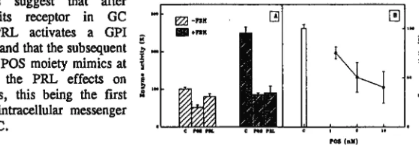
Addition of 1 µg/ml PRL (NIADDK-rPRL-B-5) to cultures (Figure 1) induced rapid (30 seconds) and transient (5 minutes) tumor GPI changes in differentiated granulosa cells. There was no effect on GPI levels in undifferentiated cells not expressing PRL receptors (Figure l inset). Parallel experiments performed with 3H-Ins-labeled cells showed no effect of PRL on the phosphoinositide tumor.
In another set of experiments (Fig. 2), GCs were cultured in the presence or absence of FSH, (buthcAMP or CTX with and without PRL or POS, the latter obtained by treating the GPI extracted from rat liver with phosphatidylinositol-specific PLC. The absence of PRL receptors in undifferentiated cells prevents its biological effects, but POS was as effective in these cells as in cells previously exposed to FSH to achieve differentiation.
IL-2) plays a central role in the immune system regulating the proliferation and differentiation of T lymphocytes
In any case, it is clear that the PI cascade is the main source (not exclusive, of course) of the. These results demonstrate that GPI lipids are an important component of the biological response to IL-2. Insulin- and glucose-dependent release of lipid-modified cAMP-binding proteins from plasma membranes and mitochondria of the yeast Saccharnyces Cerevisiae.
The anchor structure of the plasma membrane glycoprotein is characterized by the presence and specific arrangement of the components of typical glycosyl-phosphatidylinositol (GPI)-modified ectoproteins of higher eukaryotes (Müller and Bandlow, in press). The two lipid-modified cAMP-binding proteins are genetically unrelated to the regulatory subunit of the cytoplasmic protein kinase A (Müller and Bandlow, submitted). Therefore, we investigated whether insulin mediates cleavage of the lipid membrane anchors of the plasma men\brane and mitochondrial cAMP-binding proteins in Saccharomyces cereyisiae.
Incubation of cells with glucose alone (in the absence of insulin) also resulted in the formation of an even greater proportion of the hydrophilic cAMP receptor protein (up to about %), but with significantly delayed kinetics (2 to 3 h). The data indicate an insulin-dependent two-step processing of the GPI-anchor of plasma membrane cAMP-binding protein. As previously noted, release of the cAMP receptor from the plasma membrane in vitro requires not only cleavage of the GPI s· anchor.
We observed time- and concentration-dependent conversion of the PI-modified amphiphilic cAMP binding protein to its hydrophilic soluble derivative. Chemical analyzes of the PI glycans indicated the presence of fatty acids, alkyl glyceryl ethers, phosphate, inositol, glucosamine, mannose and ethanolainin.
EVIDENCE IN THESE CELLS OF INSULIN RESISTANCE AT POST- RECEPTOR LEVEL
INSULIN DOES NOT INDUCE HYDROl YSIS OF A GL YCOZYL-PHOSPHATIDYLINOSITOL (GPI) IN FETAL RAT HEPATOCYTES. In hepatocytes isolated from adult anirnals, an inverse correlation was observed between extracellular insulin and the number of insulin receptors and cellular GPI content. IPG affected the action of insulin on both forms of the enzyme in adult hepatocytes, while in fetal cells insulin did not change and IPG reduced glycogen phosphorylase activities.
These findings suggest a dissociation between insulin receptor occupancy and expected hormonal effects in fetal hepatocytes. While the frequency of oscillations depends on the dose of agonism, the time course of a single spike does not change with agonist concentration. In contrast, when vasopressin (Vp) is an agonist, elevated cA!V1P prolongs the falling phase of the transient\;;, moreover, ryancdine, which blocks the ea2+-induced Ca2+ release channel (CICR) in the open conformation, has almost no effect to the oscillation caused by Phe (l); in contrast, ryanodine inhibits Vp-evoked spikes.
The agonist specificity of the effect of cAMP could be explained by an enhancer of Ca2+. We are grateful for the collaboration with the Gobierno Vasco (A.S-il.), the Gobierno de Canarias (l.M.) and The WCllcomc Trust. Physologic concentrations of insulin (INS) regulate phospholipid metabolism in BC3H-1 myocytes in a time- and dose-dependent manner through multiple mechanisms, including the de novo synthesis of d-lacylglycerol (DAG) and through the hydrolysis of phosphatidylcholine and phosphatidylinositol glycan.
JNS activation of 03PAT was pertussis toxin sensitive and in cell-free preparations INS activation of G3PAT was blocked by treatment with NaF, a phosphatase inhibitor, and by treatment with either an antiserum raised against a PI-specific PLC (gift from Dr. 1 Fox) or an antiserum that recognizes the C-terminal decapeptide of Oía. Haley Veterans' Hospital and Department of Interna] Medicine University of South Florida 13000 Bruce Downs Boulevard Tampa, FL.
SERIE UNIVERSITARIA
Williams