Montserrat, gràcies per la teva ajuda amb els meus primers farmàcòfors i ànims en tot moment. Vane, moltes gràcies per ser un gran amic i per tots els temps que hem gaudit. Gràcies per tots els valors que m'heu transmès; i encara que no t'ho digui molt sovint, t'admiro i t'estimo.
Introduction: diabetes – an emerging epidemic of the 21 st century
Targeting Type 2 Diabetes
Peroxisome proliferator-activated receptor gamma (PPARγ)
Agonists of PPARα and PPARγ are currently approved for use in the treatment of dyslipidemia and T2DM, respectively [ 38 ]. These compounds act as partial agonists of PPARγ and exhibit different binding properties compared to full agonists. This mechanism consists in blocking the phosphorylation of PPARγ [51] and may explain how partial agonists can exhibit similar or higher antidiabetic effects than full agonists and the different side effect profiles of both types of agonists [52].
Dipeptidyl peptidase-IV
The DPP-IV binding site is highly druggable in the sense that close, specific binding to the enzyme can be achieved using small molecules with drug-like physicochemical properties [57,58]. I n t r o d u c t i o n | 23 The significant number of DPP-IV crystal structures published since 2003 provide a detailed picture of the structural characteristics of the binding site and the molecular recognition of small molecules. It is not surprising that a large number of different DPP-IV inhibitors have been discovered because the binding site offers a) a deep lipophilic pocket combined with numerous exposed aromatic side chains to achieve high-affinity binding of small molecules and b) significant access to solvents, which allows for tuning the physicochemical properties of the inhibitors for improved pharmacokinetic behavior [57].

Natural Products - Alternative medicine
Natural products have played an important role in the traditional treatment of T2DM since ancient times [66,67]. In many parts of the world, herbal remedies are still more accessible and affordable than conventional antidiabetic drugs. Each region of the world has developed a materia medica of antidiabetic agents based on the local flora [70-72].
Functional Food in Diabetes
I n t r o d u c t i o n | 27 nutritional science is just beginning, and there is the potential for exciting developments regarding the role of food in achieving optimal health and in the prevention and management of disease [78].
Computer-aided drug design methods in the discovery of antidiabetic drugs
ADMET properties
28 | Introduction Lipinski [93] studied the physico-chemical properties of 2245 World Drug Index (WDI) drugs and found that poor absorption and permeation are more likely when molecular weight < 500 g/mol, Clog P < 5, hydrogen bond donors < 5 and hydrogen bond acceptors < 10.
Virtual screening
Ligand-based approaches
- Ligand-Based Pharmacophore Modeling
- Similarity Analysis
- Quantitative Structure-Activity Relationships
1D-QSARs explain biological activity by relating it to a single value for a specific physicochemical property (eg, log P value) of a ligand. CoMFA models allow prediction of biological activity as well as 3D visualization of steric and electrostatic contributions to protein-ligand binding. An accurate representation of the bioactive conformation of ligands is crucial in 3D-QSAR to obtain correct ligand alignment.
Structure-based approaches
- Homology Modeling
- Protein-Ligand Docking
- Molecular Dynamics Simulations
- Structure-Based Pharmacophore Modeling
For docking-based VS, the open conformation of the target protein must be predicted. Uppenberg J, Svensson C, Jaki M, Bertilsson G, Jendeberg L, et al. 1998) Crystal structure of the ligand binding domain of the human nuclear receptor PPARgamma. 2005) Mexican plants with hypoglycemic effect used in the treatment of diabetes. 2009) Insulin-sensitizing activities of tanshinones, diterpene compounds of the root of salvia miltiorrhiza bunge.
DEVELOPMENT OF DOCKING-BASED 3D-QSAR MODELS FOR
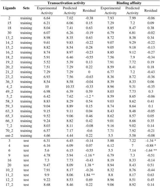
Correlation between the experimental transactivation activity and the experimental binding activity of the 49 tyrosine-based full PPARγ agonists used for the construction of the 3D-QSAR models. Scatter plots of the (A) pEC50 and (B) pIC50 models applied to the training set (colored gray) and the test set (colored black). Experimental and predicted values of the transactivation activity (pEC50) and binding affinity (pIC50) of the 49 tyrosine-based molecules used for the construction of the 3D-QSAR models.
STRUCTURAL INSIGHTS FOR THE DESIGN OF NEW PPARγ
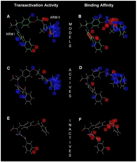
The selected conformations of the ligands, obtained with the previously described alignment protocol, were used for the generation of a pair of 3D-QSAR models (one for pIC50 and another for the percent maximal activation). Most of the pharmacophore sites are aromatic rings, highlighting the importance of hydrophobic interactions for the binding of PPARγ agonists to the receptor. This site corresponds to a carboxyl group present in the majority of the PPARγ partial agonists that forms a hydrogen bond with the Ser342 from the LBD of PPARγ.
Statistics of the best 3D-QSAR models for the analysis of the binding affinity (pIC50 model) and the transactivation activity of PPARγ (%max activation model) originate from an 80%. The favorable regions for binding are located in regions that interact with arms I and II and the right part of arm III (which includes Ser342) of the LBD of PPARγ. The representation of the 3D-QSAR model in Figure 4c suggests that when more hydrophobic interactions occur with arm I and arm II of the LBD of PPARγ, a greater binding affinity is seen in the compound.
When a hydrophobic group occupies the region of the carboxyl group responsible for the network of hydrogen bonds with the side chains of Ser289, His323, His449 and Tyr473, the transactivation activity of PPARγ decreases (see Figure 5). Other regions of the PPARγ LBD that also contribute to PPARγ transactivation activity are the regions of arms III and II that are closer to arm I. Experimental and predicted values of the pIC50 (i.e., the binding affinity measured by the displacement of a -labeled full agonist).
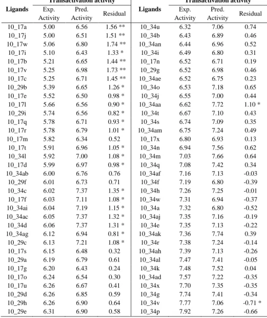
Experimental and predicted values of the % of maximal transactivation activities relative to the full agonist rosiglitazone.

IDENTIFICATION OF NOVEL PPARγ PARTIAL AGONISTS BY A
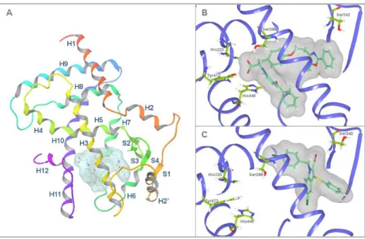
With minor exceptions, most of the currently known partial agonists interact with the LBD of PPARγ through a hydrogen bond with Ser342 [11] and several hydrophobic interactions that are similar to those that occur with full agonist binding (Figure 1B). The discriminatory power of the VS workflow to identify PPARγ partial agonists was evaluated by applying it to a group of 135 known PPARγ full agonists (Supporting Information Table 1), 19 known PPARγ partial agonists (Supporting Information Table 2) and 3,122 decoys obtained from the DUD database [27]. None of the tested compounds (C1-10) at the concentrations analyzed significantly reduced the viability or increased the cytotoxicity in HepG2 and 3T3-L1 cells (results not shown).
PDB codes of the ligand-protein complexes used for generating the structure-based pharmacophore models for PPARγ full agonists and PPARγ partial agonists. C h a p t e r 3 | 113 conformer and corresponding to the 4 sites of the partial agonist pharmacophores were initially identified as putative PPARγ partial agonists. These five partial agonists are representative of each of the five groups of PPARγ partial agonists defined in Table 4.
The ability of the VS workflow to identify partial PPARγ agonists was tested by applying it to a group of 135 known full PPARγ agonists (Supporting Information Table 1), 19 known partial PPARγ agonists (Supporting Information Table 2) and 3,122 decoys obtained from the DUD database [27]. For each step of the VS workflow, an enrichment factor (EF) and a value for sensitivity (Se) and specificity (Sp) were calculated [40]. For the rest of the steps, the set of partial agonists was considered to be the active compounds.
This value is a measure of the relative affinity of the test compound for the PPAR LBD.
IDENTIFICATION OF NATURAL EXTRACTS WITH
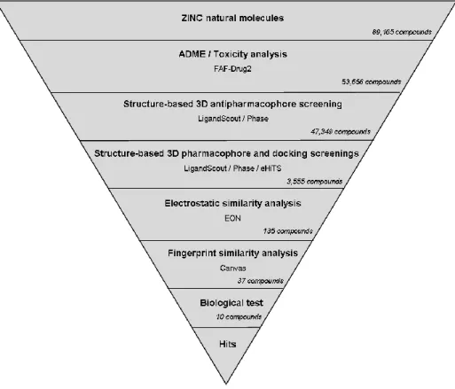
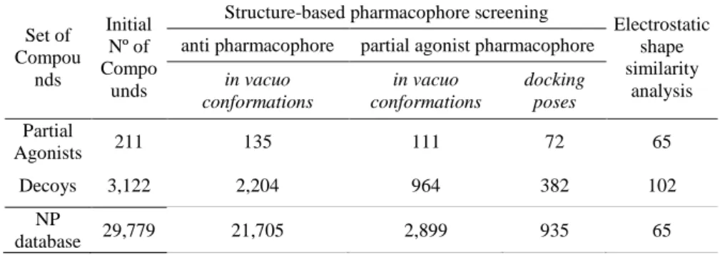
Thus, PPARγ partial agonists from natural extracts are promising candidates for the treatment of T2DM. Therefore, this VS workflow seems suitable to identify molecules with antidiabetic properties that can act as PPARγ partial agonists. A dataset of 211 known PPARγ partial agonists and 3,122 decoys extracted from the DUD database were used to validate our VS workflow.
Chemical comparison between molecules predicted as PPARγ partial agonists and molecules with described antidiabetic activity. This is in agreement with the results of our VS workflow showing that xanthoangelol F acts as a PPARγ partial agonist (Table 2). The second column represents the cluster number to which each molecule belongs when compared to a cluster of 211 synthetic PPARγ partial agonists.
None of these clusters contained any of the 211 known PPARγ partial agonists previously clustered with the VS hits. The second column represents the number of the group to which each molecule belongs when compared to a group of 211 synthetic PPARγ partial agonists. None of the 22 hits show chemical similarity to 211 known PPARγ partial agonists obtained from the literature and are therefore new chemical scaffold candidates for the development of PPARγ partial agonists.
Molecules that had at least one vacuum-generated conformer and that matched the 4 sites of the partial agonist pharmacophore were initially identified as putative PPARγ partial agonists.
IDENTIFICATION OF NOVEL HUMAN DIPEPTIDYL PEPTIDASE-
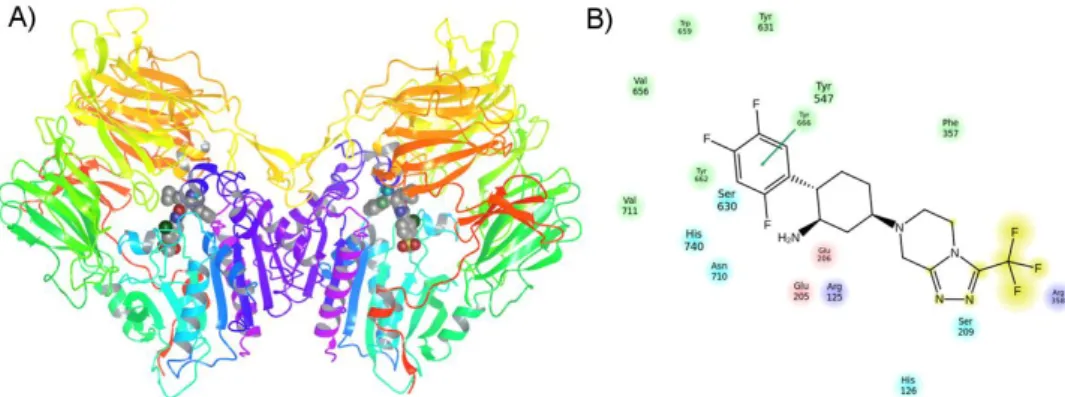
Based on the analysis of DPP-IV crystal structures [10-16] and the interpretation of structure-activity relationship data, the lipophilic S1 pocket and the Glu205/Glu206 dyad can be considered as key molecular anchors for DPP-IV inhibition [8]. Drug-like reversible DPP-IV inhibitors used to generate a common structure-based pharmacophore with corresponding IC50 values. We concluded that these two sites are essential for DPP-IV inhibition and considered that they are required in the common structural pharmacophore (see Figure 3).
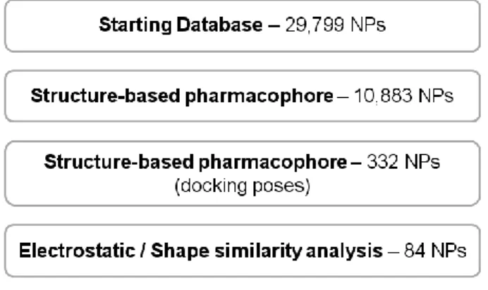
The relative location of the experimental positions of the ligands in Figure 1 after DPP-IV superposition. Schematic overview of the VS workflow and the procedure used for the selection of the VS hits tested for DPP-IV inhibitory activity. Finally, the poses for the 4,952 compounds from the second pharmacophore screen were subjected to a shape and electrostatic potential comparison with the experimental pose of the DPP-IV inhibitor at PDB file 3C45 (which has the smallest IC50 for all the non-peptide reversible inhibitors found in DPP-IV inhibitor complexes at the PDB [12]; see Figure 1).
Notably, 12 out of the 50 clusters obtained consisted exclusively of NPs previously unidentified as DPP-IV inhibitors. The relative DPP-IV inhibitory activity with or without the selected NPs (vehicle, 1% DMSO) is shown where each column represents the mean ± SEM (n=3 or 4). Criteria for the selection of the 3D structures for DPP-IV complexes used to derive the common structure-based pharmacophores.
The resulting individual energetic pharmacophores were used to construct a common structure-based pharmacophore for reversible DPP-IV inhibition.

IDENTIFICATION OF NATURAL EXTRACTS WITH POTENTIAL
The major substrates of DPP-IV are incretins, such as glucagon-like peptide-1 (GLP-1) and glucose-dependent insulinotropic polypeptide (GIP), which stimulate insulin secretion [ 13 ]. The efficacy and safety profile of DPP-IV inhibitors has so far been promising and favorable. Our results therefore suggest that the hypoglycemic properties of tecostamin could be mediated by the inhibition of DPP-IV.
From the same family as epinephrine, two additional compounds were predicted by our VS to be potential DPP-IV inhibitors. Therefore, the DPP-IV inhibition induced by this epicatechin derivative may contribute to the antihyperglycemic effect of GSPE [ 48 ]. The remaining 3 molecules predicted to be DPP-IV inhibitors through our VS workflow and found in extracts with described antidiabetic properties are hydroxysmirnovine from Galega orientalis [52], (-)-halosaline (CAS number from Haloxylon salicornicum [53] and isochanoclavine) - (I) (CAS number from Pennisetum typhoideum [54] (see Table 1).
Natural extracts with reported antidiabetic activity containing molecules predicted by our US protocol to be DPP-IV inhibitors. Natural extracts with no described antidiabetic activity (but of the same genus as plants with extracts with described antidiabetic activity) containing molecules predicted by our US protocol to be DPP-IV inhibitors. We demonstrated experimentally that our US protocol was able to identify DPP-IV inhibitors that (a) were not structurally related to a known molecule that inhibits DPP-IV and (b) that have never been reported to have antidiabetic activity.
Concomitant treatment with PPARγ partial agonists and DPP-IV inhibitors may be more effective in some cases.