Koldobika Villelabeitia-Jaureguizar, Davinia Vicente Campos, Alejandro Berenguel Senen, Ver´onica Hern´andez Jim´enez, Mar´ıa Elvira Barrios Garrido-Lestache, Jose L´opez Chicharro
PII: S0167-5273(17)32346-X
DOI: doi:10.1016/j.ijcard.2017.06.067 Reference: IJCA 25159
To appear in: International Journal of Cardiology Received date: 15 April 2017
Revised date: 10 June 2017 Accepted date: 15 June 2017
Please cite this article as: Villelabeitia-Jaureguizar Koldobika, Campos Davinia Vicente, Senen Alejandro Berenguel, Jim´enez Ver´onica Hern´andez, Garrido-Lestache Mar´ıa Elvira Barrios, Chicharro Jose L´opez, Effects of high-intensity interval versus continuous ex- ercise training on post-exercise heart rate recovery in coronary heart-disease patients, International Journal of Cardiology(2017), doi:10.1016/j.ijcard.2017.06.067
This is a PDF file of an unedited manuscript that has been accepted for publication.
As a service to our customers we are providing this early version of the manuscript.
The manuscript will undergo copyediting, typesetting, and review of the resulting proof before it is published in its final form. Please note that during the production process errors may be discovered which could affect the content, and all legal disclaimers that apply to the journal pertain.
ACCEPTED MANUSCRIPT
TITLE PAGE
EFFECTS OF HIGH-INTENSITY INTERVAL VERSUS CONTINUOUS EXERCISE TRAINING ON POST-EXERCISE HEART RATE RECOVERY IN CORONARY HEART-DISEASE PATIENTS
Koldobika Villelabeitia-Jaureguizar, MD, Hospital Universitario Infanta Elena.
Valdemoro, Madrid, Spain. E-mail: [email protected]
Correspondence to: Davinia Vicente Campos, PhD, Universidad Francisco de Vitoria.
Madrid. Spain. e-mail: [email protected] 0034627900087
Alejandro Berenguel Senen, MD, Hospital Universitario Virgen de la Salud. Toledo, Castilla La Mancha, Spain. E-mail: [email protected]
Verónica Hernández Jiménez, MD, Hospital Universitario Rey Juan Carlos. Madrid, Spain. E-mail: [email protected]
María Elvira Barrios Garrido-Lestache, PhD, MD, Hospital Universitario Rey Juan Carlos. Madrid, Spain. E-mail: [email protected]
Jose López Chicharro, PhD, MD. Grupo FEBIO. Universidad Complutense de Madrid. Madrid. Spain. E-mail: [email protected]
ACCEPTED MANUSCRIPT
ABSTRACT
Background: Heart rate recovery (HRR) has been considered a prognostic and mortality indicator in both healthy and coronary patients. Physical exercise prescription has shown improvements in VO2peak and HRR, but most of the studies have been carried out applying continuous training at a moderate intensity, being very limited the use of protocols of high intensity interval training in coronary patients.
We aimed to compare the effects of a moderate continous training (MCT) versus a high intensity interval training (HIIT) programme on VO2 peak and HRR.
Methods: Seventy three coronary patients were assigned to either HIIT or MCT groups for 8 weeks. Incremental exercise tests in a cycloergometer were performed to obtain VO2peak data and heart rate was monitored during and after the exercise test to obtain heart rate recovery data.
Results: Both exercise programmes significantly increase VO2peak with a higher increase in the HIIT group (HIIT: 4.5± 4.46 ml/kg/min vs MCT: 2.46±3.57 ml/kg/min;
P=0.039). High intensity interval training resulted in a significantly increase in HRR at the first and second minute of the recovery phase (15,44±7,04 vs 21,22 ±6,62, P
<0,0001 and 23,73±9,64 vs 31,52±8,02, p <0,0001, respectively).
Conclusions: The results of our research show that the application of HIIT to patients with chronic ischemic heart disease of low risk resulted in an improvement in VO2peak, and also improvements in post-exercise heart-rate recovery, compared with continuous training
Keywords: coronary artery disease, high interval training, heart rate recovery, aerobic functional capacity.
Abbreviations:
CHD: coronary heart disease HR: heart rate
HRR: heart rate recovery
MCT: moderate continuous training HIIT: high intensity interval training CAD: coronary artery disease CHF: congestive heart failure
CPET: cardiopulmonary exercise test
ACCEPTED MANUSCRIPT
1. Introduction
Coronary heart disease (CHD) continues to be the leading cause of morbidity and mortality in the developed countries, being the cause of approximately one third of all deaths in individuals over the age of 35 [1]. In spite of the research carried out to date, the pathophysiological basis of the disease is still not fully understood, with, among other pathogenic approaches, the chronic dysfunction of the autonomic nervous system being proposed as such a basis[2]. This has been implicated in the development of cardiovascular risk factors such as hypertension, diabetes, and dyslipidemia, and has been directly linked with mortality caused by coronary artery disease [3].
The results of recent meta-analysis [4] have confirmed that the inclusion of exercise programs in cardiac rehabilitation reduces cardiovascular mortality and hospital readmissions. The majority of the published research on the benefits of exercise for individuals with cardiovascular diseases have used constant load exercise (MCT) of between 60% and 80% of VO2peak. These studies have shown significant improvements in aerobic functional capacity (VO2peak) of between 12% and 31% [5].
In this context, for many years moderate continuous training (MCT) has been accepted as the gold standard [6]. However, for some time now, different teams have adopted a high-intensity interval training model (HIIT) as the most efficient with respect to objective adaptations of the majority of cardiac rehabilitation programs in patients with coronary artery disease (CAD), congestive heart failure (CHF) and metabolic syndrome [7].
In recent years, various revisions and meta-analysis on the implementation of HIIT for CAD and CHF have been published [8-11]. Two of these studies [10, 11] demonstrated in 260 patients with coronary heart disease the superiority of HIIT over continuous training with greater increases in VO2peak. These findings are consistent with the results of previous meta-analysis, which compared HIIT with continuous training in patients with heart failure [12] and cardio-metabolic disease [13]. Recent studies [14, 15] have even shown their effects on the diastolic dysfunction of the left ventricle that had not been effectively treated with drugs.
HIIT is usually defined as exercise of repeated intervals of a short to intermediate duration (eg. 10 s to 5 min) completed at a higher intensity than the corresponding anaerobic threshold [16] . The exercise intervals are separated by low-intensity recovery times or rest which allow for partial recovery from the efforts of the previous interval [16].
The heart rate recovery (HRR) has been proposed in many studies as an indicator of prognosis and mortality, in that a slowing of the HRR after strenuous exercise is a predictor of mortality both in healthy subjects and in patients with CHD [3, 17].
Meanwhile, other authors even consider that post-exercise HRR is a good inidcator of
ACCEPTED MANUSCRIPT
cardiac rehabilitation program effectiveness [18-20], it being a quality index to stratify patient risk upon completing a rehabilitation program [21].
Post-exercise HRR can be divided into 2 phases: fast and slow. The quick phase refers to the first minute of recovery (< 1 min) and characterises a period in which there is an abrupt and rapid decline of the HR [22, 23]. The slow phase, meanwhile, refers to the period following the fast phase (≥ 2 min) [23-25]. HRR in the first minute after exercising corresponds to vagal reactivation, especially in the first 30 seconds [22], while later recovery (≥2 min) is likely attributed to a drop in sympathetic activity [26].
To date there exists no consensus regarding the most appropriate time and method to obtain HRR data. Since the publication of Cole et al’s research [27, 28] into men and women without cardiovascular disease, measuring the HRR in the first minute after exercise is the most common method employed, with a cut-off point of ≤12 bpm to consider a reading as clinically abnormal. Readings lower than 12 bpm were associated with mortality risk for any cause 2 times higher in the population group referred to [28]. Measuring HRR 2 minutes after exercise during passive recovery in a sitting position has also shown itself to be an independent predictor of cardiovascular mortality from all causes in a long series of coronary patients, moreover providing valuable information that can be used for risk stratification in this population group [29]. Readings lower than 22 bpm 2 minutes after exercise have been considered clinically abnormal [27, 28]. Shetler et al [30] suggested that a recovery of 22 bpm 2 minutes after exercise exceed in capacity the HRR 1 prognosis. Other studies carried out on healthy subjects confirmed the prognostic value of the HRR reading at 2 min post-exercise [27, 31].
Training sessions in cardiac rehabilitation programs which often use continuous exercises of moderate intensity have shown improvements in HRR after 8 [32] and 12 [33-37] weeks of training, the studies being more limited [38, 39] which have valued modifications in the HRR employing HIIT protocols, compared with the continuous protocols of constant intensity.
Intermittent high-intensity exercise, which are matched to continuous for either energy expenditure or exercise duration, have shown improvements in different hemodynamic indicators at rest, cardiorespiratory fitness, entotelial functionality and morphology and function of the left ventricle [40-45].
The objective of this research was to compare the influence of 2 different exercise protocols (continuous and HIIT) when used with coronary patients as part of a cardiac rehabilitation program on post-exercise heart-rate recovery readings.
2. Methods
2.1 Study population
ACCEPTED MANUSCRIPT
A prospective, randomized clinical trial (NCT02168712) was conducted with patients referred by the Cardiac Rehabilitation Department who were diagnosed with stable New York Heart Association functional class I or II coronary artery disease with angina pectoris or myocardial infarction and no heart failure. To be included in the study, patients had to achieve a respiratory exchange ratio ≥ 1.10 during the initial cardiopulmonary exercise test (CPET). This respiratory exchange ratio value is often used as a criterion for achieving a maximum exercise effort [46]. Patients who had residual ischemia (by electrocardiogram [ECG] criteria or angina symptoms), severe ventricular arrhythmias, uncontrolled hypertension, permanent pacemakers, or implanted cardiac defibrillators were excluded.
After signing an informed consent form, patients were randomized on a one-to-one basis to either the MCT or the HIIT group. The mode of exercise training was a cycle ergometer with 40 minutes per sessions, 3 days per week (total of 24 sessions over 2 months).
Patients entered the study within 6 weeks from the revascularization procedure.
Selected CPET variables were recorded before and after the exercise training.
Cardiopulmonary exercise tests were administered by staff blinded as to which exercise training group the patients were assigned.
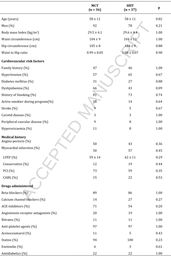
Characteristics and medication use of the patients are shown in table 1.
2.2 Cardiopulmonary exercise test (CPET)
All patients underwent exercise testing with a cycle ergometer (Ergoline900S, Ergoline GmbH, Bitz, Germany) including analysis of exhaled gases (UltimaCardiO2, Medical Graphics Corporation, St Paul, Minnesota). The exercise test protocol was tailored to each patient’s physical condition, with gradual increments of 10, 15, or 20 W/min. The same protocol was applied before and after the Exercise training program. The objective of the exercise tests was to achieve a sustained effort for 8 to 12 minutes, with the aim of proper oxygen uptake (VO2) kinetics and maintaining a linear relationship between VO2, exercise workload and heart rate (HR).
A 12-lead ECG was continuously monitored, and blood pressure was measured every 3 minutes during the exercise tests. Exercise workloads in watts and metabolic, cardiac, ventilatory and electrocardiographic parameters were analyzed. The ECG was continuously monitored during the first 5 minutes of recovery. Recorded HR was determined from the computerized test reports and was the average of the last 5 RR intervals.
HRR indices were calculated by subtracting first and second minute HR on recovery period from the maximal HR obtained during stress testing and designated as HRR-1 and HRR-2, respectively. [28, 47]. The relative change in HRR was determined as the decrease produced in HR at 1 and 2 min after finishing exercise expressed as a percentage of the peak HR (%HRR-1/HRpeak and %HRR-2/HRpeak, respectively). We also calculated the relative decrease in HR at 1 and 2 min post-exercise as a
ACCEPTED MANUSCRIPT
percentage of the increase produced from resting HR to peak HR (%HRR-1/(HRpeak − HRrest) and %HRR-2/(HRpeak – HRrest), respectively).
The first (VT1) ventilatory threshold were considered to be indicator of the aerobic threshold, and was determined after the ventilatory equivalent method described by Skinner et al. [46]. The VO2 in mL·kg-1 ·min-1 and HR in beats·min-1 at VT1 were the parameters used to determine the MCT exercise intensity.
2.3 Steep Ramp Test
To design the HIIT program, we used the steep ramp test (SRT) protocol, according to the methodology described by Meyer et al. [48]. This exercise test protocol is composed of 2 minutes of free pedaling at 25 W followed by progressive 25-W increments every 10 seconds, maintaining a constant pedal cadence of between 50 and 60 rpm. The test was stopped when the patient could not maintain continuous pedal cadence of > 40 rpm after encouragement to increase to 50 rpm and/or experienced hemodynamic and/or electrical alterations. The maximum exercise load achieved, as measured in watts, was the exercise parameter used to design the HIIT program for each patient.
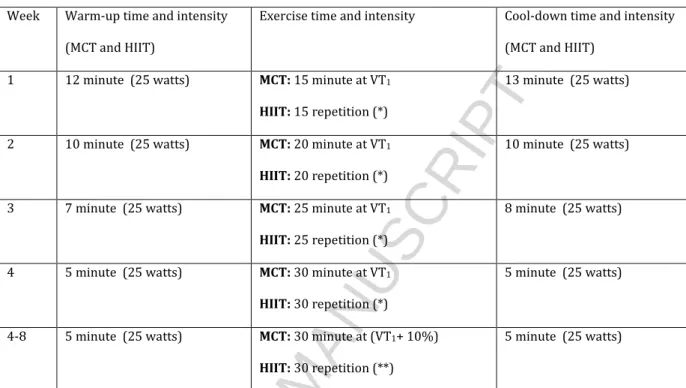
2.4 MCT and HIIT Program Designs
The metabolic parameters obtained during the pretraining CPET were used to design the MCT program. Patients were asked to keep their training HR below the HR at VT1 during the first month. During the second month, the intensity of the exercise was adjusted, increasing to a training HR that corresponded to VT1 plus 10%. The HIIT protocol used the methodology initially described by Meyer et al [48] and which was recently published by our group [49]. In this type of exercise, the intensity was established using workload (watts), without taking HR into consideration as a measure for regulating the intensity of the exercise. The training workloads depended on the maximum workload achieved during the SRT. The intervals were designed as follows.
In the first month of training, 20-second repetitions at an intensity corresponding to 50% of the maximum load reached with the SRT (peak intervals) were followed by 40- second recovery periods at 10%. In the second month of training, the intensity of exercise was adjusted using the results of a new SRT. The total duration of both types of training was 40 minutes per session throughout the exercise program (including warm-up and cool-down). Table 2 summarizes the exercise time and intensity progression for both MCT and HIIT. Patients rated the peak level of exertion during each training session using the Borg Rating of Perceived Exertion Scale [50]. Both types of exercise were reviewed and approved by the local Research Ethics Committee.
Patients enrolled in the study participated in other activities established in our cardiac rehabilitation program that were aimed at managing psychological stress and learning about cardiac health habits. They were also taught to devise a home walking program for the days on which they did not have to attend sessions in the hospital. The recommended intensity of walking was a perceived exertion of 11 to 13 on the Borg scale.
ACCEPTED MANUSCRIPT
2.5 Safety of the Exercise Training Programs
To verify the safety of using this kind of aerobic Exercise training, we made a daily record of any incidents or adverse effects that could limit the planned exercise. An incident was considered low if there were no repercussions and it was possible to start and/or restart training (eg, muscle overload, fatigue, muscular pain, and dyspnea without oxygen desaturation). A moderate incident was defined as one that limited the planned training (dyspnea with desaturation < 94%, muscle injury, vasovagal conditions), and an incident was defined as severe if it was potentially lifethreatening (ischemia, ventricular arrhythmia, hypertensive emergencies).
2.6 Statistical analysis
Quantitative variables were described using means and standard deviations. The normality of the data distribution was determined using the Kolmogorov Smirnov test.
To evaluate the effect of each exercise protocol on the quantitative variables, pre- and post-program values were compared using Student’s dependent samples t test. The effect was measured in absolute terms via the difference between the post-program values and those obtained before training. These changes were described with the mean and standard deviation. Comparisons between the 2 training programs were made using Student’s t test in the case of quantitative variables and using the χ 2 test of association or Fisher exact test for qualitative variables. All the comparisons were made using 2-tailed tests, and the level of significance was set at P < 0.05. The relationship between HRR and VO2peak was assessed by calculating Pearson correlation coefficients. All statistical tests were performed using commercially available software (SPSS, Version 19,0, Inc., Chicago, IL, USA).
3. Results
A total of 73 patients were included and studied (36 patients MCT group and 37 patients HIIT group). At the start of the study, there were no significant differences between the groups with regard to clinical characteristics and medication use.
3.1 Training Data
The intensity of exercise in the MCT group in the first month was 64.2% ± 8.5% of the VO2peak reached during the initial CPET (corresponding to the VT1) and 69.5% ± 8.7%
in the second month (corresponding to VT1 + 10%). The exercise workload applied at the peak intervals in the HIIT group using the Meyer et al methodology [48] was 104.5% ± 22.2% (first month) and 134.5% ± 29.7% (second month) of the maximum load reached in the initial CPET corresponding to 50% of the SRT in both months.
Adherence to the treatment sessions (the number of sessions attended compared with the number of sessions scheduled) was 87.5% in the MCT group and 92% in the HIIT group.
3.2 Cardiopulmonary exercise test
ACCEPTED MANUSCRIPT
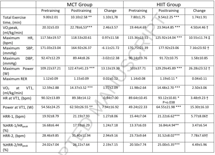
The exercise effort test results for both groups can be seen in Table 3. After 8 weeks of training, both exercise programs significantly increased their VO2peak, with a greater increase in the HIIT group (4.5 ± 4.46 ml/kg/min vs 2.46 ± 3.57 ml/kg/min, for patients of the HIIT and MCT groups respectively, P=0.039).
Both groups also showed a significant increase in the peak exercise workload achieved (MCT 13.13 ± 19.39 W vs HIIT 26.28 ± 23.52 W), with a significantly higher increase in the HIIT group (P=0.012). A significant increase was observed in maximal HR in the HIIT group only (10.55 ± 11.74 beats·min-1; P=0.001). The total time of the exercise effort test, as well as the VO2 and exercise workload at VT1 significantly increased in both groups, but HR at VT1 only increased in the HIIT group (3.48 ± 9.23; p=0.030).
3.3 Heart Rate Recovery
In Table 3, the heart-rate recovery values for both groups are reflected. With regard to the HR recovery in the first and second minute after the exercise test, the only significant change was observed in the HIIT group (15.44 ± 7.04 vs 21.22 ± 6.62; P
<0.0001 and 23.73 ± 9.64 vs 31.52± 8.02; p<0.0001, respectively). Differences were observed in changes in HRR-1 (MCT: 1,27 ± 8,06 vs HIIT: 5,77 ± 8,06; p=0,021) and in HRR-2 (MCT: 2,94 ± 9,16 vs HIIT: 7,78 ± 7,69; p=0,022) between groups.
3.4 Relationship between HRR and Peak VO2
Peak VO2 showed significant correlation with HRR-1 (r = 0.40; p < 0.001) and %HRR- 1/HRpeak (r = 0.28; p =0.014), and with HRR-2 (r = 0.43; p < 0.001) and %HRR- 2/HRpeak (r = 0.26; p =0.026), for the whole group of patients pre-training. With regard to the post-training Peak VO2 showed significant correlation with HRR-1 (r = 0.38; p < 0.01), and with HRR-2 (r = 0.53; p < 0.001) and %HRR-2/HRpeak (r = 0.37; p
<0.01), for the whole group of patients (Figure 1).
3.5 Safety of the Training Intervention
No incidents or adverse events were recorded that limited the ability of patients to perform the prescribed exercise in either of the training programs.
4. Discussion
The principle contribution of this study has been the verification of an improvement in the post-exercise heart-rate recovery (HRR1 and HRR2) associated with the group of patients that underwent training with HIIT methodology. In addition, a greater increase in post-exercise values of VO2peak in the HIIT group was also recorded.
Another interesting observation is that in the group of patients as a whole, we found a significant correlation of VO2peak between HHR1 and HHR2.
ACCEPTED MANUSCRIPT
In contrast to most previous research that analysed only HRR1 [32-37], we measured the heart rate recovery in the 1 and 2 min post-exercise. Both points have shown associations with mortality risk, although the heart rate in min 2 post-exercise has proved to be the most powerful predictor [30], which suggests the need to add systematic evaluation of HRR2 in patients with cardiovascular disease. Considering that this was a maximal exercise test, we propose that in addition to the indices of HRR as absolute values (HRpeak-HR1 and HRpeak -HR2), it is perhaps preferable to also consider HRR as a percentage of the peak HR recorded during the graded cardiopulmonary test. Thus, HRR relative to HRpeak as a measure of HR recovery applicable to all subjects regardless of age is expressed. Similarly, only the HIIT group improved the% HRR1 / HRpeak (%) (13.37 ± 6.03 to 16.84 ± 4.94 bpm; p = 0.003) and%
HRR2 / HRpeak (%) (to 20.50 ± 7.74 to 25.00 ± 5 35 bpm; p <0.0001), with average post-exercise values of % HRR1 / HRpeak (%)of 17.93% and 16.84% for MCT and HIIT groups respectively; and %HRR2 / HRpeak (%) of 26.22% and 25.00% for MCT and HIIT groups, respectively.
Our study showed average HRR1 values (pre- and post-training) above the clinical cut- off point established at <12 bpm at min 1 post-exercise for a HRR anomaly to be considered. However, it should be noted that 16 of the 73 patients (21.9%) of our study did not reach a HRR-1 of 12 in the evaluation previous to the start of the exercise program. Only 7 patients maintained their HRR1 below the limit of <12 bpm after the recovery period, emphasizing in this regard the effectiveness of HIIT, which reduced the number of patients below the clinical HRR1 limit by 81.8%. Although an HRR of <12 bpm in the 1st minute after exercise is the most commonly used index, some research has shown increases in mortality risk with HRR 1 of <25 bpm [51] and <22 bpm [52].
Different studies have shown improvements of heart-rate recovery with exercise training. Mahdavi Anari et al [53] observed, after a 12-week training period, a significant improvement of the HRR-1 (13.76 ± 1.38 bpm to 17.07±1.33 bpm). The improvement in recovery was similar in patients with coronary artery bypass graft or percutaneous transluminal coronary angioplasty. The results obtained in our HIIT group were similar in HRR-1 (15.44 ± 7.04 bpm to 21.22 ± 6.62 bpm). For their part, Ribeiro et al [54] and Tiukinhoy et al [55] also observed similar results in the improvement of HRR1 with an 8-week exercise program. Thus, HHR is a useful indicator to stratify the patient risk after completing a rehabilitation program.
Regarding the HRR-2 value, Cole et al [28] proposed a normality limit of ≤42 beats.
Based on this criterion, only 6 of our patients (8.2%) were above this limit before starting the exercise program, it being reached by 13 patients (17.8%) at the end of it.
While the MCT group managed to have 3 patients more under this limit upon completing the exercise program, in the HIIT group, 4 new patients were incorporated, suggesting greater effectiveness in the HIIT modality. Unlike the many studies that have evaluated HRR1, those that have measured HRR-2 are scarcer, meaning that their established clinical criteria of normality are less powerful.
The later recovery (≥2 min) is likely attributed to a drop in sympathetic activity (reduced activation of peripheral muscle mechanoreceptors and chemoreceptors) and to humoral factors such as catecholamines [25, 51, 56-60], and, in contrast to that
ACCEPTED MANUSCRIPT
which occurs in the fast phase of the HRR (≤1 min), the slow phase (≥2 min) is clearly dependant on the intensity and duration of the previously performed exercise[22], conditioning the normalisation of the associated metabolic stress [25, 61].
The intermittent high-intensity exercise (HIIT), which are matched to continuous for either energy expenditure or exercise duration, have shown improvements in different hemodynamic indicators at rest, cardiorespiratory fitness, entotelial functionality and morphology and function of the left ventricle [40-45]. However, Conraads et al [62]
observed in a large population of CAD patients similar improvements in exercise capacity and peripheral endothelial function following HIIT or moderate continuous training.
The physiopathological reason why an abnormal HRR is associated with a worse clinical prognosis is not clear. In accordance with Huang et al [63], there exists a close relationship between HRR and the endothelial function; thus, any delay in the HRR might indicate endothelial dysfunction, and this alteration has proved to be a powerful predictor of global mortality in CHD [63].Another factor that might contribute to HRR delay has been associated with an excessive pro-inflammatory state. In accordance with the recent concept of “cholinergic anti-inflammatory pathway”, immunity is coordinated by neural circuits that operate reflexively, and this well established neural circuit terminates excessive pro-inflammatory cytokine responses, preventing immune-mediated damage [64, 65]. Therefore, a fall in parasympathetic activity can result in a pro-inflammatory response, thereby increasing morbidity and mortality [2, 66, 67]. Similarly, Youn et al [68] confirmed that a slowing of post-exercise HRR was linked to an exaggerated pro-inflammatory response, being an independent predictor variable in patients with heart failure.
Different studies have shown that HIIT training improves VO2peak values in patients to a greater extent with respect to continuous load training [38, 43, 45]. Also of note in our research was that VO2 peak improved in both groups, but more so (p<0.05) in the HIIT group. Other investigations [62] showed no differences in VO2 peak improvement using HIIT vs aerobic continuous training in patients with coronary artery disease. In the HIIT protocol of our study, the training intensity was established using the workload in watts as a percentage of the maximum workload reached in the SRT. The workloads applied were high intensity with a range of 104% and 134% of the maximum load reached during the initial effort test (CPET). Meyers et al [69] noted that using this methodology for the HIIT design, exercise time at an intensity higher than 85%
VO2peak during prolonged training was prolonged, which justifies physiological adaptations to those associated with the improvement of the VO2peak.
In our study, we found a significant correlation between the VO2peak values of the entire group with HRR-1 (r=0.40; p<0.001) and HRR-2 (r=0.43; p<0.001), thereby confirming the relation between VO2peak and HRR suggested by the authors [70-74].
Our group [75] also found a significant correlation between VO2peak (ml/kg/min) and HRR3 (r = 0.36; p < 0.001) in adult physically active men. Together, this data indicates that cardiorespiratory fitness is linked to HRR.
ACCEPTED MANUSCRIPT
Maximum heart rate increased with training in the HIIT group, with changes in the continuous training (MCT) group not being found. This response seems to be related to the high workload achieved in the HIIT group in the effort test after the training program. Underlying these functional improvements, cellular adaptations including rate of Ca2+ cycling and Ca2+ sensitivity of the cardiomyocytes were demonstrated in animal models following HIIT.
The two groups of our study improved VO2 and the load (W) associated with VT1, with no differences between them. Similar results were found in other research studies [40, 76-78], while in others, greater improvements in VO2 associated to VT with HIIT were obtained [39, 79]. The different protocols used may justify the lack of concordance in the results. Looking at HR related to VT, this factor only increased in the HIIT group, reflecting peripheral metabolic adaptations that allow for the sustaining of a greater workload in VT1.
In line with the increase of VO2peak, the maximum load reached (Wmax) increased significantly more (p = 0.012) in the HIIT group, reflecting an improvement in the base- acid balance with peak loads.
Additionally, HIIT seems to be a safe exercise modality and did not differ in frecuency or magnitude of cardiovascular adeverse events during exercise training as compared with continuous training, as was shown previously [77].
Although it is a randomized study, we studied a small number of patients. It would be necessary large-scale, randomized clinical trials to investigate clinical end-points.
5. Conclussions
The results of our research show that the application of HIIT to patients with chronic ischemic heart of low risk resulted in an improvement in VO2peak, and also improvements in post-exercise heart-rate recovery, compared with continuous training (which showed no significant changes). Given the observed inverse relationship between the values of VO2peak and post-exercise HR recovery rates with all-cause mortality in these patients, the results of our study argue for giving preference to the interval high intensity training during the rehabilitation program in low-risk coronary patients.
Conflicts of interest
The authors report no relationships that could be construed as a conflict of interest.
ACCEPTED MANUSCRIPT
References
[1. Lloyd-Jones, D., et al., Executive summary: heart disease and stroke statistics-- 2010 update: a report from the American Heart Association. Circulation, 2010.
121(7): p. 948-54.
2. Thayer, J.F. and R.D. Lane, The role of vagal function in the risk for cardiovascular disease and mortality. Biol Psychol, 2007. 74(2): p. 224-42.
3. Grad, C. and D. Zdrenghea, Heart Rate Recovery in Patients with Ischemic Heart Disease - Risk Factors. Clujul Med, 2014. 87(4): p. 220-5.
4. Anderson, L., et al., Exercise-Based Cardiac Rehabilitation for Coronary Heart Disease: Cochrane Systematic Review and Meta-Analysis. J Am Coll Cardiol, 2016. 67(1): p. 1-12.
5. van Tol, B.A., et al., Effects of exercise training on cardiac performance, exercise capacity and quality of life in patients with heart failure: a meta- analysis. Eur J Heart Fail, 2006. 8(8): p. 841-50.
6. Hambrecht, R., et al., Physical training in patients with stable chronic heart failure: effects on cardiorespiratory fitness and ultrastructural abnormalities of leg muscles. J Am Coll Cardiol, 1995. 25(6): p. 1239-49.
7. Ito, S., T. Mizoguchi, and T. Saeki, Review of High-intensity Interval Training in Cardiac Rehabilitation. Intern Med, 2016. 55(17): p. 2329-36.
8. Kemi, O.J. and U. Wisloff, High-intensity aerobic exercise training improves the heart in health and disease. J Cardiopulm Rehabil Prev, 2010. 30(1): p. 2- 11.
9. Smart, N.A., G. Dieberg, and F. Giallauria, Intermittent versus continuous exercise training in chronic heart failure: a meta-analysis. Int J Cardiol, 2013.
166(2): p. 352-8.
10. Elliott, A.D., et al., Interval training versus continuous exercise in patients with coronary artery disease: a meta-analysis. Heart Lung Circ, 2015. 24(2): p. 149- 57.
11. Liou, K., et al., High Intensity Interval versus Moderate Intensity Continuous Training in Patients with Coronary Artery Disease: A Meta-analysis of Physiological and Clinical Parameters. Heart Lung Circ, 2016. 25(2): p. 166- 74.
12. Haykowsky, M.J., et al., Meta-analysis of aerobic interval training on exercise capacity and systolic function in patients with heart failure and reduced ejection fractions. Am J Cardiol, 2013. 111(10): p. 1466-9.
13. Weston, K.S., U. Wisloff, and J.S. Coombes, High-intensity interval training in patients with lifestyle-induced cardiometabolic disease: a systematic review and meta-analysis. Br J Sports Med, 2014. 48(16): p. 1227-34.
14. Angadi, S.S., et al., High-intensity interval training vs. moderate-intensity continuous exercise training in heart failure with preserved ejection fraction: a pilot study. J Appl Physiol (1985), 2015. 119(6): p. 753-8.
15. Hollekim-Strand, S.M., et al., High-intensity interval exercise effectively improves cardiac function in patients with type 2 diabetes mellitus and diastolic
ACCEPTED MANUSCRIPT
dysfunction: a randomized controlled trial. J Am Coll Cardiol, 2014. 64(16): p.
1758-60.
16. Buchheit, M., et al., Performance and physiological responses during a sprint interval training session: relationships with muscle oxygenation and pulmonary oxygen uptake kinetics. Eur J Appl Physiol, 2012. 112(2): p. 767-79.
17. Chou, C.L., et al., Impact of Phase II cardiac rehabilitation on abnormal heart rate recovery. J Chin Med Assoc, 2014. 77(9): p. 482-6.
18. MacMillan, J.S., et al., Exercise and heart rate recovery. Heart Lung, 2006.
35(6): p. 383-90.
19. Streuber, S.D., E.A. Amsterdam, and C.L. Stebbins, Heart rate recovery in heart failure patients after a 12-week cardiac rehabilitation program. Am J Cardiol, 2006. 97(5): p. 694-8.
20. Kligfield, P., et al., Effect of age and gender on heart rate recovery after submaximal exercise during cardiac rehabilitation in patients with angina pectoris, recent acute myocardial infarction, or coronary bypass surgery. Am J Cardiol, 2003. 92(5): p. 600-3.
21. Chen, J.T., et al., Beneficial effects of home-based cardiac rehabilitation on metabolic profiles in coronary heart-disease patients. Kaohsiung J Med Sci, 2016. 32(5): p. 267-75.
22. Imai, K., et al., Vagally mediated heart rate recovery after exercise is accelerated in athletes but blunted in patients with chronic heart failure. J Am Coll Cardiol, 1994. 24(6): p. 1529-35.
23. Coote, J.H., Recovery of heart rate following intense dynamic exercise. Exp Physiol, 2010. 95(3): p. 431-40.
24. Buchheit, M., et al., Noninvasive assessment of cardiac parasympathetic function: postexercise heart rate recovery or heart rate variability? Am J Physiol Heart Circ Physiol, 2007. 293(1): p. H8-10.
25. Perini, R., et al., Plasma norepinephrine and heart rate dynamics during recovery from submaximal exercise in man. Eur J Appl Physiol Occup Physiol, 1989. 58(8): p. 879-83.
26. Pecanha, T., N.D. Silva-Junior, and C.L. Forjaz, Heart rate recovery: autonomic determinants, methods of assessment and association with mortality and cardiovascular diseases. Clin Physiol Funct Imaging, 2014. 34(5): p. 327-39.
27. Cole, C.R., et al., Heart rate recovery after submaximal exercise testing as a predictor of mortality in a cardiovascularly healthy cohort. Ann Intern Med, 2000. 132(7): p. 552-5.
28. Cole, C.R., et al., Heart-rate recovery immediately after exercise as a predictor of mortality. N Engl J Med, 1999. 341(18): p. 1351-7.
29. Gayda, M., et al., Heart rate recovery after exercise and long-term prognosis in patients with coronary artery disease. Can J Cardiol, 2012. 28(2): p. 201-7.
30. Shetler, K., et al., Heart rate recovery: validation and methodologic issues. J Am Coll Cardiol, 2001. 38(7): p. 1980-7.
31. Savin, W.M., D.M. Davidson, and W.L. Haskell, Autonomic contribution to heart rate recovery from exercise in humans. J Appl Physiol Respir Environ Exerc Physiol, 1982. 53(6): p. 1572-5.
32. Hai, J.J., et al., Relationship between changes in heart rate recovery after cardiac rehabilitation on cardiovascular mortality in patients with myocardial infarction. Heart Rhythm, 2010. 7(7): p. 929-36.
ACCEPTED MANUSCRIPT
33. Giallauria, F., et al., Long-term effects of cardiac rehabilitation on end-exercise heart rate recovery after myocardial infarction. Eur J Cardiovasc Prev Rehabil, 2006. 13(4): p. 544-50.
34. Giallauria, F., et al., Exercise-based cardiac rehabilitation improves heart rate recovery in elderly patients after acute myocardial infarction. J Gerontol A Biol Sci Med Sci, 2006. 61(7): p. 713-7.
35. Hao, S.C., A. Chai, and P. Kligfield, Heart rate recovery response to symptom- limited treadmill exercise after cardiac rehabilitation in patients with coronary artery disease with and without recent events. Am J Cardiol, 2002. 90(7): p.
763-5.
36. Tsai, S.W., Y.W. Lin, and S.K. Wu, The effect of cardiac rehabilitation on recovery of heart rate over one minute after exercise in patients with coronary artery bypass graft surgery. Clin Rehabil, 2005. 19(8): p. 843-9.
37. Wu, S.K., et al., Cardiac rehabilitation vs. home exercise after coronary artery bypass graft surgery: a comparison of heart rate recovery. Am J Phys Med Rehabil, 2006. 85(9): p. 711-7.
38. Jaureguizar, K.V., et al., Effect of High-Intensity Interval Versus Continuous Exercise Training on Functional Capacity and Quality of Life in Patients With Coronary Artery Disease: A RANDOMIZED CLINICAL TRIAL. J Cardiopulm Rehabil Prev, 2016. 36(2): p. 96-105.
39. Currie, K.D., et al., Low-volume, high-intensity interval training in patients with CAD. Med Sci Sports Exerc, 2013. 45(8): p. 1436-42.
40. Moholdt, T., et al., Aerobic interval training increases peak oxygen uptake more than usual care exercise training in myocardial infarction patients: a randomized controlled study. Clin Rehabil, 2012. 26(1): p. 33-44.
41. Munk, P.S., N. Butt, and A.I. Larsen, High-intensity interval exercise training improves heart rate variability in patients following percutaneous coronary intervention for angina pectoris. Int J Cardiol, 2010. 145(2): p. 312-4.
42. Munk, P.S., et al., High-intensity interval training may reduce in-stent restenosis following percutaneous coronary intervention with stent implantation A randomized controlled trial evaluating the relationship to endothelial function and inflammation. Am Heart J, 2009. 158(5): p. 734-41.
43. Rognmo, O., et al., High intensity aerobic interval exercise is superior to moderate intensity exercise for increasing aerobic capacity in patients with coronary artery disease. Eur J Cardiovasc Prev Rehabil, 2004. 11(3): p. 216-22.
44. Warburton, D.E., et al., Effectiveness of high-intensity interval training for the rehabilitation of patients with coronary artery disease. Am J Cardiol, 2005.
95(9): p. 1080-4.
45. Wisloff, U., et al., Superior cardiovascular effect of aerobic interval training versus moderate continuous training in heart failure patients: a randomized study. Circulation, 2007. 115(24): p. 3086-94.
46. Skinner, J.S. and T.M. McLellan, The transition from aerobic to anaerobic metabolism. Res Q Exerc Sport, 1980. 51(1): p. 234-48.
47. Tulumen, E., et al., The reproducibility of heart rate recovery after treadmill exercise test. Ann Noninvasive Electrocardiol, 2011. 16(4): p. 365-72.
48. Meyer, K., et al., Interval training in patients with severe chronic heart failure:
analysis and recommendations for exercise procedures. Med Sci Sports Exerc, 1997. 29(3): p. 306-12.
ACCEPTED MANUSCRIPT
49. Villelabetia Jaureguizar K, D.-B.I., Vaquerizo I, Calero MJ, Mahillo I., Entrenamiento interválico en pacientes con cardiopatía isquémica: metodología y análisis de resultados ergoespirométricos. Rehabilitación, 2011. 45: p. 7.
50. G, B., Borg´s perceived exertion and pain scales. 1998: Human Kinetics.
51. Jouven, X., et al., Heart-rate profile during exercise as a predictor of sudden death. N Engl J Med, 2005. 352(19): p. 1951-8.
52. Lahiri, M.K., P.J. Kannankeril, and J.J. Goldberger, Assessment of autonomic function in cardiovascular disease: physiological basis and prognostic implications. J Am Coll Cardiol, 2008. 51(18): p. 1725-33.
53. Mahdavi Anari, L., et al., Effect of Cardiac Rehabilitation Program on Heart Rate Recovery in Coronary Heart Disease. J Tehran Heart Cent, 2015. 10(4): p.
176-81.
54. Ribeiro, F., et al., Exercise training enhances autonomic function after acute myocardial infarction: a randomized controlled study. Rev Port Cardiol, 2012.
31(2): p. 135-41.
55. Tiukinhoy, S., N. Beohar, and M. Hsie, Improvement in heart rate recovery after cardiac rehabilitation. J Cardiopulm Rehabil, 2003. 23(2): p. 84-7.
56. Kannankeril, P.J., et al., Parasympathetic effects on heart rate recovery after exercise. J Investig Med, 2004. 52(6): p. 394-401.
57. Gibbons, R.J., et al., ACC/AHA 2002 guideline update for exercise testing:
summary article. A report of the American College of Cardiology/American Heart Association Task Force on Practice Guidelines (Committee to Update the 1997 Exercise Testing Guidelines). J Am Coll Cardiol, 2002. 40(8): p. 1531-40.
58. Nilsson, G., et al., Heart rate recovery is more strongly associated with the metabolic syndrome, waist circumference, and insulin sensitivity in women than in men among the elderly in the general population. Am Heart J, 2007. 154(3):
p. 460 e1-7.
59. Huikuri, H.V., et al., Heart rate variability and progression of coronary atherosclerosis. Arterioscler Thromb Vasc Biol, 1999. 19(8): p. 1979-85.
60. Hart, E., et al., Beta-adrenergic receptor desensitization in man: insight into post-exercise attenuation of cardiac function. J Physiol, 2006. 577(Pt 2): p. 717- 25.
61. Al Haddad, H., et al., Effect of acute hypoxia on post-exercise parasympathetic reactivation in healthy men. Front Physiol, 2012. 3: p. 289.
62. Conraads, V.M., et al., Aerobic interval training and continuous training equally improve aerobic exercise capacity in patients with coronary artery disease: the SAINTEX-CAD study. Int J Cardiol, 2015. 179: p. 203-10.
63. Huang, P.H., et al., Usefulness of attenuated heart rate recovery immediately after exercise to predict endothelial dysfunction in patients with suspected coronary artery disease. Am J Cardiol, 2004. 93(1): p. 10-3.
64. Borovikova, L.V., et al., Vagus nerve stimulation attenuates the systemic inflammatory response to endotoxin. Nature, 2000. 405(6785): p. 458-62.
65. Tracey, K.J., Reflex control of immunity. Nat Rev Immunol, 2009. 9(6): p. 418- 28.
66. Haensel, A., et al., The relationship between heart rate variability and inflammatory markers in cardiovascular diseases. Psychoneuroendocrinology, 2008. 33(10): p. 1305-12.
67. Thayer, J.F. and J.E. Fischer, Heart rate variability, overnight urinary norepinephrine and C-reactive protein: evidence for the cholinergic anti-
ACCEPTED MANUSCRIPT
inflammatory pathway in healthy human adults. J Intern Med, 2009. 265(4): p.
439-47.
68. Youn, J.C., et al., Post-Exercise Heart Rate Recovery Independently Predicts Clinical Outcome in Patients with Acute Decompensated Heart Failure. PLoS One, 2016. 11(5): p. e0154534.
69. Meyer, P., et al., High-intensity interval exercise in chronic heart failure:
protocol optimization. J Card Fail, 2012. 18(2): p. 126-33.
70. Heffernan, K.S., et al., Heart rate recovery and heart rate complexity following resistance exercise training and detraining in young men. Am J Physiol Heart Circ Physiol, 2007. 293(5): p. H3180-6.
71. Darr, K.C., et al., Effects of age and training status on heart rate recovery after peak exercise. Am J Physiol, 1988. 254(2 Pt 2): p. H340-3.
72. Hirsh, D.S., et al., Association of heart rate recovery and maximum oxygen consumption in patients with chronic congestive heart failure. J Heart Lung Transplant, 2006. 25(8): p. 942-5.
73. Myers, J., et al., Effects of exercise training on heart rate recovery in patients with chronic heart failure. Am Heart J, 2007. 153(6): p. 1056-63.
74. Carnethon, M.R., et al., A longitudinal study of physical activity and heart rate recovery: CARDIA, 1987-1993. Med Sci Sports Exerc, 2005. 37(4): p. 606-12.
75. Vicente-Campos, D., et al., Heart rate recovery normality data recorded in response to a maximal exercise test in physically active men. Eur J Appl Physiol, 2014. 114(6): p. 1123-8.
76. Amundsen, B.H., et al., High-intensity aerobic exercise improves diastolic function in coronary artery disease. Scand Cardiovasc J, 2008. 42(2): p. 110-7.
77. Rognmo, O., et al., Cardiovascular risk of high- versus moderate-intensity aerobic exercise in coronary heart disease patients. Circulation, 2012. 126(12):
p. 1436-40.
78. Moholdt, T., et al., Long-term follow-up after cardiac rehabilitation: a randomized study of usual care exercise training versus aerobic interval training after myocardial infarction. Int J Cardiol, 2011. 152(3): p. 388-90.
79. Keteyian, S.J., et al., Greater improvement in cardiorespiratory fitness using higher-intensity interval training in the standard cardiac rehabilitation setting. J Cardiopulm Rehabil Prev, 2014. 34(2): p. 98-105.
ACCEPTED MANUSCRIPT
FIGURE LEYENDS
Figure 1. Correlation between HRR-1 and HRR-2 plotted against Peak VO2, pre- and post-training. a) HRR-1 pre-training values against Peak VO2 pre-training. b) HRR-1 post-training values against Peak VO2 post-training. c) HRR-2 pre-training values against Peak VO2 pre-training. d) HRR-2 post-training values against Peak VO2 post- training.
ACCEPTED MANUSCRIPT
Figure 1
ACCEPTED MANUSCRIPT
Table 1. Patients’ characteristics and medication use
MCT
(n = 36) HIIT
(n = 37) p
Age (years) 58 ± 11 58 ± 11 0.82
Men (%) 92 78 0.21
Body mass Index (kg/m2) 29.5 ± 4.1 29.6 ± 4.4 1.00
Waist circumference (cm) 104 ± 9 104 ± 11 1.00
Hip circumference (cm) 105 ± 8 104 ± 9 0.88
Waist to Hip ratio 0.99 ± 0.05 1.00 ± 0.07 0.90
Cardiovascular risk factors
Family history (%) 47 46 1.00
Hypertension (%) 57 65 0.67
Diabetes mellitus (%) 31 27 0.88
Dyslipidaemia (%) 66 43 0.09
History of Smoking (%) 81 73 0.74
Active smokier during program(%) 10 14 0.64
Stroke (%) 9 5 0.67
Carotid disease (%) 3 3 1.00
Peripheral vascular disease (%) 9 8 1.00
Hyperuricaemia (%) 11 8 1.00
Medical history Angina pectoris (%)
50 43 0.36
Myocardial infarction (%)
50 57 0.45
LVEF (%) 59 ± 14 62 ± 11 0.29
Conservative (%) 12 19 0.44
PCI (%) 73 59 0.35
CABG (%) 15 22 0.53
Drugs administered
Beta-blockers (%) 89 86 1.00
Calcium channel blockers (%) 14 27 0.27
ACE-inhibitors (%) 71 54 0.20
Angiotensin receptor antagonists (%) 20 19 1.00
Nitrates (%) 11 11 1.00
Anti-platelet agents (%) 97 97 1.00
Acenocoumarol (%) 11 5 0.43
Statins (%) 94 100 0.23
Ezetimibe (%) 6 3 0.61
Antidiabetics (%) 22 22 1.00
ACE: Angiotensin converting enzyme; CABG: Coronary artery bypass graft; HIIT: High-intensity interval training; LVEF: Left ventricular ejection fraction; MCT: Moderate continuous training; PCI: Percutaneous coronary intervention
ACCEPTED MANUSCRIPT
Table 2. Program designs for MCT group or HIIT groups
Week Warm-up time and intensity (MCT and HIIT)
Exercise time and intensity Cool-down time and intensity (MCT and HIIT)
1 12 minute (25 watts) MCT: 15 minute at VT1
HIIT: 15 repetition (*)
13 minute (25 watts)
2 10 minute (25 watts) MCT: 20 minute at VT1 HIIT: 20 repetition (*)
10 minute (25 watts)
3 7 minute (25 watts) MCT: 25 minute at VT1
HIIT: 25 repetition (*)
8 minute (25 watts)
4 5 minute (25 watts) MCT: 30 minute at VT1
HIIT: 30 repetition (*)
5 minute (25 watts)
4-8 5 minute (25 watts) MCT: 30 minute at (VT1+ 10%) HIIT: 30 repetition (**)
5 minute (25 watts)
* 20-second repetitions at 50% of the maximum load reached with the first SRT (steep ramp test) followed by 40-second of recovery period at 10% of the first SRT. ** 20-second repetitions at 50% of the maximum load reached with the second SRT followed by 40-second of recovery period at 10% of the second SRT. MCT:
moderate continuous training; HIIT: high intensity interval training; VT1: first ventilatory threshold
ACCEPTED MANUSCRIPT
Table 3. Cardiopulmonary exercise stress test variables and HRR-1 and HRR-2 values in MCT group vs. HIIT group
MCT Group HIIT Group
Pretraining Posttraining Change Pretraining Posttraining Change Total Exercise
time, (min)
9.00±2.01 10.10±2.58 ** 1.10±1,78 7.80±1,75 9.54±2.25 *** 1.74±1.91
VO2peak, (ml/kg/min)
20.32±5.03 22.78±6,53*** 2.46±3.57 19.44±4.69 23.94±4.85 *** 4.50±4.46 †
Maximum HR, (bpm)
117.56±19.57 118.53±20.61 0.97±11.58 115.36±13.51 125.92±14.04 *** 10.55±11.74 ‡
Maximum SBP, (mmHg)
171.03±23.04 164.92±26.37 -6.11±21.72 170.75±21.39 177.92±23.06 7.16±23.92 †
Maximum DBP, (mmHg)
92.47±12.29 89.44±8.26 -3.02±12.38 90.14±19.76 91.72±10.75 1.58±10.85
Maximum Power (W)
109.22±37.21 122.47±41.23 *** 13.13±19.39 103±37.71 129.29±45.89 *** 26.28±23.52 †
Maximum RER 1.12±0.09 1.15±0.09 0.02±0.12 1.14±0.08 1.19±0.11 * 0.04±0.11 VO2 at VT1,
(ml/kg/min)
12.59±2.88 14.37±3.52 *** 1.77±2.09 11.98±2.64 14.48±2.70 *** 2.50±3.06
HR at VT1, (bpm) 90.32±13.89 89.38±14.12 -0.94±7.00 89.64±10.45 93.12±10.81 * P=o.038
3.48±9.23 †
Power at VT1, (W) 54.56±24.25 62.50±26.55 ** 7.94±16.92 49.24±22.33 64.55±21.98 *** 15.30±16.10 HRR-1, (bpm) 19.92±8.79 21.19±7.93 1.27±8.06 15.44±7.04 21.22±6.62*** 5.77±8.06†
%HRR-1/HRpeak
(%)
16.68±6.44 17.93±6.29 1.24±7.18 13.37±6.03 16.84±4.94** 3.47±6.54
HRR-2, (bpm) 28.46±9.85 31.40±11.94 2.94±9.16 23.73±9.64 31.52±8.02*** 7.78±7.69†
%HRR-2/HRpeak
(%)
24.02±7.04 26.22±7.64 2.19±7.15 20.50±7.74 25.00±5.35*** 4.49±5.96
* Within-group difference <0.05; ** Within-group difference <0.01; *** Within-group difference <0.001
† Between-group difference <0.05; ‡ Between-group difference < 0.01
Abbreviations: HR: heart rate, SBP: systolic blood pressure, DBP: diastolic blood pressure, W: watios, RER: respiratory exchange ratio, VT1: ventilatory threshold 1, HRR-1: heart rate peak minus heart rate at 1min of recovery, HRpeak:
heart rate peak, HRR-2: heart rate peak minus heart rate at 3min of recovery