En la tesis de diploma estudiamos la regulación de dos enzimas del metabolismo de la glucosa. Por lo tanto, después de la caracterización del ADNc de FBP, evaluamos el efecto de la hipoxia sobre la expresión de PFK y FBP en L. Los resultados experimentales mostraron que la expresión de PFK y FBP fue inducida por el efecto de la hipoxia en el hepatopáncreas, pero no en las branquias.
PFK-1 es el que interfiere directamente con la glucólisis mientras que PFK-2 participa en la síntesis de fructosa 2,6-fosfato, un importante regulador alostérico de PFK-1 (Pegoraro et al., 2013). Por otro lado, en el pez Gillichthys mirabilis, el gen de la glucosa 6-fosfatasa (G6-Pasa) se induce en hipoxia (Gracey et al., 2001), lo que sugiere la activación de la gluconeogénesis. También se evaluó el efecto de la hipoxia sobre la expresión de FBP en el hepatopáncreas y branquias.
Por otro lado, no existen reportes sobre el efecto de la hipoxia sobre la expresión de PFK en camarones. En este capítulo, se evaluó el efecto de la hipoxia sobre la expresión de PFK en el hepatopáncreas y se encontró que aumentaba significativamente después de 24 h de hipoxia (~90 veces). En mamíferos, se ha demostrado que algunos genes para la glucólisis (Semenza, 2000) y la gluconeogénesis (Choi et al., 2005) están regulados por HIF-1 (factor inducible por hipoxia) en condiciones hipóxicas.
Finalmente, la tesis muestra la participación de HIF en la regulación de la expresión de los genes PFK y FBP.
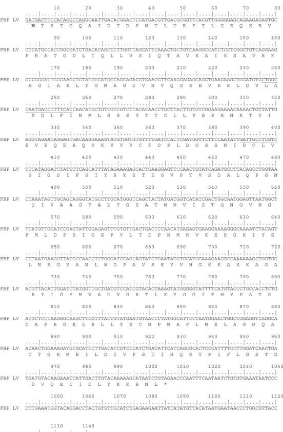
Expression of fructose 1, 6-bisphosphatase and
Interestingly, some marine species tolerate fluctuating environmental oxygen supply quite well (Gorr et al., 2010). Phosphofructokinase (PFK), pyruvate kinase (PK) and hexokinase (HK) are key regulatory enzymes of glycolysis (Fraenkel et al., 1996). FBP appears to be present in all living organisms with the exception of archaebacteria, whose FBP activity is based on a non-homologous protein (Stec et al., 2000).
The hepatopancreas of crustaceans is a center for carbohydrate metabolism as well as a site for gluconeogenesis (Rosas et al., 2001). From studies in mammals, we now know that HIF-1 binds directly to the specific PEPCK promoter region that induces transcription under hypoxic conditions (Choi et al., 2005). HIF-1 binds a core sequence of the Hypoxia Response Elements (HRE) in the promoters of hypoxia-responsive genes and induces their expressions ( Lee et al., 2004 ).
We know that HIF-1 regulates the expression of hexokinase and LDH in shrimp in hypoxia (So~nanez-Organis et al., 2012; Sonanez-Organis et al., 2011). Moreover, there is evidence that hypoxia induces the enzymatic activity of FBP in marine vertebrate species (Martínez et al., 2006). Expression of PEPCK in the grass shrimp hepatopancreas (Brown-Peterson et al., 2008) and glucose 6-phosphatase in fish liver (Gracey et al., 2001), both irreversible enzymes of gluconeogenesis, increase during hypoxia.
The functions of the hepatopancreas include the production of digestive enzymes, hemolymphatic proteins, and absorption of nutrients (Yepiz-Plascencia et al., 2000). Indeed, there is evidence in teleost fish of different cell types differing in their glycolytic and gluconeogenic capabilities (Mommsen et al., 1991). Our research group has previously reported an elevation of glucose in the hemolymph of shrimp during hypoxia (So ~ Nanez-Organis et al., 2009).
In mammals, glutathione is the most important intracellular antioxidant and inhibits some forms of apoptosis (Voehringer et al., 2000). Enzyme activity is regulated by fructose 2,6-bisphosphate (F-2, 6-BP), an inhibitor that binds to the substrate site, and adenosine monophosphate (AMP), an inhibitor that binds to the allosteric site (Stec et al. , 1996) . AMP causes a shift in the mammalian enzyme from the highly active R (relaxed) state to the inactive T (tense) state (Zhang et al., 1994).
Role of HIF-1 on phosphofructokinase and fructose 1, 6-
Recently, it was shown that the expression of glycolytic phosphofructokinase (PFK) and gluconeogenic fructose bisphosphatase (FBP) is induced in the hepatopancreas of shrimp during hypoxia (Cota-Ruiz et al., 2015). Both PFK and FBP enzymes are important regulators of their respective pathways (Al Hasawi et al., 2014; Yánez et al., 2003) and catalyze. In mammals, several genes for enzymes of glycolysis (Semenza, 2000) and gluconeogenesis pathways (Choi et al., 2005) are upregulated by Hypoxia Inducible Factor (HIF-1) under low oxygen conditions.
HIF-1 binds the core HRE (Hypoxia Responsive Element) sequence in the promoters of hypoxia-regulated genes and regulates their expression (Lee et al., 2004). HIF proteins are members of the basic helix-loop-helix/PAS (Per/ARNT/Sim) family of transcription factors ( Gorr et al., 2004 ) that recognize the sequence RCGTG ( Wang et al., 2015 ). Transcript detection for PFK, FBP, and L8 was performed exactly as reported previously (Cota-Ruiz et al., 2015), except that the final melting curve program was performed in the same manner as for HIF-1 (see below ).
HIF-1α and HIF-1β transcripts were detected in very low relative amounts in the shrimp hepatopancreas, as previously reported in the same shrimp species ( Soñanez-Organis et al., 2009 ). It is known that the efficiency of dsRNA to silence target transcripts depends, among other things, on the abundance of target transcripts (Fire et al., 1998). Similar results were found for hexokinase activity in the same shrimp species; a tissue-specific induction of enzymatic activity is reduced when HIF-1 was silenced (Soñanez-Organis et al., 2011).
This is consistent with studies in cell lines where the HIF-1α protein is rapidly degraded (b5 min half-life when oxygenated) or stabilized (by its immediate accumulation under hypoxic conditions) (Huang et al., 1998; Jewell et al. , 2001). Nevertheless, cumulative amounts of lactate are generated as an end product (Jackson et al., 2001; Nilsson and Lutz, 2004). FBP Upregulation in Prolonged Hypoxia and Involvement of HIF-1 Taking into account our previous report on FBP induction under long-term hypoxic conditions (48 h) (Cota-Ruiz et al., 2015), we were encouraged to see if HIF- 1 was involved in FBP upregulation.
In mammalian cells, HIF-1 can bind to the specific phosphoenol pyruvate kinase (PEPCK) gene promoter to control its expression (Choi et al., 2005). Furthermore, we cannot exclude the possibility that some hepatopancreas cell populations may function in a "gluconeogenic manner" (Cota-Ruiz et al., 2015), promoting the use of the anaerobic end product lactate or certain amino acids in the gluconeogenics. way. For example, when fish were subjected to 4 weeks of hypoxic conditions, a higher FBP activity was found (Martínez et al., 2006).

Revisiting the hypoxia and anoxia resilience in marine animals
A fundamental property of these organisms is their ability to obtain O2 from the environment (Fu et al., 2011). In addition, exposure to low oxygen adversely affects growth, reproduction, locomotion, and feeding (Levin et al., 2009; Wu, 2002). Surprisingly, the elephant seal can dive for at least 25 minutes without breathing (López-Cruz et al., 2014).
Exposure to hypoxia in the rainbow trout Oncorhynchus mykiss caused an increase in hemoglobin concentration through release from erythrocytes and, under prolonged hypoxia, an erythropoietin-mediated synthesis of new erythrocytes increased the oxygen/carrying capacity of the blood (Lai et al. ). The latter suggests that hypercapnia/low pH increases tissue oxygen delivery resulting in less deleterious effects of hypoxia (Rathburn et al., 2013). Crayfish Parastacus brasiliensis maintain their glycogen reserves even under hypoxic conditions (Silva-Castiglioni et al., 2011).
There is evidence that even hemocyanin can be synthesized in oxygen-deficient conditions in shrimp (Sun et al., 2014). In the shrimp Litopenaeus vannamei, hypoxia causes lactate concentration and lactate dehydrogenase (LDH) activity (Soñanez-Organis et al., 2012), while in goldfish an accumulation of lactate is reported (Fu et al., 2011). Despite the negative effects of L-lactate accumulation in crustacean blood, it binds to the extracellular pigment hemocyanin and increases its affinity with oxygen (Weber et al., 2008).
They bind a core sequence of the hypoxia responsive elements (HRE) in the promoter gene regions causing their expressions (Lee et al., 2004). HIF has been implicated in the hypoxic induction of Daphnia magna globin genes that contribute to the increase/improvement of oxygen carrying capacity of this species (Gorr et al., 2006). In the killer fish Fundulus heteroclitus, there are DNA elements in intron 2 of the LDH-B gene that are somehow similar to the HRE reported in mammalian genes, suggesting that they may interact with HIF (Rees et al., 2001).
In addition, HIF-1 silencing showed a reduction or complete decrease in lactate concentration in a tissue-specific manner in hypoxia-exposed shrimp (Soñanez-Organis et al., 2010). As shown by several mammalian studies, NO plays an important role in the regulation of vasodilation under hypoxic conditions (Ho et al., 2012). In the marine killer whale Fundulus grandis, specific activities of some gluconeogenic enzymes have been shown to increase in the liver during prolonged hypoxic exposure (Martínez et al., 2006).
Forty-one of the genes required for gluconeogenesis occur in the eurioxyfish Gillichthys mirabilis (Gracey et al., 2001). In shrimp hepatopancreas, an induction of fructose bisphosphatase (FBP) transcripts has been reported (Cota-Ruiz et al., 2015).