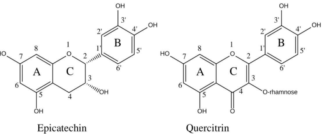
A su vez, juegan un papel importante en el color y las características sensoriales de frutas y verduras (Pereira et al., 2009). Los flavonoides son uno de los grupos de metabolitos secundarios ampliamente distribuidos en las plantas, y generalmente son responsables del color de frutos, hojas y flores (Pereira et al., 2009). Uno de los factores importantes en el potencial antioxidante de un flavonoide es el grado de hidroxilación (-OH) y la posición de estos grupos funcionales en la molécula (Bors et al., 1990; Balasundram et al., 2006).
Respecto a los extractos obtenidos de plantas, hay que tener en cuenta que su potencial antioxidante está influenciado por varios factores, que determinarán en gran medida el perfil y la concentración de los compuestos fenólicos presentes (Khan et al., 2009; Verma y Shukla, 2015). Por otro lado, factores bióticos como herbívoros, microorganismos y parásitos pueden afectar principalmente el perfil y concentración de compuestos fenólicos (Esra et al., 2010). Esto se puede lograr mediante una extracción directa de los compuestos con solventes relacionados, debido a su alta solubilidad (Wijngaard et al., 2012).
La cromatografía en columna es uno de los métodos más utilizados para la purificación de compuestos fenólicos debido a su simplicidad (Ignat et al., 2011).

Isolation and characterization of antioxidant phenolic compounds from Litsea glaucescens Kunth extract
Introduction
Therefore, it is necessary to find effective alternatives to help the human enzymatic system to reduce the high incidence of chronic human diseases. In this regard, plants represent an interesting alternative source of bioactive compounds, due to their wide variety of secondary metabolites, which are associated with different health benefits, including antioxidant properties [8]. Research on chemical constituents of Litsea plants proves the presence of a variety of bioactive compounds such as phenolic compounds, alkaloids, lactones, terpenes, terpenoids, butanolides, steroids and amides [10, 12].
One of the litsea species that has been little studied is Litsea glaucescens, which as a member of the genus Litsea, may represent a good source of bioactive compounds with potential biological activities. However, additional information is required to determine the phenolic compounds responsible for the antioxidant activity of L.
Materials and Methods 1. General information
34 Based on the above, the aim of the present study was to isolate and identify the phenolic compounds responsible for the antioxidant activity of L. The antioxidant activity of the DPPH radical scavenging activity assay was performed using the modified version of Usia et al. 280 µL of FRAP reagent was mixed with 20 µL of LGE and the absorbance was read at 630 nm after 30 min of storage in the dark.
The antioxidant activity (DPPH and FRAP) and phenol content were determined in all fractions (I-XII). One way ANOVA was used and mean comparisons were performed using the Tukey-Kramer test.
Results and discussion
Overall, stem and root extracts showed better antioxidant activity than inner bark extract from said plants. 38 can be attributed to the ability of polar solvents to extract mainly phenolic compounds, which are associated with potent antioxidant activity. On the other hand, ferric reducing activity of LGE and their fractions was determined by the FRAP assay (Figure 1).
LGE exhibited higher activity than other reported Litsea extracts, however, the antioxidant activity assessed by this method was lower than the positive control (Vit. C and Trolox). The results obtained in the FRAP evaluation are consistent with the DPPH assay, which evidenced that F-XII, F-XI and LGE extract were the most active, suggesting that these treatments are a good source of bioactive compounds with high antioxidant activity . In this regard, different authors have isolated and characterized different compounds with exceptional antioxidant activity from Litsea plants, where phenolic compounds such as flavonol and flavanone were the most active [12, 28].
The results show that the phenolic compounds present in LGE were associated with the antioxidant potential of the analyzed extracts, as F-XI and F-XII showed the highest phenolic content and antioxidant activity. The antioxidant activity of these compounds is largely related to the number and position of hydroxyl groups in the molecule and the 2-3 double bond and 4-oxo function [43, 46]. Furthermore, several studies have shown that these compounds can act synergistically, causing a significant increase in antioxidant activity [47, 48].
Quintanar-Isaias, A.; Garcia-Marquez, E.; Cruz-Sosa, F., Histochemistry, total phenolic content and antioxidant activity in leaves and wood of Litsea glaucescens Kunth (Lauraceae). Arfan, M.; Amin, H.; Kosinska, A.; Karamač, M.; Amarowicz, R., Antioxidant activity of phenolic fractions of Litsea monopetala [Persimon-leaved litsea] bark extract. Iacopini, P.; Baldi, M.; Storchi, P.; Sebastiani, L., Catechin, epicatechin, quercetin, rutin and resveratrol in red grapes: Content, in vitro antioxidant activity and interactions.

Seasonal effects on the biological activities of Litsea glaucescens Kunth extracts
Materials and Methods
DMSO did not exceed 0.5% of the total volume per well (preliminary studies have shown that DMSO does not cause cell damage at this concentration). Then, 10 µl of MTT solution (5 mg/ml) was added to each well (in the case of the M12.C3.F6 cell line culture, only MTT solution (5 mg/ml) was added). As can be observed, the clear difference between them is the height of the chromatographic peaks (related to the concentration of the phenols), which appear larger in the chromatograms corresponding to the extracts ALGE, SULGE and SLGE compared to those labeled as WLGE.
In addition, the HPLC-DAD analysis allowed us to identify two of the main phenolic compounds present in L. On the other hand, ferric reducing power of the L. glaucescens extracts was evaluated through their ability to reduce the ferric complex Fe3+-tripyridyltriazine to Fe2+- tripyridyltriazine. glaucescens extracts were in the range:. To demonstrate the correlation between both parameters, a series of plots of data obtained through DPPH, FRAP and ORAC assays against the concentration of phenolic compound (CPC) in the four L.
The regression coefficients (R) of the linear correlations for each series are presented in Figure 3. As we hypothesized, it seems that phenolic compounds were the main ones responsible for the antioxidant activity (evaluated by three different methods) of the four L. , it should be noted that purity of the CAPE used in the tests (above 95%).
However, the structural characteristics of these phenols are also important, and they are the same described above as enhancers of the antioxidant activity of L. As can be observed, there is a clear effect of the concentration of both on their antimicrobial activity. . Furthermore, higher concentrations such as 1000 and 800 µg/mL had similar activity to gentamicin (>98% inhibition), which is an evidence of strong antimicrobial activity of this extract.
Conclusions
Baquero, et al., "The global threat of antimicrobial resistance: science for intervention," New Microbes and New Infections, vol. Jagger, et al., "Diabetic cardiovascular disease caused by oxidative stress," International Journal of Molecular Sciences, vol. Castro, et al., "Chemical composition and biological activity of a new type of Brazilian propolis: red propolis," Journal of ethnopharmacology, vol.
Acosta, et al., "Antibacterial and free radical activities of Sonoran propolis," Journal of Applied Microbiology, vol. Quintero, et al., "Sonoran propolis: chemical composition and antiproliferative activity on cancer cell lines," Planta medica, vol. Dewan, et al., "Chemical group characterization and investigation of cytotoxic and antioxidant activity of Litsea glutinosa leaves," Journal of Plant Sciences, vol.
Ham, et al., "Antioxidant and anti-inflammatory activities of Litsea japonica leaves," Journal of the Korean Society for Applied Biological Chemistry, vol. Abdullah, et al., "Antiproliferative activity of primate ingested plants against MCF-7 human breast cancer cell lines," E3 J Med Res, vol. Tanaka, et al., "Active-guided fractionation of green tea extract with antiproliferative activity against human gastric cancer cells," Biological and Pharmaceutical Bulletin, vol.
Braunhut et al., "Biocatalytic oligomerized epicatechin with potent and specific anti-proliferative activity for human breast cancer cells," Molecules, vol. Sultana et al., "Pharmacological and phytochemical screening of ethanol extract of Litsea monopetala (Roxb.) Pers.", IOSR Journal of Pharmacy, vol. Soares, et al., "Fine-tuning the hydrophobicity of caffeic acid: studies of the antimicrobial activity against Staphylococcus aureus and Escherichia coli," RSC Advances, vol.
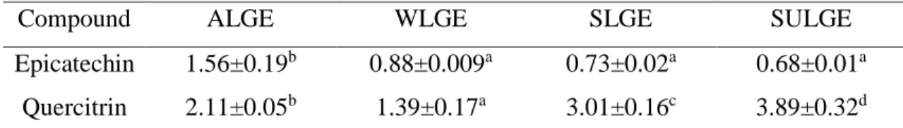
ALGE
WLGE
SLGE
SULGE
82 Figure 3. Correlation analysis. a) Correlation between content of phenolic content (CPC) and DPPH test, correlation coefficient r=0.92; b) Correlation between content of phenolic content (CPC) and FRAP test, correlation coefficient r=0.93;. Antioxidant potential of Litsea glaucescens extract and their fractions on chilled pork patties against lipid and protein.
Antioxidant potential of Litsea glaucescens extract and their fractions on refrigerated pork patties against lipid and protein
Antioxidant potential of Litsea glaucescens extract and their fractions on chilled pork patties against lipid and protein oxidation. The aim of this study was to evaluate the antioxidant effect of LG extract (ALGE) and its fractions (F-XI and F-XII) on lipid and protein oxidation of pork patties stored at 4 °C. CONCLUSION: ALGAE, F-XI and F-XII were effective in reducing the oxidative process in the evaluated pork patties and consequently extending their shelf life.
Based on the above, the aim of this study was to evaluate the effectiveness of LG extract and their fractions (F-XI and F-XII) in inhibiting lipid and protein oxidation in pork steaks stored under refrigeration at 4 °C. Total lipids from pork loin were extracted according to the method described by Bligh and Dyer,20. The effect of ALGE, F-XI and F-XII on the CD of raw pork meatballs stored under refrigeration is shown in Figure 3.
The effect of ALGE, F-XI and F-XII on the TBARS values of pork patties stored in the refrigerator is shown in Figure 4. In addition, ALGE and F-XII were also effective in slowing down the lipid oxidation process in the pork patties . This problem may indicate that the lipid oxidation products induced the protein oxidation of pork patties.
In this sense, ALGE, F-XI and F-XII showed that they were effective in retarding lipid oxidation in the pork patties and also reduced protein oxidation with the same affectation. Similarly, Rodríguez-Carpena et al.47 studied avocado extracts addition on protein oxidation of pork patties (15 days at 4 °C). Similarly, Jia et al.35 studied the effect of blackcurrant extract in pork patties during your storage (9 days at 4 °C).
This suggests that the evaluated treatments presented a protective effect on the browning parameter of pig dough. The average result of loss of fresh aroma and loss of fresh taste of pork dough during the storage period is summarized in Figure 7.
