Fourth universal definition of myocardial infarction (2018)
Kristian Thygesen* (Denmark), Joseph S. Alpert* (USA), Allan S. Jaffe (USA), Bernard R. Chaitman (USA), Jeroen J. Bax (The Netherlands), David A. Morrow (USA), Harvey D. White* (New Zealand): the Executive Group on behalf of the Joint European Society of Cardiology (ESC)/American College of Cardiology (ACC)/
American Heart Association (AHA)/World Heart Federation (WHF) Task Force for the Universal Definition of Myocardial Infarction
Authors/Task Force Members/Chairpersons: Kristian Thygesen* (Denmark), Joseph S. Alpert* (USA), Allan S. Jaffe (USA), Bernard R. Chaitman (USA), Jeroen J. Bax (The Netherlands), David A. Morrow (USA), Harvey D. White* (New Zealand), Hans Mickley (Denmark), Filippo Crea (Italy), Frans Van de Werf (Belgium), Chiara Bucciarelli-Ducci (UK), Hugo A. Katus (Germany), Fausto J. Pinto (Portugal), Elliott M. Antman (USA), Christian W. Hamm (Germany), Raffaele De Caterina (Italy), James L. Januzzi Jr (USA), Fred S. Apple (USA), Maria Angeles Alonso Garcia (Spain), S. Richard Underwood (UK), John M. Canty Jr (USA), Alexander R. Lyon (UK), P. J. Devereaux (Canada), Jose Luis Zamorano (Spain), Bertil Lindahl
(Sweden), William S. Weintraub (USA), L. Kristin Newby (USA), Renu Virmani (USA), Pascal Vranckx (Belgium), Don Cutlip (USA), Raymond J. Gibbons (USA), Sidney C. Smith (USA), Dan Atar (Norway), Russell V. Luepker (USA), Rose Marie Robertson (USA), Robert O. Bonow (USA), P. Gabriel Steg (France), Patrick T.
O’Gara (USA), Keith A. A. Fox (UK)
* Corresponding authors. Kristian Thygesen, Department of Cardiology, Aarhus University Hospital, Palle Juul-Jensens Boulevard, DK-8200 Aarhus N, Denmark. Tel:þ45 78452262, Fax:þ45 78452260, Email: [email protected]; [email protected]. Joseph S. Alpert, Department of Medicine, University of Arizona College of Medicine, 1501 N.
Campbell Ave., P.O. Box 245037, Tucson AZ 85724-5037, USA. Tel:þ1 5206262763, Email: [email protected]. Harvey D. White, Green Lane Cardiovascular Service, Auckland City Hospital, Private Bag 92024, 1030 Auckland, New Zealand. Tel:þ64 96309992, Fax: 00 64 9 6309915, Email: [email protected].
The content of this ESC/ACC/AHA/WHF Expert Consensus Document has been published for personal and educational use only. No commercial use is authorized. No part of the ESC/ACC/AHA/WHF Expert Consensus Document may be translated or reproduced in any form without written permission from the ESC or ACC or AHA or WHF.
Permission can be obtained upon submission of a written request to Oxford University Press, the publisher of the European Heart Journal and the party authorized to handle such permissions on behalf of the ESC, ACC, AHA and WHF ([email protected]).
Disclaimer. The ESC/ACC/AHA/WHF Expert Consensus Document represents the views of the ESC, ACC, AHA, and WHF and was produced after careful consideration of the scientific and medical knowledge and the evidence available at the time of their publication. The ESC, ACC, AHA, and WHF are not responsible in the event of any contra- diction, discrepancy, and/or ambiguity between the ESC/ACC/AHA/WHF Expert Consensus Document and any other official recommendations or Expert Consensus Document issued by the relevant public health authorities, in particular in relation to good use of healthcare or therapeutic strategies. Health professionals are encouraged to take the ESC/
ACC/AHA/WHF Expert Consensus Document fully into account when exercising their clinical judgment, as well as in the determination and the implementation of preventive, diagnostic, or therapeutic medical strategies; however, the ESC/ACC/AHA/WHF Expert Consensus Document does not override, in any way whatsoever, the individual responsi- bility of health professionals to make appropriate and accurate decisions in consideration of each patient’s health condition and in consultation with that patient and, where appropriate and/or necessary, the patient’s caregiver. Nor does the ESC/ACC/AHA/WHF Expert Consensus Document exempt health professionals from taking into full and careful consideration the relevant official updated recommendations or Expert Consensus Documents issued by the competent public health authorities, in order to manage each patient’s case in light of the scientifically accepted data pursuant to their respective ethical and professional obligations. It is also the health professional’s responsibility to verify the applicable rules and regulations relating to drugs and medical devices at the time of prescription.
This article has been co-published inEuropean Heart Journal, Journal of the American College of Cardiology, Circulation, andGlobal Heart. All rights reserved.
VC2018 European Society of Cardiology, American College of Cardiology, American Heart Association, Inc., and World Heart Foundation. The articles are identical except for minor stylistic and spelling differences in keeping with each journal’s style. Any citation can be used when citing this article.
doi:10.1093/eurheartj/ehy462
Downloaded from https://academic.oup.com/eurheartj/advance-article-abstract/doi/10.1093/eurheartj/ehy462/5079081 by guest on 12 November 2018
.. ..
.. ..
.. ..
.. ..
.. ..
.. ..
.. ..
.. ..
.. ..
.. ..
.. ..
.. ..
.. ..
.. ..
.. ..
.. ..
.. ..
.. ..
.
(Brazil), Emanuele Barbato (Italy), Jean-Pierre Bassand (France), Eric Bates (USA), John A. Bittl (USA), Gu¨ enter Breithardt (Germany), He´ ctor Bueno (Spain), Raffaele Bugiardini (Italy), Mauricio G. Cohen (USA), George Dangas (USA), James A. de Lemos (USA), Victoria Delgado (Netherlands), Gerasimos Filippatos (Greece), Edward Fry (USA), Christopher B. Granger (USA), Sigrun Halvorsen (Norway), Mark A. Hlatky (USA), Borja Ibanez (Spain), Stefan James (Sweden), Adnan Kastrati (Germany), Christophe Leclercq (France), Kenneth W. Mahaffey (USA), Laxmi Mehta (USA), Christian Mu¨ ller (Switzerland), Carlo Patrono (Italy), Massimo Francesco Piepoli (Italy), Daniel Pi~neiro (Argentina), Marco Roffi (Switzerland), Andrea Rubboli (Italy), Samin Sharma (USA), Iain A. Simpson (UK), Michael Tendera (Poland), Marco Valgimigli (Switzerland), Allard C. van der Wal (Netherlands), Stephan Windecker (Switzerland)
The disclosure forms of all experts involved in the development of this Expert Consensus Document are available on the ESC website www.escardio.org/guidelines
...
Keywords Expert Consensus Document
•
Myocardial infarction•
Type 1 MI•
Type 2 MI•
Type 3 MI•
Type 4aMI
•
Type 4b MI•
Type 4c MI•
Type 5 MI•
Cardiac troponin•
High sensitivity cardiac troponin•
Myocardial injury
•
Prior myocardial infarction•
Silent myocardial infarction•
Recurrent myocardialinfarction
•
Re-infarction•
Cardiac procedural myocardial injury•
Takotsubo syndrome•
Myocardial infarc- tion with non-obstructive coronary arteries (MINOCA)Table of contents
Abbreviations and acronyms . . . 3
1 What is new in the Universal Definition of Myocardial Infarction? . . . 4
2 Universal definitions of myocardial injury and myocardial infarction: summary . . . 5
3 Introduction . . . 6
4 Pathological characteristics of myocardial ischaemia and infarction . . . 7
5 Biomarker detection of myocardial injury and infarction . . . 7
6 Clinical presentations of myocardial infarction . . . 9
7 Clinical classification of myocardial infarction . . . 9
7.1 Myocardial infarction type 1 . . . 9
7.2 Myocardial infarction type 2 . . . 10
7.3 Myocardial infarction type 2 and myocardial injury . . . 12
7.4 Myocardial Infarction type 3 . . . 12
8 Coronary procedure-related myocardial injury . . . 13
9 Myocardial infarction associated with percutaneous coronary intervention (type 4a myocardial infarction) . . . 14
10 Stent/scaffold thrombosis associated with percutaneous coronary intervention (type 4b myocardial infarction) . . . 15
11 Restenosis associated with percutaneous coronary intervention (type 4c myocardial infarction) . . . 15
12 Myocardial infarction associated with coronary artery bypass grafting (type 5 myocardial infarction) . . . 15
13 Other definitions of myocardial infarction related to percutaneous coronary intervention or coronary artery bypass grafting . . . 16
14 Recurrent myocardial infarction . . . 16
15 Re-infarction . . . 16
16 Myocardial injury and infarction associated with cardiac procedures other than revascularization . . . 16
17 Myocardial injury and infarction associated with non-cardiac procedures . . . 16
18 Myocardial injury or infarction associated with heart failure . . . 17
19 Takotsubo syndrome . . . 17
20 Myocardial infarction with non-obstructive coronary arteries . . . 17
21 Myocardial injury and/or infarction associated with kidney disease . . . 17
22 Myocardial injury and/or infarction in critically ill patients . . . 17
23 Biochemical approach for diagnosing myocardial injury and infarction . . . 17
24 Analytical issues of cardiac troponins . . . 20
25 The 99th percentile upper reference limit . . . 20
26 Operationalizing criteria for myocardial injury and infarction . . . 20
27 Electrocardiographic detection of myocardial infarction . . . 21
28 Application of supplemental electrocardiogram leads . . . 22
29 Electrocardiographic detection of myocardial injury . . . 23
Downloaded from https://academic.oup.com/eurheartj/advance-article-abstract/doi/10.1093/eurheartj/ehy462/5079081 by guest on 12 November 2018
.. ..
.. ..
.. ..
.. ..
.. ..
.. ..
.. ..
.. ..
.. ..
.. ..
.. ..
.. ..
.. ..
.. ..
.. ..
.. ..
.. ..
.. ..
.. ..
.. ..
.. ..
.. ..
.. ..
.. ..
.. ..
.. ..
.. ..
.. ..
.. ..
.. ..
.. ..
.. ..
.. ..
..
30 Prior or silent/unrecognized myocardial infarction . . . 23
31 Conditions that confound the electrocardiographic diagnosis of myocardial infarction . . . 23
32 Conduction disturbances and pacemakers . . . 24
33 Atrial fibrillation . . . 24
34 Imaging techniques . . . 24
34.1 Echocardiography . . . 24
34.2 Radionuclide imaging . . . 22
34.3 Cardiac magnetic resonance imaging . . . 22
34.4 Computed tomographic coronary angiography . . . 26
35 Applying imaging in acute myocardial infarction . . . 26
36 Applying imaging in late presentation of myocardial infarction . . . 26
37 Regulatory perspective on myocardial infarction in clinical trials . . . 26
38 Silent/unrecognized myocardial infarction in epidemiological studies and quality programmes . . . 27
39 Individual and public implications of the myocardial infarction definition . . . 27
40 Global perspectives of the definition of myocardial infarction . . . 27
41 Using the Universal Definition of Myocardial Infarction in the healthcare system . . . 27
42 Appendix . . . 28
43 Acknowledgements . . . 28
44 References . . . 28
Abbreviations and acronyms
ACC American College of Cardiology ACS Acute coronary syndrome AHA American Heart Association ARC-2 Academic Research Consortium-2 AUC Area under the curve
CAD Coronary artery disease CABG Coronary artery bypass grafting CKD Chronic kidney disease CK-MB Creatine kinase MB isoform CMR Cardiac magnetic resonance
CTCA Computed tomographic coronary angiography cTn Cardiac troponin
cTnI Cardiac troponin I cTnT Cardiac troponin T
CT Computed tomography
CV Coefficient of variation EF Ejection fraction
ECG Electrocardiogram or electrocardiographic
HF Heart failure
hs-cTn High-sensitivity cardiac troponin
IFCC International Federation of Clinical Chemistry and Laboratory Medicine
ISFC International Society and Federation of Cardiology LAD Left anterior descending artery
LBBB Left bundle branch block;
LoD Limit of detection
LGE Late gadolinium enhancement
LGE-CMR Late gadolinium enhancement cardiac magnetic resonance
LV Left ventricular
LVH Left ventricular hypertrophy MI Myocardial infarction
MINOCA Myocardial infarction with non-obstructive coronary arteries
MONICA MONItoring of trends and determinants in CArdiovascular disease
MPS Myocardial perfusion scintigraphy NHLBI National Heart, Lung, and Blood Institute NSTEMI Non-ST-elevation myocardial infarction PET Positron emission tomography PCI Percutaneous coronary intervention POC Point of care
RBBB Right bundle branch block
SPECT Single photon emission computed tomography STEMI ST-elevation myocardial infarction
ST-T ST-segment–T wave
TIMI Thrombolysis in Myocardial Infarction
TTS Takotsubo syndrome
UDMI Universal Definition of Myocardial Infarction URL Upper reference limit
WHF World Heart Federation WHO World Health Organization
Downloaded from https://academic.oup.com/eurheartj/advance-article-abstract/doi/10.1093/eurheartj/ehy462/5079081 by guest on 12 November 2018
What’s new in the universal definition of myocardial infarction?
New concepts
• Differentiation of myocardial infarction from myocardial injury.
• Highlighting peri-procedural myocardial injury after cardiac and non-cardiac procedures as discrete from myocardial infarction.
• Consideration of electrical remodelling (cardiac memory) in assessing repolarization abnormalities with tachyarrhythmia, pacing, and rate-related conduction disturbances.
• Use of cardiovascular magnetic resonance to define aetiology of myocardial injury.
• Use of computed tomographic coronary angiography in suspected myocardial infarction.
Updated concepts
• Type 1 myocardial infarction: Emphasis on the causal relationship of plaque disruption with coronary athero-thrombosis; new Figure 3.
• Type 2 myocardial infarction: Settings with oxygen demand and supply imbalance unrelated to acute coronary athero-thrombosis;
new Figures 4 and 5.
• Type 2 myocardial infarction: Relevance of presence or absence of coronary artery disease to prognosis and therapy.
• Differentiation of myocardial injury from type 2 myocardial infarction; new Figure 6.
• Type 3 myocardial infarction: Clarify why type 3 myocardial infarction is a useful category to differentiate from sudden cardiac death.
• Types 4–5 myocardial infarction: Emphasis on distinction between procedure-related myocardial injury and procedure-related myocardial infarction.
• Cardiac troponin: Analytical issues for cardiac troponins; new Figure 7.
• Emphasis on the benefits of high-sensitivity cardiac troponin assays.
• Considerations relevant to the use of rapid rule-out and rule-in protocols for myocardial injury and myocardial infarction.
• Issues related to specific diagnostic change ('delta') criteria for the use of cardiac troponins to detect or exclude acute myocardial injury.
• Consideration of new non-rate-related right bundle branch block with specific repolarization patterns.
• ST-segment elevation in lead aVR with specific repolarization patterns, as a STEMI equivalent.
• ECG detection of myocardial ischaemia in patients with an implantable cardiac defibrillator or a pacemaker.
• Enhanced role of imaging including cardiac magnetic resonance imaging for the diagnosis of myocardial infarction; new Figure 8.
New sections
• Takotsubo syndrome.
• MINOCA.
• Chronic kidney disease.
• Atrial fibrillation.
• Regulatory perspective on myocardial infarction.
• Silent or unrecognized myocardial infarction.
©ESC/ACC/AHA/WHF 2018
ECG = electrocardiogram; MINOCA = myocardial infarction with non-obstructive coronary arteries; STEMI = ST-elevation myocardial infarction.
Downloaded from https://academic.oup.com/eurheartj/advance-article-abstract/doi/10.1093/eurheartj/ehy462/5079081 by guest on 12 November 2018
2 Universal definitions of myocardial injury and myocardial infarction:
summary
Universal definitions of myocardial injury and myocardial infarction
Criteria for myocardial injury
The term myocardial injury should be used when there is evidence of elevated cardiac troponin values (cTn) with at least one value above the 99th percentile upper reference limit (URL). The myocardial injury is considered acute if there is a rise and/or fall of cTn values.
Criteria for acute myocardial infarction (types 1, 2 and 3 MI)
The term acute myocardial infarction should be used when there is acute myocardial injury with clinical evidence of acute myocardial ischaemia and with detection of a rise and/or fall of cTn values with at least one value above the 99th percentile URL and at least one of the following:
• Symptoms of myocardial ischaemia;
• New ischaemic ECG changes;
• Development of pathological Q waves;
• Imaging evidence of new loss of viable myocardium or new regional wall motion abnormality in a pattern consistent with an ischaemic aetiology;
• Identification of a coronary thrombus by angiography or autopsy (not for types 2 or 3 MIs).
Post-mortem demonstration of acute athero-thrombosis in the artery supplying the infarcted myocardium meets criteria for type 1 MI.
Evidence of an imbalance between myocardial oxygen supply and demand unrelated to acute athero-thrombosis meets criteria for type 2 MI.
Cardiac death in patients with symptoms suggestive of myocardial ischaemia and presumed new ischaemic ECG changes before cTn values become available or abnormal meets criteria for type 3 MI.
Criteria for coronary procedure-related myocardial infarction (types 4 and 5 MI) Percutaneous coronary intervention (PCI) related MI is termed type 4a MI.
Coronary artery bypass grafting (CABG) related MI is termed type 5 MI.
Coronary procedure-related MI ≤ 48 hours after the index procedure is arbitrarily defined by an elevation of cTn values > 5 times for type 4a MI and > 10 times for type 5 MI of the 99th percentile URL in patients with normal baseline values. Patients with elevated pre-procedural cTn values, in whom the pre-procedural cTn level are stable (≤ 20% variation) or falling, must meet the criteria for a > 5 or > 10 fold increase and manifest a change from the baseline value of > 20%. In addition with at least one of the following:
• New ischaemic ECG changes (this criterion is related to type 4a MI only);
• Development of new pathological Q waves;
• Imaging evidence of loss of viable myocardium that is presumed to be new and in a pattern consistent with an ischaemic aetiology;
• Angiographic findings consistent with a procedural flow-limiting complication such as coronary dissection, occlusion of a major epicardial artery or graft, side-branch occlusion-thrombus, disruption of collateral flow or distal embolization.
Isolated development of new pathological Q waves meets the type 4a MI or type 5 MI criteria with either revascularization procedure if cTn values are elevated and rising but less than the pre-specified thresholds for PCI and CABG.
Other types of 4 MI include type 4b MI stent thrombosis and type 4c MI restenosis that both meet type 1 MI criteria.
Post-mortem demonstration of a procedure-related thrombus meets the type 4a MI criteria or type 4b MI criteria if associated with a stent.
Criteria for prior or silent/unrecognized myocardial infarction
Any one of the following criteria meets the diagnosis for prior or silent/unrecognized MI:
• Abnormal Q waves with or without symptoms in the absence of non-ischaemic causes.
• Imaging evidence of loss of viable myocardium in a pattern consistent with ischaemic aetiology.
• Patho-anatomical findings of a prior MI.
©ESC/ACC/AHA/WHF 2018
CABG = coronary artery bypass grafting; cTn = cardiac troponin; ECG = electrocardiogram; MI = myocardial infarction; PCI = percutaneous coronary intervention; URL = upper reference limit.
Downloaded from https://academic.oup.com/eurheartj/advance-article-abstract/doi/10.1093/eurheartj/ehy462/5079081 by guest on 12 November 2018
.. ..
.. ..
.. ..
.. ..
.. ..
.. ..
.. ..
.. ..
.. ..
.. ..
.. ..
.. ..
.. ..
.. ..
.. ..
.. ..
In the late 19th century, post-mortem examinations demonstrated a
..
possible relationship between thrombotic occlusion of a coronary artery and myocardial infarction (MI).1However, it was not until the beginning of the 20th century that the first clinical descriptions appeared describing a connection between the formation of a throm- bus in a coronary artery and its associated clinical features.2,3Despite these landmark observations, considerable time elapsed before gen- eral clinical acceptance of this entity was achieved, in part due to one autopsy study that showed no thrombi in the coronary arteries of 31% of deceased patients with an MI.4The clinical entity was referred to as coronary thrombosis, although use of the term ‘MI’ ultimately prevailed. Over the years, several different definitions of MI have been used, leading to controversy and confusion. Hence, a general and worldwide definition for MI was needed. This occurred for the first time in the 1950–70s, when working groups from the World Health Organization (WHO) established a primarily electrocardio- graphic (ECG)-based definition of MI intended for epidemiological use.5The original description, with minor modifications, is still used in epidemiological surveys (Figure1).6–8
With the introduction of more sensitive cardiac biomarkers, the European Society of Cardiology (ESC) and the American College of
by abnormal biomarkers in the setting of acute myocardial ischaemia should be labelled as MI.9The principle was further refined by the Global MI Task Force, leading to the Universal Definition of Myocardial Infarction Consensus Document in 2007, introducing a novel MI classification system with five subcategories.10This docu- ment, endorsed by the ESC, the ACC), the American Heart Association (AHA), and the World Heart Federation (WHF), was adopted by the WHO.11The development of even more sensitive assays for markers of myocardial injury made further revision of the document necessary, particularly for patients who undergo coronary procedures or cardiac surgery. As a result, the Joint ESC/ACC/AHA/
WHF Task Force produced the Third Universal Definition of Myocardial Infarction Consensus Document in 2012.12
Studies have shown that myocardial injury, defined by an elevated cardiac troponin (cTn) value, is frequently encountered clinically and is associated with an adverse prognosis.13,14Although myocardial injury is a prerequisite for the diagnosis of MI, it is also an entity in itself. To establish a diagnosis of MI, criteria in addition to abnormal biomarkers are required. Non-ischaemic myocardial injury may arise secondary to many cardiac conditions such as myocarditis, or may be associated with non-cardiac conditions such as renal failure.15 Therefore, for
Clinical Approach Epidemiological Approach
1950 1960 1970 1980 1990 2000 2010 2020
Fourth UDMI Definition Third
UDMI Definition
UDMI Definition ESC ACC
Re- definition
First WHO Definition
Fifth WHO Definition
ISFC WHO Definition
WHO MONICA Definition
AHA - ESC - WHF - NHLBI Clinical and epidemiological definition
©ESC/ACC/AHA/WHF 2018
Figure 1 History of documents on the definition of myocardial infarction. ACC = American College of Cardiology; AHA = American Heart Association; ESC = European Society of Cardiology; ISFC = International Society and Federation of Cardiology; MONICA = MONItoring of trends and determinants in CArdiovascular disease; NHLBI = National Heart, Lung, and Blood Institute; UDMI = Universal Definition of Myocardial Infarction; WHF = World Heart Federation; WHO = World Health Organization.
Downloaded from https://academic.oup.com/eurheartj/advance-article-abstract/doi/10.1093/eurheartj/ehy462/5079081 by guest on 12 November 2018
.. ..
.. ..
.. ..
.. ..
.. ..
.. ..
.. ..
.. ..
.. ..
.. ..
.. ..
patients with increased cTn values, clinicians must distinguish whether
..
patients have suffered a non-ischaemic myocardial injury or one of the MI subtypes. If there is no evidence to support the presence of myo- cardial ischaemia, a diagnosis of myocardial injury should be made.
This diagnosis can be changed if subsequent evaluation indicates crite- ria for MI. The current Fourth Universal Definition of Myocardial Infarction Consensus Document reflects these considerations through adhering to the clinical approach of the definition of MI.
4 Pathological characteristics of myocardial ischaemia and
infarction
MI is defined pathologically as myocardial cell death due to prolonged ischaemia. Diminished cellular glycogen, and relaxed myofibrils and sarcolemmal disruption, are the first ultrastructural changes and are seen as early as 10–15 min after the onset of ischaemia.16 Mitochondrial abnormalities are observed as early as 10 min after coronary occlusion by electron microscopy and are progressive.17It can take hours before myocyte necrosis can be identified by post- mortem examination in humans; this is in contrast to animal models, in which biochemical evidence of myocardial cell death due to apop- tosis can be detected within 10 min of induced myocardial ischaemia in association with myocyte death.15Experimentally, necrosis pro- gresses from the subendocardium to the subepicardium over several hours. The time course may be prolonged by increased collateral flow, reduced determinants of myocardial oxygen consumption, and intermittent occlusion/reperfusion, which can precondition the heart.18Timely implementation of reperfusion therapy, when appro- priate, reduces ischaemic injury of the myocardium.19,20
5 Biomarker detection of
myocardial injury and infarction
Cardiac troponin I (cTnI) and T (cTnT) are components of the con- tractile apparatus of myocardial cells and are expressed almost exclu- sively in the heart.21,22 Increases in cTnI values have not been
reported to occur following injury to non-cardiac tissues. The situa- tion is more complex for cTnT. Biochemical data indicate that injured skeletal muscle expresses proteins that are detected by the cTnT assay, leading to some situations where elevations of cTnT could emanate from skeletal muscle.23–27Recent data suggest that the fre- quency of such elevations in the absence of ischaemic heart disease may be higher than originally thought.28,29cTnI and cTnT are the pre- ferred biomarkers for the evaluation of myocardial injury,12,21,22,30
and high-sensitivity (hs)-cTn assays are recommended for routine clinical use.22 Other biomarkers, e.g. creatine kinase MB isoform (CK-MB), are less sensitive and less specific.31Myocardial injury is defined as being present when blood levels of cTn are increased above the 99th percentile upper reference limit (URL).12,21,22,30
The injury may be acute, as evidenced by a newly detected dynamic rising and/or falling pattern of cTn values above the 99th percentile URL,
or chronic, in the setting of persistently elevated cTn levels.
Although elevated cTn values reflect injury to myocardial cells, they do not indicate the underlying pathophysiological mechanisms, and can arise following preload-induced mechanical stretch or phys- iological stresses in otherwise normal hearts.32–34Various causes have been suggested for the release of structural proteins from the myocardium, including normal turnover of myocardial cells, apopto- sis, cellular release of cTn degradation products, increased cellular wall permeability, the formation and release of membranous blebs, and myocyte necrosis.27,35Yet, it is not clinically possible to distin- guish which increases of cTn levels are due to which mechanisms.36 However, regardless of the mechanism, acute myocardial injury, when associated with a rising and/or falling pattern of cTn values with at least one value above the 99th percentile URL and caused by myo- cardial ischaemia, is designated as an acute MI.12,21,22,30
Histological evidence of myocardial injury with myocyte death can be detected in clinical conditions associated with non-ischaemic mechanisms of myocardial injury as well37,38(Figure2).
Myocardial ischaemic or non-ischaemic conditions associated with increased cTn values are presented inTable1. The complexity of clin- ical circumstances may sometimes make it difficult to discriminate specific individual mechanism(s) of myocardial injury. In this situation, the multifactorial contributions resulting in myocardial injury should be described in the patient record.
Criteria for myocardial injury
Detection of an elevated cTn value above the 99th percentile URL is defined as myocardial injury. The injury is considered acute if there is a rise and/or fall of cTn values.
Clinical criteria for MI
The clinical definition of MI denotes the presence of acute myo- cardial injury detected by abnormal cardiac biomarkers in the set- ting of evidence of acute myocardial ischaemia.
Downloaded from https://academic.oup.com/eurheartj/advance-article-abstract/doi/10.1093/eurheartj/ehy462/5079081 by guest on 12 November 2018
Clinical evidence of acute ischaemic
myocardial injury = myocardial infarctionc myocardial injuryb
Increased cTn = No myocardial injurya
Hypotension/
shock
Hypoxaemia Anaemia
Ventricular tachyarrhythmia
Kidney disease
Heart failure
©ESC/ACC/AHA/WHF 2018
Figure 2Spectrum of myocardial injury, ranging from no injury to myocardial infarction. Various clinical entities may involve these myocardial cate- gories, e.g. ventricular tachyarrhythmia, heart failure, kidney disease, hypotension/shock, hypoxaemia, and anaemia. cTn = cardiac troponin; URL = upper reference limit.aNo myocardial injury = cTn values <_ 99th percentile URL or not detectable.bMyocardial injury = cTn values > 99th percentile URL.cMyocardial infarction = clinical evidence of myocardial ischaemia and a rise and/or fall of cTn values > 99th percentile URL.
Downloaded from https://academic.oup.com/eurheartj/advance-article-abstract/doi/10.1093/eurheartj/ehy462/5079081 by guest on 12 November 2018
.. ..
.. ..
.. ..
.. ..
.. ..
.. ..
.. ..
.. ..
.. ..
.. ..
.. ..
.. ..
.. ..
.. ..
.. ..
.. ..
.. ..
.. ..
.. ..
.. ..
.. ..
.. ..
.. ..
.. ..
.. ..
.. ..
.. ..
.. ..
.. ..
.. ..
.. ..
.. ..
.. ..
.. ..
.. ..
.. ..
.. ..
.. ..
.. ..
.. ..
6 Clinical presentations of myocardial infarction
Onset of myocardial ischaemia is the initial step in the development of MI and results from an imbalance between oxygen supply and demand. Myocardial ischaemia in a clinical setting can most often be identified from the patient’s history and from the ECG. Possible ischaemic symptoms include various combinations of chest, upper extremity, mandibular, or epigastric discomfort during exertion or at
rest, or an ischaemic equivalent such as dyspnoea or fatigue. Often, the discomfort is diffuse; not localized, nor positional, nor affected by movement of the region. However, these symptoms are not specific for myocardial ischaemia and can be observed in other conditions such as gastrointestinal, neurological, pulmonary, or musculoskeletal complaints. MI may occur with atypical symptoms such as palpitations or cardiac arrest, or even without symptoms.12Very brief episodes of ischaemia too short to cause necrosis can also cause cTn release and elevations. The involved myocytes can subsequently die due to apoptosis.42
If myocardial ischaemia is present clinically or detected by ECG changes together with myocardial injury, manifested by a rising and/
or falling pattern of cTn values, a diagnosis of acute MI is appropriate.
If myocardial ischaemia is not present clinically, then elevated cTn lev- els may be indicative of acute myocardial injury if the pattern of values is rising and/or falling, or related to more chronic ongoing injury if the pattern is unchanging.14 Similar considerations are relevant when evaluating events that are potentially related to procedures that may cause myocardial injury and/or MI. Additional evaluations may lead to a need for the initial diagnosis to be revised.
Patients with suspected acute coronary syndrome (ACS) that are ruled out for MI with normal cardiac biomarker values (<_ 99th per- centile URL) may have unstable angina or an alternative diagnosis.
These patients should be evaluated and treated accordingly.11,43
7 Clinical classification of myocardial infarction
For the sake of immediate treatment strategies such as reperfusion therapy, it is usual practice to designate MI in patients with chest dis- comfort or other ischaemic symptoms, who develop new ST- segment elevations in two contiguous leads or new bundle branch blocks with ischaemic repolarization patterns as an ST-elevation MI (STEMI) (see section 27). In contrast, patients without ST-segment elevation at presentation are usually designated non-ST-elevation MI (NSTEMI). The categories of patients with STEMI, NSTEMI, or unsta- ble angina are customarily included in the concept of ACS. In addition to these categories, MI may be classified into various types based on pathological, clinical, and prognostic differences, along with different treatment strategies.
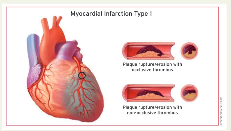
7.1 Myocardial infarction type 1
MI caused by atherothrombotic coronary artery disease (CAD) and usually precipitated by atherosclerotic plaque disruption (rupture or erosion) is designated as a type 1 MI. The relative burden of athero- sclerosis and thrombosis in the culprit lesion varies greatly, and the dynamic thrombotic component may lead to distal coronary emboli- zation resulting in myocyte necrosis.44,45 Plaque rupture may not only be complicated by intraluminal thrombosis but also by haemorrhage into the plaque through the disrupted surface (Figure3).44,45
Table 1 Reasons for the elevation of cardiac troponin values because of myocardial injury
Myocardial injury related to acute myocardial ischaemia
Atherosclerotic plaque disruption with thrombosis.
Myocardial injury related to acute myocardial ischaemia because of oxygen supply/demand imbalance
Reduced myocardial perfusion, e.g.
• Coronary artery spasm, microvascular dysfunction
• Coronary embolism
• Coronary artery dissection
• Sustained bradyarrhythmia
• Hypotension or shock
• Respiratory failure
• Severe anaemia
Increased myocardial oxygen demand, e.g.
• Sustained tachyarrhythmia
• Severe hypertension with or without left ventricular hypertrophy
Other causes of myocardial injury
Cardiac conditions, e.g.
• Heart failure
• Myocarditis
• Cardiomyopathy (any type)
• Takotsubo syndrome
• Coronary revascularization procedure
• Cardiac procedure other than revascularization
• Catheter ablation
• Defibrillator shocks
• Cardiac contusion Systemic conditions, e.g.
• Sepsis, infectious disease
• Chronic kidney disease
• Stroke, subarachnoid haemorrhage
• Pulmonary embolism, pulmonary hypertension
• Infiltrative diseases, e.g. amyloidosis, sarcoidosis
• Chemotherapeutic agents
• Critically ill patients
• Strenuous exercise
©ESC/ACC/AHA/WHF 2018
For a more comprehensive listing, see39–41
Downloaded from https://academic.oup.com/eurheartj/advance-article-abstract/doi/10.1093/eurheartj/ehy462/5079081 by guest on 12 November 2018
.. ..
.. ..
.. ..
.. ..
.. ..
.. ..
.. ..
.. ..
.. ..
.. ..
.. ..
.. ..
.. ..
.. ..
.. ..
.. ..
.. ..
.. ..
.. ..
.. ..
.. ..
.
cTn = cardiac troponin; ECG = electrocardiogram; URL = upper reference limit.
aPost-mortem demonstration of an atherothrombus in the artery supplying the infarcted myocardium, or a macroscopically large cir- cumscribed area of necrosis with or without intramyocardial hae- morrhage, meets the type 1 MI criteria regardless of cTn values.
It is essential to integrate the ECG findings with the aim of classify- ing type 1 MI into STEMI or NSTEMI in order to establish the appro- priate treatment according to current Guidelines.46,47
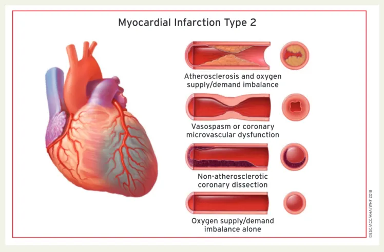
7.2 Myocardial infarction type 2
The pathophysiological mechanism leading to ischaemic myocardial injury in the context of a mismatch between oxygen supply and
demand has been classified as type 2 MI.10,12By definition, acute athe- rothrombotic plaque disruption is not a feature of type 2 MI. In patients with stable known or presumed CAD, an acute stressor such as an acute gastrointestinal bleed with a precipitous drop in hae- moglobin, or a sustained tachyarrhythmia with clinical manifestations of myocardial ischaemia, may result in myocardial injury and a type 2 MI. These effects are due to insufficient blood flow to the ischaemic myocardium to meet the increased myocardial oxygen demand of the stressor. Ischaemic thresholds may vary substantially in individual patients depending on the magnitude of the stressor, the presence of non-cardiac comorbidities, and the extent of underlying CAD and cardiac structural abnormalities.
Studies have shown variable occurrences of type 2 MI depending on criteria used for diagnosis. Some reports rely on specific predeter- mined oxygen mismatch criteria,48,49whereas others apply more lib- eral criteria. Most studies show a higher frequency of type 2 MI in women. The short- and long-term mortality rates for patients with type 2 MI are generally higher than for type 1 MI patients in most but not all studies due to an increased prevalence of comorbid con- ditions.49–57Coronary atherosclerosis is a common finding in type 2 MI patients selected for coronary angiography. In general, these patients have a worse prognosis than those without CAD.54–57 Prospective evaluations of the importance of CAD with type 2 MI using consistent definitions and approaches are needed.
It has been shown that the frequency of ST-segment elevation in type 2 MI varies from 3–24%.53In some cases, coronary embolism caused by thrombi, calcium or vegetation from the atria or ventricles, or acute aortic dissection may result in a type 2 MI. Spontaneous
Myocardial Infarction Type 1
Plaque rupture/erosion with occlusive thrombus
Plaque rupture/erosion with non-occlusive thrombus
©ESC/ACC/AHA/WHF 2018
Figure 3Myocardial infarction type 1.
Criteria for type 1 MI
Detection of a rise and/or fall of cTn values with at least one value above the 99th percentile URL and with at least one of the following:
• Symptoms of acute myocardial ischaemia;
• New ischaemic ECG changes;
• Development of pathological Q waves;
• Imaging evidence of new loss of viable myocardium or new regional wall motion abnormality in a pattern consistent with an ischaemic aetiology;
• Identification of a coronary thrombus by angiography includ- ing intracoronary imaging or by autopsy.a
Downloaded from https://academic.oup.com/eurheartj/advance-article-abstract/doi/10.1093/eurheartj/ehy462/5079081 by guest on 12 November 2018
.. ..
.. ..
.. ..
.. ..
coronary artery dissection with or without intramural haematoma is
.
another non-atherosclerotic condition that may occur, especially in young women. It is defined as spontaneous dissection of the coronary artery wall with accumulation of blood within the false lumen, which can compress the true lumen to varying degrees (Figure4).58
All of the clinical information available should be considered in dis- tinguishing type 1 MI from type 2 MI. The context and mechanisms of type 2 MI should be considered when establishing this diagnosis (Figure5). The myocardial oxygen supply/demand imbalance attribut- able to acute myocardial ischaemia may be multifactorial, related either to: reduced myocardial perfusion due to fixed coronary athe- rosclerosis without plaque rupture, coronary artery spasm, coronary microvascular dysfunction (which includes endothelial dysfunction, smooth muscle cell dysfunction, and the dysregulation of sympathetic innervation), coronary embolism, coronary artery dissection with or without intramural haematoma, or other mechanisms that reduce oxygen supply such as severe bradyarrhythmia, respiratory failure with severe hypoxaemia, severe anaemia, and hypotension/shock; or to increased myocardial oxygen demand due to sustained tachyar- rhythmia or severe hypertension with or without left ventricular
hypertrophy. In patients who undergo timely coronary angiography, description of a ruptured plaque with thrombus in the infarct-related artery may be helpful in making the distinction between type 2 MI vs.
type 1 MI, but angiography is not always definitive, clinically indicated, or required to establish the diagnosis of type 2 MI.
Myocardial Infarction Type 2
Atherosclerosis and oxygen supply/demand imbalance
Vasospasm or coronary microvascular dysfunction
Non-atherosclerotic coronary dissection
Oxygen supply/demand imbalance alone
©ESC/ACC/AHA/WHF 2018
Figure 4Myocardial infarction type 2.
Criteria for type 2 MI
Detection of a rise and/or fall of cTn values with at least one value above the 99th percentile URL, and evidence of an imbalance between myocardial oxygen supply and demand unrelated to acute coronary athero-thrombosis, requiring at least one of the following:
• Symptoms of acute myocardial ischaemia;
• New ischaemic ECG changes;
• Development of pathological Q waves;
• Imaging evidence of new loss of viable myocardium or new regional wall motion abnormality in a pattern consistent with an ischaemic aetiology.
Downloaded from https://academic.oup.com/eurheartj/advance-article-abstract/doi/10.1093/eurheartj/ehy462/5079081 by guest on 12 November 2018
.. ..
.. ..
.. ..
.. ..
.. ..
.. ..
.. ..
.. ..
.. ..
.. ..
.. ..
.. ..
.. ..
.. ..
.. ..
.. ..
.. ..
It appears advisable in the acute setting to treat the underlying ischaemic imbalance of oxygen supply and demand. This treatment may include volume adjustment, blood pressure management, admin- istration of blood products, heart-rate control, and respiratory sup- port.47,48Depending on the clinical situation, coronary evaluations may be indicated to assess the likelihood of CAD. If it is present, the MI Guidelines may be applied in accordance with the ECG findings of STEMI or NSTEMI.46,47However, if CAD is absent, the benefits of cardiovascular risk reduction strategies with type 2 MI remain uncertain.
7.3 Myocardial infarction type 2 and myocardial injury
Type 2 MI and myocardial injury are frequently encountered in clini- cal practice and both are related to a poor outcome.13,14,49,51,56
A conceptual model to facilitate the clinical distinction between acute ischaemic myocardial injury with or without an acute atherothrom- botic event (type 1 or type 2 MI) vs. conditions without acute ischae- mic myocardial injury is displayed inFigure6. Acute MI requires a rising and/or falling pattern of cTn values. Acute myocardial injury may also manifest such a pattern but if the injury is related to struc- tural heart disease, the cTn values may be stable and unchanging.
Type 2 MI and non-ischaemic myocardial injury may coexist. It should be recognized that some disease entities may be on both sides of the diagram, e. g. acute heart failure that may occur in the context of acute myocardial ischaemia. Nevertheless, abnormal cTn values in the setting of acute and/or chronic heart failure are often better cate- gorized as a myocardial injury condition. Few studies have compared the incidence and clinical features of type 2 MI vs. myocardial injury without acute myocardial ischaemia.
7.4 Myocardial infarction type 3
The detection of cardiac biomarkers in the blood is fundamental for establishing the diagnosis of MI.10,12However, patients can manifest a typical presentation of myocardial ischaemia/infarction, including pre- sumed new ischaemic ECG changes or ventricular fibrillation, and die before it is possible to obtain blood for cardiac biomarker determina- tion; or the patient may succumb soon after the onset of symptoms before an elevation of biomarker values has occurred. Such patients are designated as having a type 3 MI, when suspicion for an acute myocardial ischaemic event is high, even when cardiac biomarker evi- dence of MI is lacking.10,12This category allows the separation of fatal MI events from the much larger group of sudden death episodes that may be cardiac (non-ischaemic) or non-cardiac in origin. When a
Fixed coronary atherosclerosis
Coronary embolism
Sustained tachyarrhythmia Severe hypertension +/- Left ventricular hypertrophy
Severe bradyarrhythmia Respiratory failure
Severe anaemia Hypotension/Shock
Coronary spasm
Coronary microvascular dysfunction
Coronary artery dissection +/- Intramural haematoma
Mechanisms Context
Type 2 myocardial
infarction
Secondary to another illness or process
Main reason leading to clinical presentation (e.g. chest pain)
Oxygen supply and demand
imbalancea
aIschaemic thresholds vary substantially in relation to the magnitude of the stressor and the extent of underlying cardiac disease.
©ESC/ACC/AHA/WHF 2018
Figure 5Framework for type 2 myocardial infarction considering the clinical context and pathophysiological mechanisms attributable to acute myocardial ischaemia. The illustration above is modified from Januzzi and Sandoval.59
Downloaded from https://academic.oup.com/eurheartj/advance-article-abstract/doi/10.1093/eurheartj/ehy462/5079081 by guest on 12 November 2018
.. ..
.. ..
.. ..
.. ..
.. ..
.. ..
.. ..
.. ..
.. ..
.. ..
.. ..
.. .
type 3 MI is diagnosed and a subsequent autopsy reveals recent evi- dence of an MI, with a fresh or recent thrombus in the infarct-related artery, the type 3 MI should be reclassified to a type 1 MI. Original investigations addressing the incidence of type 3 MI are sparse, but a study showed an annual incidence below 10/100 000 person-years and a frequency of 3 – 4% among all types of MI.60
8 Coronary procedure-related myocardial injury
Cardiac procedural myocardial injury related to coronary revasculari- zation procedures, whether percutaneous coronary intervention (PCI) or coronary artery bypass grafting (CABG), may be temporally related to the procedure itself, reflecting periprocedural issues, or may occur later reflecting complications of a device, such as early or late stent thrombosis or in-stent restenosis for PCI, or graft occlusion or stenosis with CABG. Late gadolinium enhancement (LGE) cardiac magnetic resonance (CMR) allows assessment of procedural myocar- dial injury.61–63When quantifying procedural injury using LGE-CMR before and shortly after PCI or CABG, it was found that 32% of patients had evidence of procedural myocardial injury.63Furthermore, it has been shown that patients with elevation of cTnI values after PCI Atherosclerosis
+ thrombosis
Acute myocardial infarction
With acute ischaemiab
Troponin rise and/or fall
Elevated Cardiac Troponin Value(s) > 99th percentile URL
Acute myocardial injury
Without acute ischaemiab
Troponin level stablea
Chronic myocardial injury
Oxygen supply and demand
imbalance
Type 1 MI: triggers
• Plaque rupture
• Plaque erosion
Examples
• Acute heart failure
• Myocarditis Type 2 MI: examples
• Severe hypertension
• Sustained tachyarrhythmia
Examples
• Structural heart disease
• Chronic kidney disease
©ESC/ACC/AHA/WHF 2018
Figure 6 A model for interpreting myocardial injury. Ischaemic thresholds vary substantially in relation to the magnitude of the stressor and the extent of underlying cardiac disease. MI = myocardial infarction; URL = upper reference limit.aStable denotes <_ 20% variation of troponin values in the appropriate clinical context.bIschaemia denotes signs and/or symptoms of clinical myocardial ischaemia.
Criteria for type 3 MI
Patients who suffer cardiac death, with symptoms suggestive of myocardial ischaemia accompanied by presumed new ischaemic ECG changes or ventricular fibrillation, but die before blood sam- ples for biomarkers can be obtained, or before increases in car- diac biomarkers can be identified, or MI is detected by autopsy examination.
Downloaded from https://academic.oup.com/eurheartj/advance-article-abstract/doi/10.1093/eurheartj/ehy462/5079081 by guest on 12 November 2018
.. ..
.. ..
.. ..
.. ..
.. ..
.. ..
.. ..
.. ..
.. ..
.. ..
.. ..
.. ..
.. ..
.. ..
.. ..
.. ..
.. ..
lowing a coronary revascularization procedure may reflect procedural myocardial injury. Of importance, if the baseline value before the pro- cedure is above the 99th percentile URL, it is essential that cTn levels are stable prior to the evaluation in order to reliably establish the pres- ence of acute procedural myocardial injury. It is not possible to deter- mine, when intervening in a patient with an acute MI event resulting in an increased cTn level, how much of any given increase is related to the MI and how much is due to the procedure.
A large proportion of patients have abnormal values of cTn after PCI, ranging from20 – 40% in stable CAD to 40 – 50% in MI.64The occurrence of procedural myocardial injury can be detected by the measurement of cTn before the procedure and repeated 3 – 6 h later. Where the second value is rising, further sampling should be performed to document the peak cTn value. Increasing levels after the procedure can only be attributed with certainty to procedural myocardial injury when the pre-procedural cTn values are normal (<_ 99th percentile URL), or if they are stable or falling. For patients that present with an ACS and undergo a prompt coronary revascula- rization procedure resulting in only a single pre-procedural baseline value that is normal or mildly elevated, followed by subsequent post- procedural values that continue to increase, the post-procedural increase should be attributed to the index event. Recent data corrob- orate the importance of elevated pre-procedure cTn values as a prognostic marker in patients that have values that rise after the pro- cedure.65To diagnose procedural myocardial injury in the clinical set- ting of only a single pre-procedural cTn value, the cardiac Tn values would need to be stable or falling post-procedure, followed by a sub- sequent increase that exceeds the 99th percentile URL, and if the value has not returned to baseline, the increase should be > 20%
with an absolute value > the 99th percentile URL.
9 Myocardial infarction associated with percutaneous coronary
intervention (type 4a myocardial infarction)
Stand-alone post-procedural increases of cTn values are sufficient to establish a diagnosis of procedural myocardial injury but not for the diagnosis of type 4a MI. Type 4a MI requires an elevation of cTn val- ues greater than five times the 99th percentile URL in patients with
post-procedure cTn must rise > 20% to an absolute value more than five times the 99th percentile URL. In addition, there should be evi- dence of new myocardial ischaemia, either from ECG changes, imag- ing evidence, or from procedure-related complications associated with reduced coronary blood flow such as coronary dissection, occlusion of a major epicardial artery or a side branch occlusion/
thrombus, disruption of collateral flow, slow flow or no-reflow, or distal embolization. The use of hs-cTn assays to diagnose type 4a MI (and type 5 MI) is an area of active research. Many hs-cTn assays are available, which have wide dynamic ranges. Different criteria may be required for different assays. However, it has recently been shown that the optimal hs-cTnT thresholds to predict cardiovascular events at 30 days and 1 year were very close to the five-fold increase sug- gested by the Third Universal Definition of Myocardial infarc- tion.12,66,67These criteria are therefore retained because of a lack of new scientific evidence that identifies superior criteria for defining this MI subtype. Other criteria that meet the definition of type 4a MI, regardless of hs-cTn or cTn values, are the development of new pathological Q waves or autopsy evidence of recent procedure- related thrombus in the culprit artery.
Criteria for cardiac procedural myocar- dial injury
Cardiac procedural myocardial injury is arbitrarily defined by increases of cTn values (> 99th percentile URL) in patients with normal baseline values (<_ 99th percentile URL) or a rise of cTn values > 20%of the baseline value when it is above the 99th per- centile URL but it is stable or falling.
Criteria for PCI-related MI 48 h after the index procedure
(type 4a MI)
Coronary intervention-related MI is arbitrarily defined by an ele- vation of cTn values more than five times the 99th percentile URL in patients with normal baseline values. In patients with ele- vated pre-procedure cTn in whom the cTn level are stable (<_ 20%variation) or falling, the post-procedure cTn must rise by
> 20%. However, the absolute post-procedural value must still be at least five times the 99th percentile URL. In addition, one of the following elements is required:
• New ischaemic ECG changes;
• Development of new pathological Q waves;a
• Imaging evidence of new loss of viable myocardium or new regional wall motion abnormality in a pattern consistent with an ischaemic aetiology;
• Angiographic findings consistent with a procedural flow-limit- ing complication such as coronary dissection, occlusion of a major epicardial artery or a side branch occlusion/thrombus, disruption of collateral flow, or distal embolization.b
aIsolated development of new pathological Q waves meets the type 4a MI criteria if cTn values are elevated and rising but less than five times the 99th percentile URL.
bPost-mortem demonstration of a procedure-related thrombus in the culprit artery, or a macroscopically large circumscribed area of necrosis with or without intra-myocardial haemorrhage meets the type 4a MI criteria.
Downloaded from https://academic.oup.com/eurheartj/advance-article-abstract/doi/10.1093/eurheartj/ehy462/5079081 by guest on 12 November 2018
.. ..
.. ..
.. ..
.. ..
.. ..
.. ..
.. ..
.. ..
.. ..
.. ..
.. ..
.. ..
.. ..
.. ..
.. ..
.. ..
.. ..
.. .
10 Stent/scaffold thrombosis associated with percutaneous coronary intervention (type 4b myocardial infarction)
A subcategory of PCI-related MI is stent/scaffold thrombosis, type 4b MI, as documented by angiography or autopsy using the same criteria utilized for type 1 MI. It is important to indicate the time of the occur- rence of the stent/scaffold thrombosis in relation to the timing of the PCI procedure. The following temporal categories are suggested:
acute, 0–24 h; subacute, > 24 h to 30 days; late, > 30 days to 1 year;
and very late > 1 year after stent/scaffold implantation.68
11 Restenosis associated with percutaneous coronary
intervention (type 4c myocardial infarction)
Occasionally MI occurs and—at angiography, in-stent restenosis, or restenosis following balloon angioplasty in the infarct territory—is the only angiographic explanation since no other culprit lesion or thrombus can be identified. This PCI-related MI type is designated as type 4c MI, defined as focal or diffuse restenosis, or a complex lesion associated with a rise and/or fall of cTn values above the 99th percen- tile URL applying, the same criteria utilized for type 1 MI.
12 Myocardial infarction
associated with coronary artery bypass grafting (type 5 myocardial infarction)
Numerous factors can lead to procedural myocardial injury during a CABG procedure. Many of them are related to the details of the car- diac preservation, the extent of the direct traumatic injury to the myocardium, as well as any potential ischaemic injury. For that rea- son, increases in cTn values should be expected after all CABG pro- cedures,69,70which need to be taken into account when comparing the extent of procedural myocardial injury after cardiac surgery with that associated with less invasive approaches. Depending on whether it is off-pump or on-pump surgery, procedural myocardial injury is observed among 32 – 44% of CABG patients when quantified by LGE-CMR.61,63The area under the curve (AUC) and routine cTn sampling has demonstrated an excellent linear relationship with the mass of the new injury as defined by LGE-CMR. AUC for CK-MB is also good, although clearly inferior to cTnI.69However, these rela- tionships vary depending on the nature of the procedure, the nature of the cardioplegia, and the specific assay used to measure cTn. Very high cTn values are most often associated with coronary artery- related events.61,63,69 Thus, although cardiac biomarkers and
especially cTn appear robust for the detection of procedural myocar- dial injury and also, in the presence of new myocardial ischaemia, for the detection of type 5 MI, a specific cut-off value for all procedures and all cTn assays is difficult to define. However, in order to ensure consistency with the analogous standards of the preceding definition of type 5 MI12and because of the lack of new scientific evidence that identifies superior criteria for defining this MI subtype, it is suggested that a cTn value > 10 times the 99th percentile URL is applied as the cut-off point during the first 48 h following CABG, occurring from a normal baseline cTn value (<_ 99th percentile URL), for diagnosing type 5 MI. It is important that the post-procedural elevation of cTn values is accompanied by ECG, angiographic, or imaging evidence of new myocardial ischaemia/new loss of myocardial viability.71 The higher cut-off of MI after CABG than after PCI (10 times vs. 5 times the 99th percentile URL) has been arbitrarily selected due to the occurrence of more unavoidable myocardial injury during surgery than during PCI.
It should be recognized that ST-segment deviation and T wave changes are common after CABG due to epicardial injury, and are not reliable indicators of myocardial ischaemia in this setting.
However, ST-segment elevation with reciprocal ST-segment depres- sion or other specific ECG patterns may be a more reliable finding of a potential ischaemic event.
Marked isolated elevation of cTn values within the 48 h post- operative period, even in the absence of ECG/angiographic or other imaging evidence of MI, indicates prognostically significant cardiac procedural myocardial injury.72 The presence of significant proce- dural myocardial injury in patients with operative problems (e.g. diffi- culty coming off bypass, technically difficult anastomoses in a heavily calcified aorta, of perioperative evidence of myocardial ischaemia, etc.) should prompt clinical review of the procedure and/or consider- ation of additional diagnostic testing for possible type 5 MI.
Criteria for CABG-related MI 48 h after the index procedure (type 5 MI)
CABG-related MI is arbitrarily defined as elevation of cTn values
> 10 times the 99th percentile URL in patients with normal base- line cTn values. In patients with elevated pre-procedure cTn in whom cTn levels are stable (<_ 20%variation) or falling, the post- procedure cTn must rise by > 20%. However, the absolute post- procedural value still must be > 10 times the 99th percentile URL. In addition, one of the following elements is required:
• Development of new pathological Q waves;a
• Angiographic documented new graft occlusion or new native coronary artery occlusion;
• Imaging evidence of new loss of viable myocardium or new regional wall motion abnormality in a pattern consistent with an ischaemic aetiology.
aIsolated development of new pathological Q waves meets the type 5 MI criteria if cTn values are elevated and rising but < 10 times the 99th percentile URL.
Downloaded from https://academic.oup.com/eurheartj/advance-article-abstract/doi/10.1093/eurheartj/ehy462/5079081 by guest on 12 November 2018
.. ..
.. ..
.. ..
.. ..
.. ..
.. ..
.. ..
.. ..
.. ..
.. ..
.. ..
.. ..
.. ..
.. ..
.. ..
.. ..
.. ..
.. ..
.. ..
.. ..
.. ..
.. ..
.. ..
.. ..
.. ..
.. ..
.. ..
.. ..
.. ..
.. ..
.. ..
.. ..
.. ..
.. ..
.. ..
.. ..
.. ..
.. ..
.. ..
.. ..
.. ..
.. ..
infarction related to percutaneous coronary intervention or coronary artery bypass grafting
There is no universal consensus on the cTn or hs-cTn cut-off points that clearly distinguish cardiac procedural myocardial injury from MI.
The distinction is made on the basis of an injury created by a flow- limiting complication during the procedure that results in sufficient myocardial ischaemia to generate a procedure-related MI. The size of the insult will determine the magnitude of the cTn release. Various groups have used multiples of the 99th percentile URL and set thresholds to diagnose periprocedural MIs for clinical trials.68,73 Unless a standard assay is used for all analyses, given the heterogene- ity of cTn assays, this approach could lead to very different values depending on the assay used locally. The Academic Research Consortium-2 (ARC-2) suggests a post-procedural cTn value >_ 35 times the 99th percentile URL for both PCI and CABG in patients that have a normal baseline cTn value or in patients with elevated pre-procedure cTn values in whom the cTn levels are stable or falling.
ARC-2 proposes that one ancillary criterion be required in addition to the >_ 35 cTn rise to fulfill the definition of periprocedural MI. The ancillary criteria are one or more of the following: new significant Q waves (or equivalent), flow-limiting angiographic complications in a major epicardial vessel or > 1.5 mm diameter branch, or a substantial new loss of viable myocardium on echocardiography related to the procedure.68Furthermore, ARC-2 has defined stand-alone criteria for significant procedural myocardial injury if the rise in cTn is >_ 70 times the 99th percentile URL (where the baseline is lower than the URL, elevated and stable, or falling).68
14 Recurrent myocardial infarction
Incident MI is defined as the individual’s first MI. When features of MI occur in the first 28 days after an incident event, the second event is not counted as a new MI for epidemiological purposes. If characteris- tics of MI occur after 28 days following an incident MI, it is considered to be a recurrent MI.11
15 Re-infarction
The term re-infarction is used clinically for an acute MI that occurs within 28 days of an incident or recurrent MI.11The ECG diagnosis of suspected re-infarction following the initial MI may be confounded by the initial evolutionary ECG changes. Re-infarction should be consid- ered when ST-elevation >_ 1 mm recurs or new pathognomonic Q waves appear in at least two contiguous leads, particularly when asso- ciated with ischaemic symptoms. However, re-elevation of the ST- segment can also be seen in threatened myocardial rupture or in cases of pericarditis, and should lead to additional diagnostic evaluation.
In patients where re-infarction is suspected from clinical signs or symptoms following the initial MI, an immediate measurement of cTn
vated, but stable or decreasing at the time of suspected re-infarction, the diagnosis of re-infarction requires a > 20% increase of the cTn value in the second sample.74If the initial cTn concentration is nor- mal, the criteria for new