Impact of H63D mutations,
magnetic resonance and metabolic syndrome among outpatient
referrals for elevated serum ferritin in the Basque Country
Agustin Castiella,* Eva Zapata,* Leire Zubiaurre,* Jose Ma. Alustiza,† Ma. Dolores De Juan,‡ Arantxa Iribarren,* Jose I. Emparanza,§ Pedro Otazua||
*Gastroenterology, Mendaro Hospital; †Radiology, Osatek Donostia; ‡Immunology Service, Donostia Hospital; §Epidemiology Unit, Donostia Hospital; ||Gastroenterology Service, Mondragon Hospital, Spain.
ABSTRACT
Background and Aims. There are limited data on clinical and phenotypic characteristics of outpatients re-ferred for hyperferritinemia (HF). To determine the causes of HF in outpatients rere-ferred to a secondary hospital. Material and methods. A prospective study of 132 consecutive patients with HF (> 200 μg/L, women; > 300 μg/L, men) was conducted from January-December 2010. Results. Mean age, 54.42 years (SD: 13.47, range: 23-83); body mass index (BMI), 28.80 (SD: 3.96, 17-39); ferritin (SF), 579.54 ng/mL (SD: 296.575, 206-1668); transferrin saturation (TSI), 43.87% (SD: 14.09, 12-95); iron (Fe), 134 μg/dL (SD: 49.68, 55-322); overweight: 48.31%, and obese: 40.44% (89%), and most patients were men (108/132). Regarding HFE mutations, H63D/H63D genotype and H63D allele frequencies were 17.5% (vs. 7.76% in controls); and 36% (31% in controls) respectively. While 63.6% consumed no alcohol, 18.1% consumed ≥ 60 g/day, the mean being 20.83 (SD: 33.95, 0-140). Overall, 6/132 (4.5%) patients were positive for B or C hepatitis. Mean LIC by MRI was 36.04 (SD: 32.78, 5-210), 53 patients having normal concentrations (< 36 μmol/g), 22 (33%) iron over-load (37-80), and 4 (5%) high iron overover-load (> 80). Metabolic syndrome (MS) was detected in 44/80 men (55%) and 10/17 women (59%). In this group, the genotype frequency of the H63D/H63D mutation was significantly higher than in controls-21.56% vs. 7.76%- (p = 0.011); the H63D allelic frequency was 42.15% in MS group and 31% in controls (p = 0.027). Conclusion. The H63D/H63D genotype and H63D allele predispose indivi-duals to HF and MS. MRI revealed iron overload in 33% of patients.
Key words. Hyperferritinemia. HFE gene. Hemochromatosis. MRI. Metabolic syndrome.
Correspondence and reprint request: Agustin Castiella, Ph.D., M.D. Gastroenterology Service, Mendaro Hospital, Barrio Mendarozabal s/n; 20850 Mendaro, Spain.
Tel.: 00-34-032800. Fax: 00-34-032856 E-mail: [email protected]
Manuscript received: September 14, 2014. Manuscript accepted: November 23, 2014.
INTRODUCTION
SF values are frequently requested by clinicians.1 Raised levels of SF is a common finding in routine laboratory tests. Such patients are referred to an outpatient hospital consultation for testing for HF and genetic hemochromatosis.1-5 Raised SF levels may be present in various different clinical condi-tions, such as inflammatory diseases, and specifical-ly in renal, hepatic and neoplastic diseases. In patients with liver disease, raised serum ferritin is
characteristic of hereditary hemochromatosis, but may also be found in patients with chronic hepatitis C, alcoholic or non-alcoholic steatohepatitis, and non-alcoholic fatty liver disease.3,6
Epidemiological studies on hereditary hemochro-matosis (HH) in Mediterranean populations have shown that the prevalence of C282Y is lower than in Northern European countries,3,7 and that the H63D mutation is found among 15-20% of the general pop-ulation.3,8 In the Basque Country, the allele fre-quency is as high as 30%.7
There are limited data in the literature on clinical and phenotypic characteristics of outpatients re-ferred for HF.1 Specifically, there has been very lit-tle research on epidemiologic factors including alcohol, viral hepatitis, obesity and metabolic syn-drome.
MRI.9,10 When validated, with values being strongly correlated with those determined biochemically, and standardized, LIC values obtained by MRI are as useful as those obtained by liver biopsy.9-11
The objectives of our study were to prospectively analyse consecutive patients referred to the outpa-tient clinic of a secondary hospital in the Basque Country and to determine the causes of the HF as well as the relevance of HFE gene mutations and liver iron measurement by MRI in the diagnosis.
MATERIAL AND METHODS
Patients
This was a prospective study including patients from January to December 2010. The study was conducted in Mendaro Hospital, Deba Valley, a sec-ondary hospital in the Basque Health Service (Os-akidetza), with a total catchment of 70,000 people.
Inclusion criteria: consecutive outpatients re-ferred for HF (> 200 μg/L in women, > 300 μg/L in men), in accordance with the WHO criteria,1,3 by their General practitioners were eligible to partici-pate in the study provided that they were aged 18 years or older.
Exclusion criteria: systemic inflammation, infec-tions, and renal, or neoplastic diseases (excluded by clinical, laboratory and radiologic methods).
Methods
• Radiology.
a) MRI. MRI images were obtained with a 1.5-Tesla system (Philips Intera, Osatek, Donostia). The MRI technique used (SIR method) was that proposed by Alustiza, et al.10 As steatosis is frequent, we systematically perform T1-weighted in-phase and opposed-phase imaging to discard liver steatosis. All the iron quantification sequences were in-phase sequences to be sure that the fat did not interfere in signal intensity measurements.12 The LIC calculated by this model has a high correlation with biochemical measurements, obtained by atomic spectrometry, in liver biop-sy samples (r = 0.937).10 The LIC was consid-ered as: normal (< 36 μmol/g), iron overload (IO) (37-80 μmol/g), and high iron over-load (HIO) (> 80 μmol/g).10 We have also studied the presence of liver fat. Steatosis was classified as absent or present.
b) Abdominal ultrasound. Steatosis was graded by ultrasound and classified as absent or present.3
• Laboratory measurements. SF, Fe and TSI
were obtained from blood samples taken fasting from all the patients included. All were found to have raised SF values (> 300 μg/L), the laborato-ry normal range for SF being 15 to 200 μg/L in women and 30 to 300 μg/L in men.1,3 The ranges considered to be normal for Fe and TSI were 50-145 μg/dL and 15-45% respectively.
The serum values of ALT, AST, GGTP, alkaline phosphatase, bilirrubin, albumin, glucose, cho-lesterol, HDL-chocho-lesterol, triglycerides, etc., were obtained from the same blood samples.
• HFE mutation analysis. DNA was extracted
from the blood samples and HFE gene analysis was performed by multiplex real-time PCR using LightCycler technology (LC 1.0). Simultaneous detection of the HFE C282Y, H63D and S65C mutations was carried out in a single capillary using LC-Red 640, LC-Red 705 and fluorescein-labelled hybridization probes (Tibmolbiol, Berlin, Germany). Melting curve analysis was used to distinguish wild type and mutant alleles in each case.
The results were compared with a control group of blood donors from the same geographical area, Guipuzcoa.7
• Definition of metabolic syndrome (MS). We
employed established criteria,13 namely.
° Waist circumference ≥ 94 cm in men; ≥ 80 cm in women, and two of the following factors. ° Triglycerides ≥ 150 mg/dL or treatment for
this dyslipidaemia.
° HDL < 40 mg/dL women, < 50 mg/dL men or treatment for this dyslipidaemia.
° Glucose ≥ 100 mg/dL or type 2 diabetes. ° Hypertension: systolic pressure ≥ 130 mmHg;
diastolic pressure ≥ 85 mmHg, or treatment for arterial hypertension.
° Overweight or obesity, where a body mass in-dex (BMI) < 25 was considered normal; BMI > 25 and < 30, overweight; and BMI > 30, obesity.6
• Alcohol consumption. Heavy drinking was de-fined as the consumption of > 60 g/day;8,14 while > 40 g/day was considered moderate alcohol con-sumption. The information was collected from the interview with the patient.
• Statistics. SPSS 15.0 software (SSPS Inc., Chi-cago, IL, USA) was used to perform the appropri-ate statistical analyses. Mean values with range and standard deviation were calculated for con-tinuous variables and frequencies and percentag-es for categorical variablpercentag-es.
To compare HFE mutation frequency in patients with HF and the control group, we used Chi-square and Fisher’s exact tests, due to the small number of cases. In all the analysis, a p < 0.05 value was considered statistically significant. • Ethics. The study was conducted in accordance
with the Declaration of Helsinki and with the understanding and the consent of the patients. The study was approved by the Mendaro Hospi-tal Ethics Committee.
RESULTS
Patients
A total of 132 patients fulfilling the selection cri-teria were included in the study. There was a male predominance of approximately 4:1 (108 men, 24 women).
Not all the results were available for all the pa-tients.
Radiology
Abdominal ultrasound was suggestive of steatosis in 69/132 (52.3%), and normal in 44 (33.3%), with other findings in the remaining cases (14.4%).
LIC was obtained by MRI in 79/132 patients. The mean LIC was 36.04 μmol/g (SD: 32.78, 5-210), 53 patients having normal concentrations (<36 μmol/ g), 22 IO (37-80 μmol/g), and 4 HIO (> 80 μmol/g), 50% with a predisposing genotype. That is, 33% of patients had IO and 5% HIO.
Steatosis by MRI was studied in 79/132 patients: steatosis was present in 27 patients (34.17%) and 52 patients have not liver steatosis (65.82%).
Clinical and Laboratory data
The main clinical and laboratory data are pre-sented in tables 1-3. Overall, 43/89 patients (48.31%) were classified as overweight (BMI 25-29.9), 36/89 (40.44%) as obese (BMI ≥ 30), and 10/89 (11.23%) as normal (< 25), meaning that 89% were overweight or obese.
The percentage of obese patients was high, and we studied the MRI findings in this group: 16 from 36 obese patients were studied by MRI for iron and steatosis. The mean LIC of the group was 34.68 μmol/g (SD 27.72). Steatosis was present in 8 from 16 patients studied. Three patients from 8 without steatosis had an ultrasound that suggested steato-sis, but there were no discordancy cases (ultra-sound/ MRI) in the steatosis group.
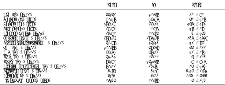
Table 1. Clinical baseline data, laboratory data and liver iron concentration (LIC) results obtained by magnetic resonance imag-ing (MRI) of the patients included in the study.
Mean SD Range
Age (yr) (n:132) 54.42 13.47 23 - 83
Weight (kg) (n:89) 83.17 15.86 43 - 137
Height (cm) (n:89) 175.28 55.21 146 - 191
BMI (kg/m²) (n:89) 28.80 3.96 17 - 39
Alcohol (g/day) (n:121) 20.83 33.95 0 - 140
Ferritin (ng/mL) (n:132) 579.54 296.57 206 - 1668
Transferrin saturation (%) (n:132) 43.87 14.10 12 - 95
Fe (μg/dL) (n:132) 133.77 49.69 55 - 322
ALT (IU/L) (n:132) 55.61 47.22 12 - 371
GOT (IU/L) (n:132) 41.01 35.33 4 - 230
GGTP (IU/L) (n:132) 94.83 141.47 8 - 806
Alkaline phosphatase (IU/L) (n:132) 73.32 20.71 34 - 160
Bilirubin (mg/dL) (n:132) 0.74 0.38 0.15 - 2.71
Albumin (g/L) (n:132) 4.60 0.32 3.50 - 5.50
LIC by MRI (μmol/g) (n:79) 36.04 32.79 5 - 210
The alcohol data (121 patients) revealed that 77 (63.6%) consumed none and 22 consumed ≥ 60 g of alcohol/day (18.1%). Applying a cut-off of > 40 g/day, 25.6% were considered drinkers. The mean consumption was 20.83 g of alcohol/day (SD: 33.95, 0-140).
Viral serology produced positive results in 6/132 patients (4.5%): for HBV in 2 cases (1.5%) and HB-sAg and HCV in 4 (3%).
A total of 50 patients were receiving treatment for hypertension and 35 for dyslipidaemia.
MS (13) was detected in 44/80 men (55%) and 10/ 17 women (59%), corresponding overall to 54/97 of the HF patients (55.67%).
No liver biopsies were taken during the study pe-riod.
HFE mutation analysis
This analysis was conducted in 120 patients. Mu-tations were found in 60.84% of cases in the study group (vs. 62.93% of the control group). The results are presented in table 3. The genotype frequency of the H63D/H63D mutation in cases was more than double that in controls (17.5 vs. 7.76%), while the allele frequency of the H63D mutation was also high-er: 36 vs. 31% controls.7 These differences were sig-nificant (p < 0.05), that is, both genotype and allele frequencies were significantly higher in the HF group.
Predisposing genotypes for liver iron overload/ HH8 were found in 27/120 patients (22.5%), H63D/ H63D in 21 and C282Y/H63D in 6 cases. Twenty pa-tients had TS > 45%, and LIC was measured by MRI in all but one case: in the H63D/H63D group, 7 patients had raised and 8 normal LIC values, while in the C282Y/H63D group, one patient was found to have raised and 3 normal LICs by MRI.
HFE mutation analysis in MS
This analysis was studied in 51 from 54 patients with MS. The results were: 2 C282Y/wt (3.92%), 11 H63D/H63D (21.56%), 15 H63D/wt (29.41%), 3 C282Y/H63D (5.88), 19 wt/wt (37.25%), and 1 S65C (1.96%). The genotype frequency of the H63D/H63D mutation in MS cases was significantly higher than in controls7 –21.56 vs. 7.76%– (p = 0.011); the H63D allelic frequency was 42.15% in MS group and 31% in controls (p = 0.027).
HFE and liver iron overload
Twenty-six patients were shown to have IO-HIO in the liver by MRI. Among these individuals, the HFE mutation analysis found: H63D/H63D in 8 cas-es; wt/wt, 7; H63D/wt, 7; S65C/wt, 2; C282Y/wt, 1; and C282Y/H63D, 1; that is, 9 of these 26 patients had predisposing genotypes.
When we analysed some special groups, namely the SF > 1,000 ng/L group, and HIO group we ob-tained the following results:
• SF > 1000 ng/L group: 12/132 patients (9%) had SF levels of > 1,000 ng/L. In this group, the gen-otypes were determined in 11 patients with the following results: H63D/wt in 5; wt/wt, 3; H63D/ H63D, 1; C282Y/ wt, 1; and S65C/wt, 1. Accord-ingly, it seems that there is no relationship between HFE gene mutations and SF > 1,000 ng/L. Further, LIC was obtained by MRI in 10/12 of the patients in the high ferritin (> 1,000 ng/L)
Table 2. Laboratory data and clinical data for metabolic syndrome.
N:97 Mean SD Range
Glucose (mg/dL) 106.95 25.03 73 - 272
Cholesterol (mg/dL) 201.71 42.92 96 - 338
HDL-CHOL (mg/dL) 54.00 17.08 28 - 162
Triglycerides (mg/dL) 151.75 93.71 40 - 477
Systolic blood pressure (mmHg) 139.26 19.93 100 - 190
Diastolic blood pressure (mmHg) 81.19 11.03 50 - 105
Waist circumference (cm) 100.06 11.70 63 - 132
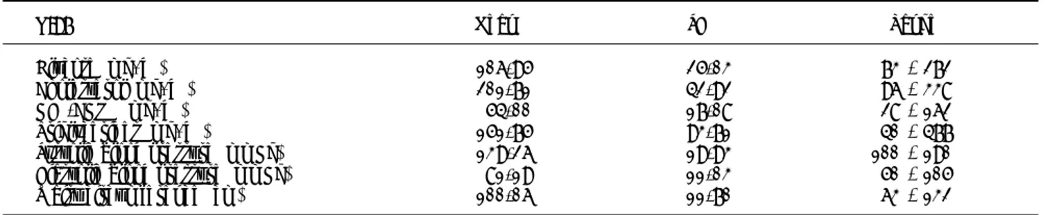
Table 3. HFE gene mutations in Basque patients from the stu-dy group and the control group.7
N:120 Study group Control group
C282Y/C282Y 0 0
C282Y/WT 6 8
C282Y/H63D 6 4
H63D/H63D 21 9
H63D/WT 38 45
WT/WT 47 43
S65C/WT 2 5
group. The LIC was raised (> 36 μmol/g), corre-sponding to IO, in 8/10 (80%) of these patients, while 2 had HIO.
• HIO group: overall, four patients had LICs of > 80 μmol/g, and their genetic findings were: H63D/wt in 2; C282Y/H63D in 1; and H63D/ H63D in 1; that is, 50% of these patients had pre-disposing genotypes. The C282y/H63D patient consumed no alcohol and had neither steatosis nor MS. One H63D patient drunk more than 80 g alcohol/day and presented steatosis; while the other patient with H63D, drank no alcohol and steatosis was not observed. Lastly, the H63D/ H63D patient drunk more than 80 g/day but did not have steatosis.
DISCUSSION
HF is a frequent finding in primary care when conducting routine laboratory studies. Patients with raised SF are referred to gastroenterology-hepatolo-gy units to study the relevance of this clinical enti-ty.1-5,15,16 Recently, Adams and Barton17 have published an article stating that no more than 10% of these HF patients have liver iron overload. In a retrospective study, Yenson, et al.5 reported an even lower rate of 5% of liver IO in Chinese and other Asian populations in British Columbia, Canada. Sev-eral studies have focused on analysing the causes of high or very high HF,18-21 but very few have explored the aetiology of patients referred for HF.1-3,5,16
Wong and Adams,1 in a retrospective series of 119 patients, found that in non-C282Y homozygotes with SF > 1,000 ng/L, IO was absent in 64% of the patients. Analysing this situation in the patients in-cluded in our study, 80% of those with a raised SF > 1,000 ng/L (12 patients) were found to have IO. Dev-er, et al.2 reviewed the clinical charts of patients with HF referred to an IO Clinic in the USA: 51% of the subjects were diagnosed with HH. The results of that study are, however, influenced by the mixed characteristics of subjects included (not only those referred for HF, but also some with known HH and their relatives) and hence are not useful for compar-ison with our data. In Italy, Licata, et al.3 studied the presence of hepatic siderosis in liver biopsies of 54 patients, finding it in 17 (32%) of cases. Perez-Aguilar, et al.15 in Valencia, Spain, studied the pres-ence of iron in liver biopsies of 14 patients with SF > 1,000 ng/L, but for this series no quantitative measurements were reported. In our study group, as many as 33% of patients were found to have liver
iron overload by a validated method using MRI.9-11 And more importantly, overall 5% of the patients showed high liver iron overload (> 80 μmol/g), like HH patients.
Wong, et al.1 reported that 71% of the patients with raised LIC had a TSI > 50%. In our series, > 50% patients had raised TSI in the IO-HIO group.
In the Basque Country, the prevalence of HFE gene mutations in HH and the general population differs from other populations in Europe and else-where across the world.7 In the HH group, only 57% of patients had the C282Y/C282Y mutation, while the H63D mutation is very prevalent even in the general population, with a frequency > 30%.
There is evidence of the H63D mutation contrib-uting to iron overload, increasing serum iron and transferrin, and also that its relationship with HH is independent of the presence of the C282Y muta-tion.8,21 In a control group,7 the H63D allele fre-quency was 31%. In this study of patients referred for HF, 61% presented mutations in the HFE gene, similar to the rate in controls (63%). The allele fre-quency was 36 vs. 31% in the control group, and the genotype frequency of the H63D/H63D mutation was 17.5%, compared to only 7.75% in the control group (both p < 0.05). This implies that rate of HF patients with this genotype is more than double that in the general population. Our results are consistent with the suggestion that this genotype could be re-sponsible for HF.22,23
In a previous study by Aguilar-Martinez, et al.,23 the frequency of the H63D homozygous genotype was found to be higher among patients who had been referred with a personal or family history of IO than in the general population. The causative role of the H63D/H63D mutation in HH or IO has been demonstrated,22-24 but with less penetrance and a considerable variation in phenotypic expression.23 Assessing the relevance of HFE mutations in our HIO and IO groups, no significant differences were encountered. Further, no HFE genotype differences were seen when studying the SF > 1,000 ng/L group.
Patients with a history of chronic alcohol con-sumption or features of MS often present with hy-perferritinemia.6 Heavy drinking (> 60 g/day of alcohol) was detected as a cause,8,13 in 18% of the population studied (25.6% if considering a cut-off of > 40 g/day). Overweight (BMI: 25-29.9) and obesity (BMI > 30), however, emerge as the most important causes of this entity: 89% of the patients were over-weight or obese at the time of HF diagnosis. The obese patients presented a LIC by MRI of 34.68 μmol/g, not different from that of the whole study group, and 50% revealed fat in their livers, steato-sis. Further, liver ultrasound revealed signs of stea-tosis, with suspicion of non-alcoholic fatty liver disease, in 52% of the patients. When we studied for the presence of fat in the liver by MRI, steatosis was present in 34.17% of the 79 studied patients. MS is present in 25% of adults in Western countries, and HF is found in only 15% of MS patients.26-30 In a recent study for detection of HH and biochemical iron overload in primary care, developed in Spain, 45% of patients with elevated iron measures (TSI and/or SF) had MS.31 Notably, in our study, 56% of patients with HF met the criteria for MS. Consider-ing the high percentage of H63D mutation in the population, we have studied HFE mutations in MS group, to determine if these mutations predispose to MS. The H63D/H63D mutation in MS cases was significantly higher than in controls7 –21.56 vs. 7.76%– (p = 0.011) and the H63D allelic frequency was 42.15% in the MS group and 31% in controls (p = 0.027). These results point out that H63D/H63D genotype and H63D allele predispose the patients with HF to develop a MS.
Analyzing the high SF group, we found no relationship between HFE gene mutations and SF > 1,000 ng/L. Of 12 patients with high SF, LIC was determined by MRI in 10. The LIC was raised in 8 of these 10 patients (80%), indicating that they had IO.
CONCLUSION
In conclusion, we can say from this study that the H63D/H63D genotype predisposes to HF. Obesi-ty and overweight are very important etiological fac-tors, affecting 89% of our HF patients. In our region, alcohol may be a cause of HF but viruses were rare as an underlying factor. Not all HF repre-sents iron overload6 but MRI revealed iron overload in 33% of the patients. MS is present in 56% of the patients and may be induced by H63D/H63D geno-type and H63D allele.
ABBREVIATIONS
• BMI: body mass index. • Fe: iron.
• HF: hyperferritinemia.
• HH: hereditary hemochromatosis. • HIO: high iron overload.
• IO: iron overload.
• LIC: liver iron concentration. • MRI: magnetic resonance imaging. • MS: metabolic syndrome.
• SF: ferritin.
• TSI: transferrin saturation.
CONFLICT OF INTEREST
The authors report no conflicts of interest. The authors are responsible for the content and writing of the paper.
AUTHOR’S CONTRIBUTIONS
All authors met the criteria for authorship as stablished by the International Committee of Medi-cal Journal Editors. The manuscript represents hon-est work and all authors are able to verify the validity of the results reported.
Congress: presented as abstract at the Liver In-ternational meeting (EASL) 2012; Barcelona, Spain, April 18-22/ 2012.
ACKNOWLEDGEMENT
The authors thank the translation department of the Instituto Vasco de Investigaciones Sanitarias (BIOEF) for their help.
REFERENCES
1. Wong K, Adams PC. The diversity of liver diseases among outpatient referrals for elevated serum ferritin. Can J Gastroenterol 2006; 20: 467-70.
2. Dever JB, Mallory MA, Mallory JE, Wallace D, Kowdley KV. Phenotypic characteristics and diagnoses of patients re-ferred to an iron overload clinic. Dig Dis Sci 2010; 55: 803-7.
3. Licata A, Nebbia ME, Cabibbo G, Lo Iacono G, Barbaria F, Brucato V, Alessi N, et al. Hyperferritinemia is a risk fac-tor for steatosis in chronic liver disease. World J Gas-troenterol 2009; 15: 2132-8.
4. Hearnshaw S, Thompson NP, McGill A. The epidemiology of hyperferritinemia. World J Gastroenterol 2006; 12: 5866-9.
Columbia experience. Can J Gastroenterol 2008; 22: 37-40.
6. Beaton MD, Adams PC. Treatment of hyperferritinemia. Ann Hepatol 2012; 11: 294-300.
7. De Juan MD, Reta A, Castiella A, Pozueta J, Prada A, Cua-drado E. HFE gene mutations analysis in Basque Country hereditary haemochromatosis patients and controls. Eur J Hum Genet 2001; 9: 961-4.
8. Castiella A, Zapata E, De Juan MD, Otazua P, Fernandez J, Zubiaurre L, Arriola JA, et al. Significance of H63D homo-zygosity in a Basque Country population with hemochro-matosis. J Gastroenterol Hepatol 2010; 25: 1295-8. 9. Alustiza JM, Emparanza JI, Aldazabal P, Garrido A, Garcia
N, Salvador E. Standardization of the quantification of iron concentration in the liver by magnetic resonance imaging. Radiología 2012; 54: 149-54.
10. Alustiza JM, Artetxe J, Castiella A, Aguirre C, Emparanza JI, Otazua P, Garcia-Bengoechea M, et al. MR quantifica-tion of hepatic iron concentraquantifica-tion. Radiology 2004; 230: 479-84.
11. Castiella A, Alústiza JM, Zapata E, Emparanza JI, Otazua P, Zubiaurre L, Aguirre A. Mild iron overload in dysmetabo-lic hyperferritinemia: MRI may overestimate the liver iron concentration values. Ann Hematol 2012; 91: 961. 12. Alústiza JM, Castiella A. Liver fat and iron at in-phase and
opposed-phase MR imaging. Radiology 2008; 246: 641. 13. Alberti KG, Zimmet P, Shaw J; IDF Epidemiology Task Force
Consensus Group. The metabolic syndrome-a new world-wide definition. Lancet 2005; 366: 1059-62.
14. Fletcher LM, Powell LW. Hemochromatosis and alcoholic li-ver disease. Alcohol 2003; 30: 131-6.
15. Olynyk JK, Gan E, Tan T. Predicting iron overload in hyper-ferritinemia. Clin Gastroenterol Hepatol 2009; 7: 359-62. 16. Pérez-Aguilar F, Benlloch S, Berenguer M. Estudio de pa-cientes remitidos por elevación de la ferritina y/o satura-ción de la transferrina: importancia del hígado graso no alcohólico. Gastroenterol Hepatol 2004; 27: 508-14. 17. Adams PA, Barton JC. A diagnostic approach to
hyperfe-rritinemia with a non-elevated transferring saturation. J Hepatol 2011; 55: 453-8.
18. Lee MH, Means RT Jr. Extremely elevated serum ferritin levels in a University Hospital: associated diseases and cli-nical significance. Am J Med 1995; 98: 566-71.
19. Ramírez C, Rubio C, Fernandez de la Puebla RA, Aguilera C, Espejo I, Fuentes F. Significado clínico de los valores altos de ferritina sérica. Med Clin (Barc) 2004; 122: 532-4.
20. Crook MA, Walker PL. Extreme hyperferritinemia; clinical causes. J Clin Pathol 2013; 66: 438-40.
21. McKenzie SW, Means RT Jr. Extreme hyperferritinemia in patients infected with human immunodeficiency virus is not a highly specific marker for disseminated histoplasmo-sis. Clin Infect Dis 1997; 24: 519-20.
22. De Diego C, Opazo S, Murga MJ, Martinez-Castro P. H63D homozygotes with hyperferritinaemia: is this genotype the primary cause of iron overload? Eur J Haematol 2006; 78: 66-71.
23. Aguilar-Martínez P, Bismuth M, Picot MC, Thelcide C, Pa-geaux GP, Blanc F, Schved JF, et al. Variable phenotypic presentation of iron overload in H63D homozygotes: are genetic modifiers the cause? Gut 2001; 48: 836-42. 24. Tomatsu S, Orii KO, Fleming RE, Holden CC, Waheed A,
Britton RS, Gutierrez MA, et al. Contribution of the H63D mutation in HFE to murine hereditary hemochromatosis. PNAS 2003; 100: 15788-93.
25. Harrison-Findik DD. Gender-related variations in iron metabolism and liver diseases. World J Hepatol 2010; 2: 302-10.
26. Chen LY, Chang SD, Sreenivasan GM, Tsang PW, Broady RC, Li CH, Zypchen LN. Dysmetabolic hyperferritinemia is associated with normal transferrin saturation, mild hepa-tic iron overload, and elevated hepcidin. Ann Hematol 2011; 90: 139-43.
27. Adams LA, Angulo P, Abraham SC, Torgerson H, Bran-dhagen D. The effect of the metabolic syndrome, he-patic steatosis and steatohepatitis on liver fibrosis in hereditary hemochromatosis. Liver Int 2006; 26: 298-304.
28. Fargion S, Mattioli M, Fracanzani AL, Sampietro M, Tavazzi D, Fociani P, Taioli E, et al. Hyperferritinemia, iron over-load, and multiple metabolic alterations identify patients at risk for nonalcoholic steatohepatitis. Am J Gastroente-rol 2001; 96: 2448-55.
29. Dongiovanni P, Fracanzani AL, Fargion S, Valenti L. Iron in fatty liver and in the metabolic syndrome: a promising therapeutic target. J Hepatol 2011; 55: 920-32.
30. Corradini E, Pietrangelo A. Iron and steatohepatitis. J Gastroenterol Hepatol 2012; 27(Suppl. 2): 42-6.