International Journal of
Molecular Sciences
ISSN 1422-0067 www.mdpi.com/journal/ijms ArticleMolecular Characterization of
α
- and
β
-Thalassaemia among
Malay Patients
Nur Fatihah Mohd Yatim 1, Masitah Abd. Rahim 1, Kavitha Menon 2, Faisal Muti Al-Hassan 3, Rahimah Ahmad 4, Anita Bhajan Manocha 5, Mohamed Saleem 1 and Badrul Hisham Yahaya 1,*
1 Regenerative Medicine Cluster, Advanced Medical and Dental Institute, Universiti Sains Malaysia,
Bertam, Kepala Batas, Penang 13200, Malaysia; E-Mails: [email protected] (N.F.M.Y.); [email protected] (M.A.R.); [email protected] (M.S.)
2 Healthy Lifestyle Cluster, Advanced Medical and Dental Institute (AMDI),
Universiti Sains Malaysia, Kepala Batas, Penang 13200, Malaysia; E-Mail: [email protected]
3 Allianze University College of Medical Sciences (AUCMS), Waziria Medical Square,
Jalan Bertam 2, Mukim 6, Kepala Batas, Penang 13200, Malaysia; E-Mail: [email protected]
4 Haematology Unit, Cancer Research Centre, Institute for Medical Research, Jalan Pahang,
Kuala Lumpur 50588, Malaysia; E-Mail: [email protected]
5 Department of Medicine, Hospital Seberang Jaya Jalan Tun Hussein Onn, Seberang Prai 13700,
Malaysia; E-Mail: [email protected]
* Author to whom correspondence should be addressed; E-Mail: [email protected]; Tel.: +604-5622-539; Fax: +604-5622-349.
Received: 11 February 2014; in revised form: 6 March 2014 / Accepted: 13 March 2014 / Published: 19 May 2014
Keywords:α-thalassaemia; β-thalassaemia; Malay; Penang
1. Introduction
Thalassaemias are autosomal recessive disorders characterised by quantitative defects in globin chain synthesis. These biosynthetic defects can be classified according to the globin chain or chains involved in deficient synthesis. Best characterised are α- and β-thalassaemia [1], which result from defective synthesis of alpha and beta chains, respectively [2].
Thalassaemia is one of the most common inherited disorders of haemoglobin [3] and is currently a public health problem affecting Malaysia’s multi-ethnic population [4]. Studies have shown that 3%–5% of the 28.3 million people in Malaysia are carriers of the thalassaemia gene [5,6]. The major ethnic groups are Bumiputras who make up the majority, followed by the Chinese and Indians [7]. In 2010, there were 4768 registered thalassaemia patients in Malaysia and between 600,000 and 1,000,000 carriers of the thalassaemia trait [4]. Current statistics show that 1 in 20 Malaysians are carriers of the β-thalassaemia trait [8]. It was further estimated that 120–350 infants are born each year with transfusion-dependent thalassaemia [8].
In most of the existing clinical set ups in Penang, the diagnosis of thalassaemia is based on data obtained from clinical findings, blood picture and haemoglobin analysis rather than molecular characterisation. It would be prudent to perform molecular characterisation for the genotype before clinical management of thalassaemia is begun. Characterising the most common α- and β-thalassaemia mutations in the Malay population should make the subsequent diagnostic approach simpler, more cost effective and quicker by focusing on the small number of ethnically predominant alleles instead of a wide range of rare alleles. Since the frequencies of α- and β-thalassaemia alleles vary considerably with geographic location and ethnic group, this study was performed to characterise α- and β-thalassaemia mutations at the molecular level among Malay patients from Penang, a northern state of Peninsula Malaysia.
2. Results
methods. Some of the representative agarose gel electrophoresis are shown in Figures 1 and 2 for multiplexed ARMS-PCR and Gap-PCR, respectively.
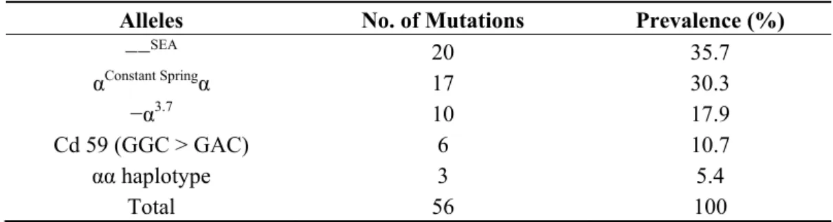
Table 1. Spectrum of α-thalassaemia gene defects identified in Malay patients by multiplex Gap- and ARMS-PCR.
Alleles No. of Mutations Prevalence (%)
−−SEA 20 35.7
αConstant Springα 17 30.3
−α3.7 10 17.9
Cd 59 (GGC > GAC) 6 10.7
αα haplotype 3 5.4
Total 56 100
Table 2. Prevalence of α-thalassaemia genotypes in Malay patients.
Genotypes No. of Patients n = 28 Prevalence (%)
−−SEA/αCSα 13 46.4
−α3.7/−−SEA 6 21.4
αCd 59α/αCSα 4 14.3
−α3.7/−α3.7 2 7.1
αCd 59α/αα 2 7.1
−−SEA/αα 1 3.6
Total 28 100
Figure 2. Multiplex ARMS PCR genotype analysis of α globin gene cluster on agarose gel electrophoresis. Lane 1 ladder; Lanes 2–4 are positive controls for Cd 59 (G > A), Cd 125 (T > C), and term Cd TAA > CAA (Hb CS) with their respective bands; Lane 5 non-template control; Lanes 6 and 7 Hb CS; and Lane 8 Cd 59 (G > A); 930 bp bands on Lanes 2–4 and 6–8 are internal control bands amplifying a segment of 3' UTR of LIS1 gene.
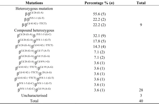
Table 3. Frequency of β-thalassaemia mutations identified in Malay patients using Multiplex-ARMS.
Mutations Percentage % (n) Total Heterozygous mutation
β/βCd 26 (G-A)
β/βIVS 1-1 (G-T) β/βCd 41/42 (−TTCT)
55.6 (5) 22.2 (2)
22.2 (2) 9
Compound heterozygous
βCd 26 (G-A)/βIVS 1-5 (G-C) βCd 26 (G-A)/βIVS 1-1 (G-T) βCd 26 (G-A)/βCd 41/42 (−TTCT)
βCd 26 (G-A)/βCd 17 (A-T) βCd 26 (G-A)/βCd 15 (G-A) βCd 26 (G-A)/βCd 8/9 (+G) βCd 41/42 (−TTCT)/βCd 19 (A-G)
βCd 41/42 (−TTCT)/β-28 (A-G) βCd 41/42 (−TTCT)/βIVS 1-1 (G-T)
βIVS 1-5 (G-C)/βIVS 1-1 (G-T) βIVS 1-5 (G-C)/βCd 19 (A-G)
32.1 (9) 17.8 (5) 14.3 (4) 7.1 (2) 7.1 (2) 3.6 (1) 3.6 (1) 3.6 (1) 3.6 (1) 3.6 (1)
3.6 (1) 28
Uncharacterised 3
Total 40
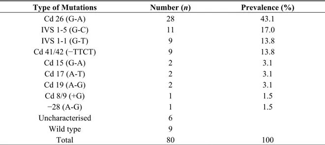
Table 3, nine patients were heterozygous for β-thalassaemia, 28 patients were compound heterozygous for β-thalassaemia and three patients remained uncharacterised by this procedure using the 20 different mutations described above. As summarised in Table 4, Cd 26 (G > A) and IVS 1-5 (G > C) were the two most common mutations observed and they accounted for a little over 60% of the patients. IVS 1-1 (G > T) and Cd 41/42 (−TTCT) had equal prevalence cumulatively accounting for a quarter of all the mutations observed. The remaining alleles, Cd 15 (G > A), Cd 17 (A > T), Cd 19 (A > G), Cd 8/9 (+G) and −28 (A > G) were rare alleles and were observed either on two chromosomes or on singletons. Capillary gel electrophoresis for the common β globin gene alleles is shown in Figure 3.
Table 4. Distribution of β-thalassaemia mutations identified in Malay patients.
Type of Mutations Number (n) Prevalence (%)
Cd 26 (G-A) 28 43.1
IVS 1-5 (G-C) 11 17.0
IVS 1-1 (G-T) 9 13.8
Cd 41/42 (−TTCT) 9 13.8
Cd 15 (G-A) 2 3.1
Cd 17 (A-T) 2 3.1
Cd 19 (A-G) 2 3.1
Cd 8/9 (+G) 1 1.5
−28 (A-G) 1 1.5
Uncharacterised 6
Wild type 9
Total 80 100
3. Discussion
The reported frequencies of α- and β-thalassaemia mutations and deletions vary considerably between ethnic groups and with geographic locations [7,8]. Consequently, this study was performed to characterise the α- and β-thalassaemia determinants in the mainland of Penang where the majority of the population is Malay. Samples were taken from Malay patients who were previously diagnosed with α- or β- thalassaemia by the HPLC method at the Hospital Seberang Jaya (HSJ), Penang. All the 68 patients had a mean corpuscular volume below 80 fL and mean corpuscular haemoglobin less than 27 pg.
The present study showed −−SEA to be the most prevalent α-thalassaemia determinant in Penang, with a prevalence of 35%, followed by αCSα (30%) and − α3.7 (18%). The inflated prevalence observed here clearly contradicts the nationwide prevalence reported for α-thalassaemia amongst Malays at large [9–11]. Several studies have shown plausible evidence that −−SEA cis gene deletion as the most common amongst Malaysian Chinese, while this deletion is not as common as the − α3.7 allele in Malays [3,9–11]. On average, throughout the population, the −−SEA deletion is characteristically four times more common in Malaysian Chinese than in Malays [3,10,11]. Moreover, Hb CS which was observed at a prevalence of 30% in this study, by contrast, has a nationwide frequency ranging between 1.1% and 3.6% (95%CI) in the ethnic Malays [11]. The most likely explanation for this remarkable overrepresentation of the α-thalassaemia genes in this study is the non-random nature of samples, representing symptomatic patients attending for regular follow-ups at Hospital Seberang Jaya. As is confirmed here, nearly 82% of these patients had HbH disease—out of which over 46% of patients had non-deletional HbH disease (−−SEA/αCSα); and 21% of patients had deletional HbH disease (−α3.7/−−SEA). Another 14% of subjects had genotype αCd 59α/αCSα presenting phenotypically as thalassaemia intermedia or HbH disease, presumably because haplotype αCd 59α has α0 phenotype [2]. Therefore, the contact clinical sampling method adopted and the small sample size used for characterising the α-thalassaemia gene are likely to be the two main reasons for the discordance in allelic prevalence reported here. Furthermore, since no clinical disturbances are generally associated with either α+-thalassaemia homozygotes (−α/−α) or α0 heterozygotes (−−/αα) these benign α-thalassaemia minor individuals are not well represented and neither are the alleles carried by them. The aforementioned genotypes are incidentally diagnosed during routine haematological examinations or as a result of family screening to diagnose a symptomatic thalassaemia in a relative. It is therefore necessary to obtain a simple random sample that is representative of the Malay subpopulation in the Penang district to consolidate these observations. The results were similar to a study from the Guangdong Province in Southern China. It was noted that the −−SEA deletion was the most common mutation detected (48.54%) followed by the −α3.7 deletions [12].
though considerable variation exists from one geographic location to another [13–16]. Characterisation of β-thalassaemia alleles on a contemporary multiethnic sample by Hassan et al. and George et al. revealed that HbE among the Malay ethnic group were 23% and 28.8%, respectively [9,13]. A similar study conducted a decade earlier by Tan et al. demonstrated the frequency of HbE in ethnic Malay was 19% [6].
It is observed here that the prevalence of IVS 1-5 (G > C) among Malays from Penang is three-fold lower than the most common HbE allele. By contrast, Hassan et al. reported an equal distribution of both alleles among Malays throughout Malaysia [13], while Tan et al. reported a posteriori elevation of the former by an approximate ratio of 2:1 [6]. This empirical increase in the prevalence of HbE among Malays from Penang could be attributed to its clinical severity as the mutation is known to be associated with the β0-thalassaemia phenotype, especially when the gene is in compound heterozygosity with a second β-thalassaemia allele (βE/β0 or βE/β+). In fact, all the 23 compound heterozygous patients with βE (Table 4) characterised here had a history of red cell transfusions at variable frequencies. This exemplified the clinical severity and disease heterogeneity of the disease when the Hb E allele is in transposition with another β mutation. Pathologically, this point mutation, commonly known as haemoglobin variant, simultaneously produces a structural and a quantitative defect in globin chain synthesis; hence, it is better known as thalassaemia haemoglobinopathy [17].
The third most common β-thalassaemia mutation characterised here is IVS I-1 (G > T). As with the other observations, this mutation is prominently only found in Malays, among whom it has reached a high heterozygous carrier frequency and has given rise to a β0 phenotype [12,14–16].
A relatively high prevalence of Cd 41/42 (−TTCT) is observed in Penang. This allele is prominent in the Malaysian-Chinese and less frequent amongst ethnic Malays in the general population [13,14]. This remarkable incongruity could be due to miscegenation between Malays and Chinese in Penang undergoing a demographic process that changes the allelic boundaries. This phenomenon could be acting on the distribution of many other thalassaemia alleles in this multiethnic subpopulation as Malaysians live side-by-side, breaking the norms of endogamy. Even a low level of gene flow over generations can lead to substantial changes, producing a heterogeneous pattern of allele frequencies.
Interestingly, Cd 15 (G-A) and Cd 8/9, which are usually the rare types of β-thalassaemia mutations among Malays, are also observed in this study. In addition, Cd 15 (G-A) was found in the Thai population, at a prevalence of 0.3% [18]. In Malaysia, Cd 8/9 has been found in the Kedayan population, while Cd 15 (G-A) was found in Malaysian Indian patients but at a very low frequency [13].
4. Experimental Section
4.1. Sample Collections
(JEPeM), Universiti Sains Malaysia and Research and Ethics Committee (MREC, Kuala Lumpur, Malaysia), Ministry of Health. Approximately 2 mL of whole blood was collected into EDTA vacutainer (Bio Lab, Shah Alam, Malaysia) from each patient after obtaining informed consent.
4.2. DNA Extraction
Genomic DNA was extracted from 200 µL whole blood using a commercially available DNA extraction kit, QIAamp DNA Blood Mini Kit (Qiagen GmbH, Hilden, Germany). The concentration of DNA was determined by NanoDrop Spectrophotometer ND-1000 (NanoDrop Technologies, Wilmington, DE, USA). The extraction was carried out according to the manufacturer’s instructions. To perform PCR in this study, a concentration of DNA around 50 ng/μL was required.
4.3. Molecular Analysis for α- and β-Thalassaemia Alleles
α-thalassaemia genotypes were tested using 13 different determinants commonly found in Malaysia and those reported from the Southeast Asia region. The panel included four double α globin gene deletions (−−SEA, −−MED, −−FIL, and −−THAI), three single gene deletions (−α3.7 rightward deletion, −α4.2 leftward deletion, and −α20.5) and six non-deletion α2 globin gene mutations namely initiation codon (ATG > A-G), codon 30 (ΔGAG), codon 35 (TCC > CCC), codon 59 (GGC > GAC), codon 125/Hb Quang Zhe (CTG > CCG) and termination codon/Hb Constant Spring (TAA > CAA). In-house optimised multiplexed Gap-PCR and multiplexed amplification refractory mutation system (M-ARMS) methods were used in parallel for gene deletion and mutation testing, respectively [11,19,20].
For β-thalassaemia genotyping, 20 different mutations were tested: 19 by M-ARMS and one by simple ARMS technique. Genotyping of the 19 β-thalassaemia determinants were carried out in five separate multiplexed PCR reactions based on the shared thermo-cycling conditions of respective allele-specific primers as described by Hassan et al. (2013) with slight modification [13]. In the first M-ARMS-A reaction, allele specific primers for four mutations IVS 1-5 (G > C), Cd 41/42 (−TTCT), Cd 17 (A > T) and Cd 26 (G > A) were multiplexed, while IVS 1-1 (G > T), Cd 8/9 (+G), −28 (A > G) and Cd 71/72 (+A) mutations were amplified in M-ARMS-B reaction. In the third M-ARMS-C reaction, alleles IVS 1-1 (G > A), Cd 43 (G > T), Cd 16 (−C), and Poly A (A > G) were multiplexed, while M-ARMS-D had allele-specific primers targeted at −88 (C > T), initiation codon (ATG > AGG), Cd 15 (G > A) and −29 (A > G) mutations. Finally, allele specific primers used in M-ARMS-E reaction were to screen mutations −86 (C > G), Cd 19 (A > G) and Cap + 1 (A > C) [13]. IVS 2-654 (C > T) was tested in a separate ARMS reaction.
4.4. Gel Electrophoresis
The PCR products for α-thalassaemia were analysed by using agarose gel electrophoresis. PCR products of multiplex gap PCR were run on a 1.5% agarose gel electrophoresis using 1× Tris-Borate-EDTA (TBE) buffer. Whereas, PCR products for α-thalassaemia from multiplex ARMS PCR were run on a 1.2% agarose gel.
Madrid, Spain) in 1X TBE buffer (Biobasic, Markham, ON, Canada). DNA band was visualized under transilluminator (Vilber Lourmat, Sud Marne-la-Vallée, France). QIAxel Advanced System (QIAGEN GmBH, Hilden, Germany) was used to analyse MARMS-A, MARMS-B, and MARMS-F which used automated capillary electrophoresis to separate the products based on their sizes and visualised on the interfaced computer system using its dedicated QIAxcel ScreenGel Software (QIAGEN GmBH, Hilden, Germany).
5. Conclusions
In this study, we have demonstrated the α- and β-thalassaemia gene frequency in the Penang population and further described its allelic distributions in the Malay populations. The heterogeneity of the α globin defects in the present study population is different from the other states in Malaysia as well as the presence of rare types of β-thalassaemia mutations. This may be attributed to a unique situation where there is a higher proportion of miscegenation among Malay with other ethnicities.
The results from this study will serve as a baseline for further investigations into the genetic defects. This information would provide health care professionals better awareness of the possible clinical spectrum of α- and β-thalassaemia in this geographic locality, thus facilitating better diagnostic and management of the disease. The prevention of severe α- and β-thalassaemia syndrome is very much dependent upon the availability of molecular characterisation, supported by adequate genetic counselling, and targeted public awareness programmes. The molecular diagnosis of α- and β-thalassaemia patients followed by genetic counselling of at risk couples should be made available as it is essential for the accurate diagnosis of both carrier and disease states.
In addition, such an effort would reduce the economic burden, and comprehensive and effective management of this problem in our country will be better achieved. Thus, a coordinated and interactive collaboration between the relevant stakeholders is necessary to ensure the effectiveness and success of the National Prevention and Control Programme for Thalassaemia in Malaysia.
Acknowledgments
The authors are grateful to the Advanced Medical and Dental Institute for providing AMDI Research Student Fund (CIPPT.1000), the Director of Hospital Seberang Jaya, Penang and to all staff at Molecular Genetic Laboratory, Haematology unit, Institute of Medical Research (IMR), Kuala Lumpur for their support and allowing us to conduct our study at their places. The authors would also like to thank all staff at the Regenerative Medicine Cluster, Advanced Medical and Dental Institute (AMDI), Universiti Sains Malaysia (USM) for their excellent help. The authors would also like to thank the Director General of Health, Ministry of Health, for his kind permission to conduct the molecular analyses at the Institute for Medical Research. (Research ID NMRR-12-1200-14169 and Research ID NMRR-12-1198-14167).
Conflicts of Interests
References
1. Hoffbrand, A.V.; Moss P.A.H.; Pettit, J.E. Essential Haematology, 5th ed.; Blackwell: Malden, MA, USA, 2006.
2. Harteveld, C.; Higgs, D. Alpha-thalassaemia. Orphanet J. Rare Dis. 2010, 1750–1172.
3. Rosnah, B.; Rosline, H.; Zaidah, A.W.; Haslina, M.N.N.; Marini, R.; Shafini, M.Y.; Nurul Ain, F.A. Detection of common deletional alpha-thalassemia spectrum by molecular technique in Kelantan, Northeastern Malaysia. ISRN Hematol. 2010, doi:10.5402/2012/462969. 4. Association of Clinical Registries, Malaysia Malaysian Thalassaemia Registry. Available online:
http://www.acrm.org.my/affiliatedDatabases_thalassamia.htm (accessed on 15 November 2012). 5. George, E. Beta-thalassemia major in Malaysia, an on-going public health problem. Med. J. Malays.
2001, 56, 397–400.
6. Tan, J.A.; George, E.; Tan, K.L.; Chow, T.; Tan, P.C.; Hassan, J.; Chia, P.; Subramanium, R.; Chandran, R.; Yap, S.F. Molecular defects in the beta-globin gene identified in different ethnic groups/populations during prenatal diagnosis for beta-thalassemia: A Malaysian experience. Clin. Exp. Med. 2004, 4, 142–147.
7. Department of Statistics Malaysia. Population. Malaysia: The Official Website of the Department of Statistics. Available online: http://www.statistics.gov.my/portal/Index.php?option=com_content& view=article&id=54%3Apopulation-updated-31072009&catid=35%3Akey-statistics&Itemid=53& lang=en (accessed on 13 November 2012).
8. Ministry of Health. Go For Thalassaemia Screening (2010). Available online: http://www.infosihat. gov.my/media/risalah/Ris_Thalasemia/Ris_Thalasemia_02_BI/Risalah%20BI.pdf (accessed on 13 November 2012).
9. George, E.; Teh, L.; Rosli, R.; Lai, M.; Tan, J. Beta Thalassaemia mutations in Malays: A simplified cost-effective strategy to identify the mutations. Malays. J. Med. Health Sci. 2013, 8, 1–8.
10. Wee, Y.C.; Tan, K.L.; Chow, T.W.; Yap, S.F.; Tan, J.A. Heterogeneity in alpah-thalassaemia interactions in Malays, Chinese and Indians in Malaysia. J. Obstet. Gynaecol. Res. 2005, 31, 540–546. 11. Ahmad, R.; Saleem, M.; Aloysious, N.S.; Yelumalai, P.; Mohamed, N.; Hassan, S. Distribution of alpha thalassaemia gene variants in diverse ethnic populations in Malaysia: Data from the Institute for Medical Research. Int. J. Mol. Sci. 2013, 14, 18599–18614.
12. Xu, X.; Zhou, Y.; Luo, G.; Al, E. The prevalence and spectrum of α- and β- thalassaemia in Guangdong province: Implications for the future health burden and population screening. J. Clin. Pathol. 2004, 57, 517–522.
13. Hassan, S.; Ahmad, R.; Zakaria, Z.; Zulkafli, Z.; Abdullah, W.Z. Detection of beta-globin gene mutations among beta-thalassaemia carriers and patients in malaysia: Application of multiplex amplification refractory mutation system-polymerase chain reaction. Malays. J. Med. Sci. 2013, 20, 13–20.
15. Thong, M.K.; Soo, T.L. The spectrum of beta-globin gene mutations in children with beta-thalassaemia major from Kota Kinabalu, Sabah, Malaysia. Singap. Med. J. 2005, 46, 340–343.
16. Abdullah, W.A.; Jamaluddin, N.B.; Kham, S.K.; Tan, J.A. The spectrum of beta-thalassemia mutations in Malays in Singapore and Kelantan. Southeast Asian J. Trop. Med. Public Health 1996, 27, 164–168.
17. Olivieri, N.F. Treament strategies for haemoglobin E beta thalassaemia. Blood Rev. 2012, 1, 28–30.
18. Winichagoon, P.; Fucharoen, S.; Wilairat, P.; Fukumaki, Y. Molecular mechanisms of thalassemia in southeast Asia. Southeast Asian J. Trop. Med. Public. Health 1995, 26 (Suppl. 1), 235–240.
19. Chong, S.S.; Boehm, C.D.; Higgs, D.R.; Cutting, G.R. Single-tube multiplex-PCR screen for common deletional determinants of alpha-thalassaemia. Blood 2000, 95, 360–362.
20. Eng, B.; Patterson, M.; Walker, L.; Chui, D.H.K.; Waye, J.S. Detection of severe nondeletional α-thalassemia mutations using a single-tube multiplex ARMS assay. Genet. Test. 2001, 5, 327–329.