O R I G I N A L A R T I C L E
Dental iron precipitates in patients with Type 2 diabetes
Miguel Angel Ortiz
‐
Arrambide
1|
Karla Isabel Juarez
‐
Ibarra
1|
Guadalupe Ismael Malagón
‐
Santiago
1|
Norma Cruz
‐
Fierro
1|
Myriam Angelica De La Garza
‐
Ramos
1,21
Facultad de Odontologia, Universidad Autonoma de Nuevo Leon, Monterrey, Mexico 2
Centro de Investigación y Desarrollo en Ciencias de la Salud (CIDICS), Universidad Autonoma de Nuevo Leon, Monterrey, Mexico
Correspondence
Myriam A. De la Garza‐Ramos, Facultad de Odontologia, Universidad Autonoma de Nuevo Leon, Calle Dr. Eduardo Aguirre Pequeño s/n, Colonia Mitras Centro, Monterrey 64460, Nuevo Leon, Mexico. Email: [email protected]
Abstract
Diabetes mellitus (DM) is a frequent worldwide disease. There are currently more
than 46 million people who suffer this disease in North America and the Caribbean.
The objective of this study was to determine if there is an association between DM
and the presence of iron precipitates (Fe
2+) in dental structure. The third molar was
extracted for reasons that merit extraction from 40 individuals with and without
DM to analyze dentin tissue. Horizontal and longitudinal slices of tooth samples were
made and later stained with 10% potassium cyanoferrate. The samples were observed
by optical microscope to identify basophilic elements. A nonparametric Spearman
cor-relation was performed to find an association between the quantitative (gender,
group, and dentinal tissue) and qualitative variables (gender). The Mann
–
Whitney U
test was used to find differences in the means of the nonparametric variables in
two different groups in relation to the
P
value (<0.05). Iron elements were found in
the predentin and circumpulpal dentin areas, and the results obtained showed a
sta-tistically significant difference between dentin tissue from patients with diabetes
and those without. Individuals with Type 2 DM are prone to present iron precipitates
in predentin and circumpulpal dentin tissue. Few iron elements were found in dental
organs of individuals without DM.
K E Y W O R D S
dentin, diabetes mellitus, iron overload, precipitation
1
|I N T R O D U C T I O N
Diabetes mellitus (DM) is a frequent worldwide disease. There are
cur-rently more than 46 million people who suffer this disease in North
America and the Caribbean, and these figures are increasing
(Interna-tional Diabetes Federation, 2017). Type 2 DM is a metabolic syndrome
that generally develops in adults. It is characterized by a hyperglycemic
state that depends or not on the administration of insulin and that
causes diverse consequences in the individuals who suffer the disease
(American Diabetes Association, 2014). Iron is a fundamental mineral
that promotes adequate cell maintenance and proliferation, and it also
participates in oxygenation processes in all cells of the body (Oexle,
Gnaiger, & Weiss, 1999). There are multiple benefits when there are
optimal levels of iron, but when the human body has an elevated
amount of iron, this is associated with a greater risk of glucose
metabo-lism disorders (Lee, Kim, & Kim, 2011) and Type 2 DM (Bao, Rong, Rong,
& Liu, 2012). One of the effects of iron is to generate reactive oxygen
species and nitrogen which can produce oxidative stress that releases
free radicals that induce cell damage especially in pancreatic beta cells
which are susceptible to oxidative stress (Asmat, Abad, & Ismail, 2016).
-This is an open access article under the terms of the Creative Commons Attribution License, which permits use, distribution and reproduction in any medium, provided the original work is properly cited.
©2018 The Authors. Clinical and Experimental Dental Research published by John Wiley & Sons Ltd. DOI: 10.1002/cre2.150
The human hemochromatosis protein controls the interaction
between the transferrin receptor and transferrin regulating the
absorp-tion of iron by the intestine. When it binds to the transferrin receptor, it
reduces its affinity for transferrin, and when it reaches the pancreas,
there is deposition of iron in the islets. This causes the production of
reactive oxygen species that produce β‐cell failure and insulin
resis-tance with this being a possible cause of DM (Simcox & McClain, 2013).
Dentin is a mineralized avascular tissue that forms the bulk of the
tooth. It is live tissue that produces the shape of the tooth. Dentin is
collagen that is mineralized after organization of the collagen matrix
in predentin. Predentin is unmineralized dentin that is 20‐to 30‐μm
wide and that is found adjacent to the pulp surface. It represents the
formation of dentin before its calcification and maturation (Tjäderhane
& Haapasalo, 2009).
There is an association between iron overload and Type 2 DM. It
has been found that moderate increases in iron are associated with
glucose and insulin elevations (Bao et al., 2012). The nonmineral
struc-ture of bone (osteoid) and dentin are similar; therefore, there is a
rela-tionship with iron deposits in dentin, specifically in predentin (Hess &
Villanueva, 1982). Potassium ferrocyanide is a histological stain that is
commonly used to detect the presence of iron in tissue samples
(Crudo, Erramouspe, Sueldo, & Arias, 2016).
The objective of this research is to determine if there is an
asso-ciation between DM and the presence of iron precipitates (Fe2+) in
dental structure.
2
|M E T H O D S
For this study, third molars from each subject were analyzed. The
teeth were extracted for diverse reasons in individuals over 18 years
and less than 40 years of age with and without Type 2 diabetes. Teeth
had to be healthy with Grade 1 caries with enamel restoration and
coronary integrity. Exclusion criteria were teeth with trauma, with
extensive restoration, without coronary integrity, and with external
or internal radicular resorption. Elimination criteria included teeth with
fractures during the study or any failure during the experimental
pro-cess. This study was previously approved by the Ethics Committee
with registration number SPSI 0106113—folio 00113.
A total of 40 upper and lower uni and multiradicular total or partially
erupted third molars were extracted and later placed in a Falcon tube with
artificial saliva and a drop of hypochlorite at 37°C. The teeth were divided
into two groups of 20 third molars each: experimental (A) and control (B).
They were later embedded in crystal resin, placing each specimen in a
heavy silicon mold to form cubes and later perform hard tissue slices.
Tooth slices were made from the upper portion of the clinical
crown in a coronal manner to the apical portion separating both parts
and obtaining 1‐mm thick flat slices; this was done with a water‐
cooled diamond disc with a Marathon III electric micromotor (Saeyang
Microtech Co., Shanghai, China) to obtain a highly polished surface.
A solution was prepared by mixing equal parts of 20%
hydrochlo-ric acid (Sigma‐Aldrich, Inc. St. Louis, MI, USA) and 10% potassium
fer-rocyanide (K4Fe(CN)6·3H2O; Sigma‐Aldrich, Inc.) diluted in distilled
water for 40 min obtaining half a liter of solution in a glass receptacle.
Each slice was placed individually in a screw‐top glass container.
Afterwards, the slices were washed in distilled water three times each
for 5 min removing the excess liquid of the slices. After staining, the
slices were placed on glass slides for observation under an optic
microscope (LSM510, Carl Zeiss Co. Ltd., Jena, Germany). Images of
the specimens were observed at a magnification of 4x/0.10/0.17,
10/0.25/0.17, and 40x/0.65/0.17, identifying the areas of tissue and
visually counting the number of iron precipitates and obtaining
photo-graphs in each magnification with a Canon EOS Rebel T5 camera
(Canon U.S.A., Inc. Huntington, NY, USA).
2.1
|Statistical analysis
The data obtained were captured in an Excel 2016 database with which
frequency tables of the variables capillary glucose/iron tissue dental
precipitates were compared with the remaining variables. A
nonpara-metric Spearman correlation test was applied to the results to find an
association between the original variables. The Mann–Whitney U test
was applied to find differences in the means of nonparametric variables
in the two groups with aP< 0.05 being significant.
3
|R E S U L T S
The results of the experimental and control group in the clinical crown
are shown in Table 1. No significant difference was found between the
experimental group and the control with regard to age (P= 0.295);
however, a significant difference was found in capillary glucose
(P < 0.001) and in the predentin and circumpulpal dentin tissue
(P< 0.001), individually and as a group.
The results of the variables of the experimental and control group
in dental tissue of the root are shown in Table 2. There was no
signif-icant difference between the experimental group and the control with
regard to age (P= 0.295). A significant difference was found in
capil-lary glucose (P< 0.001) and the respective tissue areas of the dental
root both individually and as a group (P< 0.001).
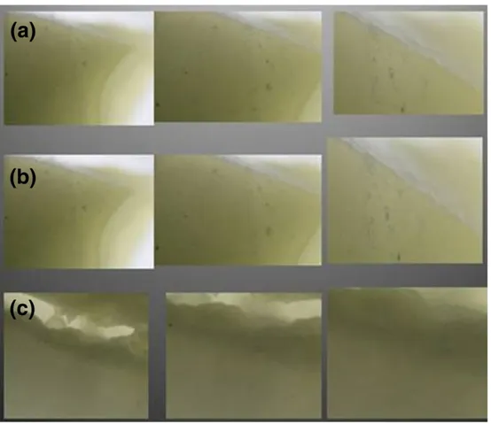
With regard to the presence of iron precipitates in the areas of
dental tissue both in the clinical crown and in the dental root, a
signif-icant amount was found in the areas of the predentin and circumpulpal
dentin, with this being less in the dentin layer (Figure 1a–c) in contrast
to the same areas in the control group where no precipitates were
seen (Figure 2a–c).
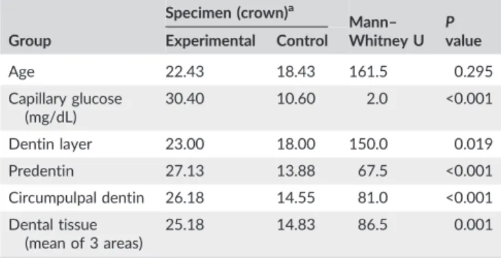
TABLE 1 Analysis of the experimental and control group with regard to iron precipitates found and observed in dental tissue of the clinical crown
Group
Specimen (crown)a
Mann– Whitney U
P value Experimental Control
Age 22.43 18.43 161.5 0.295
Capillary glucose (mg/dL)
30.40 10.60 2.0 <0.001
Dentin layer 23.00 18.00 150.0 0.019
Predentin 27.13 13.88 67.5 <0.001
Circumpulpal dentin 26.18 14.55 81.0 <0.001
Dental tissue (mean of 3 areas)
25.18 14.83 86.5 0.001
The iron necessary for the formation of dentin as well as for the
diverse systemic effects caused by DM is deposited in dental tissue
with this being greater in the predentin and circumpulpal dentin areas.
In relation to negative associations in the group variables, a figure of
−85% was obtained in the experimental group versus the control group in high capillary glucose levels.
4
|D I S C U S S I O N
It was possible to identify iron precipitates in dental tissue in
individ-uals with DM using 10% potassium hexacyanideferrate. Iron elements
were found in a greater concentration in circumpulpal dentin and in
predentin with a statistically significant difference between the area
of the dental root and the area of the clinical crown in comparison
with a control group of people without diabetes. This demonstrates
that the observation of iron precipitates in hard tissue is associated
with patients with Type 2 DM with high glucose levels.
Elevated iron stores have been associated with a risk of Type 2
diabetes. Higher serum ferritin levels were more prevalent in a South
Korean population with metabolic syndrome and DM in the 2008
Korean National Health and Nutrition Examination Survey (Lee et al.,
2011) demonstrating a positive association between elevated iron
stores (measured by serum ferritin levels) and the prevalence of
met-abolic syndrome and DM after adjustment for age, sex, educational
level, smoking, alcohol intake, and body mass index. This has also been
seen in patients with hereditary hemochromatosis and transfusional
iron overload as in beta thalassemia major and bone marrow
trans-plantation (Simcox & McClain, 2013). The pathogenesis of diabetes
has been associated with elevated iron deposits (Thomas, MacIsaac,
Tsalamandris, & Jerums, 2004).
Fernandez‐Cao et al. (2013) carried out an observational cohort
analysis of individuals without diabetes but with cardiovascular risk
followed for 1–8 years in the PREDIMED Trial. Of the initial sample,
12.2% developed diabetes with a median follow‐up of 4.8 ± 1.3 years.
These participants had a greater intake of total iron and heme iron in
the form of meat consumption. Red meat, especially processed meat,
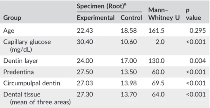
TABLE 2 Analysis of the experimental and control group with regard to iron precipitates found and observed in dental tissue of the root
Group
Specimen (Root)a
Mann– Whitney U
p value Experimental Control
Age 22.43 18.58 161.5 0.295
Capillary glucose (mg/dL)
30.40 10.60 2.0 <0.001
Dentin layer 24.00 17.00 130.0 0.004
Predentina 27.50 13.50 60.0 <0.001
Circumpulpal dentin 27.03 13.98 69.5 <0.001
Dental tissue
(mean of three areas)
27.30 13.70 64.0 <0.001
a
Data are presented as means.
has been implicated, in addition to Type 2 diabetes, in cancer,
Alzheimer's disease, and cardiovascular disease.
Elevated iron stores cause insulin resistance and possibly
defec-tive secretion of insulin. Patients with DM and elevated serum iron
concentrations have higher glucose levels (Misra, Bhatter, Kumar,
Gupta, & Khan, 2016).
Another problem with iron deposits in dentin is that they can act
as an intrinsic stain altering the color of teeth (Bonilla, Mantín,
Jiménez, & Llamas, 2007). The clinical complications of intrinsic stains
of dentin can compromise aesthetics and dentin adhesion capabilities.
Because intrinsic stains mainly affect aesthetics, the intensity of the
pigment must be identified. This alteration ranges from mild to severe.
Diagnosing the magnitude of the damage as well as the etiological
fac-tor will determine the appropriate treatment to achieve predictable
results (Haro‐Velastegui, 2012).
Because of the scarce information regarding diabetes in relation
to the hard tissues of dental organs, more research is needed in the
subject because these changes are not observed by simple clinical
inspection and often not even radiographically; however, the reports
in this study show that there are adverse processes that can only be
observed by microscope in a hard tissue specimen of an extracted
dental organ.
More research on this subject could be useful to prevent early
stage Type 2 DM because these precipitates are present in people
with diabetes and in people susceptible to diabetes. This finding could
be useful to identify susceptibility to DM and to confirm the disease
(Hess & Villanueva, 1982).
Based on the results in this study, the presence of iron
precipi-tates in dental tissue in the predentin and circumpulpal dentin layers
of the clinical crown, and the tooth root was statistically significant
in study subjects with Type 2 DM in which hard dental tissue slices
were stained to identify iron precipitates. It would be interesting to
see if this also correlates with serum ferritin levels; however, further
research is needed to clarify this point.
5
|C O N C L U S I O N S
DM affects some of the independent variables of the crown. Staining
with 10% potassium ferrocyanide identified iron precipitates in dental
tissue in individuals with Type 2 diabetes. Iron precipitates were found
in dental tissue with this being greater in individuals with high blood
glucose levels.
D A T A A V A I L A B I L I T Y
Data are available by contacting Dr. Miguel Angel Ortiz‐Arrambide,
email: [email protected].
C O M P E T I N G F I N A N C I A L I N T E R E S T S
No specific funding was received for this study.
C O N F L I C T O F I N T E R E S T
The authors declare that they have no conflicts of interest in this
study.
A U T H O R C O N T R I B U T I O N S
Miguel Angel Ortiz‐Arrambide—Project conception and design,
acqui-sition, analysis, and data interpretation, drafted the manuscript. Karla
Isabel Juarez‐Ibarra—Reviewed data acquisition and analysis, the
intel-lectual content, and the draft of the manuscript and approved the final
version of the manuscript. Guadalupe Ismael Malagón‐Santiago—
Reviewed data acquisition and analysis and provided statistical
sup-port. Norma Cruz‐Fierro—Reviewed data acquisition and analysis and
approved the final version of the manuscript. Myriam Angelica De La
Garza‐Ramos—Reviewed data acquisition and analysis, the intellectual
content, helped draft the manuscript, and approved the final version
of the manuscript.
O R C I D
Myriam Angelica De La Garza‐Ramos
https://orcid.org/0000-0003-0792-3695
R E F E R E N C E
American Diabetes Association (2014). Diagnosis and classification of dia-betes mellitus.Diabetes Care,37(Supplement 1), S81. https://doi.org/ 10.2337/dc14‐S081–S90.
Asmat, U., Abad, K., & Ismail, K. (2016). Diabetes mellitus and oxidative stress—A concise review. Saudi Pharmaceutical Journal, 24(5), 547–553. https://doi.org/10.1016/j.jsps.2015.03.013
Bao, W., Rong, Y., Rong, S., & Liu, L. (2012). Dietary iron intake, body iron stores, and the risk of type 2 diabetes: A systematic review and meta‐ analysis. BMC Medicine, 10(1), 119. https://doi.org/10.1186/1741‐ 7015‐10‐119
Bonilla, V., Mantín, J., Jiménez, A., & Llamas, R. (2007). Alteraciones del color de los dientes.Revista Europea de Odonto‐Estomatología, 1–12.
Crudo, C., Erramouspe, B., Sueldo, E., & Arias, M. (2016). Tinción de hierro medular. Coloración de Perls.Revista Argentina de Hematología,20(2), 243–246.
Fernandez‐Cao, J. C., Arija, V., Aranda, N., Bullo, M., Basora, J., Martínez‐ González, M. A.,…Salas‐Salvadó, J. (2013). Heme iron intake and risk of new‐onset diabetes in a Mediterranean population at high risk of cardiovascular disease: An observational cohort analysis.BMC Public Health,13(1), 1042. https://doi.org/10.1186/1471‐2458‐13‐1042
Haro‐Velastegui, S. C. (2012). Causas y tratamientos de la pigmentación dental por medios intrínsecos y extrínsecos. (Bachelor's), Universidad de Guayaquil, Guayaquil. Retrieved from http://repositorio.ug.edu.ec/ handle/redug/3573
Hess, J., & Villanueva, A. (1982). The accumulation of predentin iron: A possible relationship with diabetes mellitus.Journal of Histotechnology, 5(3), 119–122. https://doi.org/10.1179/his.1982.5.3.119
International Diabetes Federation. (2017). IDF Diabetes Atlas 8th edn. In. Retrieved from http://www.diabetesatlas.org
Lee, B.‐K., Kim, Y., & Kim, Y.‐I. (2011). Association of serum ferritin with metabolic syndrome and diabetes mellitus in the South Korean general population according to the Korean National Health and Nutrition Examination Survey 2008. Metabolism, Clinical and Experimental, 60(10), 1416–1424. https://doi.org/10.1016/j.metabol.2011.02.008
Misra, G., Bhatter, S. K., Kumar, A., Gupta, V., & Khan, M. Y. (2016). Iron profile and glycaemic control in patients with type 2 diabetes mellitus. Medical Science,4(4), 22.
Oexle, H., Gnaiger, E., & Weiss, G. (1999). Iron‐dependent changes in cel-lular energy metabolism: Influence on citric acid cycle and oxidative phosphorylation. Biochimica et Biophysica Acta (BBA)‐Bioenergetics, 1413(3), 99–107. https://doi.org/10.1016/S0005‐2728(99)00088‐2
Simcox, J. A., & McClain, D. A. (2013). Iron and diabetes risk.Cell Metabo-lism,17(3), 329–341. https://doi.org/10.1016/j.cmet.2013.02.007
Thomas, M. C., MacIsaac, R. J., Tsalamandris, C., & Jerums, G. (2004). Ele-vated iron indices in patients with diabetes.Diabetic Medicine,21(7), 798–802. https://doi.org/10.1111/j.1464‐5491.2004.01196.x
Tjäderhane, L., & Haapasalo, M. (2009). The dentin–pulp border: A dynamic interface between hard and soft tissues. Endodontic Topics, 20(1), 52–84. https://doi.org/10.1111/j.1601‐1546.2012.00266.x
How to cite this article: Ortiz‐Arrambide MA, Juarez‐Ibarra
KI, Malagón‐Santiago GI, Cruz‐Fierro N, De La Garza‐Ramos
MA. Dental iron precipitates in patients with Type 2 diabetes.
Clin Exp Dent Res. 2019;5:14–18. https://doi.org/10.1002/