P
Controversies and Limitations in the
Diagnosis of Chronic Obstructive
Pulmonary Disease
Alejandro Reyes-García, Luis Torre-Bouscoulet and Rogelio Pérez-Padilla*
Department of Research on Tobacco and COPD, Instituto Nacional de Enfermedades Respiratorias Ismael Cosío Villegas, Mexico City, Mexico
Received for publication: 07-06-2018 Approved for publication: 21-09-2018 doi: 10.24875/RIC.18002626
ABSTRACT
Chronic obstructive pulmonary disease (COPD) is a major cause of chronic morbidity and mortality worldwide. While the cut-off point to define airflow obstruction has been controversial, it is widely accepted that the spirometry test is vital, as well as performing it after using a bronchodilator. The 6-second spirometry and the forced expiratory volume in 1 second/forced ex-piratory volume in 6 seconds (FEV1/FEV6) have demonstrated validity for defining obstruction, and it would be advisable to incorporate them in the definitions of obstruction. Another relevant issue is that spirometry with borderline obstruction can vary over time, changing to above or below the cut-off point. Thus, surveillance should be considered over time, repeating the spirometry to have a greater certainty in the diagnosis. The objective of this article was to conduct an in-depth review of the controversies in the diagnosis of COPD. During the past years, COPD definition has been updated in different times; however, it is now considered more as a complex syndrome with systemic participation, requiring a multidimensional assessment, and not only a spirometry. (REV INVEST CLIN. 2019;71:28-35)
Key words: Chronic obstructive pulmonary disease. Case finding. Screening.
Corresponding author: *Rogelio Pérez-Padilla
Department of Research in Tobacco and COPD Instituto Nacional de Enfermedades Respiratorias Ismael Cosío Villegas (INER)
Calzada de Tlalpan, 4502 Col. Sección XVl, Del. Tlalpan C.P. 14080, Mexico City, Mexico E-mail: [email protected]
INTRODUCTION
Sixty years ago, the term “emphysema” in the Unit-ed States was equivalent to “chronic bronchitis” in Great Britain; to avoid confusion, the former was considered an anatomopathological diagnosis, while the latter was a clinical diagnosis. This underscores
the controversy that has since existed to define and diagnose the disease named chronic obstructive pul-monary disease (COPD). COPD is one of the main causes of morbidity and mortality in the world1.
Today, it is the third most important non-transmis-sible disease, representing 5.3% of all deaths world-wide2.
INFORMATION SEARCH
We searched for manuscripts published and indexed in PubMed under the following terms: “COPD” or
“COPD” and “diagnosis” and “definition” excluding “asthma” and “overlap syndrome,” considering review
articles and clinical practice guidelines published dur-ing the past 10 years. We obtained 97 articles rele-vant for the purpose.
Definition
According to the definition proposed by the Global Initiative for Chronic Obstructive Lung Disease (GOLD) in the Global Strategy for the Diagnosis, Man-agement, and Prevention of Chronic Obstructive Lung Disease, 2017 update, “COPD is a common,
prevent-able, and treatable disease that is characterized by persistent respiratory symptoms and airflow limita-tion that is due to airway and/or alveolar abnor-malities usually caused by significant exposure to noxious particles or gases1.”
COPD definition has been changed during the years. Today, it is accepted that COPD is not only a single clinical entity but also is considered a complex syn-drome, resulting from the chronic exposure to one or more noxious agents that are known and that gener-ate a different clinical course1. The definition of COPD
has included a functional component, centered origi-nally on the progressive and accelerated decline in lung function in individuals who smoke, and now on irrevers-ible airflow obstruction, as clearly airflow obstruction may occur by the hastened decline of forced expira-tory volume in 1 second (FEV1) or by abnormal growth and development of the lung3. At least in
epidemio-logical studies, individuals with irreversible airflow ob-struction without relevant exposures are included in the COPD category, which may give rise to confusion. In addition to functional abnormalities, individuals with COPD present varying degrees of emphysema and chronic bronchitis. This complex syndrome is charac-terized by inflammation not only of the lungs and air-ways but also systemic4, which leads to an increased
risk of comorbidity, functional deterioration, as well as limitations in performing daily life activities and de-crease in the health-related quality of life5.
Risk factors for COPD include a deficiency of natu-ral antiproteases (α1-antitrypsin or antiprotease);
exposure to tobacco, biomass, or industrial smoke; previous pulmonary infections; asthma; and abnormal pulmonary development caused by prenatal or early life events6. The majority of cases of COPD in the
developed world are related to tobacco consumption; thus, its importance should be emphasized as a defi-nite and preventable cause of the disease. However, about one-third of patients with COPD, or more cor-rectly with irreversible airflow obstruction, are indi-viduals who never smoked7.
COPD has been clinically defined by the presence of some cardinal symptoms that include dyspnea, cough, and sputum production6,8,9. GOLD’s Global Strategy
for the Diagnosis, Management, and Prevention of COPD, in its most recent 2017 update, mentions that the diagnosis of COPD should be considered in all individuals with dyspnea, chronic cough, or phlegm and/or exposure to any of the risk factors for the disease1. However, recent studies have demonstrated
that some subjects who are smokers experience symptoms similar to those observed in patients with COPD - they even exhibit episodes resembling a COPD exacerbation - but without airflow obstruction6. In
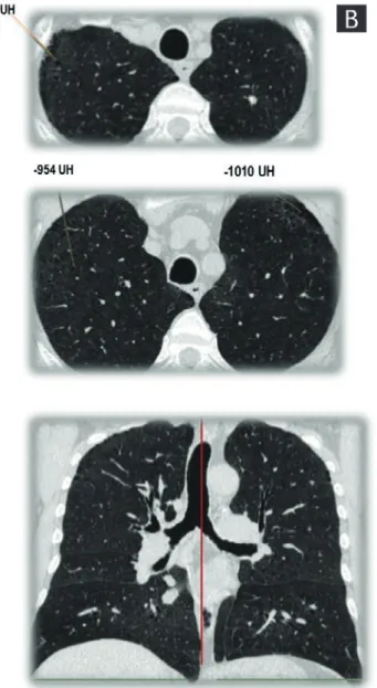
some of these patients, computed tomography (CT) of the thorax has demonstrated pulmonary emphy-sema; although these individuals do not fulfill the di-agnostic criteria of COPD proposed by GOLD, they have a pulmonary disease associated with smoking or with another exposure (Fig. 1)10. Due to the possible
progression to airflow limitation, this disorder was named “pre-COPD6,” which was recognized by GOLD
since 2006 as Stage 0. However, this questionable term disappeared in future revisions, since not all sub-jects with GOLD 0 (or pre-COPD) will develop airflow obstruction11.
We aimed for the identification of COPD endotypes, i.e., groups of similar patients according to a multidi-mensional evaluation of the disease, including several aspects: clinical, physiological, immunological, patho-logical, genetic, exposure, prognostic, and different response to treatment6,12.
SPIROMETRIC DEFINITION OF COPD
forced expiratory volume in 1 second and forced vital capacity (FEV1/FVC) quotient. There is controversy regarding the cut-off point of this quotient that should be used to define obstruction. GOLD defines obstruction as a FEV1/FVC < 0.70, whereas the most common proposed alternative is to use the lower limit of normal (LLN) (the lower 5th percentile),
de-rived from reference values that adjust for age and
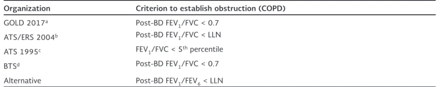
sex. Table 1 shows some definitions of airflow ob-struction used in the past.
The 6-s spirometry has been proposed as a simplified alternative to an FVC maneuver because it has the advantage of standardizing the measurement time of the denominator, since the FVC may be measured using good-quality tests at different times and give
different results13. The quotient of the FEV
1/FEV6 test
is nearly equivalent to the FEV1/FVC for COPD diag-nosis14,15; however, the former is more reproducible,
the required maneuver causes less fatigue, and it is possibly more specific than the FEV1/FVC. There are fewer available reference values for FEV6 and FEV1/ FEV6 in comparison with the current gold standards FVC and FEV1/FVC.
GOLD CRITERION VERSUS THE LLN
According to GOLD 2017, an FEV1/FVC quotient of < 0.70 after the bronchodilator test confirms the exis-tence of a persistent airflow limitation and identifies the presence of COPD in patients with compatible symptoms and risks1. This criterion has been
em-ployed in numerous clinical assays and is independent of reference values16. However, in healthy persons,
FEV1/FVC decreases with age, a situation not consid-ered by that criterion. The fixed cut-off point of 0.70 can cause errors in diagnosis at the extremes of life, resulting in underdiagnosis in young adults (false neg-atives) and overdiagnosis in older adults (false posi-tives that increase disproportionately with age)17-19.
Thus, the prevalence of COPD by that definition is higher than the one estimated by the statistical crite-rion of <LLN or less than the 5th percentile (20.1% vs.
14.7% in the PLATINO study)20. The high rate of false
positives in older adults may cause drug overprescrip-tion, adverse effects of medicaoverprescrip-tion, excessive use of resources confirming or ruling out the diagnosis, and disease labeling of healthy individuals. The FEV1/FVC
< LLN criterion to identify obstruction is more spe-cific, reducing the rate of false-positives; however, the LLN depends on the equation of reference used for the post-bronchodilator values that are being employed. In some populations, it will be necessary to develop spirometry reference values if those currently avail-able do not adequately fit the population18.
The criteria for defining obstruction should influence prognosis and not only be based on a statistical cri-terion. In this perspective, the cut-off point would be that which identifies an increased benefit or treat-ment and a deteriorated prognosis without a dispro-portionate rise in false-positives. The FEV1, per se, has demonstrated to be a strong prognostic indicator, and in groups with reduced FEV1 (such as GOLD stages 2-4), a worse prognosis would be expected. Patients with FEV1/FVC < 0.7 tend to have a lower FEV1 than those with an FEV1/FVC of > 0.7, and adults with FEV1/FVC <LLN an FEV1 lower than individuals with FEV1/FVC < 0.7.
PRE- OR POST-BRONCHODILATOR
SPIROMETRY?
The assessment of airflow obstruction should be done with the spirometry performed after the use of bronchodilators, to lower the contribution of asthma and other causes of reversible obstruction. In the PLA-TINO study, it was demonstrated that the bronchodi-lator test reduced by 35% (from 21.7% to 14%) the prevalence of COPD by using the FEV1/FVC % < 0.70
Table 1: Spirometric definitions of COPD
Organization Criterion to establish obstruction (COPD)
GOLD 2017a Post-BD FEV
1/FVC < 0.7
ATS/ERS 2004b Post-BD FEV1/FVC < LLN
ATS 1995c FEV1/FVC < 5th percentile
BTSd Post-BD FEV1/FVC < 0.7
Alternative Post-BD FEV1/FEV6 < LLN
FEV1: forced expiratory volume in 1 second; FVC: forced vital capacity, FEV6: forced expiratory volume in 6 seconds; LLN: lower limit of normal (5th percentile).
aGlobal initiative for chronic obstructive lung disease. Global strategy for the diagnosis, management, and prevention of chronic obstructive pulmonary disease. 2017.
bStandards for the diagnosis and treatment of patients with COPD: a summary of the ATS/ERS position paper. Eur Respir J. 2004;23(6):932-46. ATS; American Thoracic Society. ERS; European Respiratory Society.
criteria, while when using the FEV1/FVC < LLN crite-rion, the reduction was 37% (from 17.4% to 10.8%)20.
The latter provides more certainty in the clinical diag-nosis of COPD. Despite what has been discussed, the pre-bronchodilator spirometry is frequently used to assess bronchial obstruction in patients in whom COPD is suspected.
ONE OR MORE SPIROMETRY TESTS
FOR THE DIAGNOSIS OF COPD?
In daily clinical practice, the diagnosis of COPD is based on the results obtained in the initial spirometry. To date, there is no recommendation by GOLD guide-lines, to repeat the forced spirometry to increase con-sistency and certainty in the diagnosis. However, the FEV1 and the FVC, their ratio, and all tests, for that matter, vary over time even in healthy subjects. For example, the annual variability reported for the FEV1 and the FVC is of ± 15%. In up to 22% of subjects with a baseline spirometry showing obstruction, their tests normalized during the 1st year of follow-up and,
after 2 years, the percentage increased to 24-32%21.
These results demonstrate that a longitudinal spiro-metric evaluation could increase certainty in the diag-nosis, mainly in patients with borderline values inde-pendently of the reference values and diagnostic criteria employed.
CT SCANNING AND DIFFUSING
CAPACITY TO IDENTIFY EMPHYSEMA
AND ITS RELATION WITH COPD
Emphysema is defined as “an abnormal and
perma-nent dilation of the air spaces that occur distally to the terminal bronchioles and that is accompanied by the destruction of the interalveolar septa, without evidence of fibrosis10.” High-resolution CT (HRCT) of
the thorax and low-dose tomography of the thorax allow to identify areas of attenuation with <−950 UH, which are consistent with emphysema.
HRCT is the method of choice for diagnosing pulmo-nary emphysema in vivo22, due to greater spatial
res-olution compared with the conventional tomography of the thorax23. Quantitative analysis of lung density
measured by HRCT permits to evaluate the degree of extension of emphysema; however, this depends to a
great extent on the subjective and visual evaluation of the radiologist24. It is now possible to perform a
more objective evaluation using tools and software to carry out measurements such as the emphysema in-dex (EI), which defines the relation between the vol-ume of emphysema and total lung volvol-ume after a three-dimensional reconstruction. Other indicators include the pixel index (PI), defined as the percentage of pixels with an attenuation of <−900 UH, as well as the EI in expiration (EIex), PI in maximum expiration (PIex), and the pulmonary blood flow (BF)25,26. The
sensitivity of the EI in the HRCT is 0.80 (95% confi-dence interval [CI], 0.74-0.84), while when the tomo-graphic signs (EI, PIex, EIex, and BF) are combined, the sensitivity for detecting emphysema rises up to 0.87 (95% CI, 0.64-0.96)25. In comparison with inspiration,
CT measurements in expiration are tightly correlated with airflow obstruction; however, this exposes the patients to additional radiation26.
The use of low-dose CT (20-40 mA) with a slice thickness of 1.25 mm allows for the identification of pulmonary emphysema with accuracy, in addition to permitting the graduation of its extension and correlating it with the histopathological pattern (centrilobular or paraseptal). Likewise, it is possible to identify other typical findings in smoker patients, such as interstitial lung disease, bronchiectasis, and calcification in the coronary arteries or aorta. Low-dose CT can decrease the amount of global radia-tions for the quantitative evaluation of emphysema, without losing diagnostic value10,26. In a recent
study that included smokers with normal spirome-try, it was demonstrated that 75% of the partici-pants had emphysema detected in the low-dose CT of the thorax. Although the extension of the em-physema was mild, those findings were associated with a lower quality of life, low DLCO, a greater num-ber of exacerbations in the previous year, and a significant fall in oxygen saturation during the 6-minute walk test (6MWT)10. Despite the benefits
of the CT, no guidelines, to our knowledge, recom-mend its routine use, due to the exposure to radia-tion and to its considerable cost.
A single-breath carbon monoxide diffusing capacity (DLCO) and a spirometry below LLN values suggest the presence of emphysema27. In patients with pulmonary
and correlates with lower resting PaO2, as well as a greater requirement of supplementary oxygen, fewer meters in the 6MWT, and lower maximum exercise capacity28.
RESPIRATORY SYMPTOMS
AND COPD
Recently, the relevance of respiratory symptoms has been emphasized since they can predict a poor prog-nosis, an accelerated decrease in lung function, and exacerbations. Various symptom questionnaires have been developed, including COPD assessment test (CAT) and COPD questionnaire score29. The
CAT questionnaire is a sensitive, simple, and quick tool for assessing the respiratory status of COPD patients30. The new GOLD classification incorporates
the symptoms (CAT score > 10 and mMRC dyspnea score > 2) and the frequency or severity of exacer-bations for therapeutic decisions1. The combination
of a acting bronchodilator (LABA) and long-acting muscarinic antagonist (LAMA) is recommend-ed for patients classifirecommend-ed as GOLD Groups B or C with persistent symptoms after bronchodilator mono-therapy with LAMA or LABA31. This combination
re-duces symptoms and exacerbations compared with LAMA or LABA monotherapy32. However, the
clas-sification by symptoms is more unstable and less of a prognostic factor than that based on spirometry33
and, according to current GOLD classification1, can
give rise to the prescription of expensive LABA to persons with minimal obstruction, with borderline obstruction, or even to false positives, very frequent in mild COPD such as GOLD stage 1 in older indi-viduals. Respiratory symptoms (cough, phlegm, dys-pnea, and wheezing) may have a variety of causes and should be investigated as any other symptom before assuming that they are caused solely by COPD and that will respond to bronchodilators. In population-based studies, those symptoms are as-sociated with smoking, passive smoking, and expo-sure to occupational agents, as well as with asthma diagnosis, with spirometry abnormalities (obstruc-tive and restric(obstruc-tive), and with self-reported cardiac disease, but a long list of causes is known. The use of questionnaires applied by the clinician, compared to those self-answered by the patient, reduces the number of diagnostic evaluations necessary to iden-tify a COPD patient2,34.
MULTIDIMENSIONAL INDICATORS
IN COPD
Multidimensional indicators are increasingly used not only for the diagnosis of COPD but also for prognosis. These indexes have greater prognostic value in com-parison with the isolated spirometry measurement35.
The body mass index, airflow obstruction, dyspnea score, and exercise (BODE) index (acronym for BODE capacity) has demonstrated to be better than FEV1 for predicting the risk of death due to any cause and due to respiratory causes in patients with COPD36,
which is to be expected up to a certain point in that it incorporates multiple domains of the disease and is expected to vary less over time37,38.
DETECTION OF COPD IN THE
COMMUNITY AND PRIMARY CARE
In the general population, there is an enormous un-derdiagnosis of COPD that can rise to 90% when the spirometry definition is used39. The number of
spi-rometry tests conducted in primary care has increased very little, even in developed countries, and in some reports, in only 12.2% of patients with clinical symp-toms suggestive of COPD, a spirometry is performed to confirm the diagnosis40-42.
There is a lack of scientific evidence to define the best procedures for the timely detection of COPD, espe-cially in high-risk groups, i.e., “case finding.” Case find-ing is a strategy whereby resources are focused on individuals or groups suspected of being at risk for a specific disease, instead of considering the whole population. It implies the active and systematic search for persons at risk instead of waiting for the presenta-tion of the symptoms or signs of active disease. Ac-cumulated smoking (pack-years) is the most impor-tant risk factor for airflow obstruction; therefore, the presence of smoking (especially in older men) is a common requisite for selecting individuals for case finding, given that the higher the level of smoking, the prevalence of COPD will also rise in the selected group43,44. The best detection strategy will probably
vary according to the country, region, and character-istics of the population and of the health system34.
considered at high risk for the spirometry. An inter-mediate step with a simplified lung function test (peak flow or 6-s spirometry) can increase availability and reduce the number of spirometry tests, a strat-egy that is, especially, efficient if the objective is to identify moderate-to-severe obstruction39.
A program for the active search of COPD in its pre-clinical stage requires an important assignment of resources; thus, at present, it is considered that, in asymptomatic never smokers or in individuals unex-posed to other noxious factors, a screening spirom-etry is not recommended. Spiromspirom-etry should be performed preferably on symptomatic patients, older than 40 years of age, with risk factors such as smoking, especially if they smoked > 10 (or 20) pack-years or had other exposure risks such as substantial exposure to biomass smoke or occupa-tional dusts or smokes. In this scenario, up to one in five subjects will have COPD, a number that increas-es to one of every three subjects in those of higher age and with greater exposure to tobacco. With fewer symptoms, age, or exposures, the number of spirometries performed to identify one individual with airflow obstruction increases progressively and can be cost-ineffective.
In Latin America, the PLATINO study (Proyecto Lati-noamericano de Investigación de la Enfermedad Pul-monar Obstructiva, Latin-American Project of Inves-tigation in Obstructive Pulmonary Disease) showed a prevalence of COPD of 14.3% with the GOLD cri-terion (FEV1/FVC < 0.70), of whom nearly 90% had no medical diagnosis, that is, 90% of patients with COPD did not know that they had the disease33,45.
The main factors for an underdiagnosis of COPD were a younger age, mild obstruction, fewer respira-tory symptoms, and importantly, the lack of a spi-rometry test42.
In the PUMA study (prevalence study and regular practice, diagnosis, and treatment, among general practitioners in populations at risk of COPD in Latin America), at-risk subjects were included if they were ≥ 40 years old, current or ex-smokers (≥ 10 pack-years), and/or with exposure to biomass smoke (wood or coal, for cooking or heating; exposure ≥ 100 h/year). The COPD prevalence in this study was 20.1% and 14.7% using post-BD FEV1/FVC < 0.70 and LLN definitions, respectively43.
CONCLUSION
The current definition of COPD includes a post-bron-chodilator spirometry in subjects with exposures and risk factors, although the cut-off point to define ob-struction varies, generating definitions with more or less specificity. While the present-day guidelines rec-ommend a single spirometry test, the variability of the latter, particularly in borderline tests, requires an ob-servation over time and the performance of repeated tests. The FEV1/FEV6 index is more reliable than the FEV1/FVC, specifically when groups with spirometries with a different expiratory time are compared.
REFERENCES
1. Vogelmeier CF, Criner GJ, Martinez FJ, et al. Global strategy for the diagnosis, management, and prevention of chronic obstruc-tive lung disease 2017 report. Gold execuobstruc-tive summary. Am J Respir Crit Care Med. 2017;195:557-82.
2. Haroon S, Jordan R, Takwoingi Y, Adab P. Diagnostic accuracy of screening tests for COPD: a systematic review and meta-analysis. BMJ Open. 2015;5:e008133.
3. Lange P, Celli B, Agustí A, et al. Lung-function trajectories lead-ing to chronic obstructive pulmonary disease. N Engl J Med. 2015;373:111-22.
4. Eagan TM, Ueland T, Wagner PD, et al. Systemic inflammatory markers in COPD: results from the Bergen COPD cohort study. Eur Respir J. 2010;35:540-8.
5. Agustí A, Edwards LD, Rennard SI, et al. Persistent systemic in-flammation is associated with poor clinical outcomes in COPD: a novel phenotype. PLoS One. 2012;7:e37483.
6. Celli BR, Agustí A. COPD: time to improve its taxonomy? ERJ Open Res. 2018;4:00132-2017.
7. Pérez-Padilla R, Fernández R, López Varela MV, et al. Airflow obstruction in never smokers in five Latin American cities: the PLATINO study. Arch Med Res. 2012;43:159-65.
8. Woodruff PG, Barr RG, Bleecker E, et al. Clinical significance of symptoms in smokers with preserved pulmonary function. N Engl J Med. 2016;374:1811-21.
9. Rodriguez-Roisin R, Han MK, Vestbo J, Wedzicha JA, Woodruff PG, Martinez FJ. Chronic respiratory symptoms with normal spi-rometry. A reliable clinical entity? Am J Respir Crit Care Med. 2017;195:17-22.
10. Alcaide AB, Sanchez-Salcedo P, Bastarrika G, et al. Clinical fea-tures of smokers with radiological emphysema but without air-way limitation. CHEST. 2017;151:358-65.
11. Vestbo J, Lange P. Can GOLD stage 0 provide information of prognostic value in chronic obstructive pulmonary disease? Am J Respir Crit Care Med. 2002;166:329-32.
12. Loscalzo J, Barabasi AL. Systems biology and the future of medicine. Wiley Interdiscip Rev Syst Biol Med. 2011;3:619-27. 13. Pérez-Padilla R, Wehrmeister FC, Celli BR, et al. Reliability of
FEV1/FEV6 to diagnose airflow obstruction compared with FEV1/FVC: the PLATINO longitudinal study. PLoS One. 2013; 8:e67960.
14. Jing J, Huang T, Cui W, Xu F, Shen H. Should FEV1/FEV6 replace FEV1/FVC ratio to detect airway obstruction?: a metaanalysis. Chest. 2009;135:991-8.
15. Swanney MP, Jensen RL, Crichton DA, Beckert LE, Cardno LA, Crapo RO. FEV6 is an acceptable surrogate for FVC in the spiro-metric diagnosis of airway obstruction and restriction. Am J Respir Crit Care Med. 2000;162:917-9.
16. Bhatt SP. Diagnosis of chronic obstructive pulmonary disease: breathing new life into an old debate. Ann Am Thorac Soc. 2018;15:163-5.
18. Güder G, Brenner S, Angermann CE, et al. GOLD or lower limit of normal definition? A comparison with expert-based diagnosis of chronic obstructive pulmonary disease in a prospective co-hort-study. Respir Res. 2012;13:13.
19. Wollmer P, Engström G. Fixed ratio or lower limit of normal as cut-off value for FEV1/VC: an outcome study. Respir Med. 2013;107:1460-2.
20. Pérez-Padilla R, Hallal PC, Vázquez-García JC, et al. Impact of bronchodilator use on the prevalence of COPD in population-based samples. COPD. 2007;4:113-20.
21. Pérez-Padilla R, Wehrmeister FC, Montes de Oca M, et al. Insta-bility in the COPD diagnosis upon repeat testing vary with the definition of COPD. PLoS One. 2015;10:e0121832.
22. Choromańska A, Macura KJ. Role of computed tomography in quantitative assessment of emphysema. Pol J Radiol. 2012; 77:28-36.
23. Milne S, King GG. Advanced imaging in COPD: insights into pul-monary pathophysiology. J Thorac Dis. 2014;6:1570-85. 24. Lee YK, Oh YM, Lee JH, et al. Quantitative assessment of
em-physema, air trapping, and airway thickening on computed to-mography. Lung. 2008;186:157-65.
25. Li J, Zhang H, Bai Y, et al. Diagnostic value of computed tomog-raphy in chronic obstructive pulmonary disease: a systematic review and meta-analysis. COPD J Chronic Obstruct Pulm Dis. 2012;9:563-70.
26. Xie X, Jong PA de, Oudkerk M, et al. Morphological measure-ments in computed tomography correlate with airflow obstruc-tion in chronic obstructive pulmonary disease: systematic re-view and meta-analysis. Eur Radiol. 2012;22:2085-93. 27. Cotton DJ, Soparkar GR, Graham BL. Diffusing capacity in the
clinical assessment of chronic airflow limitation. Med Clin. 1996; 80:549-64.
28. Mohsenifar Z, Lee SM, Diaz P, et al. Single-breath diffusing ca-pacity of the lung for carbon monoxide: a predictor of PaO2, maximum work rate, and walking distance in patients with em-physema. Chest. 2003;123:1394-400.
29. Sundh J, Janson C, Lisspers K, Montgomery S, Ställberg B. Clini-cal COPD questionnaire score (CCQ) and mortality. Int J Chron Obstruct Pulmon Dis. 2012;7:833-42.
30. Negro RW, Bonadiman L, Turco P. Sensitivity of the COPD as-sessment test (CAT questionnaire) investigated in a population of 681 consecutive patients referring to a lung clinic: the first Italian specific study. Multidiscip Respir Med. 2014;9:15. 31. Anzueto A, Miravitlles M. Considerations for the correct
diag-nosis of COPD and its management with bronchodilators. Chest. 2018;154:242-8.
32. Thomas M, Halpin DM, Miravitlles M. When is dual bronchodila-tion indicated in COPD? Int J Chron Obstruct Pulmon Dis. 2017; 12:2291-305.
33. Menezes AM, Pérez-Padilla R, Jardim JR, et al. Chronic obstruc-tive pulmonary disease in five Latin American cities (the PLA-TINO study): a prevalence study. Lancet. 2005;366:1875-81. 34. Haroon SM, Jordan RE, O’Beirne-Elliman J, Adab P. Effectiveness
of case finding strategies for COPD in primary care: a system-atic review and meta-analysis. NPJ Prim Care Respir Med. 2015; 25:15056.
35. Agustí A, Celli B. Natural history of COPD: gaps and opportuni-ties. ERJ Open Res. 2017;3:00117-2017.
36. Celli BR, Cote CG, Marin JM, et al. The body-mass index, airflow obstruction, dyspnea, and exercise capacity index in chronic obstructive pulmonary disease. N Engl J Med. 2004;350: 1005-12.
37. Puhan MA, Garcia-Aymerich J, Frey M, et al. Expansion of the prognostic assessment of patients with chronic obstructive pul-monary disease: the updated BODE index and the ADO index. Lancet. 2009;374:704-11.
38. Casanova C, Aguirre-Jaíme A, Torres JP de, et al. Longitudinal assessment in COPD patients: multidimensional variability and outcomes. Eur Respir J. 2014;43:745-53.
39. Pérez-Padilla R, Thirion-Romero I, Guzmán N. Underdiagnosis of chronic obstructive pulmonary disease: should smokers be of-fered routine spirometry tests? Expert Rev Respir Med. 2018; 12:83-5.
40. Luize AP, Menezes AM, Pérez-Padilla R, et al. Assessment of five different guideline indication criteria for spirometry, including modified GOLD criteria, in order to detect COPD: data from 5,315 subjects in the PLATINO study. NPJ Prim Care Respir Med. 2014;24:14075.
41. Moreira GL, Gazzotti MR, Manzano BM, et al. Incidence of chron-ic obstructive pulmonary disease based on three spirometrchron-ic diagnostic criteria in Sao Paulo, Brazil: a nine-year follow-up since the PLATINO prevalence study. Sao Paulo Med J. 2015; 133:245-51.
42. Herrera AC, Montes de Oca M, López Varela MV, et al. COPD under diagnosis and misdiagnosis in a high-risk primary care population in four Latin American countries. A key to ehance disease diagnosis: the PUMA study. PLoS One. 2016;11: e0152266.
43. López Varela MV, Montes de Oca M, Rey A, et al. Development of a simple screening tool for opportunistic COPD case finding in primary care in Latin America: the PUMA study. Respirology. 2016;21:1227-34.
44. Dirven JA, Tange HJ, Muris JW, van Haaren KM, Vink G, van Schayck OC. Early detection of COPD in general practice: imple-mentation, workload and socioeconomic status. A mixed-meth-ods observational study. Prim Care Respir J. 2013;22:338-43. 45. Tálamo C, de Oca MM, Halbert R, et al. Diagnostic labeling of