Impact of Sustained Viral Response in the
Evolution of Minimal Hepatic Encephalopathy:
a Prospective Pilot Study
Marta Hernández-Conde, Carlos Fernández-Carrillo, Elba Llop, Christie Perelló, Marta López-Gómez, José L. Martínez-Porras, Natalia Fernández-Puga, José L. Calleja
Department of Gastroenterology and Hepatology, Hospital Universitario Puerta de Hierro Majadahonda, Majadahonda, Madrid, Spain. January-February, Vol. 18 No. 1, 2019: 203-208
The Official Journal of the Mexican Association of Hepatology, the Latin-American Association for Study of the Liver and
the Canadian Association for the Study of the Liver
Manuscript received: Manuscript received:Manuscript received:
Manuscript received:Manuscript received: Fabruary 09, 2018. Manuscript accepted:Manuscript accepted:Manuscript accepted:Manuscript accepted:Manuscript accepted: July 31, 2018.
DOI:10.5604/01.3001.0012.7914 A B S T R A C T A B S T R A C T A B S T R A C T A B S T R A C T A B S T R A C T
Introduction and aims. Introduction and aims.Introduction and aims. Introduction and aims.
Introduction and aims. To determine the prevalence of minimal hepatic encephalopathy (MHE) in patients with liver cirrhosis (LC) due to hepatitis C virus (HCV) infection and to evaluate the impact of sustained viral response (SVR) on MHE. MaterialMaterialMaterialMaterialMaterial and
and and and
and methods.methods.methods.methods.methods. We performed a prospective study using MHE screening and follow-up on patients with HCV and LC. The patients were evaluated at the beginning of treatment and 24 weeks after treatment. Results.Results.Results.Results.Results. 64 patients were included. 51.6% were male, the median age was 62years, Child-Pugh classification A/B/C 93.8%/4.7%/1.6% and median MELD was 8.3. Prior hydropic decom-pensation was present in 11 patients. Median values of liver stiffness, as measured by transient elastography (TE) were 22.8 KPa. Indirect signs of portal hypertension (PH) were present in 53.1% of patients, with a mean of 11.9 mmHg among the ones with a measurement of the hepatic venous pressure gradient. The prevalence of MHE before treatment was 26.6%. After treatment, 98.4% of patients achieved SVR. The presence of MHE at 24weeks post-treatment had an statistically significant association with the presence of pre-treatment MHE (80% vs. 21.6%; p < 0.01), higher MELD scores at 24-weeks post-treatment (9.8 vs. 8; p = 0.02), higher Child-Pugh scores at 24-weeks post-treatment (p = 0.04), higher baseline INR levels (1.4 vs. 1.1; p < 0.001) and with the presence of indirect signs of PH (100% vs. 47.1%; p = 0.02). During follow-up, those patients without MHE at 24weeks post-treat-ment had a higher probability of experiencing an improvepost-treat-ment in post-treatpost-treat-ment TE (80.9% vs. 40%, p = 0.04). Conclusion.Conclusion.Conclusion.Conclusion.Conclusion. We found that SVR may lead to MHE resolution in a considerable proportion of patients, which has potential implications for disease prognosis.
Key words. Key words.Key words. Key words.
Key words. HCV. MHE. Direct-acting antivirals. Critical flicker frequency.
INTRODUCTION
Minimal hepatic encephalopathy (MHE) is primarily characterized by a subtle impairment of neuro-cognitive abilities which is not readily detectible via standard mental status testing or neurological examination.1 MHE is the earliest stage of hepatic encephalopathy (HE) and is a clin-ically relevant condition that affects the quality of life and job performance of patients with liver cirrosis.2
The actual prevalence of MHE is unknown and varies among different studies depending on the criteria used for diagnosis and the study populations. In cirrhotic patients with good liver function (Child-Pugh class A), MHE prevalence is low (< 15%), while in those with decom-pensated or advanced cirrhosis (Child-Pugh class B or C),
MHE may affect half of the patients.3 MHE is not restrict-ed to liver cirrhosis (LC); patients with other liver-relat-ed conditions such as portal vein thrombosis or portosystemic shunt can also present with MHE. It is also notable that MHE appears to be more frequent in patients with hepatitis C virus (HCV) infection, irrespectively of the stage of liver fibrosis. HCV-positive patients display abnormalities in verbal learning, attention span, executive function and memory when they are evaluated with suita-ble neuropsychological tests.4
standard’ assessment. The 2 most widely used tests are the critical flicker frequency (CFF) and the psychometric he-patic encephalopathy score (PHES) test battery, along with its variants.5
The PHES is a selection of 5 psychometric test batter-ies copyrighted and validated in Italian, German and Span-ish patient cohorts. PHES assesses attention span, motor speed and accuracy, visual orientation and visual-spatial construction.1 PHES has been shown to be influenced by age, education level and cultural issues. In addition, per-forming this battery can be time consuming and prone to bias from disturbance, mood and interaction with the test-er.
The CFF has the advantage of not being dependent on age, gender, cultural or educational factors and not show-ing a learnshow-ing effect.6 However, the CFF depends signifi-cantly on the experimental setting (the color and luminance of the stimuli, the visual angle, etc.). CFF showed a moderate pooled sensitivity of 61% yet a reason-able specificity of 79%.7
Several studies have shown quick improvement in liver function and other parameters in cirrhotic patients treated with anti-HCV direct-acting antivirals (DAA), which are molecules targeting specific nonstructural proteins of the virus, allowing interferon-free treatment.8-13 To date, pa-tients with HCV infection and papa-tients with LC have been reported as more susceptible to MHE, although it re-mains unknown whether the natural course of MHE may be altered after interferon-free DAA treatment. Therefore, our objective was to prospectively explore the potential role of HCV clearance in the course of MHE among pa-tients with LC and HCV infection after DAA treatment.
MATERIAL AND METHODS
The inclusion criteria for this study were i) LC and ii) HCV infection with an indication for DAA treatment (par-itaprevir/ritonavir+ ombitasvir + dasabuvir, ledipasvir + sofosbuvir, daclatasvir + sofosbuvir and elbasvir/grazo-previr). A total of 181 consecutive outpatients with LC due to HCV infection were treated with DAA from May 2015 until March 2017 in our tertiary care hospital. Of these patients, 64 met inclusion criteria and were selected for our study before initiating treatment. All of them had a baseline evaluation immediately before starting treatment. Fifty-six of them were prospectively followed-up and evaluated at two time points; at baseline and at 24 weeks after treatment. LC was diagnosed on a clinical basis using semiology, laboratory parameters, ultrasound-Doppler as-sessments and liver stiffness measurements by TE using FibroScan® (Echosens, Paris, France) (cut-off in 14Kpa). Liver function was assessed using the model for end-stage liver disease (MELD) and Child-Pugh scores. All patients
were offered hepatic venous pressure gradient (HVPG) measurements, but only 27 patients accepted. HVPG ≥ 10 mm Hg was defined as clinically significant portal hyper-tension (CSPH). HCV infection was diagnosed by the presence of serum anti-HCV antibodies and detectable HCV RNA. Sustained viral response (SVR) was defined as undetectable HCV RNA 24 weeks after the end of antivi-ral treatment and was analyzed by intention to treat. The exclusion criteria were overt HE, acute gastrointestinal hemorrhage, hepatorenal syndrome or spontaneous bacte-rial peritonitis in the 7 days prior to inclusion, significant non-hepatic diseases such as decompensated heart, respi-ratory or renal failure, decompensated or poorly control-led diabetes mellitus, and anamnestic neurological diseases such as Alzheimer’s disease or Parkinson’s dis-ease. Likewise, unwillingness to comply with study re-quirements, obvious alcohol abuse, ophthalmologic disorders and red-green visual blindness were exclusion criteria. Patients on medication including psychoactive drugs such as antidepressants or sedatives were also ex-cluded.
The study protocol was approved by the Ethics Com-mittee of the Hospital Universitario Puerta de Hierro-Majadahonda (PI-48-16. 2016) and written informed consent was obtained from all the patients prior to inclu-sion.
The PHES is comprised of 5 paper-pencil tests (Number Connection Tests A and B, the Digit Symbol coding Test, the Serial Dotting Test and the Line Draw-ing Test). The Serial DottDraw-ing Test consists of 10 rows of 10 circles, and the subject is timed on how quickly he or she can place a dot in the center of each circle. The Line Drawing Test requires the subject to draw a continuous line between two parallel (winding) lines, and scores in-clude completion time and errors. The on-line software developed by the Spanish Network for Hepatic Encepha-lopathy was used (available at http://www.redeh.org), and its results were adjusted for age and educational level.14 The CFF was measured using a HEPAtonorm analyzer (Accelab GmbH, Kusterdingen, Germany). This test measures the frequency at which the patient perceives that a fused light becomes a flickering light. The device causes a stepwise decrease in frequency from 60 to 25Hz. After a brief instruction and training period, the flicker frequen-cies were measured 10 times and the mean value and stand-ard deviation (SD) were calculated. CFF was measured in a quiet, semi-darkened room to avoid interferences. Both tests were performed by the same investigator in a quiet room. Patients were classified as having MHE when the PHES score was below -4 points or the CFF score was below the cut-off value (39Hz).15,16
Con-tinuous variables were displayed as means and SD, where-as discrete variables were expressed where-as percentages. Com-parisons between groups were made using the Mann-Whitney U test, the Student’s t test, the χ2 test or the Fisher’s exact test (when expected values in any of the cells of a contingency table were < 5) for categorical data. Correlations between psychometric tests and CFF were calculated by using Spearman-Rho rank correlation. P-val-ues < 0.05 were considered as statistically significant.
RESULTS
Sixty-four patients were included in this study (patient characteristics in table 1).
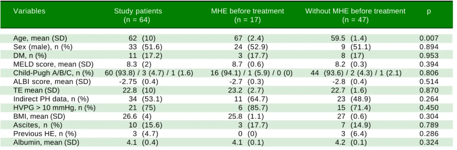
A slight majority were male (33 patients, 51.6%) and the mean age was 62 years (SD 10). The distribution of Child-Pugh scores was as follows: 60 patients were class A (93.8%), 3 patients were class B (4.7%) and 1 patient was class C (1.6%). The median MELD score was 8.3 (SD 2). Previous hepatic decompensation (ascites) was present in 11 patients (17.2%). Three patients (4.7%) had a previous episode of HE grade I almost one year prior to initiation of antiviral treatment, at the time of assessments asympto-matic. The median value of liver stiffness was 22.8 KPa (SD 10). Thirty-four patients (53.1%) had indirect signs of portal hypertension, e.g., platelet count < 100,000 103/μL, splenomegaly by ultrasound or esophageal varices in gas-troscopy. Among the 27 patients for whom measurement of the HVPG was available, the mean value was 11.9 mmHg (SD 3.7) and 21 of them had CSPH.
MHE was diagnosed in 17 patients (26.6 %) before treatment and its frequency varied significantly according to age (p < 0.01). No association was found between the presence of MHE and TE or the value of HVPG (Table 1). After treatment, 62 patients (96.9%) achieved SVR (pa-tient characteristics are included in table 2). MHE at 24
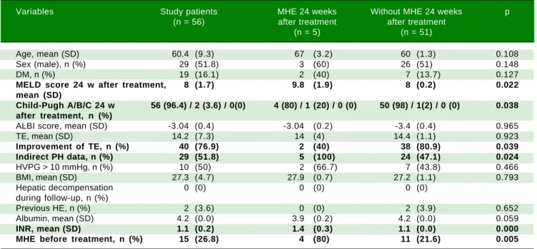
weeks post-treatment was evaluated in a total of 56 tients (87.5%). This assessment was not possible in 8 pa-tients (12.5%) due to the following reasons: 2 papa-tients (3.1%) died during follow-up (one patient died due to hepatocellular carcinoma 20 weeks after treatment; the other patient died from non-liver disease during treat-ment), 4 patients (6.3%) were lost to follow-up and 2 pa-tients (3.1%) were treated with antidepressants. Of the patients who died, only one of them had MHE before treatment.
At 24 weeks post-treatment, MHE was diagnosed in 5 patients (8.9%), 4 of which had MHE before treatment. The presence of MHE at 24 weeks post-treatment had a statistically significant association with the presence of pre-treatment MHE (80% vs. 21.6%; p<0.01), higher MELD scores at 24-weeks post-treatment (9.8 vs. 8, p = 0.02), higher Child-Pugh scores at 24-weeks post-treat-ment (5 score vs. 5.6 score, p = 0.004), higher baseline INR levels (1.4 vs. 1.1; p < 0.001) and with the presence of indirect signs of PH (100% vs. 47.1%; p = 0.02). In addi-tion, those patients without MHE at 24 weeks post-treat-ment had a higher probability of experiencing an improvement in post-treatment TE (80.9% vs. 40%, p = 0.04) (Table 2).
Fifteen of the 17 patients with pre-treatment MHE were evaluated at 24 weeks post-treatment. Of them, 11 pa-tients experienced MHE resolution (73.3%; p = 0.01) (Ta-ble 3).
The patients with MHE resolution after treatment, in comparison with those who did not, had higher baseline albumin levels (4.2 vs. 3.8 g/dL, p < 0.01), lower baseline INR levels (1.1 vs. 1.5; p < 0.05) and lower ALBI score (-2.9 vs. -2.5; p < 0.01). Although the baseline and 24-weeks post-treatment MELD scores were lower in patients with MHE resolution, this difference was not statistically sig-nificant (7.9 vs. 10.8; p = 0.06 and 8 vs. 10.3; p = 0.1).
De-Table 1. Baseline characteristics of the study patients (n = 64).
Variables Study patients MHE before treatment Without MHE before treatment p
(n = 64) (n = 17) (n = 47)
Age, mean (SD) 62 (10) 67 (2.4) 59.5 (1.4) 0.007
Sex (male), n (%) 33 (51.6) 24 (52.9) 9 (51.1) 0.894
DM, n (%) 11 (17.2) 3 (17.7) 8 (17) 0.953
MELD score, mean (SD) 8.3 (2) 8.7 (0.6) 8.2 (0.3) 0.394
Child-Pugh A/B/C, n (%) 60 (93.8) / 3 (4.7) / 1 (1.6) 16 (94.1) / 1 (5.9) / 0 (0) 44 (93.6) / 2 (4.3) / 1 (2.1) 0.806
ALBI score, mean (SD) -2.75 (0.4) -2.7 (0.3) -2.8 (0.4) 0.514
TE mean (SD) 22.8 (10) 23.2 (2.7) 22.7 (1.6) 0.870
Indirect PH data, n (%) 34 (53.1) 11 (64.7) 23 (48.9) 0.264
HVPG > 10 mmHg, n (%) 21 (75) 6 (85.7) 15 (71.4) 0.450
BMI, mean (SD) 26.6 (4) 25.8 (1.1) 27 (0.6) 0.304
Ascites, n (%) 10 (15.6) 3 (17.7) 7 (14.9) 0.789
Previous HE, n (%) 3 (4.7) 0 (0) 3 (6.4) 0.286
Table 3. Characteristics of the study patients according to MHE resolution after SVR (n = 15).
Variables MHE resolution (n = 11) No MHE resolution (n = 4) p
Age, mean (SD) 64 (3.8) 68 (2.8) 0.463
Sex (male), n (%) 5 (45.5) 3 (75) 0.310
DM, n (%) 1 (9.1) 2 (50) 0.08
Baseline MELD score, mean (SD) 7.9 (0.4) 10.8 (2.1) 0.055
MELD score 24-w post-treatment, mean (SD) 8 (0.3) 10.3 (2.4) 0.137
Child-Pugh A/B/C 24-w post treatment, n (%) 11 (100) 3 (75) / 1(25) 0.086
ALBI score, mean (SD) -2.9 (0.2) -2.5 (0.3) 0.007
TE, mean (SD) 20.5 (3.1) 21.5 (4.8) 0.868
Improvement of TE, n (%) 8 (80) 2 (50) 0.262
Indirect PH data, n (%) 5 (45.5) 4 (100) 0.057
Ascites, n (%) 1 (9.1) 1 (25) 0.423
Albumin, mean (SD) 4.2 (0.1) 3.8 (0.2) 0.003
INR, mean (SD) 1.1 (0.0) 1.5 (0.3) 0.044
spite not finding any association between pre-treatment MHE and pre-treatment CSPH (p > 0.05), we found that an improvement of HVPG 24 weeks after treatment was more frequent in patients without pre-treatment MHE (28.6% vs. 70%, p = 0.03).
Remarkably, we did not find any correlation between the results of CFF and PHES tests either before (r = 0.02, p = 0.86) or 24 weeks after treatment (r = -0.02, p = 0.88).
DISCUSSION
To the best of our knowledge, this is the first study as-sessing MHE outcomes after SVR. In our cohort, 73.3% of patients experienced MHE resolution, which suggests that MHE may improve and mirror liver function
ameliora-tion after viral clearance. In the present study, some fac-tors related to the severity of pre-treatment liver disease were associated with the presence of MHE at 24 weeks post-treatment (MHE before treatment, MELD and Child Pugh scores, INR levels and indirect signs of PH). Simi-larly, improvement of TE after SVR was associated with the absence of MHE after treatment. These findings are consistent with previous published literature and may have prognostic implications. As previously discussed, MHE could help in predicting survival and outcomes of patients with MELD scores of 10 or higher.17 Although frequently overlooked, MHE is of outmost importance because of its relationship with falls, impairment of motor vehicle driving abilities, overt HE, and even survival.17-20 Patients who recover from MHE would then be expected
Table 2. Characteristics of the study patients after achieve SVR (n = 56).
Variables Study patients MHE 24 weeks Without MHE 24 weeks p
(n = 56) after treatment after treatment
(n = 5) (n = 51)
Age, mean (SD) 60.4 (9.3) 67 (3.2) 60 (1.3) 0.108
Sex (male), n (%) 29 (51.8) 3 (60) 26 (51) 0.148
DM, n (%) 19 (16.1) 2 (40) 7 (13.7) 0.127
MELD score 24 w after treatment, 8 (1.7) 9.8 (1.9) 8 (0.2) 0.022
mean (SD)
Child-Pugh A/B/C 24 w 56 (96.4) / 2 (3.6) / 0(0) 4 (80) / 1 (20) / 0 (0) 50 (98) / 1(2) / 0 (0) 0.038 after treatment, n (%)
ALBI score, mean (SD) -3.04 (0.4) -3.04 (0.2) -3.4 (0.4) 0.965
TE, mean (SD) 14.2 (7.3) 14 (4) 14.4 (1.1) 0.923
Improvement of TE, n (%) 40 (76.9) 2 (40) 38 (80.9) 0.039
Indirect PH data, n (%) 29 (51.8) 5 (100) 24 (47.1) 0.024
HVPG > 10 mmHg, n (%) 10 (50) 2 (66.7) 7 (43.8) 0.466
BMI, mean (SD) 27.3 (4.7) 27.9 (0.7) 27.2 (1.1) 0.793
Hepatic decompensation 0 (0) 0 (0) 0 (0)
during follow-up, n (%)
Previous HE, n (%) 2 (3.6) 0 (0) 2 (3.9) 0.652
Albumin, mean (SD) 4.2 (0.0) 3.9 (0.2) 4.2 (0.0) 0.059
INR, mean (SD) 1.1 (0.2) 1.4 (0.3) 1.1 (0.0) 0.000
to improve their quality of life and to experience a delay in the progression of liver disease, yet further studies are needed to support this hypothesis.
Regarding age, no association between age and post-treatment MHE or MHE resolution (p = 0.11 and p = 0.9, respectively) was found. Moreover, we were unable to locate any reported association between age and MHE in current literature. The apparent association between age and pre-treatment MHE might be spurious and influenced by a limited sample size.
The frequency of MHE was lower in our study than in other previous studies (27% vs. 30-50%).21 However, the high proportion of Child Pugh class A patients in our pa-tient cohort (93.8%) may account for this finding. PHES and CFF tests are widely accepted for detecting MHE and have shown high reproducibility and little bias due to training effect or daytime variability.5 Furthermore, we performed the follow-up tests 9-12 months after the pre-treatment visit, which should be sufficient amount of time to avoid training effect, and always in the morning. There-fore, our results should be minimally influenced by these weak yet potential sources of bias. However, no correla-tion was detected in our research between both tests. In fact, PHES and CFF are considered to be complementary tests because they explore different pathways and mecha-nisms of MHE, with continuous and dichotomous varia-bles assessing either psychometric or neurophysiological factors, respectively. Although there is some controversy regarding the most appropriate method to detect MHE, some experts have advocated using PHES and CFF simul-taneously as a reasonable approach to raise sensitivity.22 Our results support such recommendations.
This real-life exploratory study has some inherent limi-tations. First, our work is based on single-center recruit-ment of consecutive outpatients who need to meet very restrictive inclusion and exclusion criteria for assessing MHE. This precluded from obtaining a sample size with greater statistical power; some patients could not perform the tests due to visual impairment, inability to understand the fundamentals or were on treatments with anti-depres-sants or sedatives. In this setting, obtaining a significant number of HVPG measurements is also very difficult. Sec-ondly, there was no control group, for instance patients without HCV infection or without cirrhosis. We were una-ble to obtain a control group without SVR due to the high effectiveness of DAA treatment. In addition, patients with more advanced liver disease were underrepresented in the study. These patients are known to have poorer outcomes with regards to MHE.21 Therefore, the population with a more advanced disease could benefit the most from treat-ment with DAA when it comes to MHE, although our re-sults suggest that HE resolution may be more probable at earlier stages of liver disease. Finally, long term follow-up
is unavailable at the moment even though it would be valua-ble for assessing quality of life and liver disease outcomes.
Treatment of HCV with IFN-free DAA has led to a revolution which has resulted in high SVR figures in a wide range of populations, including many cirrhotic pa-tients. In this proof-of-concept prospective study, which included HCV-infected cirrhotic patients with MHE, we found that SVR may lead to MHE resolution in a considerable proportion of patients, which may have potential implications for disease prognosis. Pa-tients with more compensated liver disease may have a greater probability of experiencing MHE resolution. In addition, we recommend performing both PHES and CFF tests to assess MHE due to their poor individual performance. New studies in this scarcely studied field are encouraged.
ABBREVIATIONS
• CSPH: clinically significant portal hypertension. • CFF: critical flicker frequency.
• DAA: direct-acting antivirals. • HE: hepatic encephalopathy.
• HVPG: hepatic venous pressure gradient. • HCV: hepatitis C virus.
• ISHEN: International Society for Hepatic Encepha-lopathy and Nitrogen Metabolism.
• LC: liver cirrhosis.
• MHE: minimal hepatic encephalopathy. • MELD: model for end-stage liver disease.
• PHES: psychometric hepatic encephalopathy score. • SVR: sustained viral response.
• TE: transient elastography.
CONFLICT OF INTEREST
Jose Luis Calleja: Consultant and speaker: Gilead Sciences, Abbvie.
Rest of the authors: no conflict of interest.
NONE FINANCIAL SUPPORT
None.
TRIAL REGISTRATION NUMBER
Not applicable.
REFERENCES
2. Vilstrup H, Amodio P, Bajaj J, Cordoba J, Ferenci P, Mullen KD, Weissenborn K, et al. Hepatic encephalopathy in chronic liver disease: 2014 Practice Guideline by the American As-sociation for the Study of Liver Diseases and the European Association for the Study of the Liver. Hepatology 2014; 60: 715-35.
3. Groeneweg M, Moerland W, Quero JC, Hop WC, Krabbe PF, Schalm SW. Screening of subclinical hepatic encephalopa-thy. J Hepatol 2000; 32: 748-53.
4. Solinas A, Piras MR, Deplano A. Cognitive dysfunction and hepatitis C virus infection. World J Hepatol 2015; 7: 922-5. 5. Kircheis G, Hilger N, Haussinger D. Value of critical flicker
frequency and psychometric hepatic encephalopathy score in diagnosis of low-grade hepatic encephalopathy. Gastro-enterology 2014; 146: 961-9.
6. Romero-Gomez M, Cordoba J, Jover R, del Olmo JA, Ramirez M, Rey R, de Madaria E, et al. Value of the critical flicker fre-quency in patients with minimal hepatic encephalopathy. Hepatology 2007; 45: 879-85.
7. Torlot FJ, McPhail MJ, Taylor-Robinson SD. Meta-analysis: The diagnostic accuracy of critical flicker frequency in mini-mal hepatic encephalopathy. Alimentary pharmacology & therapeutics 2013; 37: 527-36.
8. Curry MP, O’Leary JG, Bzowej N, Muir AJ, Korenblat KM, Fenkel JM, Reddy KR, et al. Sofosbuvir and Velpatasvir for HCV in Patients with Decompensated Cirrhosis. New Eng J Med 2015; 373: 2618-28.
9. Manns M, Samuel D, Gane EJ, Mutimer D, McCaughan G, Buti M, Prieto M, et al. Ledipasvir and sofosbuvir plus ribavirin in patients with genotype 1 or 4 hepatitis C virus infection and advanced liver disease: a multicentre, open-label, ran-domised, phase 2 trial. The Lancet Infectious Diseases 2016; 16: 685-97.
10. Charlton M, Everson GT, Flamm SL, Kumar P, Landis C, Brown RS Jr, Fried MW, et al. Ledipasvir and Sofosbuvir Plus Ribavirin for Treatment of HCV Infection in Patients With Advanced Liver Disease. Gastroenterology 2015; 149: 649-59.
11. Foster GR, Irving WL, Cheung MC, Walker AJ, Hudson BE, Verma S, McLauchlan J, et al. Impact of direct acting antiviral therapy in patients with chronic hepatitis C and decompen-sated cirrhosis. J Hepatol 2016; 64: 1224-31.
12. Cheung MC, Walker AJ, Hudson BE, Verma S, McLauchlan J, Mutimer DJ, Brown A, et al. Outcomes after successful di-rect-acting antiviral therapy for patients with chronic hepati-tis C and decompensated cirrhosis. J Hepatol 2016; 65: 741-7.
13. Fernandez Carrillo C, Lens S, Llop E, Pascasio JM, Crespo J, Arenas J, Fernandez I, et al. Treatment of hepatitis C virus
infection in patients with cirrhosis and predictive value of model for end-stage liver disease: Analysis of data from the Hepa-C registry. Hepatology 2017.
14. Romero Gomez M, Cordoba J, Jover R, del Olmo J, Fernan-dez A, Flavia M, Company L, et al. Normality tables in the Spanish population for psychometric tests used in the diag-nosis of minimal hepatic encephalopathy. Medicina Clínica 2006; 127: 246-9.
15. Weissenborn K, Ennen JC, Schomerus H, Ruckert N, Hecker H. Neuropsychological characterization of hepatic encepha-lopathy. J Hepatol 2001; 34: 768-73.
16. Kircheis G, Wettstein M, Timmermann L, Schnitzler A, Haussinger D. Critical flicker frequency for quantification of low-grade hepatic encephalopathy. Hepatology 2002; 35: 357-66.
17. Ampuero J, Simon M, Montoliu C, Jover R, Serra MA, Cordo-ba J, Romero-Gomez M. Minimal hepatic encephalopathy and critical flicker frequency are associated with survival of pa-tients with cirrhosis. Gastroenterology 2015; 149: 1483-9. 18. Romero-Gomez M. Critical flicker frequency: it is time to
break down barriers surrounding minimal hepatic encepha-lopathy. J Hepatol 2007; 47: 10-1.
19. Bajaj JS, Saeian K, Schubert CM, Hafeezullah M, Franco J, Varma RR, Gibson D, et al. Minimal hepatic encephalopathy is associated with motor vehicle crashes: the reality beyond the driving test. Hepatology 2009; 50: 1175-83.
20. Soriano G, Roman E, Cordoba J, Torrens M, Poca M, Torras X, Villanueva C, et al. Cognitive dysfunction in cirrhosis is associated with falls: a prospective study. Hepatology 2012; 55: 1922-30.
21. Das A, Dhiman RK, Saraswat VA, Verma M, Naik SR. Preva-lence and natural history of subclinical hepatic encephalopa-thy in cirrhosis. J Gastroenterol Hepatol 2001; 16: 531-5. 22. Bajaj JS, Cordoba J, Mullen KD, Amodio P, Shawcross DL,
Butterworth RF, Morgan MY. Review article: the design of clinical trials in hepatic encephalopathy—an International So-ciety for Hepatic Encephalopathy and Nitrogen Metabolism (ISHEN) consensus statement. Alimentary pharmacology & therapeutics 2011; 33: 739-47.
Correspondence and reprint request: Professor José L. Calleja
Department of Gastroenterology and Hepatology. Hospital Universitario Puerta de Hierro Majadahonda. Manuel de Falla, 1, 28222 Majadahonda, Madrid, Spain.