Hernández-Pedro N, Soca-Chafre G, Alaez-Versón C, Carrillo-Sánchez K, Aviles-Salas A, Vergara E, Arrieta O. Mutational profile by targeted next generation sequencing of non-small cell lung cancer in the Mexican population. Salud Publica Mex. 2019;61:308-317. https://doi.org/10.21149/10113
Hernández-Pedro N, Soca-Chafre G, Alaez-Versón C, Carrillo-Sánchez K, Aviles-Salas A, Vergara E, Arrieta O. Perfil mutacional por secuenciación de
nueva generación en cáncer de pulmón de células no pequeñas en población mexicana. Salud Publica Mex. 2019;61:308-317. https://doi.org/10.21149/10113
Mutational profile by targeted next
generation sequencing of non-small cell
lung cancer in the Mexican population
Norma Hernández-Pedro, PhD,(1) Giovanny Soca-Chafre, PhD,(1) Carmen Alaez-Versón, PhD,(2) Karol Carrillo-Sánchez, BsC,(2)
Alejandro Avilés-Salas, MD,(3) Edgar Vergara, PhD,(4) Oscar Arrieta, MSc.(4)
(1) Laboratorio de Medicina Personalizada, Instituto Nacional de Cancerología. Mexico City, Mexico. (2) Laboratorio de Diagnóstico Genómico, Instituto Nacional de Medicina Genómica. Mexico City, Mexico. (3) Departamento de Patalogía, Instituto Nacional de Cancerología. Mexico City, Mexico.
(4) Unidad Funcional de Oncología Torácica, Instituto Nacional de Cancerología. Mexico City, Mexico.
Received on: October 23, 2018 • Accepted on: April 5, 2019
Corresponding author: MSc. Oscar Arrieta. Instituto Nacional de Cancerología. Av. San Fernando 22, Sección XVI. 14080, Mexico City, Mexico. E-mail: [email protected]
Abstract
Objective. Targeted next-generation sequencing (t-NGS)
has revolutionized clinical diagnosis allowing multiplexed de-tection of genomic alterations. This study evaluated the profile of somatic mutations by t-NGS in Mexican patients with non-small cell lung cancer (NSCLC). Materials and methods. Genomic DNA was extracted from 90 lung adenocarcinomas and sequences were generated for a panel of 48 cancer ge-nes. Epidermal Growth Factor Receptor (EGFR) mutations were detected in parallel by quantitative PCR. Results. The mutational profile of NSCLC revealed alterations in 27 genes, where TP53 (47.8%) and EGFR (36.7%) exhibited the highest mutation rates. EGFR Q787 mutations were present in 14 cases (15.6%), 10 cases had exon 19 deletions (11.1%), seven cases had L858R (7.8%). The mutational frequency for genes like EGFR, MET, HNF1A, HER2 and GUSB was different compared to caucasian population. Conclusion. t-NGS improved NSCLC treatments efficacy due to its sensitivity and specificity. A distinct pattern of somatic mutations was found in Mexican population.
Keywords: mutational analysis, DNA; adenocarcinoma; lung; DNA sequencing
Resumen
Objetivo. La secuenciación dirigida de nueva generación
(SNG) permite la detección múltiple de mutaciones. Este estudio evalúa el perfil de mutaciones somáticas por SNG en pacientes mexicanos con cáncer de pulmón de células no pequeñas (CPCNP). Material y métodos. Se aisló ADN de 90 muestras de pacientes con CPCNP y se analizarón 48 genes relacionados con cáncer. Las mutaciones del receptor del factor de crecimiento epidérmico (EGFR) se detectaron por PCR cuantitativa. Resultados. Se detectaron alteracio-nes en 27 gealteracio-nes. Las mutacioalteracio-nes más frecuentes fueron TP53 (47.8%) y EGFR (36.7%). En el gen EGFR, 14 casos fueron mu-taciones Q787 (15.6%), 10 presentaron microdeleciones en el exón 19 (11.1%), y siete en L858R (7.8%). La frecuencia de mutación en EGFR, MET, HNF1A, HER2 y GUSB fue diferente en comparación con población caucásica. Conclusión. NGS modifica el tratamiento del paciente con CPCNP por su sensibilidad y especificidad para detectar mutaciones. La población mexicana presenta un perfil mutacional particular.
L
ung cancer (LC) is the most common cause of glo-bal cancer-related mortality, with over a million deaths each year.1 Non-small cell lung cancer (NSCLC)represents 85% of the cases, the most frequent histo-logical subtypes are adenocarcinoma, squamous cell carcinoma, and large cell carcinoma.2,3 NSCLCs are
cha-racterized by a unique pattern of genomic aberrations including mutations, amplifications, deletions, and rearrangements/fusions. Genetic profiling has identi-fied driver mutations in over 80% of adenocarcinomas and approximately 47% of squamous cell carcinoma, many of which are relevant for clinical diagnosis and targeted therapy.4,5
Currently, lung adenocarcinomas are treated on the basis of genomic aberrations to ensure better ob-jective responses. Genomic testing of EGFR and ALK alterations is part of the standard diagnosis in NSCLC.6
Patients harboring EGFR mutations in advanced NSCLC benefit from receiving EGFR tyrosine kinase inhibitors (TKIs), like erlotinib, afatinib, gefitinib and osimertinib.7
Moreover, crizotinib has shown efficacy for patients with ALK-positive fusions. However, patients harboring sensitizing EGFR mutations develop TKIs resistance within one year.8,9
Next-generation sequencing (NGS) has improved the diagnosis in NSCLC, and has increased the re-cognition of mutations like MET, BRAF and HER2 as novel targets for personalized therapies.10 Furthermore,
around 37% of the patients receive targeted therapy based in genomic profile.11 Recently, the evaluation of
tumor mutational burden by NGS has been a useful predictor of response to treatment.12
The mutation profile of many potentially actiona-ble NSCLC genes in the Mexican population of patients remains largely unexplored. Previous studies by our group have described different NSCLC mutations and their relationship with clinical characteristics, such as never-smokers, female gender, wood smoke exposure and prognosis.13,14 EGFR mutations frequency vary
among ethnic groups with 50-60% incidence in Asian patients, 10-15% in Caucasians, and 25-30% in Hispa-nics, in this population is particularly associated with female gender and never-smoker.4,15 This molecular
heterogeneity of NSCLC is particularly high in Latin American countries, including Mexico as shown by the Latin-American Consortium for the Investigation of Lung Cancer (CLICaP, by its acronym in Spanish).3,4
The aim of this study was to characterize the presen-ce of potentially actionable mutations in NSCLC in Mexican patients by targeted NGS, and compare the mutation frequencies among other populations.
Materials and methods
Study population
A prospective, two-center study was conducted in patients with locally advanced NSCLC (clinical stage IIIA, IIIB) or oligometastatic disease (clinical stage IV) treated at the Thoracic Oncology Clinic of the Instituto Nacional de Cancerología (INCAN) and Instituto Nacional de Enfermedades Respiratorias (INER) from April 2015 to April 2018.
Eligible patients were over 18 years old with histo-logically confirmed locally advanced NSCLC (clinical stage IIIA and IIIB) or oligometastatic disease (clinical stage IV) according to the eight edition lung cancer stage classification.16 All recruited patients were required to
have a white blood cell count ≥3 000/mm3, platelets ≥100 000/mm3, hemoglobin ≥12 gr/dl, creatinine ≤1.5 mg/dl, total bilirubin levels ≤1.5, transaminase levels (TGO, TGP) ≤2.5 the upper limit of normal (ULN), alka-line phosphatase <5 lower ULN. Patients were excluded if they had a history of prior RT or CT at the primary site, were pregnant or lactating, those using anticoagulants in therapeutic doses, patients with concurrent malignant diseases. All patients signed informed consent. Clinical characteristics such as age, sex, smoking status, tumor stage, histology, metastasis and response to treatment were recorded in a database. The protocol was appro-ved by the scientific committee and ethics committee (15/049/ICI and CEI/1023/15).
Samples
From 2015 to 2018, a total of 90 tumor clinical speci-mens were analyzed as fresh-frozen and formalin-fixed paraffin-embedded (FFPE) samples. Lung adenocar-cinomas were classified by a pathologist according to histology as follows: low (lepidic), intermediate (acinar and papillary) and high grade (solid).
DNA extraction
USA). DNA extracted from paraffin embedded tissues were analyzed by quantitative PCR (qPCR), with the FFPE QC Kit (Illumina) for detection of inhibitors prior to library preparation.
Library construction and Next-Generation Sequencing
The TruSeq Cancer Panel (llumina, CA) for 212 am-plicons and 48 cancer-related genes was employed as previously described.13 Quality control for concentration
and size of genomic libraries was performed with a Quantus fluorometer and a 2100 Bioanalyzer (Agilent Technologies, CA, USA). Targeted sequencing was performed on a MiSeq instrument, with an average sequencing depth per base of 1000X.
Sequence analysis and variant calling
Sequences analysis and variant calling were performed using a bioinformatic pipeline developed for this project. The quality of FASTQ files generated in the sequencer was tested by the FASTQC software. High quality se-quences were aligned with BWA against hg19 as human reference genome, and processed with the PICARD tools package, which prepares the alignment to work with the GATK program. The GATK program consists of several modules, the first is responsible for realigning the sites of the genome with high propensity to insertions or deletions. The second module recalibrates the quality of reads and alignments variant calls were made with the muTect software. Statistical filters were applied to the variants to distinguish actual mutations from any possible artifacts. The filtered variants were marked re-garding their possible functional consequence by snpEff and Variant Studio. EGFR and KRAS mutations were analyzed in parallel by qPCR using the Rotor-Gene Q and the Scorpions and ARMS technologies.
Statistical tests
Statistical tests were performed using SPSS version 24 (SPSS Inc., Chicago, IL, USA). Continuous variables were summarized as arithmetic mean and standard deviation. Nominal variables were shown as ratios and 95% confidence intervals (CI). The association between categorical variables were assessed using X2 or Fisher
exact tests, the Bonferroni correction was used for multiple comparisons. The Student t or Mann-Whitney U test were used for comparison of population means depending on data distribution. Progression free sur-vival (PFS) and overall sursur-vival (OS) was calculated
by Kaplan-Meier method and compared between mutations using the Log Rank or Breslow tests. The multivariate analysis was based on Cox proportion ha-zard model. A p value <0.05 was considered significant in two- tailed tests.
Results
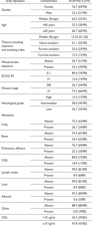
Demographic characteristicsThe clinical-pathological characteristics of the patients are the following: female gender in 76.7% of the cases, median age of 64.5 years with a range of 33-81 years, and 66.7% of 60 years or older (table I). Smokers repre-sented 38.9% of the patients, 43.3% had wood-smoke exposure. The performance status of the patients was predominantly ECOG 0-1 in 84.4%. All cases were adenocarcinomas, 73.3% at advanced stage (IV), pre-dominantly with intermediate histological grade (50%). Metastatic NSCLC was present in 26.7 % of the patients in contralateral lung, followed by bones (24.4%), pulmo-nary effusion (22.2%) and central nervous system (CNS) (18.9%). The carcinoembryonic antigen was elevated (≥10 ng/ml) in 47.8% of the patients. Chemotherapy was the treatment in 66.2% of the patients, while 33.8% received tyrosine kinase inhibitors (TKIs).
Somatic mutations
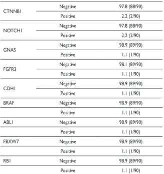
Mutations were found in 27 of 48 cancer-related genes sequenced (table II). TP53 mutations were detected in 43 patients (47.8%). In 36.7% of cases (33/90) muta-tions in the EGFR gene were found. The most frequent EGFR mutations were Q787 (15.6%), exon 19 deletions (11.1%), L858R mutation in exon 21 (7.8%), and T790M mutation in exon 20 (1.1%). Additional EGFR mutations were A750P and G719A (1.1%). Exon 19 deletions were identified in seven patients by qPCR, while by NGS ten cases were detected. Other mutations were identified, such as in KRAS, MET and PDGFRA (20%), HNF1A (14.4%), APC (12.2%) HER2 (11.1%) and MSH6 (10%). Alterations in lower frequency (<10%) were found in PIK3CA, GUSB, ALK rearrangements, KSR1, KIT, STK11, FLT3, ERBB4, VHL, CTTNB1, NOTCH1, GNAS, FGFR3, CDH1, BRAF, ABL1, FBXW7 and RB1.
Table I
CliniCalCharaCteristiCsoflungadenoCarCi
-nomapatients (n=90). mexiCo, 2018
Study Population Characteristics All (N=90) % (n/N)
Gender Female 76.7 (69/90)
Male 23.3 (21/90)
Age
Median (Range) 64.5 (33-81) <60 years 33.3 (30/90)
≥60 years 66.7 (60/90)
Tobacco-smoking exposure and smoking index
Median (Range) 12.25 (0-120) Never-smokers 61.1 (55/90) Former-smokers 25.6 (23/90) Current-smokers 13.3 (12/90)
Wood-smoke exposure
Absent 56.7 (51/90) Present 43.3 (39/90)
ECOG PS 0-1 84.4 (76/90)
2+ 15.6 (14/90)
Disease stage IIIB 26.7 (24/90)
IV 73.3 (66/90)
Histological grade
High 13.3 (12/90) Intermediate 50.0 (45/90) Low 36.7 (33/90)
Metastasis
Lung Absent 72.2 (65/89)
Present 26.7 (24/89)
Bone Absent 74.4 (67/89)
Present 24.4 (22/89)
Pulmonary effusion Absent 76.7 (69/89) Present 22.2 (20/89)
CNS Absent 80.0 (72/89)
Present 18.9 (17/89)
Lymph nodes Absent 90.0 (81/89)
Present 8.9 (8/89)
Liver Absent 90.0 (81/89)
Present 8.9 (8/89)
Adrenal Absent 93.3 (84/89)
Present 5.6 (5/89)
Other Absent 88.9 (80/89)
Present 10.0 (9/89)
CEA <10 ng/ml 43.3 (39/82)
≥10 ng/ml 47.8 (43/82)
ECOG PS: Eastern Cooperative Oncology Group Performance Status; CNS: Central Nervous System, CEA: Carcinoembryonic Antigen
Table II
moleCularprofileofsomatiCmutations
inmexiCanpatientswith nsCls (n=90).
mexiCo, 2018
Gene All (N=90)% (n/N)
TP53 Negative 52.2 (47/90) Positive 47.8 (43/90) EGFR Negative 63.3 (57/90) Positive 36.7 (33/90)
EGFR exons
Exon 19 (Deletion) 11.1 (10/90) Exon 21 (L858R) 7.8 (7/90) Exon 20 (T790M) 1.1 (1/90) Exon 20 (Q787) 15.6 (14/90)
Other 2.2 (4/90) KRAS Negative 80.0 (72/90)
Positive 20.0 (18/90) MET Negative 80.0 (72/90) Positive 20.0 (18/90) PDGFRA Negative 80.0 (72/90) Positive 20.0 (18/90) HNF1A Negative 85.6 (77/90) Positive 14.4 (13/90) APC Negative 87.8 (79/90) Positive 12.2 (11/90) HER2 Negative 88.9 (80/90) Positive 11.1 (10/90) MSH6 Negative 90.0 (84/90) Positive 10.0 (9/90) PIK3CA Negative 91.1 (82/90)
Positive 8.9 (8/90) GUSB Negative 91.1 (82/90)
Positive 8.9 (8/90) ALK fusions Negative 87.8 (79/85)
Positive 6.7 (6/85) KSR1 Negative 94.4 (85/90)
Positive 5.6 (5/90) KIT Negative 96.7 (87/90)
Positive 3.3 (3/90) STK11 Negative 96.7 (87/90)
Positive 3.3 (3/90) FLT3 Negative 98.7 (88/90)
Positive 2.2 (2/90) ERBB4 Negative 97.8 (88/90)
Positive 2.2 (2/90) VHL Negative 97.8 (88/90)
Positive 2.2 (2/90)
Associations between mutations and clinical characteristics
There was an association between EGFR (p=0.005) and HER2 (p=0.026) mutations with intermediate his-tological grade. MET (p= 0.046), APC (p=0.0051) and PDGFRA (p=0.009) mutations were more frequently in women. MET (p= 0.012) and HNF1A (p= 0.036) were predominant in patients with pulmonary effusion. TP53 mutations were common in former smokers (p=0.041), while never smokers presented higher incidence of APC mutations (p=0.030) and PDGFRA alterations correlated with ECOG 0-1 (p=0.042).
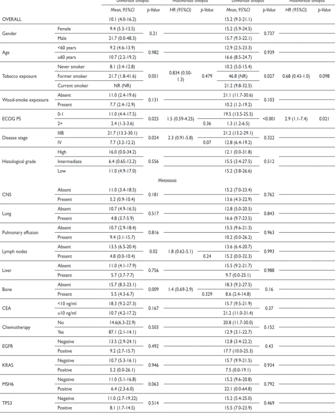
Survival of NSCLC patients
Table III shows that better PFS was associated with ECOG-PS ≤1, 11.0 vs. 2.4 months, (p=0.025); disease stage
IIIB compared to IV, 21.7 vs. 7.7 months, (p=0.024); lym-ph nodes absent 13.5 vs. 4.8 months (p=0.020); absence of bone metastasis, 15.7 vs. 5.5 months (p=0.009); and no APC mutations, 11.0 vs. 6.4 months (p=0.057). In the multivariate analysis, the only significant factor of poor prognosis for PFS was the presence of APC mutations (HR 3.1 [1.1-8.8], p= 0.032).
Factors associated with OS in univariate analysis (table III) were smoking status where current smokers had a median OS of 21.2 months (95%CI 9.8-32.5), former smokers 46.8 months (95%CI not reached) and never smokers 10.2 months (95%CI 5.0-15.4), p=0.027. Another factor was ECOG-PS PS ≤1, 19.5 vs. 1.3 months in, p< 0.001. Multivariate analysis of OS showed that ECOG was the only independent factor with HR 2.9 (1.1-7.4),
p=0.021.
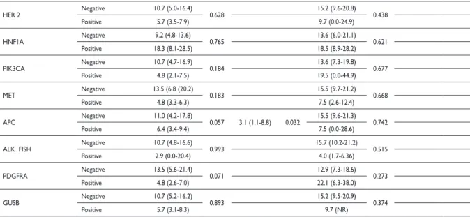
According to data from the cBioportal database,17,18
we performed a comparison to determine differences in prevalence between the frequencies of somatic mutations in our population with respect to data for Caucasian patients. The Mexican population had a different prevalence of mutations in several genes in-cluding EGFR, MET, HNF1A, HER2 and GUSB (table IV). EGFR mutations were present in 36.7 vs. 17% in our population compared to Caucasians, while in MET gene the frequencies were 20 vs. 4%, HNF1A 14.4 vs. 2.7%, GUSB 8.9 vs. 0.5% (p< 0.001), respectively, whereas in HER2 the mutation frequencies found were 11.1 vs. 2.2% (p=0.003).
Discussion
Lung cancer is the human neoplasm with the highest mutation rate after melanoma, with over 10 mutations/ Mb for smokers.19 The presence of specific driver
muta-tions has led to the development of targeted therapies for specific subsets of patients.20,21 In this study, we
analyzed the mutation profile of NSCLC in the Mexican population, the association with clinical-pathological characteristics, therapeutic response and the contrast with other ethnic groups.
TP53 was the most frequently mutated gene in almost 50% of the patients and it was associated with former tobacco consumption. TP53 mutations had no prognostic value for OS of NSCLC patients. This tumor suppressor gene ranks first among the highly mutated genes in human cancers according to the lung cancer ge-nome database.22 To date, TP53 is not a therapeutic target,
nevertheless, it represents a prognostic factor of response. Recently, it has been shown that TP53 mutations corre-late with resistance to chemotherapy, worse therapeutic responses and reduced OS of NSCLC patients depending
CTNNB1 Negative 97.8 (88/90) Positive 2.2 (2/90) NOTCH1 Negative 97.8 (88/90)
Positive 2.2 (2/90) GNAS Negative 98.9 (89/90)
Positive 1.1 (1/90) FGFR3 Negative 98.1 (89/90)
Positive 1.1 (1/90) CDH1 Negative 98.9 (89/90)
Positive 1.1 (1/90) BRAF Negative 98.9 (89/90)
Positive 1.1 (1/90) ABL1 Negative 98.9 (89/90)
Positive 1.1 (1/90) FBXW7 Negative 98.9 (89/90)
Positive 1.1 (1/90) RB1 Negative 98.9 (89/90)
Positive 1.1 (1/90) TP53: Tumor Protein 53, EGFR: Epidermal Growth Factor Receptor 1, KRAS: Kirsten Rat Sarcoma Viral Oncogene Homolog, MET: Mesenchymal Epithelial Transition/Hepatocyte Growth Factor Receptor (HGFR),PDGFRA: Platelet-derived growth factor receptor alpha,HNF1A:Hepatocyte Nuclear Factor 1-Alpha, APC: Adenomatous Polyposis Coli protein, HER2: Epidermal Growth Factor Receptor 2, MSH6: MutS Homolog 6, PIK-3CA: Phosphatidylinositol-4,5-Bisphosphate 3-Kinase Catalytic Subunit Alpha, GUSB: Glucuronidase Beta, ALK: Anaplastic Lymphoma Kinase, KSR1: Kinase suppressor of Ras 1, KIT: V-Kit Hardy-Zuckerman 4 Feline Sarcoma Viral, STK11: Serine/Threonine kinase 11,FLT3: FMS-like Tyrosine Kinase 3, ERBB4:Epidermal Growth Factor Receptor 4, VHL: Von Hippel-Lindau tumor suppressor, CTNNB1: Catenin Beta 1, NOTCH1: Neurogenic Locus Notch Homolog Protein 1, GNAS: Guanine Nucleotide Binding Protein (G Protein) Alpha, FGFR3: Fibroblast growth factor receptor 3, CDH1: E-cadherin, BRAF: V-Raf Murine Sarcoma Viral Oncogene Homolog B1, ABL1: Abelson Murine Leukemia Viral Oncogene Homolog 1, FBXW7: F-Box And WD Repeat Domain Containing 7, RB1: Retinoblastoma-Associated Protein.
Table III
univariateandmultivariateanalysisofthefaCtorsassoCiatedwithprogression-freesurvival
andoverallsurvivalin mexiCanpatientswith nsClC (n=90). mexiCo, 2018
Progression-free survival Overall survival
Univariate analysis Multivariate analysis Univariate analysis Multivariate analysis Mean, 95%CI p-Value HR (95%CI) p-Value Mean, 95%CI p-Value HR (95%CI) p-Value
OVERALL 10.1 (4.0-16.2) 15.2 (9.3-21.1) Gender Female 9.4 (5.3-13.5) 0.21 15.2 (5.9-24.5) 0.737
Male 21.7 (0.0-48.3) 15.7 (9.3-22.1) Age <60 years 9.2 (4.6-13.9) 0.982 12.9 (2.5-23.3) 0.939
≥60 years 10.7 (2.2-19.2) 16.6 (8.5-24.7) Tobacco exposure
Never smoker 8.1 (3.4-12.8)
0.051 0.834 (0.50-1.3) 0.479
10.2 (5.0-15.4)
0.027 0.68 (0.43-1.0) 0.098 Former smoker 21.7 (1.8-41.6) 46.8 (NR)
Current smoker NR (NR) 21.2 (9.8-32.5) Wood-smoke expossure Absent 11.0 (2.4-19.6) 0.131 21.1 (11.7-30.6) 0.103
Present 7.7 (2.4-12.9) 10.2 (1.2-19.2)
ECOG PS 0-1 11.0 (4.4-17.5) 0.025 1.5 (0.59-4.25) 19.5 (13.5-25.5) <0.001 2.9 (1.1-7.4) 0.021 2+ 2.4 (1.3-3.6) 0.36 1.3 (1.2-6.5)
Disease stage IIIB 21.7 (13.3-30.1) 0.024 2.3 (0.91-5.8) 21.2 (13.2-29.1) 0.322 IV 7.7 (3.2-12.2) 0.07 12.8 (6.4-19.2) Histological grade
High 16.0 (0.0-34.2) 0.556
12.1 (0.0-31.8) 0.512 Intermediate 6.4 (0.65-12.2) 15.5 (3.4-27.5) Low 11.0 (4.9-17.0) 15.2 (3.8-26.6)
Metastasis
CNS Absent 11.0 (3.4-18.5) 0.181 15.2 (7.0-23.4) 0.762 Present 5.2 (0.9-10.4) 13.6 (4.3-22.9) Lung Absent 10.7 (4.9-16.5) 0.517 12.8 (5.0-20.5) 0.843
Present 4.8 (3.7-5.9) 16.6 (9.7-23.5) Pulmonary effusion Absent 10.7 (2.9-18.4) 0.816 15.5 (9.6-21.3) 0.963
Present 9.4 (3.1-15.7) 10.2 (0.0-26.2) Lymph nodes Absent 13.5 (6.5-20.4) 0.02 1.8 (0.62-5.1) 13.6 (6.4-20.7) 0.993
Present 4.8 (0.0-10.4) 0.24 15.2 (0.0-32.3) Liver Absent 11.0 (4.1-17.9) 0.756 15.5 (9.2-21.7) 0.988
Present 5.7 (3.7-7.7) 9.7 (0.0-25.1) Bone Absent 15.7 (8.3-23.1) 0.009 1.4 (0.69-2.9) 18.3 (9.2-27.5) 0.16
Present 5.5 (4.3-6.7) 0.329 8.6 (2.4-14.8) CEA <10 ng/ml 18.3 (9.2-27.3) 0.167 15.7 (9.5-21.9) 0.37
≥10 ng/ml 10.7 (4.2-17.2) 21.2 (11.0-31.4) Chemotherapy No 14.6(6.3-22.9) 0.503 20.8 (11.7-30.0) 0.152
Yes 87.1 (2.1-14.1) 12.9 (3.1-22.7) EGFR Negative 13.5 (2.9-24.1) 0.492 12.8 (3.4-22.2) 0.43
Positive 9.2 (2.7-15.7) 17.7 (10.0-25.3) KRAS Negative 10.7 (5.3-16.1) 0.946 15.7 (9.9-21.5) 0.934
Positive 5.2 (0.0-26.1) 7.5 (0.0-19.1) MSH6 Negative 11.0 (5.1-16.8) 0.063 15.2 (9.6-20.8) 0.792
Positive 6.4 (2.3-6.0) 22.1 (0.0-64.8) TP53 Negative 11.0 (2.7-19.22) 0.514 15.2 (5.4-25.0) 0.469
Positive 8.1 (1.7-14.5) 15.5 (7.0-23.9)
(continuation)
HER 2 Negative 10.7 (5.0-16.4) 0.628 15.2 (9.6-20.8) 0.438 Positive 5.7 (3.5-7.9) 9.7 (0.0-24.9) HNF1A Negative 9.2 (4.8-13.6) 0.765 13.6 (6.0-21.1) 0.621
Positive 18.3 (8.1-28.5) 18.5 (8.9-28.2) PIK3CA Negative 10.7 (4.7-16.9) 0.184 13.6 (7.3-19.8) 0.677
Positive 4.8 (2.1-7.5) 19.5 (0.0-44.9) MET Negative 13.5 (6.8 (20.2) 0.183 15.5 (9.7-21.2) 0.668
Positive 4.8 (3.3-6.3) 7.5 (2.6-12.4) APC Negative 11.0 (4.2-17.8) 0.057 3.1 (1.1-8.8) 0.032 15.5 (9.6-21.3) 0.742
Positive 6.4 (3.4-9.4) 7.5 (0.0-28.6) ALK FISH Negative 10.7 (4.8-16.6) 0.993 15.7 (10.2-21.2) 0.515
Positive 2.9 (0.0-20.4) 4.0 (1.7-6.36) PDGFRA Negative 13.5 (5.6-21.4) 0.071 12.9 (7.3-18.6) 0.273
Positive 4.8 (2.6-7.0) 22.1 (6.3-38.0) GUSB Negative 10.7 (5.2-16.2) 0.893 15.2 (9.5-20.9) 0.374
Positive 5.7 (3.1-8.3) 9.7 (NR)
ECOG PS: Eastern Cooperative Oncology Group Performance Status; CNS: Central Nervous System; CEA: Carcinoembryonic Antigen; EGFR: Epidermal Growth Factor Receptor; KRAS: Kirsten Rat Sarcoma Viral Oncogene Homolog; MSH6: MutS Homolog 6; TP53: Tumor Protein 53; HER2: Epidermal Growth Factor Receptor 2; HNF1A: Hepatocyte Nuclear Factor 1-Alpha; PIK 3CA: Phosphatidylinositol-4,5-Bisphosphate 3-Kinase Catalytic Subunit Alpha; MET: Mes-enchymal Epithelial Transition; APC: Adenomatous Polyposis Coli protein; ALK: Anaplastic Lymphoma Kinase; PDGFRA: Platelet-derived growth factor receptor alpha; GUSB: Glucuronidase Beta.
Table IV
Comparisonbetween hispaniCand CauCasian
populationwith nsClC. mexiCo, 2018
Hispanics N= 90 Caucasians N=183
Genes n % n % P Value*
TP53 43 47.8 93 51 0,636
EGFR 33 36.7 31 17 <0.001*
KRAS 18 20 49 27 0,221
MET 18 20 7 4 <0.001*
PDGFRA 18 20 15 8 0,005
HNF1A 13 14.4 5 2.7 <0.001*
APC 11 12.2 9 5 0,029
HER2 10 11.1 4 2.2 0.003*
MSH6 9 10 4 2.2 0,012
PIK3CA 8 8.9 7 4 0,084
GUSB 8 8.9 1 0.5 <0.001*
TP53: Tumor Protein 53; EGFR: Epidermal Growth Factor Receptor 1; KRAS: Kirsten Rat Sarcoma Viral Oncogene Homolog; MET: Mesenchymal Epithelial Transition/Hepatocyte Growth Factor Receptor (HGFR); PDG-FRA: Platelet-derived growth factor receptor alpha; HNF1A: Hepatocyte Nuclear Factor 1-Alpha; APC: Adenomatous Polyposis Coli protein; HER2: Epidermal Growth Factor Receptor 2; MSH6: MutS Homolog 6; PIK 3CA: Phosphatidylinositol-4,5-Bisphosphate 3-Kinase Catalytic Subunit Alpha; GUSB: Glucuronidase Beta.
Chi-square or Fisher´s Exact test (n≤5) * Significant after Bonferroni correction
on disease stage and sequencing platforms. Likewise, the biological role of TP53 mutations can be different according to tumor histology and smoking history.23
TP53 mutations were concurrent with mutations in EGFR, MET, KRAS, and PDGFRA, while they were mutually exclusive with mutations in HNF1, APC and HER2. Concurrent mutations in TP53 and EGFR are frequent in NSCLC and may have impact on response rate, nonetheless, these results are diverse depending on the therapeutic approach. Recently, a comprehen-sive study by the lung cancer mutation consortium showed the adverse effect of concomitant mutations in TP53-mutated patients with targeted treatments for alterations in EGFR, ALK and ROS1, by developing resistance to chemotherapeutic agents. Moreover, the alterations imposed by TP53/KRAS co-mutations on cell-cycle regulation, control of DNA replication and repair result in higher neoantigen expression of neoan-tigens including PD-L1 upregulation, thereby increasing tumor immunogenicity.24,25 Double-mutant tumors with
TP53/KRAS co-mutations had significantly shorter OS and developed resistance to chemotherapy compared to wild type tumors. In contrast, TP53/KRAS co-mutation status was predictive of clinical benefit and better PFS in response to PD-1 immunotherapy.26 The incidence of
EGFR mutations are currently the main targetable oncogenic driver in the treatment of NSCLC patients, with improved response rates and less secondary effects than cytotoxic chemotherapy.27 As previously reported,
we found high prevalence of EGFR mutations, mainly exon 19 microdeletions and L858R point mutations in exon 21. In our study there was high concordance between results with qPCR based on Scorpions/ARMS technologies and the NGS platform in the detection of EGFR exon 19 deletions. EGFR mutations were associa-ted with intermediate histological grade in advanced NSCLC. Consistently, a recent study indicated a correla-tion between moderately differentiated tumor grade and a higher frequency of EGFR mutations in metastatic lung adenocarcinomas. This modifies treatment selection, since patients with high grade or poorly differentiated tumors can be treated with chemotherapy or immu-notherapy as first line treatment previous to obtaining the EGFR mutational status.28
Other EGFR alterations found with low frequency were T790M mutation in exon 20 plus the A750P and G719A. Uncommon mutations are present in less than 10% of EGFR-mutant lung cancer and associated with high grade tumors, i.e. poorly differentiated, more ag-gressive phenotypes and could represent mechanisms of resistance to EGFR-TKI treatments.8 There are no
targeted therapies for uncommon EGFR mutations, as their responses to TKIs are variable and the role in tumor biology is still unresolved.
In this study, MET exon 14 mutations were present in 20% of the patients, associated with female gender, pulmonary effusion and TP53 mutations. MET gene encodes a tyrosine kinase receptor that binds the hepa-tocyte growth factor. MET exon 14 codes for a portion of the juxtamembrane domain containing the binding site for ubiquitin ligases that participate in MET protein degradation. Mutations in MET exon 14 mutations cause exon 14 skipping, leading to constitutive signaling and oncogenicity.8 A recent study described a relationship
between MET mutations, female gender and never smokers. Both MET mutations and amplifications show clinical benefit in NSCLC patients treated with TKIs not specifically designed for MET, having durable partial response with crizotinib and capmatinib and complete metabolic response with cabozantinib independently of histological subtype. Selective MET inhibitors have been developed such as tivantinib, onartuzumab and emibetuzumab with modest clinical benefits.8,29
HER2 exon 20 mutations constitute 96% of the mutations in this gene and have been the most studied alterations in NSCLC. Conversely, other HER2 muta-tions have been described having prognostic value, including S310F/Y, D277G/H/V/Y and I655V.30 Among
the HER2 mutations in our study, I655V (exon 17) was detected with high frequency. This amino acid change in the transmembrane region increases tyrosine kinase activity leading to oncogenic signaling. I655V is present in different malignancies including breast, gastric and lung cancer. It correlates with aggressive tumor pheno-type, poor prognosis and risk of cardiotoxicity during trastuzumab treatment of breast cancer.8,31 However,
there are few studies in lung cancer and without prog-nostic value.
In the present study, we found concurrence of HER2 mutations with intermediate histological grade and MET variants, but they were mutually exclusive with TP53 and EGFR mutations. There are no reports describing the association between HER2 mutations and tumor grade, although there is association with female gender, never smokers, adenocarcinomas, it has been reported that it confers a low sensitivity to traditional EGFR-TKIs.8,30 HER2 mutations in NSCLC
have a prevalence of 4% and may have higher clinical impact than gene amplification. Preliminary results from selective inhibitors for HER2 exon 20 mutations such as poziotinib had marked radiologic and clinical response. Additionally, several pan-EGFR irreversible inhibitors such as afatinib, neratinib, and dacomitinib have shown activity in NSCLC patients with HER2 mutations.8,30,32
Adenomatous polyposis coli (APC) is a tumor suppressor gene mutated in 80% of colon carcinomas and less frequently in other malignancies including liver, breast and lung cancer.32 In our study, we found
APC mutations in NSCLC that correlated with never smokers, were mutually exclusive with TP53 muta-tions and predicted poor prognosis. APC is part of the B-catenin degradation complex in the Wnt pathway. APC mutations induce nuclear B-catenin accumulation leading oncogene activation. APC mutations have been associated with nonsmokers in colorectal cancer.33 These
mutations are generally insertions/deletions that modi-fy frameshifts, introduce premature stop codons and loss of function via truncation of APC protein. Current the-rapies for APC loss in cancer inhibit signaling through the canonical Wnt/B-catenin pathway downstream of APC or aim to restore normal APC expression. APC and TP53 mutations occur early in the initiation of car-cinogenesis, but they are not documented as mutually exclusive alterations.34 Although these alterations alone
may not be sufficient to have a significant impacto on OS concomitant TP53 and APC mutations have been described as a more aggressive molecular phenotype with implications for worse prognosis in PFS/OS.4
Euro-pean patients, 40% of Asian patients, and between 2 to 14% of Afro-American patients, the frequency of EGFR mutations in Mexico is 27%.4,35 In our study, the
preva-lence of EGFR mutations was 36.7% compared to 17% in Caucasians. Other important differences in mutation frequency between the two populations were present in known oncogenic drivers of NSCLC such as MET and HER2, while the frequency of KRAS mutation was hig-her in Caucasians as expected although not statistically significant. The incidence of KRAS mutations in NSCLC in Latin America is approximately 14-17% as reported by the CLICaP.4 MET mutation profile also differs in
type and frequency according to ethnicity. In our study, HER2 mutations were present in 11.1% of the Mexican patients in contrast to 2.2% in Caucasians and 3.9% in Asians. Similarly to EGFR mutations, HER2 mutations are associated with adenocarcinoma histology, female gender and never smokers and have favorable response to TKI and antibody treatments.23 A higher incidence of
HER2 mutations in our population opens the opportu-nity to improve response rates and overall survival with novel targeted therapies.
Conclusions
This study provides a profile of somatic mutations for NSCLC in the Mexican population. The main genomic alterations were present in TP53, EGFR, KRAS, MET, PDGFRA, HNF1A, APC, HER2 and MSH6. This muta-tion profile shows differences with other ethnic groups. Further studies are warranted to evaluate the germline molecular features underlying the relationship between ethnicity, somatic mutation rates, clinical responses and survival of NSCLC patients.
Declaration of conflict of interests. The authors declare that they have no conflict of interests.
References
1. Torre LA, Siegel RL, Jemal A. Lung Cancer Statistics. Adv Exp Med Biol. 2016;893:1-19. https://doi.org/10.1007/978-3-319-24223-1_1
2. Arrieta O, Villarreal-Garza C, Zamora J, Blake-Cerda M, de la Mata MD, Zavala DG, et al. Long-term survival in patients with non-small cell lung cancer and synchronous brain metastasis treated with whole-brain radio-therapy and thoracic chemoradiation. Radiat Oncol. 2011;6(1):166. https:// doi.org/10.1186/1748-717X-6-166
3. Raez LE, Santos ES, Rolfo C, Lopes G, Barrios C, Cardona A, et al. Challenges in facing the lung cancer epidemic and treating advanced disease in Latin America. Clin Lung Cancer. 2016;18(1):e71-9. https://doi. org/10.1016/J.CLLC.2016.05.003
4. Arrieta O, Cardona AF, Martín C, Más-López L, Corrales-Rodríguez L, Bramuglia G, et al. Updated frequency of EGFR and KRAS muta-tions in nonsmall-cell lung cancer in Latin America: The Latin-American
consortium for the investigation of lung cancer (CLICaP). J Thorac Oncol. 2015;10(5):838-43. https://doi.org/10.1097/JTO.0000000000000481 5. Arrieta O, Cardona AF, Federico-Bramuglia G, Gallo A, Campos-Parra AD, Serrano S, et al. Genotyping non-small cell lung cancer (NSCLC) in Latin America. J Thorac Oncol. 2011;6(11):1955-9. https://doi.org/10.1097/ JTO.0B013E31822F655F
6. Herbst RS, Morgensztern D, Boshoff C. The biology and management of non-small cell lung cancer. Nature. 2018;553(7689):446-54. https://doi. org/10.1038/nature25183
7. Richer AL, Friel JM, Carson VM, Inge LJ, Whitsett TG. Genomic profiling toward precision medicine in non-small cell lung cancer: getting beyond eGFR. Pharmacogenomics Pers Med. 2015;8:63-7. https://doi.org/10.2147/ PGPM.S52845
8. Arrieta O, Cardona AF, Corrales L, Campos-Parra AD, Sánchez-Reyes R, Amieva-Rivera E, et al. The impact of common and rare EGFR muta-tions in response to EGFR tyrosine kinase inhibitors and platinum-based chemotherapy in patients with non-small cell lung cancer. Lung Cancer. 2015;87(2):169-75. https://doi.org/10.1016/j.lungcan.2014.12.009 9. Barrón F, Cardona AF, Corrales L, Ramirez-Tirado LA, Caballe-Perez E, Sanchez G, et al. Characteristics of progression to tyrosine kinase inhibi-tors predict overall survival in patients with advanced non-small cell lung cancer harboring an EGFR mutation. J Thorac Dis. 2018;10(4):2166-78. https://doi.org/10.21037/jtd.2018.03.106
10. Reungwetwattana T, Weroha SJ, Molina JR. Oncogenic pathways, mo-lecularly targeted therapies, and highlighted clinical trials in non–small-cell lung cancer (NSCLC). Clin Lung Cancer. 2012;13(4):252l66. https://doi. org/10.1016/j.cllc.2011.09.004
11. Tan O, Shrestha R, Cunich M, Schofield DJ. Application of next-generation sequencing to improve cancer management: A review of the clinical effectiveness and cost-effectiveness. Clin Genet. 2018;93(3):533-44. https://doi.org/10.1111/cge.13199
12. Cheng ML, Oxnard GR. Does TMB impact the effectiveness of TKIs in EGFR-mutant NSCLC? Clin Cancer Res. 2019;25(3):899-900. https://doi. org/10.1158/1078-0432.CCR-18-2368
13. Soca-Chafre G, Hernández-Pedro N, Aviles-Salas A, Versón CA, Sán-chez KC, Cardona AF, et al. Targeted next generation sequencing identified a high frequency genetic mutated profile in wood smoke exposure-related lung adenocarcinoma patients. Oncotarget. 2018;9(55):30499-512. https:// doi.org/10.18632/oncotarget.25369
14. Arrieta O, Ramírez-Tirado LA, Báez-Saldaña R, Peña-Curiel O, Soca-Chafre G, Macedo-Perez EO. Different mutation profiles and clinical char-acteristics among Hispanic patients with non-small cell lung cancer could explain the “Hispanic paradox.” Lung Cancer. 2015;90(2):161-6. https://doi. org/10.1016/j.lungcan.2015.08.010
15. Cardona AF, Zatarain-Barrón ZL, Rojas L, Arrieta O. The reality of complexity: concomitant genomic alterations in patients with EGFR mutations. J Thorac Dis. 2018;10(2):597-9. https://doi.org/10.21037/ jtd.2017.12.136
16. Detterbeck FC, Boffa DJ, Kim AW, Tanoue LT. The Eighth Edition Lung Cancer Stage Classification. Chest. 2017;151(1):193-203. https://doi. org/10.1016/j.chest.2016.10.010
17. Cerami E, Gao J, Dogrusoz U, Gross BE, Sumer SO, Aksoy BA, et al. The cBio cancer genomics portal: An pen platform for exploring multidi-mensional cancer genomics data. Cancer Discov. 2012;2(5):401-4. https:// doi.org/10.1158/2159-8290.CD-12-0095
18. Gao J, Aksoy BA, Dogrusoz U, Dresdner G, Gross B, Sumer SO, et
al. Integrative analysis of complex cancer genomics and clinical profiles using the cBioPortal. Sci Signal. 2013;6(269):1-34. https://doi.org/10.1126/ scisignal.2004088
20. Raez LE, Cardona AF, Santos ES, Catoe H, Rolfo C, Lopes G, et al. The burden of lung cancer in Latin-America and challenges in the access to genomic profiling, immunotherapy and targeted treatments. Lung Cancer. 2018;119:7-13. https://doi.org/10.1016/J.LUNGCAN.2018.02.014 21. Tafe LJ, Pierce KJ, Peterson JD, De Abreu F, Memoli VA, Black CC,
et al. Clinical genotyping of non–small cell lung cancers using targeted next-generation sequencing: utility of identifying rare and co-mutations in oncogenic driver genes 1. Neoplasia. 2016;18(9):577-83. https://doi. org/10.1016/j.neo.2016.07.010
22. Cancer Genome Atlas Research Network. Comprehensive molecular profiling of lung adenocarcinoma. Nature. 2014;511(7511):543-50. https:// doi.org/10.1038/nature13385
23. Halvorsen AR, Silwal-Pandit L, Meza-Zepeda LA, Vodak D, Vu P, Sagerup C, et al. TP53 mutation spectrum in smokers and never smoking lung cancer patients. Front Genet. 2016;7:85. https://doi.org/10.3389/ fgene.2016.00085
24. Dearden S, Stevens J, Wu YL, Blowers D. Mutation incidence and coinci-dence in non small-cell lung cancer: meta-analyses by ethnicity and histol-ogy (mutMap). Ann Oncol. 2013;24(9):2371-6. https://doi.org/10.1093/ annonc/mdt205
25. Skoulidis F, Byers LA, Diao L, Papadimitrakopoulou VA, Tong P, Izzo J,
et al. Co-occurring genomic alterations define major subsets of KRAS-mutant lung adenocarcinoma with distinct biology, immune profiles, and therapeutic vulnerabilities. Cancer Discov. 2015;5(8):861-78. https://doi. org/10.1158/2159-8290.CD-14-1236
26. Aisner DL, Sholl LM, Berry L, Rossi M, Chen H, Fujimoto J, et al. The im-pact of smoking and TP53 mutations in lung adenocarcinoma patients with targetable mutations - the Lung Cancer Mutation Consortium (LCMC2). Clin Cancer Res. 2018;24(5):1038-47. https://doi.org/10.1158/1078-0432. CCR-17-2289
27. Arrieta O, Cruz-Rico G, Soto-Perez-de Celis E, Ramírez-Tirado LA, Caballe-Perez E, Martínez-Hernández JN, et al. Reduction in hepatocyte
growth factor serum levels is associated with improved prognosis in advanced lung adenocarcinoma patients treated with Afatinib: a Phase II trial. Target Oncol. 2016;11(5):619-29. https://doi.org/10.1007/s11523-016-0425-x
28. Levy M, Lyon L, Barbero E, Wong J, Suga M, Sam D, et al. Histologic grade is predictive of incidence of epidermal growth factor receptor mutations in metastatic lung adenocarcinoma. Med Sci. 2017;5(4):1-12. https://doi.org/10.3390/medsci5040034
29. Gkolfinopoulos S, Mountzios G. Beyond EGFR and ALK: targeting rare mutations in advanced non-small cell lung cancer. Ann Transl Med. 2018;6(8):142. https://doi.org/10.21037/atm.2018.04.28
30. Peters S, Zimmermann S. Targeted therapy in NSCLC driven by HER2 insertions. Transl Lung Cancer Res. 2014;3(2):84-8. https://doi. org/10.3978/j.issn.2218-6751.2014.02.06
31. Garrido-Castro AC, Felip E. HER2 driven non-small cell lung cancer (NSCLC): potential therapeutic approaches. Transl Lung Cancer Res. 2013;2(2):122-7. https://doi.org/10.3978/j.issn.2218-6751.2013.02.02 32. Sullivan I, Planchard D. Next-Generation EGFR tyrosine kinase inhibi-tors for treating EGFR-mutant lung cancer beyond first line. Front Med. 2017;3:76. https://doi.org/10.3389/fmed.2016.00076
33. De Nicola F, Goeman F, Pallocca M, Sperati F, Pizzuti L, Melucci E, et
al. Deep sequencing and pathway-focused analysis revealed multigene oncodriver signatures predicting survival outcomes in advanced colorectal cancer. Oncogenesis. 2018;7(7):1-10. https://doi.org/10.1038/s41389-018-0066-2
34. Rieth J, Subramanian S. Mechanisms of intrinsic tumor resistance to immunotherapy. Int J Mol Sci. 2018;19(5):1-13. https://doi.org/10.3390/ ijms19051340