Genetic Analysis of the prehistoric peopling
of Western Europe: ancient DNA and
the role of contamination
Mª Lourdes Sampietro Bergua
Mª Lourdes Sampietro Bergua
Mª Lourdes Sampietro Bergua
Mª Lourdes Sampietro Bergua
Barcelona, Noviembre de 2006
TESIS DOCTORAL
TESIS DOCTORAL
TESIS DOCTORAL
TESIS DOCTORAL
Evolutionary Biology Unit
Department of Experimental and Health Science
Universitat Pompue Fabra
Genetic Analysis of the prehistoric peopling
of Western Europe: ancient DNA and
the role of contamination
Memoria presentada por Mª Lourdes Sampietro Bergua para optar al titulo de doctor en Ciencias de la Salud y de la Vida. Esta tesis doctoral ha sido realizada bajo la codirección del Dr Carles Lalueza-Fox (Universidad de Barcelona) y del Dr Jaume Bertranpetit i Busquets (Universitat Pompue Fabra) en la Unidad de Biología Evolutiva, Departamento de Ciencias Experimentales y de la Salud, Universitat Pompeu Fabra. PhD Program in Health and Life Science (2002-2004).
Carles Lalueza-Fox Director
Jaume Bertranpetit i Busquets Director
Mª Lourdes Sampietro Bergua
ABREVIATIONS
aDNA: ancient DNA A: adenina
AMH: anatomically modern humans AP: apurinic site
Asp: Aspartic Bp: base pairs BP:Before present
BSA: bovine serum albumine C: Citosina
Ct: Cycle threshold parameter
CRS: Cambridge Reference Sequence DNA: Deoxyribonucleic acid.
EDTA: ethylenediaminetetra-acetic acid dNTP´s: deoxiribonucleotides triphosphate. ddNTP´s:dideoxiribonuclleotides triphosphate G: Guanina
GC: Gas Chromatrography H strand: heavy strand H2O2: hydrogen peroxide
HX:hypoxantine
HVR: Hyper variable region KYA: kilo years ago
L strand: Light strand MS: Mass spectrometry mtDNA: mitochondrial DNA MYA: million year ago MW: molecular weight NA: Avogadro´s number
Nfe: Effective population size
NUMTs: nuclear mitochondrial sequences ·O2: peroxide radicals
·OH: hydroxy radicals PC: Principal components
PCA: Principal Component Analysis PCR: polymerese chain reaction Ppm: parts per million
PTB: N-phenacyltiazolium bromide RT-PCR: Real Time PCR
SAM: S-adenosylmethionine SDS: sodium dodecyl sulphate
SNP: Single Nucleotide Polimorphism T: Timina
TE: Tris-Edta
Taq polymerase: Thermus aquaticus DNA polymerase TMCR: Time Most Recent Common Ancestor
UNG: Uracil N-Glycosylase UV: Ultraviolet light
1 INTRODUCTION... 1
1.1 HISTORY OF ANCIENT DNA ... 3
1.2 DNA PRESERVATION... 7
1.2.1 DNA MOLECULE ... 7
1.2.2 DNA STABILITY IN VIVO ... 8
1.2.2.1 HIDROLYTIC DAMAGE ... 9
1.2.2.2 OXIDATIVE DAMAGE ... 9
1.2.2.3 NONENZYMATIC DNA METHYLATION ... 10
1.2.3 DNA REPAIR ... 10
1.2.3.1 DIRECT REVERSAL OF DNA DAMAGE:... 10
1.2.3.2 EXCISION REPAIR:... 10
1.2.4 DNA DAMAGE AFTER CELL DEATH... 11
1.2.4.1 DNA FRAGMENTATION ... 11
1.2.4.2 NUCLEOTIDE MODIFICATION... 11
1.2.5 DNA SURVIVAL ... 13
1.3 DNA RETRIEVAL FROM FOSSIL REMAINS ... 14
1.3.1 DNA COPY NUMBER ... 16
1.3.2 DNA FRAGMENTATION ... 20
1.3.3 JUMPING PCR ... 21
1.3.4 INHIBITORS ... 23
1.3.5 CONTAMINATION... 24
1.3.6 MISCODING LESSION ... 26
1.3.7 MOLECULAR CLONING ... 28
1.4 AUTHENTICITY CRITERIA... 30
1.5 ORIGIN AND MAINTENEANCE OF THE CURRENT HUMAN GENETIC DIVERSITY ... 32
1.5.1 MUTATION ... 32
1.5.2 NATURAL SELECTION ... 33
1.5.3 GENETIC DRIFT ... 33
1.5.4 MIGRATION ... 34
1.6 THE MITOCHONDRIA ... 35
1.6.1 HUMAN MITOCHONDRIAL DNA ... 36
1.7 HUMAN POPULATION HISTORY... 41
1.7.1 HUMAN AS A PRIMATE SPECIES ... 41
1.7.2 THE FOSSIL RECORD OF HOMINIDS... 41
1.7.3 THE ORIGINS OF MODERN HUMANS ... 45
1.7.3.1 MULTIREGIONAL MODEL... 45
1.7.3.2 OUT OF AFRICA MODEL... 46
1.7.4 THE GENETIC FINGERPRINT IN THE HISTORY OF HUMAN POPULATIONS ... 47
1.7.4.1 EVIDENCE FOR THE MITOCHONDRIAL GENOME: “The mitochondria Eve”... 48
1.7.4.1.1 DISTRIBUTION OF THE MtDNA LINAGES IN THE HUMAN POPULATIONS... 50
1.7.4.2 EVIDENCE FOR Y CHROMOSOME ... 51
1.7.4.3 EVIDENCE FROM AUTOSOMIC LOCUS... 52
1.7.4.4 EVIDENCE FROM ANCIENT DNA ... 52
1.8.1 UPPER PALEOLITHIC: THE NEANDERTALS... 55
1.8.2 THE NEOLITHIC PERIOD ... 58
1.8.2.1 EVIDENCE FROM THE ARCHAEOLOGY... 59
1.8.2.2 EVIDENCE FROM THE GENETICS ... 61
1.8.2.2.1 EVIDENCE FROM AUTOSOMIC LOCUS ... 62
1.8.2.2.2 EVIDENCE FROM mtDNA ... 64
1.8.2.2.3 EVIDENCE FROM Y-CHROMOSOME ... 66
1.8.2.2.4 EVIDENCE FROM ANCIENT DNA ... 66
1.8.3 POST-NEOLITHIC PERIOD ... 67
2 OBJETIVES ... 71
3 MATERIALS AND METHODS ... 75
3.1 DNA EXTRACTION ... 77
3.1.1 DNA ISOLATION FROM TEETH ... 77
3.1.2 DNA ISOLATION FROM SOIL SEDIMENTS ... 78
3.1.3 DNA ISOLATION FROM HAIR ... 80
3.2 PCR AMPLIFICATIONS... 80
3.2.1 ANCIENT TEETH PCR ... 82
3.2.2 SOIL SEDIMENT PCR ... 83
3.3 DNA PURIFICATION ... 83
3.3.1 PCR PRODUCTS PURIFICATION ... 84
3.3.2 GEL BAND PURIFICATION... 85
3.4 CLONING OF PCR PRODUCTS ... 86
3.5 QUANTIFICATION OF MTDNA BY MEANS OF RT-PCR ... 87
3.6 SEQUENCING ... 89
3.7 SEQUENCING ANALYSIS ... 90
4 RESULTS ... 91
4.1 CHAPTER 1: TRAKING DOWN HUMAN CONTAMINATIONS IN ANCIENT HUMAN TEETH 93 4.2 CHAPTER 2: DIRTY NEANDERTAL DNA... 102
4.3 CHAPTER 3: NEANDERTAL EVOLUTIONARY GENETICS: MITOCHONDRIAL DNA DATA FROM THE IBERIAN PENINSULA... 119
4.4 CHAPTER 4:THE MITOCHONDRIAL HYPERVARIABLE REGION I OF AN IBERIAN NEANDERTAL SUGGESTS A POPULATION AFFINITY WITH OTHER EUROPEAN NEANDERTALS... 126
4.5 CHAPTER 5: MITOCHONDRIAL DNA FROM A LATE NEOLITHIC SITE SUPPORTS A LONG TERM GENETIC CONTINUITY IN THE IBERIAN PENINSULA. ... 135
4.6 CHAPTER 6: THE GENETICS OF THE PRE-ROMAN IBERIAN PENINSULA: A MTDNA STUDY OF ANCIENT IBERIANS... 147
5 DISCUSSION ... 164
5.1 PRE-LABORATORY-DERIVED CONTAMINATIONS IN ANCIENT HUMAN TEETH... 165
5.2 HUMAN POPULATION HISTORY IN WESTERN EUROPE... 169
5.2.1.2 DEMOGRAPHIC HISTORY OF THE NEANDERTAL LINEAGE IN EUROPE (Teeth Neandertal remain mtDNA extraction) ... 172
5.2.2 THE NEOLITHIC PERIOD IN EUROPE: aDNA FROM AN IBERIAN NEOLITHIC POPULATION. ... 176 5.2.3 POST-NEOLITHIC PERIOD IN THE IBERIAN PENINSULA: A mtDNA STUDY OF THE ANCIENT IBERIANS ... 179
1
1
1
1.1 HISTORY OF ANCIENT DNA
The history of ancient DNA (aDNA) starts only twenty-two years ago when Higuchi and
collaborators (Higuchi et al., 1984) extracted and sequenced, by means of molecular cloning,
DNA fragments from a museum specimen of the quagga (an Equid from South Africa that
became extinct in the nineteen century). This specimen died 150 years ago. This finding
revolutionised the field of molecular biology because it showed that it was possible to retrieve
DNA from an organism from the distanct past. A year later, Svante Pääbo (Paabo, 1985),
retrieved DNA molecules from 2,500 year old human Egyptian mummies dated using the
same methodology,. Nevertheless, retrieving DNA sequences from ancient samples was
difficult and methodologically laborious and the number of publications related to the field
increased slowly at that time.
The improvement of aDNA research is associated with the discovery in 1987 of the PCR
(Polimerase Chain Reaction) technology (Mullis and Faloona, 1987). In contrast to molecular
cloning, PCR allows the specific exponential amplification of little amounts of DNA of
interest, discarding other DNA sequences from the pool (i.e DNA from micro organisms).
Bone and teeth were quickly found to be better sources of aDNA than soft tissues (Hagelberg
et al., 1989) and this meant that museums suddenly become recognised as storehouses of
preserved genetic information from the past.
Since the discovery of the PCR, the list of publications related to aDNA increased
exponentially. High profile journals started publishing studies claiming that aDNA from
specimens that were millions years old could be successfully extracted and sequenced.
Lindhal denominated this phenomenon “the antidiluvian DNA” (Lindahl, 1993a). Examples
of this period are the retrieval of aDNA from organisms preserved in amber (Cano et al.,
1993, DeSalle et al., 1992), plants sediments dated from the Miocene (Golenberg et al., 1990)
and even the retrieval of the cytochrome b mitochondrial gene from a dinosaur bone dated to
over 80 million years ago (MYA) (Woodward et al., 1994b). However and, as we will see
below (see chapter 1.3) the field is plagued by numerous technical problems; later, some of
the most extraordinary claims that had been published so far, like the retrieval of DNA from
the dinosaur or from the insects preserved in amber, have been proven to be false (impossible
to reproduce or have been shown to derive from an identifiable source of contamination) and
Despite this, recent advantages in knowledge about the tempo and mode of DNA template
damage, sample contamination and biochemical diagenesis of the DNA molecule have
improved aDNA studies to the extent that aDNA is now emerging as a viable scientific
discipline (Willerslev and Cooper, 2005). Nevertheless, unimaginable technical
improvements apart, studies of DNA sequences should be confined to the past one million
years and more probably to the past 100,000 years in order to achieve credible results
(Hofreiter et al., 2001b).
Several studies have begun to reveal the potential of aDNA retrieval to record the methods
and processes of evolution. These studies provide a unique way to test models and
assumptions commonly used to reconstruct patterns of evolution, population genetics and
palaeoecological changes (Willerslev and Cooper, 2005). This includes studying the
phylogenetic relationships between extinct species with extant species. Australian marsupial
wolves (Thomas et al., 1989); New Zealand moa (Cooper et al., 2001), American ground
sloth (Hoss et al., 1996a) and Myotragus balearicus (Lalueza-Fox et al., 2005a) are examples
of about 50 extinct animal species for which this has been done (Paabo et al., 2004).
Moreover, studying ancient populations provides the opportunity to track genetic changes in
the population over the time. Examples of ancient population genetics are rabbits (Hardy et
al., 1995), penguins (Lambert et al., 2002) or mice (Pergams et al., 2003). In addition, the
discovery that diverse mitochondrial DNA (mtDNA) and chloroplast DNA (cpDNA)
sequences may be preserved in permafrost and cave sediments (Willerslev et al., 2003) opens
up the exciting possibility of studying ancient organisms even when no macroscopically
identifiable remains are present (Paabo et al., 2004). Another source of aDNA investigated so
far has been animal or human coprolites (Poinar, 2002). This source of aDNA has yielded
insight into the diet and behaviour both humans and animal species in the past (Paabo et al.,
2004).
Another important applications of the aDNA retrieval have been the study of the evolution of
current pathogens such the bacteria Mycobacterium tuberculosis (Zink et al., 2001) or the
virus Yersinia pestis (Gilbert et al., 2004). This is a potentially very exciting field because the
evolution of some pathogens can be expected to be fast enough to allow genetic change to be
follow over decades or centuries (Paabo et al., 2004).The study of the origins of
domestications of animals such a cattle (Beja-Pereira et al., 2006) and plant species such as
maize (Jaenicke-Despres et al., 2003) around 10,000 years ago has been also possible with the
As we will see below (see chapter 1.6), DNA sequences from mtDNA or cpDNA, that are
present in hundreds of copies in each cell, are easier retrievable from ancient specimens than
the single genome copies of nuclear DNA sequences. Therefore, this delimits, for example,
the ability to resolve phylogenies of species that either diverged recently in time or so rapidly
that different parts of the genome have different phylogenies (Paabo et al., 2004). However, a
few studies have been reported so far where the successful retrieval of nuclear DNA genes has
been possible (Greenwood et al., 1999, Jaenicke-Despres et al., 2003, Poinar et al., 2003,
Poinar et al., 2006). The retrieval of nuclear genes of extinct species has opened up the
possibility of knowing even the phenotypic characteristics of the species that inhabited our
planet in the past (Rompler et al., 2006).
This improvement of the aDNA field in other species contrasts with the obtained on
understanding the recent history of the human species (basically due to the big threat
associated to modern human contamination when working with ancient human specimens; see
chapter 1.3.5); ancient DNA has, however yielded insights into the peopling of the Americas
(Stone and Stoneking, 1993, Stone and Stoneking, 1998), peopling of the Caribbean
(Lalueza-Fox et al., 2001, Lalueza-(Lalueza-Fox et al., 2003), peopling of Central Asia (Lalueza-(Lalueza-Fox et al.,
2004), peopling of Japan (Oota et al., 1995) or even the peopling of the Canary Islands
(Maca-Meyer et al., 2004). Moreover, recently, aDNA has been useful to yield insights into
the continuous debate about if the current European populations have a Palaeolithic or a
Neolithic origin. The genetic analysis of ancient Neolithic remains from Central Europe
supports a Palaeolithic origin of the current European population (Haak et al., 2005).
However, one of the most exciting achievements in the human aDNA field was produced
when the team supervised by Svante Pääbo recovered 380 base pair (bp) of the hypervariable
mtDNA region from a Neanderthal specimen (Krings et al., 1997). This sequence showed that
this individual carried a mitochondrial type quite different from those of contemporary
humans and that this mtDNA fell outside the genetic variation of modern humans in
phylogenetics trees. This result has been subsequently corroborated by more mtDNA
sequences from the same individual (Krings et al., 1999) and from sequences of another
eleven Neanderthal specimens spread all over Europe (Ovchinnikov et al., 2000, Krings et al.,
2000, Schmitz et al., 2002, Serre et al., 2004, Lalueza-Fox et al., 2005b, Lalueza-Fox et al.,
2006, Caramelli et al., 2006, Beauval et al., 2005, Orlando et al., 2006).
The breakthrough in ancient DNA sequencing came only one year ago when Krause et al
Pleistocene woolly mammoth, Mammuthus primigenius by using a powerful variant of the
PCR technology known as multiplexing. Multiplex PCRs differs from standard PCRs by
simultaneously amplifying multiple genetic targets instead of just one. Consequently, this
study indicates that the entire mitochondrial DNA genome from extinct species can
potentially be determined with just the same amount of DNA as the used in a standard-single
locus PCR.
It seems clear that this fast improvement of the DNA technology will revolutionise the field
of aDNA in the near future, and that it is going to be focused mainly on the retrieval of
nuclear DNA sequences of extinct species. In fact, last year Margulies et al (Margulies et al.,
2005) developed a new sequencing DNA technology that was able to sequence and
assemblage 25 million bases in fragments of ~100bp in a four hour run. Taking advantage of
this new technique, Poinar et al (Poinar et al., 2006) sequenced 13 million nuclear and
mitochondrial base pairs from an extinct well-preserved mammoth dated 28,000 years ago.
Recently, Svante Pääbo and his team launched the “Neanderthal genome project”, announcing
that using the same DNA technology as Poinar et al did, the entire genome of the first extinct
homo species will be available in the next two years.
Such kind of projects was unthinkable only a few years ago and it gives us an idea of how fast
is moving the aDNA field. However, despite these technical advances, researchers must be
aware that reliable results were not obtained unless many precautions and experimental
controls are implemented in their studies; especially when working with ancient human
remains. Future developments focused mainly on removing or repairing chemical damages in
the ancient DNA templates or in discarding all kind of possible modern contaminations in the
1.2 DNA PRESERVATION
1.2.1 DNA MOLECULE
The DNA molecule is a polymer composed of four nucleic acid bases - two purines (Adenine
[A] and Guanine [G]) and two pyrimidines (Citosine [C] and Thymine [T]) - linked to
phosporilated sugars by means of glycosilic bonds. The phosporilated sugars are
2’deoxirribose units linked to each other by means of phospodiester bonds shaping a double
helix. Sugar and phosphate backbones are placed outside, whereas the bases are placed inside
the DNA molecule. Hydrogen bonds are formed between purines and pyrimidines on opposite
chains, always matching A with T and G with C (see
Figure 1).
The DNA molecule carries the information for making one organism from one
generation to the next one. Because DNA uniquely serves as a permanent copy of the cell
genome, changes in its structure have strong impact in the fitness of the cell. DNA damage
can block replication or transcription and can result in a high frequency of mutations,
consequences that are unacceptable from the standpoint of cell reproduction (Cooper, 1997) .
1.2.2 DNA STABILITY IN VIVO
However, the DNA is one of the least stable molecules within the cells (see Table 1)
and, as a consequence, it is labile and prone to many forms of damage. To protect the DNA
molecule against that damage, cells have developed structural mechanisms of protection. In
vivo, the molecule is protected by water molecules in the major grooves of the double helix.
Furthermore, nuclear DNA (but not mtDNA) is linked to histones; these proteins have an
important role in the folding of the DNA molecule inside the nucleus of the cells and
presumably avoid the exposition of the DNA to the surrounding environment. (Poinar, 2002).
Table 1. Cellular compounds, their susceptibility bonds and groups and their preservation potential. – to ++++ (weakest to strongest), a rough estimate of the preservation potential for an unaltered molecule base upon relative bond strengths (adapted from (Poinar, 2002).
Compounds class Susceptible bonds Susceptible groups Preservation potential DNA, RNA Phosphate esters,
gycosidic bond, 6 C-C bond pyrimidines
Heterocyclic rings Amino groups, Metil groups
-
Proteins Peptide bond Side chain, chiral center
-/+
Carbohydrates Acetal Hydroxy, amide +
Lipids Ester, ether,
amide
Hydroxil, carboxil, ester
++
Cutin Ester, ether Hydroxil, carboxil +++
Lignin Ether Metoxil aromatic
rings
1.2.2.1 HIDROLYTIC DAMAGE
The DNA molecule is particularly prone to hydrolytic damage – damage related to the
presence of water- due to the following reasons:
1.) The phospodiester bond is quite labile because of the lack of 2’-OH group in
ribose units and subject to quick hydrolytic cleavage generating single-stranded
nicks in the double helix. This event take place about once every 2.5 hours in a
hydrated system while under dry conditions this rate drop some 20-fold (Lindahl,
1993a, Poinar, 2002).
2.) The glycosidic bond is prone to base protonation. Nucleotide bases become a
likely leaving group thus causing the cleavage of the bond. The process is termed
depurination and forms what it is know as an apurinic/apyrimidinic site (AP site).
Apurinic sites are much more frequent than the apyrimidinic ones. Once a
nucleotide base is released from DNA, the AP site can undergo cleavage of the
DNA molecule and thus result in a single-stranded nick (Lindahl, 1993a, Poinar,
2002).
3.) Deamination: bases with amino groups such as adenine, cytosine and guanine can
undergo deamination resulting in hypoxanthine, uracil, and xanthine, respectively
(Poinar, 2002).
1.2.2.2 OXIDATIVE DAMAGE
The oxidative damage is believed to occur through the action of free radicals such as peroxide
radicals (·O2), hydrogen peroxide (H2O2) and hydroxy radicals (·OH) (Lindahl, 1993a). These
radicals are endogenously generated by the cell but they may also derive from exogenous
sources such as ionising radiation, UV light (UV light can cause also pyrimidine dimmers in
the DNA double helix) and cellular processes during bacterial and fungal degradation (Poinar,
1.2.2.3 NONENZYMATIC DNA METHYLATION
In addition to oxygen, living cells contain several other small reactive molecules that
might cause DNA damage and act as an endogenous genotoxic agent. The best characterized
is S-adenosylmethonine (SAM), a metil group donor that act mainly over the ring nitrogen’s
of purine residues. 3-Methyladenine, one of the products derived of this reaction, is a
citotoxic that blocks replication. In living cells, this lesion is rapidly repaired (Lindahl,
1993a).
1.2.3 DNA REPAIR
Cells had to develop mechanisms to repair damaged DNA to maintain the integrity of their
genomes. We can distinguish two types of mechanisms:
1.2.3.1 DIRECT REVERSAL OF DNA DAMAGE:
Some lesions in DNA can be repaired by direct reversal of the damage. This is the most
efficient way of dealing with frequent specific types of DNA damage. Two important
examples are the repair of alkilated guanine residues and pyrimidine dimers. The former is
repaired by the action of a metyltransferase enzyme, which is present in all eukariotes,
including humans. The later is repaired by means of a process called photo- reactivation that
uses the energy derived from visible light to break the pyrimidine dimer (Cooper, 1997).
1.2.3.2 EXCISION REPAIR:
Although direct repair is an efficient way of dealing with particular types of DNA damage,
excision repair is a more general mean of repairing a wide variety of chemical alterations of
the DNA. In excision repair, the damage is recognised and removed. The resulting gap is then
filled in by synthesis of a new DNA strand, using the undamaged complementary strand as a
1.2.4 DNA DAMAGE AFTER CELL DEATH
Once an organism dies, its decomposition starts due to the unchecked cellular activity
of lipases, proteases, amylases and nucleases.
Concerning to DNA, the most important factor in its long-term preservation is the rate at
which nuclease activity can be stopped. Since the activity of these enzymes is
oxygen-dependent, this occurs once the oxygen inside the cell is finished. Despite particular
conditions that can allow DNA escape from these enzymes (for example low temperatures
(Hoss et al., 1996b)), other processes can damage the DNA molecule. In the cells of a living
person, DNA is continually protected from damage by sophisticated repair systems (Cooper,
1997). However, after death, not only these repair mechanisms stop working but also the
double helix is subjected to unfavourable environmental conditions that increase the DNA
damage rate. As a consequence, the retrieval of DNA from bones and/or other tissues of
long-dead organisms can be extremely difficult (Poinar, 2002). The post mortem biochemical
modifications that the DNA molecule can suffer are believe to be analogous to those seen in
vivo (see former sections) and act via the fragmentation of the molecule’s chemical backbone
and/or the alteration of individual nucleotide bases (Hoss et al., 1996b).
1.2.4.1 DNA FRAGMENTATION
The most obvious type of post-mortem DNA damage is its fragmentation into small
sequences from 100 bp to 500 bp (Paabo, 1989). That type of degradation is due to the action
of endonucleases that occur shortly after death (see before), the action of micro organisms and
the hydrolytic or oxidative cleavage of phosphodiester bonds (Lindahl, 1993a).
1.2.4.2 NUCLEOTIDE MODIFICATION
Post mortem DNA nucleotide modifications are mainly due to hydrolytic and
oxidative damage. Regarding to oxidative damage, Hoss et al (Hoss et al., 1996b) were able
to determine the amounts of eight oxidative base modifications in extracted DNA from bones
chromatography/mass spectrometry (GC/MS). The hydantoin derivatives of pyrimidines
compounds were the most frequent (see
Figure 2).
Figure 2: Structure of eight oxidative base modifications detected in ancient DNA extracts (from (Hoss et al., 1996b).
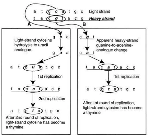
And regarding to hydrolytic nucleotide modification, the main modifications described
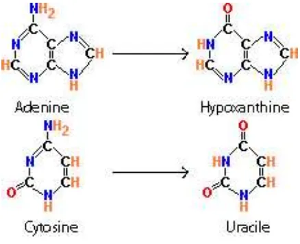
so far are the hydrolytic deamination of thymine or of adenine. The result of these processes
is the base change in the DNA backbone to uracil or hypoxantine respectively (Hofreiter et
[image:24.612.176.395.460.638.2]al., 2001b) (see Figure 3).
1.2.5 DNA SURVIVAL
If the time after death is long enough, the cumulative effects of damage to the DNA
could be so extensive that could destroy all the DNA molecules; all these molecules would be
transformed to mononucleotides or derivates. Theoretical considerations based on the
chemistry and physics of DNA, also taking into account favourable environmental conditions,
indicate that it cannot survive any longer than one million years, and probably not longer than
100,000 years in most cases (Lindahl, 1993a). Nevertheless, in exceptional circumstances,
this degradation can be significantly reduced. Such conditions include a fast inactivation of
the nucleases activity, inhibition of the action of micro organisms, fast desiccation, low
temperatures (Hoss et al., 1996a, Smith et al., 2003) and high salt concentration (Hofreiter et
al., 2001b). Of such conditions, the temperature is the most important environmental factor in
the preservation of the genetic material; due to chemical organic reactions that lead to DNA
damage take place with low rate at low temperatures. Hoss et al (Hoss et al., 1996b) showed
that a reduction of 20ºC in the average temperature of the fossil remain lead to a reduction of
1.3 DNA RETRIEVAL FROM FOSSIL REMAINS
As we have already seen, it is likely that DNA from a fossil remain is undergoing
some kind of post mortem DNA damage. Furthermore, fossil remains tend to be rare and
precious, and recovering DNA from them implies using destructive techniques. Thus, it seems
logical that the first question that an ancient DNA researcher may wonder before starting the
study of an ancient sample is: “How likely is retrieving DNA for this sample?”. Some
empirical rules that can help us on knowing if it is worth trying to extract DNA from a fossil
remain has been described. First, DNA preservation from a fossil correlates with the
environmental characteristics of the archaeological site where it has been found (temperature,
pH...). For instance, the possibility of retrieving DNA from a sample found in permafrost is
higher than in a sample found in a desert (Willerslev et al., 2003). Smith et al (Smith et al.,
2003) argued that the thermal history of hominid fossil is a key parameter for long term
survival of bio molecules in the fossil record. The thermal age of a hominid fossil was
defined as the time taking to produce a given degree of DNA degradation (assuming DNA
depurination as the principal mechanism of degradation) when temperature is held constant at
10ºC. This analysis suggests that 17,000 years at 10ºC may be a practical upper limit for DNA
survival. Second, the macroscopic appearance of the fossil remain, specially the degree of
porosity of the bone (Gilbert et al., 2005). Third, and contrary to what could intuitively be
expected, the age of the sample is not a good indicator of the state of DNA preservation
(Paabo et al., 1989, Hoss et al., 1996b). In other words, a young fossil remain does not
necessarily imply a better DNA preservation than an older fossil remain.
Thus, as we have already seen, many factors are involved in the DNA survival. An
indirect practical test has been introduced to compute the degree of damage of a fossil DNA
sample (Poinar et al., 1996). It only requires a small amount of the specimen to be analysed
(about 50 times less that would be used for a DNA extraction). This test measures changes in
the three dimensional structures of aminoacids which correlates with the degree of DNA
damage rate (see Figure 4). In all aminoacids used in proteins, except glycine, there are four
different chemical groups attached to one carbon atom know as an alpha carbon. These groups
are arranged in a tetrahedral shape, with the carbon at the centre. There are two different ways
to arrange the groups, which are chemically identical, but mirror images of each other, called
the L-form. After death, however, transitions to the D-form occur (a process known as
recemization), and eventually a dynamic equilibrium is reached in which the proportions of
L- and D- forms are equal. The rate at which racemization takes place differs for each
aminoacid and is dependent on the presence of water, the temperature and the chelation of
certain metal ions to proteins. Racemization is thus affected by some of the same factors that
affect depurination of DNA, the major hydrolytic reaction responsible of the spontaneous
degradation of nucleic acids (Lindahl, 1993a). The racemization of Aspartic Acid (Asp),
which has one of the fastest racemization rates, has an activation energy and rate constants
over a wide temperature range (at neutral pH) that are similar to those for DNA depurination
(Lindahl, 1993a). Poinar et al (Poinar et al., 1996) observed that no DNA sequences could be
retrieved from samples in which the D/L Asp ratio was higher than 0.10. Furthermore, given
that the amount of fossil aminoacids is extremely small, contamination with modern
aminoacids will tend to substantially modify the D/L ratio of all of them. Thus, since the
racemization rate of the Asp is faster than any other aminoacid of the same age, a D/L ratio
for Asp that is lower than the rate for any other aminoacid should be an indication of
contamination by more recent aminoacids.
Figure 4: Amino acid racemization. R indicates the amino acid side chain, which varies between aminoacids.
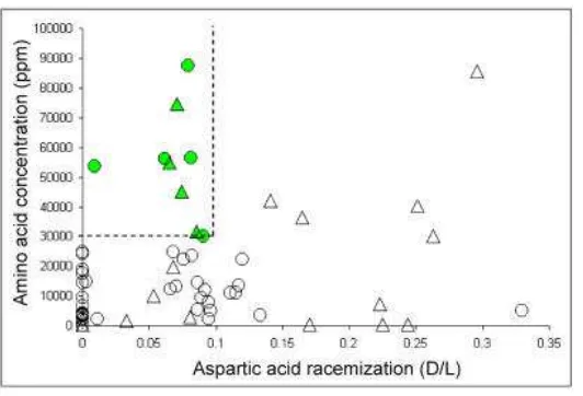
However, Serre et al (Serre et al., 2004) found that the preservation of endogenous
DNA in fossils is correlated not only with the degree of aminoacid racemization content but
also with the amount and composition of the amino acids in the ancient sample. They found,
in addition to the previously defined D/L cut off, that endogenous DNA from a Pleistocene
remain can be amplified when the amino acid content is higher than 30,000 parts per million
Figure 5: Amino Acid analysis of 64 Hominid remains. For each bone, the extend of Aspartic Acid racemization (D/L) and the aminoacid concentration (ppm) is given. The dash lines delimit the area of amino acid preservation compatible with DNA retrieval. Circles and triangles represents early modern humans and Neandertals respectively (from (Serre et al., 2004).
Nevertheless, DNA retrieval from a well-preserved sample (environmental conditions,
positive Asp racemization test and large amino acid concentration) is not going to be an easy
task. The aDNA researcher will face to a big amount of technical problems:
1.3.1 DNA COPY NUMBER
The number of DNA molecules is a crucial factor when trying to recover aDNA from an
ancient specimen (e.g. (Handt et al., 1994a, Handt et al., 1994b, Krings et al., 1997, Handt et
al., 1996). The researcher has to be sure that the number of endogenous molecules in an
ancient DNA extract is high enough in order to be confident about the final results. The
exponential nature of the PCR process implies that if the starting number of aDNA molecules
is extremely low, amplified aDNA molecules could be biased towards particular subsets of
copies of putatively damaged template. Thus, conclusions based on these results could be
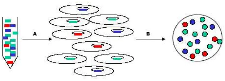
erroneous (see section 1.3.6). Furthermore, knowing the number of molecules in the extract is
extremely important due to the problem of modern human contamination (see section 1.3.5);
the lower the amount of endogenous DNA molecules in the extract, the more likely the PCR
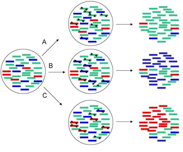
will be contaminated by exogenous DNA (see Figure 6). However, when working with
ancient human remains, since the endogenous sequences and the putatively contaminant
sequences are very similar, when quantifying them it would not be possible to differentiate
[image:28.612.136.402.64.245.2]The limitation of the number of endogenous DNA molecules is one of the main reasons for
using mitochondrial DNA (mtDNA) instead of nuclear DNA (Paabo et al., 2004). Since each
cell contains several mitochondria (up to hundreds) and each mitochondria contain several
genomes (see section 1.6), it is easier to be successful in amplifying sequences from the
mtDNA genome than from the nuclear DNA genome (e.g. (Paabo, 1989, Handt et al., 1994b);
the ratio can be 1 to 10,000 (Robin and Wong, 1988).
Figure 6: A) PCR result if the first steps of the PCR reaction are based on damaged molecules. B) PCR result if the first step of the PCR reaction are based on contaminants sequences. C) PCR result if the first step of the PCR reaction are based on the endogenous molecules
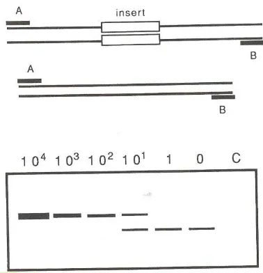
To find out if the number of DNA molecules is high enough to obtain reproducible results,
two types of approach has been done so far. In the first studies (e.g. (Krings et al., 1997), the
amount of endogenous DNA molecules were measured by using a molecular biology
technique called quantitative PCR. The basic principle of the quantitative PCR is the
construction of a “competitor construct”. This construct is a sequence equal to the endogenous
we are trying to quantify. The only difference is that it has a deletion in the sequence. Once
we have the construct, several PCRs are set up, each of which carries different dilution of the
“competitor construct”. The primers are specific for the putative endogenous sequence. As the
construct is smaller than the putative endogenous sequence, it is amplified easier in the first
[image:29.612.130.430.203.452.2]
the concentration of molecules of the putative endogenous DNA is higher than the
concentration of the construct, we start having results of the two sizes. Since the concentration
of the construct is known in each PCR reaction, the concentration of the putative endogenous
sequence can be extrapolated (see Figure 7).
Figure 7: Schematic illustration of quantitative PCR. Above the template from a tissue extract which is amplify by primers A and B and the same template in which an insert has been introduced. To a constant amount of extract, a dilution series of a known amount of the insert template is given. Above the lanes, the number of added molecules is indicated. It can be seen that there are approximately ten copies of the target sequence in the extract added to the PCR. (adapted from (Handt et al., 1994a)
There are two main pitfalls of this technique. On one hand, this technique is tedious and on
the other hand the results that we can obtain are only an approximation. For this reason,
nowadays another technique is used to quantify the endogenous DNA molecules more
accurately. This technique is based on the RT-PCR (real time PCR) reaction (Whelan et al.,
2003, Alonso et al., 2004, Malmstrom et al., 2005) and it is the most accurate method to
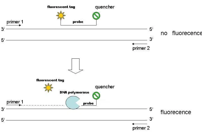
quantify a specific segment of DNA so far. In such RT-PCR, a probe lies downstream of the
forward primer and has a fluorescent tag at the 5’ end and a quencher at the 3’ end. As the
PCR proceeds, taq polymerase uses its 5’-3’ exonuclease activity and destroys the probe thus
generating fluorescence, which is proportional to the amount of amplified target DNA
present. Hence, if fluorescence is rapidly detected, then large amounts of DNA are present
and vice versa when release is small (see Figure 8). Despite the exquisite sensitivity of this
reaction, it is necessary to use some form of calibration in order to know the number of target
[image:30.612.98.287.166.362.2]Figure 8: Real Time PCR experiment. A probe lies downstream of the forward primer and has a fluorescent tag at the 5’ end and a quencher at the 3’ end. As the fluorescent tag and the quencher are closed, the fluorescent emited by the tag is annulled by the quencher.Since the taq polymerase has exonuclease 3’-5’ activity, when it is elongating and find the probe it is able to digest it. When the probe is degested, the fluorescent tag and the quencher are far away so, fluorescent is emitted. The fluorescent emmited is proportional to the number of molecules in the sample.
It is known that if a PCR starts from 1,000 or more molecules, then an experiment does not
need to be repeated to verify that nucleotide misincorporation does not influence the final
result. Nevertheless, several repetitions become necessary when fewer molecules initiate a
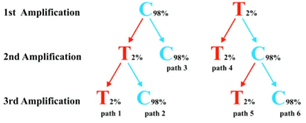
reaction. Hofreiter et al, (Hofreiter et al., 2001a) claimed that at least two independent
amplification products are needed when the extracts of ancient specimens contain few
template molecules in the PCR. In the case of obtaining discrepancy results then, at least, one
more amplification should be performed to determine which of the two sequences is
reproducible (see Figure 9). They argued that when such precautionary measurements are
taken, errors induced by damage to the DNA template are unlikely to be more frequent than
0.12% even under the unlikely scenario where each amplification starts from a single template
[image:31.612.131.468.87.309.2]
Figure 9: Schematic illustration of the strategy where two or three amplifications are used to determine an ancient DNA sequence. The correct base (C) is blue and the incorrect base (T) red. An error rate of 2% is assumed and the probability of determining an incorrect base is the sum of the three paths that arrive at the incorrect base after two or three amplifications (from (Hofreiter et al., 2001a).
1.3.2 DNA FRAGMENTATION
Several studies have been published so far (Paabo, 1989, Handt et al., 1994a, Handt et al.,
1994b, Poinar et al., 2006) showing that it is almost impossible to obtain long amplification
products from an ancient template. Furthermore, it has been observed an inverse relationship
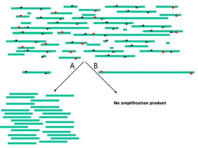
between amplification efficiency and length of the amplification products (e.g. (Handt et al.,
1994a, Malmstrom et al., 2005). In practice, this means that long sequences can only be
retrieved by means of short sequences using overlapping primers (see Figure 10). The
maximum length of the overlapping amplification will depend on the state of preservation of
the sample. For instance, Lalueza et al (Lalueza-Fox et al., 2006) were able to amplify 300 bp
of the mitochondria control region of a Neanderthal specimen by using overlapping fragments
of only 70-80 bp. In contrast, Cooper et al in 2001 (Cooper et al., 2001) were able to amplify
the complete mitochondrial genome sequence of two extinct moas as a series of 400-600bp
Figure 10: A representation of fragmented strands. A) A situation in which we try to amplify an enough short fragment so both primers can hybridise and therefore the PCR amplifiction is possible. B) A situation in which we don not get any amplification product due to one of the primers can not hybridise.
1.3.3 JUMPING PCR
It is a phenomenon that can occur during the PCR reaction due to the presence of
fragmentation in the molecule templates; it occurs when one template “recombines in vitro”
with another that shares a similar sequence during the PCR reaction. It was described for the
first time in 1990 by Svante Pääbo and collaborators (Paabo et al., 1990). They designed
specific pairs of templates partially sharing the same sequence and tried to amplify them by a
pair of primers, one primer specific to one template and the other primer specific to the other
template (so neither of the templates contained both primer sites). They showed that the only
way to obtain PCR products was by fragmenting the templates with restriction enzymes, so
jumping PCR happens (Paabo et al., 1990). Furthermore, they found that lesions such as
breaks or AP sites can cause the extending primer to jump to another similar template during
the PCR. Consequently, amplification products from damaged templates such as
archaeological DNA could be made up of a high proportion of chimerical molecules between
endogenous and contaminant sequences or endogenous with post mortem damage and without
analysing such results, since the chimerical products of this reaction could be erroneously
taken as a novel ancient DNA sequence.
Molecular cloning and sequencing of multiple clones is highly recommended in order to
detect such kind of phenomenon in our sample; these analyses will help us to sort out the
different types of molecules present in the amplification products and distinguish chimeric
products from those that are not. Nevertheless, it has been argued that the presence of a
jumping PCR phenomenon in the PCR products could be used as an indication that the DNA
may be of ancient origin (Handt et al., 1994a).
[image:34.612.103.469.240.531.2]1.3.4 INHIBITORS
It is well-known for the aDNA community that when nucleic acids are extracted from ancient
specimens, the extracts often contain components that can inhibit the activity of the DNA
polymerises (Hanni et al., 1995); furthermore, it is known that that inhibition affects longer
amplification fragments more than shorter fragments (Pusch and Bachmann, 2004). However,
after the extraction protocol and in order to avoid the presence of these inhibitors, the
resulting aqueous phase is carefully removed (as carryover of organic solvents to subsequent
extraction and amplification stages may inhibit PCR), and desalted and concentrated using
usually filtration (Cooper et al., 2001). While this procedure usually removes possible PCR
inhibitors of less than 30,000 molecular weight (MW), in some samples (such as these buried
in the soil and paleofaeces) it is common to find some co-extraction of PCR inhibitors along
with the DNA. One simple solution to deal with this problem is the incorporation of bovine
serum albumin (BSA) (or other molecules that bind to inhibitory chemicals) to PCR reactions
(Paabo et al., 1988). BSA binds to inhibitors and thus prevents them from inhibiting the
polymerase enzymes activity. Another way of stopping the activity of these inhibitors
wide-used by the aDNA community is by making serial dilutions of the extracts until we find one
dilution for which the PCR is successful (e.g. (Paabo, 1989, Woodward et al., 1994a,
Binladen et al., 2006).
Nevertheless, little is known about the nature of those inhibitors; it has been proposed that
they are soil components such us humic or fluvic acids (Paabo et al., 1989) or sub products
derived from organic reactions, such the volatile compound products of the Maillard reaction
(Poinar, 2002). The Maillard reaction is defined as the reaction of carbonyl groups on
reducing sugars (such as those that belong to the backbone skeletal of the DNA double helix)
with the primary amines of aminoacid and forms a structure known as cross links. It has been
demonstrated that treatments with the reagent PTB (N-phenacyltiazolium bromide) breaks the
cross links DNA-protein and allows DNA sequences to be amplified from some ancient
1.3.5 CONTAMINATION
We define a contaminant sequence as an exogenous (and probably modern) DNA molecule in
the pool of aDNA sequences. Contamination is one of the most common pitfalls in aDNA
studies and represents one of the biggest threads when working with samples from ancient
humans. The problems regarding ancient human DNA and modern human contamination,
often detected of more than one haplotype of one single individual has been known several
years ago(Handt et al., 1994a, Handt et al., 1996, Kolman and Tuross, 2000, Gilbert, 2005b).
As the retrieval of tiny amounts of DNA from ancient remains is a multi-step process,
contaminants can enter in multiple stages:
1.) Sample Handling: This kind of contamination is extremely important in the study
of ancient human remains (the main objective of the current thesis) because
usually cannot be monitored or controlled (only it can be monitored in
contemporaneous excavations). Unprotected handling of the remains may
impregnate the samples with the handlers sweat or skin cells, and thus exogenous
DNA could penetrate into the remains(Gilbert, 2005a). Bone and teeth remains are
extremely porous and thought very susceptible to contamination by handling. The
pulp cavity of the tooth is directly connected to the exterior by numerous dental
tubules. Therefore, once a tooth is washed or the root is directly handled, the
impervious nature of enamel is of no help in limiting contamination by handling
(Gilbert, 2005b). Mercury porosimetry demonstrates that the minimum total
interconnected porosity in human bones is higher than 8% of the bone volume, the
majority of witch is derived from the Harvesian canals(Gilbert, 2005b). We can
distinguish different potential sources of handling contaminants depending on the
step of DNA retrieval:
- Archaeologists and anthropologists: they are responsible of recovering,
washing and macroscopically analysing the fossil remains and interacts
directly with the fossil material.
- Geneticists: Once the sample arrives to the aDNA laboratory, the genetic
team must decontaminate the sample using bleach or UV light before
mask face and coverall inside the aDNA laboratory in order to avoid
contamination by handling.
2.) Extraction procedure: Contaminants can be introduced in the aDNA extract
during the preparation of all the reagents that are going to be used in the extraction
process as well as in the place were the extraction is going to be performed. In
order to avoid this kind of contamination, it is very important that the place where
the aDNA extraction is carried out is physically separated from the main
laboratory (molecular biology laboratory) with positive air pressure, UV light at
night and continually bleach cleaning of the bench surfaces (Cooper and Poinar,
2000). In addition, the manipulation of all reagents must be done in a flow cabinet
and the use of coverall, gloves, facemask and sterile tips is compulsory.
3.) PCRs setting up: Contaminants can be introduced in this stage by two main ways:
First, due to the low specificity of the primers. PCR reaction are based upon the
premise that primers will bind to specific loci not present in unrelated organism,
and thus will selectively amplify only the DNA of interest under specific PCR
conditions (e.g. annealing temperature) However, there could be DNA from some
soil micro organisms present in the aDNA extract. The low amount of aDNA
forces using unspecific conditions (e.g. high number of cycles and low
temperature) in the PCR. For this reason, it could be that the primers can also bind
to those similar sequences that are present in the extract and therefore amplify
them. Second, contaminants can be introduced while researchers are setting up the
PCR reaction. As in the extraction step, contaminants can be in the used reagents
and in the environment where researchers are working. All the precautions that are
described in the previous step are compulsory here as well. The setting up of the
PCR reaction must be done in the ancient laboratory and then carried to the main
laboratory. Material interchange between the aDNA laboratory and the main
laboratory should be strictly forbidden because the risk of contamination is
extremely high due to the large amount of amplicons that are generated in the
1.3.6 MISCODING LESSION
Unfortunately, although the majority of the damage that the DNA molecule can suffer after
cell death inhibits the activity of PCR enzymes (for example, strand fragmentation or
intermolecular cross-links; see sections 1.3.2 and 1.3.4), a small proportion of damage events
do not inhibit the polymerase activity but generate miscoding lesions. Miscoding lesions are
defined as base modifications in the amplified sequence that change the appearance of aDNA
template (Fattorini et al., 1999) and potentially generate misleading haplotype analysis
(Gilbert et al., 2003a, Gilbert et al., 2003b). It has been reported several times (for example,
see (Paabo, 1989, Hofreiter et al., 2001a, Binladen et al., 2006) that the majority of miscoding
lesions arise from the deamination of C to U or from A to hipoxantina (HX); this process is
particularly fast in the case of cytosine (Lindahl, 1993a). When a DNA molecule containing
such lesions is used as a PCR template, C→T and G→A transitions will be introduced erroneously in the final sequences. However, because either of the complementary DNA
strands can be sequenced after amplification when using Sanger sequencing technology, each
of these transitions can produce two observable phenotypes (see Figure 12). Hansen et al
(Hansen et al., 2001) termed each set of miscoding lesions as type I (A→G and T→C) and type
II (G→A and C→T).
Interestingly, the recent development of the sequencing-by-synthesis technology (see
(Margulies et al., 2005) offers the possibility of going deeper into the nature of those
miscoding lesions (Gilbert et al., 2006, Stiller et al., 2006). The advantage of this new
technique is that the nature of the date generation process is such that DNA sequence data can
be assigned to individual, originally single-stranded molecules (Gilbert et al., 2006).
Basically, DNA extracted from an ancient remain is ligated to biotynilated linkers and single
DNA strands are attached to Sepharose beads, amplified by PCR and subjected to
pyrosequencing (Margulies et al., 2005). Therefore, each read sequence derives from one
single-stranded DNA molecule, and the read sequence allows the actual template strand to be
inferred. (Gilbert et al., 2006) using this new technology demonstrated thought comparative
analyses on 390 965 bp modern chloroplast and 131 437 bp ancient woolly mammoth
sequence date that type 2 (G→A and C→T) miscoding lesions represent the majority of damage derived miscoding lesions. Furthermore, they suggested, in contrast to previous
studies (Paabo et al., 1989, Hofreiter et al., 2001a, Gilbert et al., 2003a, Binladen et al., 2006)
that the predominant cause of Type II transition is not the cytosine to uracil deamination, but
Nevertheless, Stiller et al (Stiller et al., 2006) using the same DNA technology as (Gilbert et
al., 2006) found that the predominant cause of Type II transitions are the cytosine residues
when analysing an ancient mammoth remain. They suggest that the lesion affecting cytosine
residues is very likely to be by deamination.
Given the importance of the control region of human mtDNA studies (see chapter 1.6),
Gilbert et al (Gilbert et al., 2003a, Gilbert et al., 2003b) studied the post mortem damage
distribution of this region. Contrary to what a priori is expected, they found that postmortem
DNA damage is not randomly distributed across the control region, but there are “hotspots” of
postmortem damage. In other words, there are some positions that are more prone to suffer
postmortem damages than others. Furthermore, DNA damage occurs preferentially at those
positions that have been detected as fast evolving in human population studies. Gilbert et al
(Gilbert et al., 2003b) hypothesise that these mutational hotspots may be hyper mutable
because they are more exposed to environmental damage than the others or they are not
binding sites of proteins that can protect them. They concluded that postmortem damage in
the control region could difficult population genetic analysis of ancient humans due to the
impossibility to distinguish whether a mutation is due to a postmortem damage or it was
already in the sequence.
The use of the enzyme UNG (Uracil N-Glycosylase) has been highly recommended in order
to avoid the presence of miscoding lesions in the final sequence. This enzyme excises the
uracil bases in the DNA molecule (produced due to the deamination of cytosine) and therefore
Figure 12: Determination of a strand of origin for postmortem-DNA-damage events by using type 2 (C→T/G→A) transitions as an example. A, L-strand C→T transitions after two cycles of amplifications, resulting in a permanent L-strand change. B, A theoretical H-strand G→A change, producing the L-strand phenotype of C→T change following one cycle of amplification. However, since a direct G→A postmortem modification is chemically impossible, the example depicted in this panel is not possible. Thus, all C→T changes observed on the L strand must have occurred as L-strand C→T postmortem damage, and all G→A changes on the L strand must have occurred as H-strand C→T postmortem damage (From (Gilbert et al., 2003a)
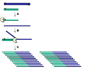
1.3.7 MOLECULAR CLONING
Molecular cloning is a well-known molecular technique in aDNA field. It allows us to detect
sequence heterogeneity in a single PCR reaction, which can be associated to jumping PCR,
contamination events and/or miscoding lesions by endogenous DNA damage or Taq
polymerase errors. Cloning PCR products consist in inserting a single PCR product inside a
bacterial plasmid, which is subsequently inserted into a bacterial cell (Escherichia.coli). After
growing up the bacteria, a posterior screening will allow us to select those bacteria that carry
the insert. Colonies derived from successfully transformed cells can be identified through
[image:40.612.151.389.64.281.2]Since each bacterium incorporates a single amplicon in its genome, postmortem DNA
damage, jumping PCR and contamination can be assessed (see Figure 13) by screening a
reasonable number of clones (see(Bower et al., 2005).
Nowadays, when working with ancient templates (mostly when working with human ancient
templates) cloning each PCR product is crucial to detect sequence heterogeneity in the
[image:41.612.111.493.227.365.2]extract.
1.4 AUTHENTICITY CRITERIA
As we have seen (see section 1.3), aDNA research presents extreme technical problems
(specially when working with human remains), which can lead to erroneous conclusions.
In the nineties, several aDNA works published in high profile journal were proved to be
erroneous (see section 1.1). It was in 1994 when Hand et al (Handt et al., 1994a) published
the first list of authenticity criteria that should be followed by all researchers in order to make
aDNA a respectable and credible scientific field. Cooper and Poinar updated that list in 2000
(Cooper and Poinar, 2000) and then Paabo et al in 2004 (Paabo et al., 2004):
1.) Physically isolated work areas: the best case scenario is having the aDNA laboratory
(where the extractions and the setting up of the PCRs reactions take place) and the
main laboratory (where all the PCR amplifications are run) in different buildings in
order to avoid contaminations from former amplified products.
2.) Negative control extractions and amplifications to detect sporadic or low-copy number
contaminations during each stage. Positive controls should generally be avoided as
they provide a contamination risk.
3.) Appropriate molecular behaviour: Due to DNA degradation, PCR amplification
strength should be inversely related to product size. In general, if shorter fragments are
not easier amplified than longer ones, it is an indication that the source of DNA is
likely to be a modern contamination (Paabo et al., 1989, Handt et al., 1994a).
Therefore, very long PCR products (> 500 bp) are suspected to be contaminants.
4.) Reproducibility: Multiple PCRs and extractions from the same sample should yield
consistent results. First, they are useful to detect contamination of a particular
extraction or amplification and second, nucleotide misincorporation leading to
consisted changes can be detected only when multiple amplifications are performed.
Overlapping primers are also highly recommended in order to detect for instance
NUMTs (see section 1.6.1)
5.) Cloning of amplification products and sequencing of multiple clones: In order to
detect post mortem damage, contamination, jumping PCR events and to unravel
6.) Independent replication: separate samples of a specimen are extracted and sequenced
in independent laboratories in order to detect intra laboratory contamination.
7.) Biochemical preservation: Biochemical assays of macromolecular preservation serves
two purposes: First, they support the claim that a specimen is well enough preserved
to allow the preservation of DNA. Second, they may be used as rapid screening
techniques to identify specimens that, according to their general state of preservation,
may contain DNA. Several techniques have been suggested (see section 1.3), although
the most widely used is the analysis of the rate of racemization of Aspartic aminoacid
(Poinar et al., 1996).
8.) Quantization of DNA templates by means of quantitative PCR or RT-PCR (see section
1.3.1). If a large number of molecules is present (>1000), there is no need to perform
several amplifications since consistent changes are extremely unlikely to occur.
However, when the number of starting templates is low, several amplifications are
needed in order to exclude the possibility of sporadic contamination.
9.) Associated remains: In studies of human remains where contamination is especially
problematic, the presence of similar DNA targets survive in associated faunal material
is one additional supporting evidence of DNA preservation because it shows that the
environment is favourable to it.
Mainly papers related to ancient humans or Neandertals remains (e.g. (Caramelli et al., 2003,
Vernesi et al., 2004) fulfil completely the list since its publication, whereas papers related to
extinct animals do not (e.g. (Shapiro et al., 2004, Bunce et al., 2005). In studies related to
ancient animals, the phylogenetic criterion is more deterministic because it is easier, in this
particular case, to discard modern contamination. Furthermore, this stringent list has been
severely criticised by some authors (see (Gilbert et al., 2005, Hebsgaard et al., 2005, Bandelt,
2005)that argue that getting a reliable result does not only mean that the researchers have only
fulfil the nine criteria but that the researcher should be more cognitive and self-critical in their
results. In other words, instead of checking if the list of criteria has been completely fulfilled,
researchers should pay more critical attention to the way the data was obtained and why the
results should be considered authentic in the particular context and conditions of the samples
analysed.
Some current results (see (Malmstrom et al., 2005) claimed that the list of the well-known
nine authenticity (Cooper and Poinar, 2000) criteria should be reviewed and subsequently
1.5 ORIGIN AND MAINTENEANCE OF THE CURRENT
HUMAN GENETIC DIVERSITY
It is clear that humans are all different from each other. The diversity that we observe
between individuals is due to both genetic and environmental factors. Current estimates of the
human genetic diversity say that if we take two not related individuals at random,
approximately 1 of each 1000 nucleotides will be different (Reich et al., 2002). These genetic
differences exist as well between human populations. Contrary to the what would be expected
based on the phenotypical variability observed between human populations, different studies
(Excoffier et al., 1992, Barbujani et al., 1997, Romualdi et al., 2002, Jorde et al., 2000) point
out that if the human individuals are hierarquicaly clustered in populations and continents,
approximately 80% of the variance of the model is explained because of differences between
individuals of the same population. Only 5% to 10% is explained because of differences
between populations of the same continent and a 10% to 15% of the variance is explained by
genetic differences between continents. This result has been traditionally used as a prove of
the lack of sense of clustering the individuals according to races (Kittles and Weiss, 2003)
The current genetic variation we observe in the human populations is the result of the
complex interaction between four evolutive forces: mutation, natural selection, genetic drift
and migration. Disentangling the effect of each one in shaping the genetic variability of our
genome can help us on understanding both past demographic and selective events that
occurred in the human species.
1.5.1 MUTATION
Mutation is defined as a structural change in the DNA molecule. It is the ultimate source of
genetic variation and thus, allows the evolution to be possible (Crow, 1997). The term
mutation covers a broad range of structural events: from substitution of a single base to
insertions and deletions of a few bases or even chromosomal rearrangements. The molecular
mechanisms that generate that big spectrum go from chemical mechanism (for instance,
cytosine deamination), physical mechanisms (for instance, breaking of the double helix for