Relative Adrenal Insufficiency is
Associated with the Clinical Outcome in Patients
with Stable Decompensated Cirrhosis
Evangelos Cholongitas,* Ioannis Goulis,* Eirini Pagkalidou,** Anna B. Haidich,** Apostolos K.A. Karagiannis,* Theodora Nakouti,* Chrysoula Pipili,*** Theodora Oikonomou,* Spyros Gerou,* Evangelos Akriviadis*
* 4th Department of Internal Medicine, Hippokration General Hospital, Medical School Aristotle University of Thessaloniki, Greece. ** Department of Hygiene and Epidemiology, School of Medicine, Thessaloniki, Greece. *** Division of Nephrology, Queen Elizabeth University Hospital, Glasgow, UK.
July-August, Vol. 16 No. 4, 2017: 584-590
INTRODUCTION
Over the last decades decompensated cirrhosis (DeCi) has been accepted as a chronic disease affecting, except for the liver itself, many extra-hepatic organ systems.1 In view
of that, additional relative adrenal insufficiency diagnosis among patients with DeCi (i.e. hepato-adrenal syndrome) perplex management and prognosis of this patient popula-tion and trigger further research in this field.2 Although AI
was firstly described in patients with liver diseases several decades ago,3 the evidence on its exact pathogenetic
mech-anisms among cirrhotic patients are not satisfactory. While several lines of evidence exist for cirrhotic patients with sepsis or acute complication of cirrhosis4,5 minimal
de-tailed studies have been published for clinically stable pa-tients with DeCi.6
The most crucial issue requiring further exploration, is the clinical impact of AI on the prognosis of stable cir-rhotic patients. So far, AI in critically ill circir-rhotic patients (with septic shock or variceal bleeding) has been found to correlate with higher mortality, with the corticosteroid administration not to account for survival improvement in all cases.2 In regards to non-critically ill cirrhotic patients,
the published studies remain controversial. While a recent study showed that AI was associated with worse outcome in patients with DeCi7 after their complication [with at
least one episode of ascites, hepatic encephalopathy, bacte-rial infection, variceal bleeding or hepatorenal syndrome The Official Journal of the Mexican Association of Hepatology,
the Latin-American Association for Study of the Liver and the Canadian Association for the Study of the Liver
Manuscript received: Manuscript received: Manuscript received: Manuscript received:
Manuscript received: August 17, 2016. Manuscript accepted:Manuscript accepted:Manuscript accepted: December 19, 2016.Manuscript accepted:Manuscript accepted:
DOI: 10.5604/01.3001.0010.0298
A B S T R A C T A B S T R A C T A B S T R A C T A B S T R A C T A B S T R A C T
Background. Background.Background. Background.
Background. The clinical impact of relative adrenal insufficiency (AI) on patients with stable decompensated cirrhosis (DeCi) has not been yet elucidated. Aim. Aim. Aim. Aim. Aim. Explore the association between AI and outcome [death or liver transplantation (LT)] in patients with DeCi. Material and methods.Material and methods.Material and methods.Material and methods. Patients with DeCi presenting no active complication have been included. Clinical and laboratoryMaterial and methods. data, including serum levels of corticosteroid-binding globulin (CBG), interleukin (IL)-1b, IL-6 and tumor necrosis factor (TNFα) were recorded in each participant. Salivary cortisol (SC) and serum total cortisol (STC) were assessed at (T0) and 1 h (T60) after intrave-nous injection of 250 μg corticotropin. Results.esults.esults.esults.esults. 113 consecutive patients were totally tested. Median SC was 3.9 ng/mL and 15.5 ng/mL and median STC was 10.7 μg/dL and 22.7 μg/dL at T0 and T60 respectively. The patients with AI [group 1, n = 34 (30%)] had significantly lower systolic blood pressure (106 ± 12 vs. 113 ± 13 mmHg, p = 0.05), serum sodium (133 ± 7 vs. 137 ± 12 mEq/ L, p = 0.04), HDL (29.9 ± 14 vs. 38.6 ± 18 mg/dL, p = 0.034) and albumin (2.7 ± 0.5 vs. 3.1 ± 0.5 g/dL, p = 0.002), but higher di-rect bilirubin (median: 1.6 vs. 0.8 mg/dL, p = 0.029) compared to those without AI [group 2, n = 79 (70%)]. Moreover, group 1 pa-tients presented more frequently past history of spontaneous bacterial peritonitis (SBP) [10/34 (29.4%) vs. 6/79 (7.5%), p = 0.002]. AI was significantly associated with death [HR = 2.65, 95% C.I.: 1.55 - 4.52, p = 0.003 over a follow up period of 12 (6-48) months.] Conclusions.
Conclusions.Conclusions. Conclusions.
Conclusions. The presence of AI in patients with stable DeCi predispose to obvious clinical implications since it is associated with circulatory dysfunction, previous history of SBP and worse survival.
Key words. Key words.Key words. Key words.
(HRS)] this was not confirmed in other two studies.8,9 As
demonstrable causes for this contrast is the lack of con-sensus for the appropriate definition of adrenal function evaluation (e.g. using total serum vs saliva cortisol) and the various cortisol cut offs which result in fluctuations on prevalence and clinical significance of AI.2 The aim of the
present study was to evaluate the prevalence of AI -by measuring the total and salivary cortisol using the short synacthen test (i.e. before and after the administration of 250 μg corticotrophin intravenously)- and the impact of AI on patients with clinically stable DeCi.
MATERIAL AND METHODS
All the consecutive adult patients with stable DeCi, ad-mitted for liver transplant (LT) evaluation to our Depart-ment from March 2011 till December 2013 were studied prospectively. DeCi diagnosis was based on laboratory and/or ultrasound evidences. All the patients suffering from hypothalamus-pituitary-adrenal axis diseases, who had been receiving steroids or other immunosuppressive medications over the last 6 months before admission and had hepatocellular carcinoma (or other types of cancer), variceal bleeding, encephalopathy or infection, such as spontaneous bacterial peritonitis (SBP), during the last month before admission were excluded. Furthermore, de-tailed clinical evaluation, laboratory measurements (white blood cells, C-reactive protein, procalcitonin, blood cul-tures and ascitic fluid paracentesis) and radiological exams (chest x-ray, upper abdominal ultrasound) if necessary, were performed to exclude active infection on admission.
Demographic details registration included the cause and duration of liver disease, the past complications of DeCi [i.e. variceal bleeding, hepatic encephalopathy, as-cites, SBP], the medication dosage and duration and the vital signs (blood pressure, pulse rate, body temperature) on admission. The short synacthen test (SST) was per-formed after an overnight bed rest. In details, an intrave-nous catheter was inserted from the previous night and strict instructions to avoid tooth-brushing, eating or drinking 4 h prior to AI evaluation were given to all pa-tients. Basal total serum cortisol was taken at 08:00 am (time T0, baseline) through the intravenous catheter al-most in parallel with the salivary cortisol evaluation per-formed through a specific oral cotton (Plain Salivette; Sarstedt, Newton, North Carolina, USA) insertion. The cotton was left for 2-3 min in the oral cavity following prompt test for blood contamination.
Subsequently, 250 μg corticotrophin (Synacthen, No-vartis Pharma, Basel, Switzerland) was given bolus intra-venously and the patients remained recumbent for an hour until a second blood sample obtained, for the (Τ60, 09:00 am) serum total cortisol level, and a suitable cotton placed
in the oral cavity, for the (Τ60, 09:00 am) salivary cortisol evaluation. Each cotton was collected in a plastic tube and the saliva was separated by centrifugation according to the manufacturer instructions. Basic (T0) and T60 salivary cortisol values were estimated by using an electrochemi-luminescence immunoassay (Roche Diagnostics Ltd, Rotkreuz, Switzerland). AI was defined when serum total cortisol at T60 minus serum total cortisol at T0 was < 9 μg/dL providing that serum total cortisol at T0 was < 35 μg/dL.7 In addition, based on salivary cortisol levels, AI
was defined as a value of T0 < 1.8 ng/mL or < 12.7 ng/mL at T60 or an increase between T0 and T60 less than 3 ng/ mL. Serum levels of cortisol binding globulin (CBG) (competitive radioimmunoassay; Biosource, Belgium) were measured and used for calculation of free cortisol in-dex (FCI) at T0 and T60, which stands for the ratio be-tween serum total cortisol and CBG.
At the same day, a full biochemical and lipid profile, blood count, coagulation and proinflammatory cytokines including serum interleukin (IL)-1b, IL-6 and tumor necrosis factor (TNF)-α were all prospectively recorded. 24-hour urine collections for urine sodium excretion (UNa24h) and urine samples for random “spot” sodium and potassium in order to calculate UNa/K ratio were ob-tained before or after the completion of the urine collec-tion. The severity of liver disease was estimated by the Child-Pugh (CTP) and the model for end stage liver dis-ease (MELD) scores.
Renal function was assessed by estimated glomerular filtration rate (eGFR) using CKD-EPI (chronic kidney disease-epidemiology) creatinine-based equation.10
“True” GFR was evaluated with 51Chromium-EDTA
(51Chr-EDTA) using the slope-intercept technique, cor-recting for body surface area, and the fast exponential curve recommended by the British Nuclear Medicine So-ciety guidelines11 after intravenous injection of a tracer, at
2, 4, and 6 h. Finally, cardiac echo was performed to search for diastolic dysfunction, which was classified into three categories depending on severity (Grade I, II, III) and cur-rent guidelines.12 The study protocol was approved by our
Institutional Review Board and conformed to the ethical guidelines of the 2013 Declaration of Helsinki.
Statistical analysis
pa-tients with and without AI and χ2 test for categorical
varia-bles.
Multivariable analysis was performed using backward selection of variables, starting with all variables with p < 0.1 in univariate analysis to find the independent factors signifi-cantly associated with the presence of AI. The discrimina-tive ability of the independent variables to predict the presence of AI in patients with DeCi was evaluated by using the area under a receiver operating characteristic curve (AUC). At the best cut off point (in which the sum of sensi-tivity plus specificity is maximal), sensisensi-tivity, specificity, positive (PPV) and negative (NPV) predictive values were
calculated. Cox proportional hazards regression analysis was used to identify the factors significantly associated with the outcome. The patients' survival according to the pres-ence of AI was calculated using Kaplan-Meier analysis and compared with the log rank sum test, while competing risk analysis was performed with the R project (version 3.2.2). A p value ≤ 0.05 was considered statistically significant.
RESULTS
Patients' characteristics
One hundred and thirteen patients with stable DeCi were included in the study (Table 1). The mean CTP and MELD scores were 7.8 ± 2 and 13.7 ± 4.6, respectively. The mean total serum cortisol was 10.7 ± 4.8 μg/dL and 22.7 ± 7 μg/dL at T0 and T60, respectively. The median salivary cortisol was 3.9 ng/mL (range: 1.6-17.8) and 15.5 ng/mL (range: 7.1-47.7) at T0 and T60, respectively. Free cortisol index (FCI) was 9.1 (range: 1.2-67) and 19.1 (range: 5.2-139) μg/dL at T0 and T60, respectively. CBG plasma concentrations were 38.7 ± 11.5 μg/mL and they were higher in patients with Child-Pugh class A patients, compared to those with Child-Pugh class B or C (42.6 ± 12.2 μg/mL vs. 31.4 ± 9.3 μg/mL, p = 0.02). The CBG and albumin plasma concentrations had poor correlation (Spearman r2: 0.063, p = 0.65).
Salivary and serum cortisol were significantly correlat-ed at T0 (Spearman r2: 0.48, p < 0.001) and T60 (Spearman
r2: 0.56, p < 0.001) in patients with serum albumin > 2.5 Table 1. Baseline clinical and laboratory characteristics of
pa-tients with decompensated cirrhosis in our cohort.
Variable Patients, n = 113
Age (mean ± SD, years) 55 ± 12
Sex, male, n (%) 81 (72)
Cause of cirrhosis, n (%)
Viral hepatitis 59 (52)
Alcohol 26 (23)
Non-alcoholic steatohepatitis/Cryptogenic 14 (12.5)
Others 14 (12.5)
CKD-EPI-estimated GFR (mean ± SD, mL/min) 73 ± 24 GFR by 51Crhomium-EDTA (mean ± SD, mL/min) 67.2 ± 21
Child-Pugh score (mean ± SD) 7.8 ± 2
MELD score, (mean ± SD) 13.7 ± 4.6
CKD-EPI: chronic kidney disease-epidemiology. GFR: glomerular filtration rate.
Spearman r2: 0.48, p < 0.001
Spearman r2: 0.56, p < 0.001
Figure 1. Figure 1. Figure 1. Figure 1.
Figure 1. A, B. Correlation between salivary and total serum cortisol at baseline (T0min) and after 1 h (T60min) for albumin values > 2.5 mg/dL (Spearman r: 0.48, p < 0.001 καιr: 0.56, p < 0.001, relatively).
A A A A A
Salivary cortisol at T0
40
30
20
10
0
0 5 10 15 20 25 30
Total serum cortisol at T0
B BB BB
Salivary cortisol at T60
100
80
60
40
20
0
0 10 20 30 40 50
Table 2. Clinical and laboratory characteristics of the patients with decompensated cirrhosis and with or without relative adrenal in-sufficiency (AI) based on total serum cortisol levels.
Variable Patients without AI Patients with AI P value
(n = 79, 70%) (n = 34, 30%)
Age (mean ± SD, years) 56 ± 11 54 ± 12 0.33
Systolic Blood Pressure (mean ± SD, mmHg) 113 ± 13 106 ± 12 0.05
Hematocrit 36 ± 6 34 ± 6 0.72
Albumin (mean ± SD, g/dL) 3.1 ± 0.5 2.7 ± 0.5 0.002
Protein (mean ± SD, g/dL) 7.0 ± 0.9 6.4 ± 1 0.003
Bilirubin, median (range), mg/dL 2.2 (0.5-52) 2.5 (0.9-24) 0.69
Direct Bilirubin, median (range), mg/dL 0.8 (0.1-21) 1.6 (0.7-23) 0.029
Prothrombine time (mean ± SD, sec) 15.7 ± 4 15.8 ± 3 0.77
INR (mean ± SD) 1.36 ± 0.39 1.4 ± 0.2 0.70
Creatinine (mean ± SD, mg/dL) 1.1 ± 0.5 1.0 ± 0.5 0.63
Na (mean ± SD, mmol/L) 137 ± 12 133 ± 7 0.04
K (mean ± SD, mmol/L) 4.3 ± 0.5 4.3 ± 0.6 0.91
Plasma Renin Activity (mean ± SD, ng/mL*h) 11 ± 3 17 ± 4 0.05
Aldosterone (mean ± SD, ng/mL) 44 ± 17 61 ± 12 0.044
Ferritin, median (range), mg/dL 148 (15-3,300) 193 (8-1650) 0.88
HDL (mean ± SD, mg/dL) 38.6 ± 18 29.9 ± 14 0.034
CPK, median (range), IU/L 58 (25-565) 61 (21-189) 0.71
GFR by 51Crhomium-EDTA (mean ± SD, mL/min) 69 ± 23 mL/min 63.5 ± 22 0.49
24 h urine sodium excretion (24 UNa) (mean ± SD, mmoL/day) 63 ± 24 48 ± 15 0.63
UNa/K ratio (mean ± SD) 1.6 ± 2 1.3 ± 1.1 0.47
Cytokines (IL-1b/IL-6/TNFα) (mean ± SD, pg/mL) 101 ± 42/218± 98/36 ± 12 69 ± 32/244 ± 102/52 ± 17 >0.05
Cortisol binding globulin (mean ± SD), μg/mL 41 ± 15 39 ± 12 0.74
Presence of cardiac diastolic dysfunction, n (%) 47 (59) 17 (50) 0.22
Child-Pugh score 8. 1± 3 7.7 ± 4 0.63
MELD score (mean±SD) 14 ± 5 12 ± 7 0.41
Na: sodium. K: potassium. GFR: glomerular filtration rate. CPK: creatine phosphokinase. UNa/K: ratio of Na/K in spot urine. IL: interleukin. TNF: tumor necrosis factor.
mg/dL (Figures 1A and 1B). No similar correlation was observed at both time points among patients with serum albumin ≤ 2.5 mg/dL (T0 and T60: Spearman r2: 0.012 and
0.12, respectively, p > 0.05). Based on total serum cortisol levels 34 (30%) patients presented AI (group 1) and 79 (70%) patients had not AI (group 2). When AI was defined by salivary cortisol, 25 (22%) had AI and 88 (78%) of 113 patients had not AI. Patients (n = 82) with concordant se-rum and salivary assays had statistically significant lower triglyceride levels compared to those with discordant tests (n = 31) (58 ± 10 mg/dL vs. 87 ± 22 mg/dL, p = 0.022).
Characteristics of patients
with or without AI based on total serum cortisol
Based on total serum cortisol levels, the patients with AI (n = 34, group 1) presented similar incidence of pre-vious cirrhotic complications compared to those (n = 79, group 2) without AI: moderate to severe ascites (22% vs. 18%, respectively, p = 0.87), encephalopathy (27% vs. 28%, respectively, p = 0.98) or variceal bleeding (23.5% vs. 24% respectively, p = 0.96) except for previous history of SBP which was more frequent in group 1
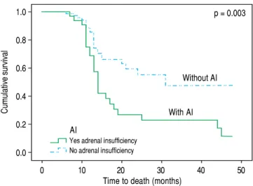
(death or LT) (HR: 0.96, 95%CI: 0.94-0.99, p = 0.05) in multivariable Cox proportional hazards regression model (the covariates included were “true” GFR, urea, AI and MELD score). Based on competing risk analysis we found that AI was significantly associated with death (using liver transplantation as a competing risk) (HR = 2.65, 95%C.I.: 1.55 - 4.52, p = 0.003) (Figure 2). Finally, the patients with AI, based on salivary cortisol, had similar mortality with those without AI [in the total cohort: log rank p = 0.94; excluding the patients who underwent LT: log rank p = 0.45].
DISCUSSION
During the last decade there is an increased interest regarding the presence of AI, which is found not only in critically ill cirrhotic patients but also in cirrhotics with stable cirrhosis indicating that AI is a feature of liver dys-function per se.2 So far, there is no consensus regarding
the appropriate definition of AI in cirrhosis; total serum cortisol measurements are considered to overestimate AI as the low serum albumin and CBG -which are both common findings in cirrhotic patients- are not taken into account.13 This study demonstrates that the prevalence of
AI in cirrhotics is greater when serum total based definition is used compared with salivary cortisol-based definition (30% vs. 22%) and the correlation between salivary (a surrogate marker of free serum corti-sol) and total serum cortisol is significant only in pa-tients with albumin concentrations > 2.5 mg/dL (Figures 1A and 1B) which is seemingly consistent with the re-sults of earlier studies.9,14 Although principal findings
emerge from our study, their clinical impact remain un-known, and thus, total serum cortisol is still the mainstay for assessment of AI.2
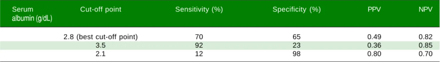
Table 4. Prediction of relative adrenal insufficiency using serum albumin in 113 consecutive patients with stable decompensated cir-rhosis.
Serum Cut-off point Sensitivity (%) Specificity (%) PPV NPV
albumin (g/dL)
2.8 (best cut-off point) 70 65 0.49 0.82
3.5 92 23 0.36 0.85
2.1 12 98 0.80 0.70
PPV: positive predictive value. NPV: negative predictive value.
Table 3. Multivariable analysis of independent variable that are correlated with the presence of relative adrenal insufficiency.
Variable Odds Ratio P value 95% Confidence 95% Confidence
Interval lower Interval upper
Serum Albumin 4.5 0.032 1.1 15.1
Factors associated with AI: multivariable regression analysis
All variables with p < 0.1 in the univariate statistics were included in multivariable regression analysis. Only serum albumin (OR: 4.5, 95%C.I.: 1.1-15.1, p = 0.032) was independently associated with the presence of AI (Table 3). Based on the area under the ROC curve, serum albumin had relatively low discriminative ability for the presence of AI (AUC = 0.68, 95% C.I.: 0.59 to 0.77). The best cut off point for serum albumin was < 2.8 g/dL giving a sensi-tivity 70%, specificity 65%, PPV 49% and NPV 82%. Inter-estingly, for albumin < 3.5 g/dL sensitivity, specificity, PPV and NPV were 92%, 23%, 36% and 85%, respectively, while for albumin < 2.1g/dL sensitivity, specifici-ty, PPV and NPV were 12%, 98%, 80% and 70%, respec-tively (Table 4).
Clinical impact of AI in patients with stable decompensated cirrhosis
Over the follow up period [median 12 (6-48) months], 83 patients died or underwent LT and 30 remained alive without LT. All deaths (n = 48) were liver related (liver/ multiorgan failure: 29, encephalopathy: 8, variceal bleed-ing/liver failure = 11) and no significant association was found between causes of death and AI. In univariate analy-sis, patients who died or underwent LT had lower “true” GFR (mean: 64 ± 25 mL/min vs. 81 ± 19 mL/min, p = 0.023), but higher serum urea [median: 38 mg/dL (11-130) vs. 34 (14-93), p = 0.041] and MELD score (18.5 ± 6 vs. 12 ± 3, p = 0.03), while they presented more frequently AI at baseline [χ2 test: 36% (30/83) vs. 13% (4/30), p = 0.02]
p = 0.003
Without AI
With AI
Based on the same criteria applied in the study of Acevedo, et al.7 the diagnosis of AI in our study was 30%
(34/113) in patients with stable DeCi. Despite the initial correlation of AI with several factors in the univariate anal-ysis, only serum albumin was independently associated with the presence of AI in the multivariable model (OR: 4.5, 95%C.I.: 1.1-15.1, p = 0.032) (Table 3); showing though relatively low discriminative ability (AUC = 0.68, 95% C.I.: 0.59 to 0.77). The sensitivity if serum albumin < 3.5 g/dL was 92%, and the specificity if serum albumin < 2.1 g/dL was 98% (Table 4).
The exact pathogenetic mechanisms implicated in the development of AI in patients with cirrhosis have not been fully elucidated. Since cirrhotic patients have low levels of lipids, AI could be attributed to cortisol production im-pairment at the level of adrenal glands particularly under conditions of stress.15 Moreover, the high levels of
circu-lating cytokines, such as TNF-α, IL-6, IL-1b which is a common finding in cirrhosis, may have a negative impact on normal adrenal function directly or indirectly via re-duced lipid delivery to adrenal glands.16 Nevertheless, no
correlation between AI and serum levels of IL-1b, IL-6 and TNFα was confirmed in our study, similarly to previ-ous studies.7 Concomitantly, no association between
cir-rhotic cardiomyopathy and development of AI was detected, as generally has been hypothesized, since diasto-lic dysfunction has not been found more commonly in pa-tients with AI.17 However, patients who developed AI
presented greater circulatory dysfunction and activation of renin-angiotensin system than those without AI as is veri-fied by the lower systolic blood pressure (106 ± 12 vs. 113 ± 13 mmHg, p = 0.05), and the higher plasma renin ac-tivity (17 ± 4 vs. 11±3 ng/mL/h, p = 0.05) and aldoster-one (61 ± 12 vs. 44 ± 17 ng/mL, p = 0.044).
Contrary to previous studies,18 we found that the pres-ence of AI was not associated with the severity of liver dis-ease (based on Child-Pugh and MELD scores). Furthermore, the patients with AI presented significantly lower serum sodium (133 ± 7 vs. 137 ± 12 mEq/L, p = 0.04), lower albumin (2.7 ± 0.5 vs. 3.1 ± 0.5 g/dL, p = 0.002) and greater direct bilirubin (median: 1.6 vs. 0.8 mg/ dL, p = 0.029) compared to patients without AI (Table 2) as it has been highlighted in earlier literature data.19
Nota-bly patients with AI suffered more often from SBP in the past compared to the group without AI [10/34 (29.4%) vs. 6/79 (7.5%), p = 0.002], which could indicate the associa-tion of AI with increased susceptibility in bacterial infec-tion. Indeed, Acevedo, et al.7 found that AI was linked with
the development of infection/severe sepsis and hepatore-nal syndrome during the follow up period possibly due to the compensatory increase of sympathetic tone which is translated to increased translocation of bacterial products from the intestinal lumen to the systemic circulation.20 However, this is not confirmed in our study, as no associ-ation between AI and complicassoci-ations of cirrhosis (includ-ing SBP) or causes of death (includ(includ-ing sepsis or hepatorenal syndrome) was demonstrated. In the same di-rection, only “true” GFR was independently associated with the outcome (death or LT) in our study during the follow up period. Nevertheless, as compared with those with normal adrenal function, patients with AI had higher mortality based on competing risk analysis [HR = 2.65, 95%C.I.: 1.55-4.52, p = 0.003) (Figure 2).
The results of the study should be viewed in the light of some limitations. The present data used a single centre design, including mainly patients with viral-related de-compensated cirrhosis, did not evaluate the free serum cortisol, which is the optimal for AI evaluation and did not assess any therapeutic manipulation of AI such as corti-sol substitution. Despite these limitations, our study is the first to our knowledge which evaluated AI in patients with well-established stable decompensated cirrhosis by using both serum and salivary cortisol and in parallel assessed the impact of AI on the outcome.
In conclusion, the present study demonstrates relative-ly high prevalence of AI in patients with DeCi accounting for essential clinical implication as it is associated with circulatory impairment, previous history of SBP and worse outcome. Subsequently the study emphasizes on the need of AI assessment in patients with stable DeCi on daily clinical basis and decision on the appropriate thera-peutic management.
ABBREVIATIONS
• AI: relative adrenal insufficiency.
• aPTT: activated partial thromboplastin time. Figure 2.
Figure 2.Figure 2.
Figure 2.Figure 2. Patients with adrenal insufficiency (AI) had significantly lower survival, compared to those without AI (HR = 2.65, 95%C.I.: 1.55-4.52, p = 0.003).
Cumulative survival
1.0
0.8
0.6
0.4
0.2
0.0
AI
Yes adrenal insufficiency No adrenal insufficiency
0 10 20 30 40 50
• AUC: receiver operating characteristic curve. • CBG: corticosteroid-binding globulin.
• CKD-EPI: Chronic Kidney Disease-Epidemiology. • CTP: child-Turcotte-Pugh.
• DeCi: decompensated cirrhosis. • eGFR: estimated GFR.
• GFR: glomerular filtration rate. • HRS: hepatorenal syndrome. • IL: interleukin.
• INR: International Bormalised Ratio • LT: liver transplantation.
• MELD: Model for End-stage Liver Disease.
• PPV and NPV: positive and negative predictive values. • PT: prothrombin time.
• SBP: spontaneous bacterial peritonitis. • SC: salivary cortisol.
• SST: short synacthen test. • STC: serum total.
• TNF: tumor necrosis factor.
• UNa/K: “spot” sodium and potassium ratio. • UNa24h: urine sodium excretion.
GRANTS AND FINANCIAL SUPPORT
Nothing to disclose.
CONFLICTS OF INTEREST
None.
REFERENCES
1. Tsochatzis EA, Bosch J, Burroughs AK. Liver cirrhosis. Lan-cet 2014; 383: 1749-61.
2. Karagiannis AK, Nakouti T, Pipili C, Cholongitas E. Adrenal in-sufficiency in patients with decompensated cirrhosis. World J Hepatol 2015; 7: 1112-24.
3. Peterson RE. Adrenocortical steroid metabolism and adrenal cortical function in liver disease. J Clin Invest 1960; 39: 320-31.
4. Arabi YM, Aljumah A, Dabbagh O, Tamim HM, Rishu AH, Al-Abdulkareem A, Knawy BA, et al. Low-dose hydrocortisone in patients with cirrhosis and septic shock: a randomized controlled trial. CMAJ 2010; 182: 1971-7.
5. Thevenot T, Borot S, Remy-Martin A, Sapin R, Cervoni JP, Ri-chou C, Vanlemmens C, et al. Assessment of adrenal func-tion in cirrhotic patients using concentrafunc-tion of serum-free and salivary cortisol. Liver Int 2011; 31: 425-33.
6. Trifan A, Chiriac S, Stanciu C. Update on adrenal insufficien-cy in patients with liver cirrhosis. World J Gastroenterol 2013; 19: 445-56.
7. Acevedo J, Fernández J, Prado V, Silva A, Castro M, Pavesi M, Roca D, et al. Relative adrenal insufficiency in decompen-sated cirrhosis: Relationship to short-term risk of severe sepsis, hepatorenal syndrome, and death. Hepatology 2013; 58: 1757-65.
8. Tan T, Chang L, Woodward A, McWhinney B, Galligan J, Macdonald GA, Cohen J, et al. Characterising adrenal func-tion using directly measured plasma free cortisol in stable severe liver disease. J Hepatol 2010; 53: 841-8.
9. Fede G, Spadaro L, Tomaselli T, Privitera G, Scicali R, Va-sianopoulou P, Thalassinos E, et al. Comparison of total corti-sol, free corticorti-sol, and surrogate markers of free cortisol in diagnosis of adrenal insufficiency in patients with stable cir-rhosis. Clin Gastroenterol Hepatol 2014; 12: 504-12. 10. Levey AS, Bosch JP, Lewis JB, Greene T, Rogers N, Roth
D. A more accurate method to estimate glomerular filtration rate from serum creatinine: a new prediction equation. Modi-fication of Diet in Renal Disease Study Group. Ann Intern Med 1999; 130: 461-70.
11. Fleming JS, Nunan TO. The new BNMS guidelines for meas-urement of glomerular filtration rate. Nucl Med Commun 2004; 25: 755-7.
12. Kazankov K, Holland-Fischer P, Andersen NH, Torp P, Sloth E, Aagaard NK, Vilstrup H. Resting myocardial dysfunction in cirrhosis quantified by tissue Doppler imaging. Liver Int 2011; 31: 534-40.
13. Fede G, Spadaro L, Tomaselli T, Privitera G, Germani G, Tso-chatzis E, Thomas M, et al. Adrenocortical dysfunction in liver disease: a systematic review. Hepatology 2012; 55: 1282-91. 14. Galbois A, Rudler M, Massard J, Fulla Y, Bennani A, Bonne-font-Rousselot D, Thibault V, et al. Assessment of adrenal function in cirrhotic patients: salivary cortisol should be pre-ferred. J Hepatol 2010; 52: 839-45.
15. Marik PE. Adrenal-exhaustion syndrome in patients with liver disease. Intensive Care Med 2006; 32: 275-80.
16. Bornstein SR. Predisposing factors for adrenal insufficien-cy. N Engl J Med 2009; 360: 2328-39.
17. Theocharidou E, Krag A, Bendtsen F, Moller S, Burroughs AK. Cardiac dysfunction in cirrhosis - does adrenal function play a role? A hypothesis. Liver Int 2012; 32: 1327-32. 18. Kharb S, Garg MK, Puri P, Nandi B, Brar KS, Gundgurthi A,
Pandit A. Assessment of adrenal function in liver diseases. Indian J Endocrinol Metab 2013; 17: 465-71.
19. Fede G, Spadaro L, Tomaselli T, Privitera G, Piro S, Rabuaz-zo AM, Sigalas A, et al. Assessment of adrenocortical re-serve in stable patients with cirrhosis. J Hepatol 2011; 54: 243-50.
20. Acevedo J, Fernandez J. New determinants of prognosis in bacterial infections in cirrhosis. World J Gastroenterol 2014; 20: 7252-9.
Correspondence and reprint request:
Evangelos Cholongitas, Assistant Professor of Internal Medicine4th Department of Internal Medicine.
Medical School of Aristotle University, Hippokration General Hospital of Thessaloniki. 49, Konstantinopoleos Street, 54642. Thessaloniki, Greece.