biomedical engineering
The fields of biological and medical physics and biomedical engineering are broad, multidisciplinary and dynamic. They lie at the crossroads of frontier research in physics, biology, chemistry, and medicine. The Biological and Medical Physics, Biomedical Engineering Series is intended to be comprehensive, covering a broad range of topics important to the study of the physical, chemical and biological sciences. Its goal is to provide scientists and engineers with textbooks, monographs, and reference works to address the growing need for information.
Books in the series emphasize established and emergent areas of science including molecular, membrane, and mathematical biophysics; photosynthetic energy harvesting and conversion; information processing; physical principles of genetics; sensory communications; automata networks, neural networks, and cellular automata. Equally important will be coverage of applied aspects of biological and medical physics and biomedical engineering such as molecular electronic components and devices, biosensors, medicine, imaging, physical principles of renewable energy production, advanced prostheses, and environmental control and engineering.
Editor-in-Chief:
Elias Greenbaum, Oak Ridge National Laboratory, Oak Ridge, Tennessee, USA
Editorial Board:
Masuo Aizawa, Department of Bioengineering, Tokyo Institute of Technology, Yokohama, Japan Olaf S. Andersen, Department of Physiology, Biophysics & Molecular Medicine, Cornell University, New York, USA Robert H. Austin, Department of Physics, Princeton University, Princeton, New Jersey, USA James Barber, Department of Biochemistry, Imperial College of Science, Technology and Medicine, London, England Howard C. Berg, Department of Molecular and Cellular Biology, Harvard University, Cambridge, Massachusetts, USA
Victor Bloomfield, Department of Biochemistry, University of Minnesota, St. Paul, Minnesota, USA Robert Callender, Department of Biochemistry, Albert Einstein College of Medicine,
Bronx, New York, USA
Britton Chance, Department of Biochemistry/ Biophysics, University of Pennsylvania, Philadelphia, Pennsylvania, USA Steven Chu, Department of Physics, Stanford University, Stanford, California, USA Louis J. DeFelice, Department of Pharmacology, Vanderbilt University, Nashville, Tennessee, USA Johann Deisenhofer, Howard Hughes Medical Institute, The University of Texas, Dallas, Texas, USA
George Feher, Department of Physics, University of California, San Diego, La Jolla, California, USA
Hans Frauenfelder, CNLS, MS B258, Los Alamos National Laboratory, Los Alamos, New Mexico, USA
Ivar Giaever, Rensselaer Polytechnic Institute, Troy, New York, USA
Sol M. Gruner, Department of Physics, Princeton University, Princeton, New Jersey, USA Judith Herzfeld, Department of Chemistry, Brandeis University, Waltham, Massachusetts, USA Pierre Joliot, Institute de Biologie
Physico-Chimique, Fondation Edmond de Rothschild, Paris, France
Lajos Keszthelyi, Institute of Biophysics, Hungarian Academy of Sciences, Szeged, Hungary
Robert S. Knox, Department of Physics
and Astronomy, University of Rochester, Rochester, New York, USA
Aaron Lewis, Department of Applied Physics, Hebrew University, Jerusalem, Israel Stuart M. Lindsay, Department of Physics and Astronomy, Arizona State University, Tempe, Arizona, USA
David Mauzerall, Rockefeller University, New York, New York, USA
Eugenie V. Mielczarek, Department of Physics and Astronomy, George Mason University, Fairfax, Virginia, USA
Markolf Niemz, Klinikum Mannheim, Mannheim, Germany
V. Adrian Parsegian, Physical Science Laboratory, National Institutes of Health, Bethesda, Maryland, USA
Linda S. Powers, NCDMF: Electrical Engineering, Utah State University, Logan, Utah, USA Earl W. Prohofsky, Department of Physics, Purdue University, West Lafayette, Indiana, USA Andrew Rubin, Department of Biophysics, Moscow State University, Moscow, Russia
Michael Seibert, National Renewable Energy Laboratory, Golden, Colorado, USA David Thomas, Department of Biochemistry, University of Minnesota Medical School, Minneapolis, Minnesota, USA
U.R. M
uller D.V. Nicolau (Eds.)
¨
Microarray Technology
and Its Applications
With 123 Figures
Including 16 Color Plates
V.P., Applied Science Nanosphere, Inc. 4088 Commercial Avenue Northbrook, IL 60062 USA
e-mail: [email protected]
Prof. Dan V. Nicolau
Swinburne University of Technology 533-545 Burwood Rd.
Hawthorn, Victoria 3122 Australia
e-mail: [email protected]
Library of Congress Cataloging in Publication Data: 2004113284
ISSN 1618-7210
ISBN 3-540-22931-0 Springer Berlin Heidelberg New York
This work is subject to copyright. All rights are reserved, whether the whole or part of the material is concerned, specifically the rights of translation, reprinting, reuse of illustrations, recitation, broadcasting, reproduction on microfilm or in any other way, and storage in data banks. Duplication of this publication or parts thereof is permitted only under the provisions of the German Copyright Law of September 9, 1965, in its current version, and permission for use must always be obtained from Springer. Violations are liable to prosecution under the German Copyright Law.
Springer is a part of Springer Science+Business Media springeronline.com
© Springer-Verlag Berlin Heidelberg 2005 Printed in Germany
The use of general descriptive names, registered names, trademarks, etc. in this publication does not imply, even in the absence of a specific statement, that such names are exempt from the relevant protective laws and regulations and therefore free for general use.
Cover concept by eStudio Calamar Steinen
Typesetting by the authors using a Springer LATEX-macro package Cover production:design & productionGmbH, Heidelberg Production: LE-TEX Jelonek, Schmidt & Vöckler GbR, Leipzig
It has been stated that our knowledge doubles every 20 years, but that may be an understatement when considering the Life Sciences. A series of discoveries and inventions have propelled our knowledge from the recognition that DNA is the genetic material to a basic molecular understanding of ourselves and the living world around us in less than 50 years. Crucial to this rapid progress was the discovery of the double-helical structure of DNA, which laid the foundation for all hybridization based technologies. The discoveries of restriction enzymes, ligases, polymerases, combined with key innovations in DNA synthesis and sequencing ushered in the era of biotechnology as a new science with profound sociological and economic implications that are likely to have a dominating influence on the development of our society during this century. Given the process by which science builds on prior knowledge, it is perhaps unfair to single out a few inventions and credit them with having contributed most to this avalanche of knowledge. Yet, there are surely some that will be recognized as having had a more profound impact than others, not just in the furthering of our scientific knowledge, but by leveraging commercial applications that provide a tangible return to our society.
still a limit to the number of laboratories that have access to this technology-its impact is truly remarkable, especially when compared, for example, to the emerging and much touted field of Nanotechnology.
Number of Articles
Number of Reviews
Fig. 1. Comparative evolution of publications regarding microarrays and
nanobiotechnology
Amidst the pace of such rapid knowledge expansion, there is a challenge in trying to compose a book that does not face obsolescence by the time of its first publication. Alas, the breadth of this field is driving the growing knowledge base into many new directions, generating the need for different books at different levels and each with a different and unique focus.
As early participants in the development of microarray technology the edi-tors have learned to appreciate the need for contributions from many different areas in the basic sciences and engineering that were crucial to its birth and continued healthy growth. In turn we have observed how the involvement in this particular scientific endeavour has affected many careers, turning physi-cist into oncologists, physicians into bioinformaticians, and chemists and biol-ogists into optical engineers. Provided the diverse nature of backgrounds that are required to further propel this field, we thought it appropriate to aggre-gate this book around three aspects of microarray technology:fundamentals, designed to provide a scientific base;fabrication, which describes the current state of the art and compares ‘old’ and new ways of building microarrays; and
applications, that are aimed to highlight only the amazing variety and options provided by these techniques. As an aid to the practitioner we have also asked the authors to provide a detailed method section wherever appropriate.
Chaps. 4 and 5 describe the principal techniques used for array manufac-ture. Chapter 6 explores the limits of miniaturization with nanoarrays, and Chap. 7 illuminates various aspects of microfluidics for automation. Finally, Chaps. 8 and 9 deal with the principles of labelling and detection method-ologies. The next parts are concerned with application of these fundamental techniques toward the development and use of specific types of microarrays. Part 2 describes DNA based microarrays in 4 chapters, covering SNP detec-tion, high sensitivity expression profiling, comparative genomic hybridizadetec-tion, and the analysis of regulatory circuits. Part 3 contains 3 chapters that deal with microarrays for protein and small molecule detection, describing array technology for antibodies, aptamers, and lipid bound proteins, respectively. The final part comprises 4 chapters that introduce the most esoteric arrays, those that contain high information content in each feature (whole cells or tissues), and the capability of performing biological reactions, such as trans-fections. How the combination of these types of arrays generates new insights into the molecular basis of normal and malignant cell function is summarized in the last chapter.
It appears that given the dynamics of microarray technology any book would be a ‘work in progress’. Rather than fighting this, the editors and the authors of this book embrace this concept: chances are that this book will grow in time in line with the new developments in microarray technology.
June, 2004 Uwe M¨uller
The initial idea for this book emerged during a serendipitous meeting between the Editors and a representative from Springer Verlag during a Conference on Microarrays, Fundamentals, Fabrication and Applications that was chaired and organized by the Editors as part of the International Society for Optical Engineering (SPIE) Meeting in January 2001 in San Jose, CA. In fact, sev-eral Chaps. of this book were authored by people present at that Conference. The Editors wish to thank the organizers of SPIE, and in particular Mar-ilyn Gorsuch and Annie Gerstl, who helped with the organisation of these Conferences in the last four years. Thanks also to the Conference co-Chairs, Ramesh Raghavachari and David Dunn. The Editors also wish to thank Pe-ter Livingston and Gerardin Solana for the tedious work of converting the manuscripts into a camera-ready format.
Many contributors have specific acknowledgements.
The authors of Chap. 1 are grateful to Stephen Felder, Ph.D. and Richard Kris, Ph.D. of NeoGen, LLC. (Tucson, AZ), the inventors of the multiplexed nuclease protection assay, for proof-of-principle work on the mRNA assay and for the software for reagent design, image analysis and data interpretation.
The authors of Chap. 4 would like to thank Innovadyne Technologies for use of Fig. 4.5 and Peter Hoyt for helpful discussions. The research pre-sented here was sponsored by the Laboratory Directed Research and Devel-opment Program of Oak Ridge National Laboratory (ORNL), managed by UT-Battelle, LLC for the U. S. Department of Energy under Contract No. DE-AC05-00OR22725 and by NIH Grant R01 HL62681-02. The manuscript has been authored by a contractor of the U.S.Government under contract DE-AC05-00OR22725. Accordingly, the U.S. Government retains a nonexclu-sive, royalty-free license to publish or reproduce the published form of this contribution, or allow others to do so, for U.S. Government purposes.
One of the authors of Chap. 6 (DVN) wishes to thank Dan V. Nicolau Jr. for discussions regarding the computational applications of nanoarrays.
Pankaj Singhal (Motorola Life Sciences) for useful discussions on hybridiza-tion kinetics. This work has been sponsored in part by NIST ATP contract #1999011104A and DARPA contract #MDA972-01-3-0001.
Some of the authors of Chap. 8, i.e. JJS and SSM, acknowledge the NIH for support. CAM acknowledges the AFOSR, DARPA, and the NSF for support of this work.
The authors of Chap. 9 are very grateful to Gabriele G¨unther for excellent technical assistance, to Dr. K. B¨ohm, Jena, for kindly providing us with mi-crotubules and kinesin samples, and to Dr. Wolf, PicoRapid GmbH Bremen, for help in spotting protein samples by an automatic arrayer.
The authors of Chap. 13 thank Dr. Tae Hoon Kim and Miss Sara Van Calcar for critical reading of the manuscript. We are also grateful to Drs. Hieu Cam, Yasuhiko Takahashi, Brian Dynlacht, Richard Young, and Mr. Tom Volkert for their help during the development of the technology described in this chapter. B.R. is supported by the Ludwig Institute for Cancer Research and a Sidney Kimmel Foundation for Cancer Research Scholar Award.
The authors of Chap. 14 wish to acknowledge the great support by Dr. Ronald Frank.
Part I General Microarray Technologies
1 Array Formats
Ralph R. Martel, Matthew P. Rounseville, Ihab W. Botros,
Bruce E. Seligmann . . . . 3
1.1 Introduction . . . 3
1.2 Reasons to Use Arrays . . . 4
1.3 Arrays for Nucleic Acid Analysis . . . 6
1.4 Protein Arrays . . . 8
1.5 The ArrayPlateTM . . . . 9
1.6 Conclusion . . . 19
References . . . 20
2 Biomolecules and Cells on Surfaces – Fundamental Concepts Kristi L. Hanson, Luisa Filipponi, Dan V. Nicolau . . . . 23
2.1 Introduction . . . 23
2.2 Types of Immobilization . . . 23
2.3 DNA Immobilization on Surfaces . . . 28
2.4 Protein Immobilization on Surfaces . . . 32
2.5 Carbohydrate Immobilization . . . 36
2.6 Immobilization of Cells on Surfaces . . . 38
2.7 Conclusions . . . 41
References . . . 42
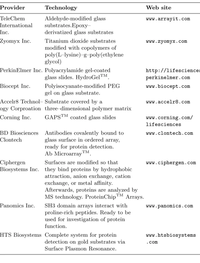
3 Surfaces and Substrates Alvaro Carrillo, Kunal V. Gujraty, Ravi S. Kane. . . . 45
3.1 Introduction . . . 45
3.2 DNA Microarrays . . . 46
3.4 Conclusion . . . 55
References . . . 56
4 Reagent Jetting Based Deposition Technologies for Array Construction Mitchel J. Doktycz. . . . 63
4.1 Introduction . . . 63
4.2 Reagent Jetting – Technology Overview . . . 63
4.3 Thermal Jet Based Dispensing . . . 65
4.4 Piezo Jet Based Dispensing . . . 67
4.5 Solenoid Jet Based Dispensing . . . 68
References . . . 71
5 Manufacturing of 2-D Arrays by Pin-printing Technologies Uwe R. M¨uller, Roeland Papen . . . . 73
5.1 Introduction . . . 73
5.2 Definition of ‘Contact’ Pin–Printing . . . 73
5.3 Overview of Different Pin Technologies . . . 74
5.4 Other System Components and Environmental Factors . . . 79
5.5 Pin Printing Process . . . 81
5.6 Example of a High Throughput Pin–Printing System for Manufacturing of 2D Arrays – the Corning GENII System . . . 84
5.7 Conclusion . . . 86
References . . . 87
6 Nanoarrays Dan V. Nicolau, Linnette Demers, David S. Ginger. . . . 89
6.1 Introduction . . . 89
6.2 Passive Nano–scale Arrays . . . 91
6.3 Computational Nanoarrays . . . 105
6.4 Dynamic Nanoarrays . . . 109
6.5 Conclusion . . . 115
References . . . 115
7 The Use of Microfluidic Techniques in Microarray Applications Piotr Grodzinski, Robin H. Liu, Ralf Lenigk, Yingjie Liu. . . .119
7.1 Introduction . . . 119
7.2 Biochannel Hybridization Arrays . . . 120
7.3 Chips with Cavitation Microstreaming Mixers – Kinetics Studies . . . 128
7.4 Integrated Microfluidic Reactors for DNA Amplification and Hybridization . . . 135
References . . . 142
8 Labels and Detection Methods James J. Storhoff, Sudhakar S. Marla, Viswanadham Garimella, Chad A. Mirkin. . . .147
8.1 Introduction . . . 147
8.2 Fluorophore Labelling and Detection Methods . . . 148
8.3 Enhanced Fluorescence-Based Assays . . . 151
8.4 Phosphor Reporters . . . 154
8.5 Electrochemical Detection . . . 156
8.6 Metal Nanoparticle Labels and Metal Thin Films for Microarrays . . . 159
8.7 Conclusions . . . 172
References . . . 174
9 Marker-free Detection on Microarrays Matthias Vaupel, Andreas Eing, Karl-Otto Greulich, Jan Roegener, Peter Schellenberg, Hans Martin. Striebel, Heinrich F. Arlinghaus. . . .181
9.1 Introduction . . . 181
9.2 Imaging Ellipsometry and Imaging Surface Plasmon Resonance on Biochips . . . 181
9.3 Intrinsic UV Fluorescence for Chip Analysis of Rare Proteins . . . 190
9.4 Genetic Diagnostics with Unlabelled DNA . . . 197
References . . . 204
Part II DNA Microarrays 10 Analysis of DNA Sequence Variation in the Microarray Format Ulrika Liljedahl, Mona Fredriksson, Ann-Christine Syv¨anen . . . .211
10.1 Introduction . . . 211
10.2 Principles of Genotyping . . . 213
10.3 Performing the Assays in Practice . . . 217
10.4 Conclusion . . . 222
References . . . 223
11 High Sensitivity Expression Profiling Ramesh Ramakrishnan, Paul Bao, Uwe R. M¨uller. . . .229
11.1 Introduction . . . 229
11.2 Oligonucleotide Expression Arrays . . . 230
11.3 cDNA-based Expression Arrays . . . 239
References . . . 245
12 Applications of Matrix-CGH (Array-CGH) for Genomic Research and Clinical Diagnostics Carsten Schwaenena, Michelle Nesslinga, Bernhard Radlwimmera, Swen Wessendorf, Peter Lichtera. . . .251
12.1 Introduction . . . 251
12.2 Technical Aspects . . . 253
12.3 Applications . . . 256
References . . . 260
13 Analysis of Gene Regulatory Circuits Zirong Li . . . .265
13.1 Introduction . . . 265
13.2 An Experimental Protocol for Genome Wide Location Analysis . . . 268
13.3 Example: Identifying the Target Genes of Human E2F4 . . . 273
13.4 Summary . . . 275
References . . . 275
Part III Protein Microarrays 14 Protein, Antibody and Small Molecule Microarrays Hendrik Weiner, J¨orn Gl¨okler, Claus Hultschig, Konrad B¨ussow, Gerald Walter . . . .279
14.1 Introduction . . . 279
14.2 Protein Microarrays . . . 280
14.3 Antibody Microarrays . . . 283
14.4 Peptide and Other Synthetic Arrays . . . 287
References . . . 290
15 Photoaptamer Arrays for Proteomics Applications Drew Smith, Chad Greef. . . .297
15.1 Introduction . . . 297
15.2 Overview of Photoaptamer Discovery and High Throughput Production . . . 298
15.3 Using Photoaptamer Microarrays . . . 301
15.4 Discussion . . . 303
References . . . 305
16 Biological Membrane Microarrays Ye Fang, Anthony G. Frutos, Yulong Hong, Joydeep Lahiri. . . .309
16.2 Biospecific Binding Studies Using Membrane Microarrays . . . . 313
16.3 Conclusions . . . 318
References . . . 319
Part IV Cell & Tissue Microarrays 17 Use of Reporter Systems for Reverse Transfection Cell Arrays Brian L. Webb . . . .323
17.1 Introduction . . . 323
17.2 Reporter Systems for Reverse Transfection . . . 325
17.3 Reagents and Protocols . . . 332
References . . . 333
18 Whole Cell Microarrays Ravi Kapur . . . .335
18.1 Introduction . . . 335
18.2 The Need . . . 336
18.3 The Solution . . . 336
18.4 Challenges and Opportunities for Cellular Micrroarrays . . . 341
References . . . 343
19 Tissue Microarrays for Miniaturized High-Throughput Molecular Profiling of Tumors Ronald Simon, Martina Mirlacher, Guido Sauter. . . .345
19.1 Introduction . . . 345
19.2 The TMA Technology . . . 346
19.3 The Representativity Issue . . . 346
19.4 TMA Applications . . . 349
19.5 Future Directions . . . 351
19.6 Protocol . . . 352
References . . . 354
20 Application of Microarray Technologies for Translational Genomics Spyro Mousses, Natasha Caplen, Mark Basik, Anne Kallioniemi, Olli Kallioniemi. . . .361
20.1 Introduction . . . 361
20.2 High Throughput Clinical Target Validation Using Tissue Microarrays . . . 363
20.4 High Throughput Characterization
of Gene Function Using Live Cell Microarrays . . . 368
20.5 Conclusions . . . 370
References . . . 372
Heinrich F. Arlinghaus
Physikalisches Institut der Universit¨at M¨unster Wilhelm-Klemm-Str. 10 D-48149 M¨unster, Germany
Paul Bao
Nanosphere, Inc.
4088 Commercial Avenue Northbrook, IL 60062, USA
Mark Basik
Translational Genomics Research Institute (TGen)
20 First Field Road
Gaithesburg, MA 20878, USA
Ihab W. Botros
High Throughput Genomics, Inc. 6296 East Grant Road
Tucson, AZ 85712, USA
Konrad B¨ussow
Max Planck Institute of Molecular Genetics
Ihnestrasse 73
D-14195 Berlin, Germany
Natasha Caplen
Medical Genetics Branch
National Human Genome Research Institute
National Institutes of Health Building 10, Room 10C103 10 Center Drive
Bethesda, MD 20892 USA
Alvaro Carrillo
Rensselaer Polytechnic Institute Howard P. Isermann Department of Chemical Engineering
Ricketts Building, 110 8th Street Troy, NY 12180, USA
Linnette Demers
NanoInk, Inc.
1335 W. Randolph Street Chicago, IL 60607, USA
Mitchel J. Doktycz
Life Sciences Division and Condensed Matter Sciences Division
Oak Ridge National Laboratory P.O. Box 2008
Oak Ridge, TN 37831-6123, USA
Andreas Eing
Nanofilm Technologie Anna-Vadenhoeck-Ring 5 D-37081 G¨ottingen, Germany
Ye Fang
Corning, Inc.
Biochemical Sciences, Science and Technology Division
Corning, NY 14870, USA
Luisa Filipponi
Swinburne University of Technology Industrial Research Institute Swinburne
533-545 Burwood Road
Hawthorn, VIC 3122, Australia
Anthony G. Frutos
Corning, Inc.
Biochemical Sciences, Science and Technology Division
Corning, NY 14870, USA
Mona Fredriksson
Uppsala University Hospital Dept Medical Sciences S-751 85 Uppsala, Sweden
Viswanadham Garimella
Nanosphere, Inc.
4088 Commercial Avenue Northbrook, IL 60062, USA
Kunal V. Gujraty
Rensselaer Polytechnic Institute Howard P. Isermann Department of Chemical Engineering
Ricketts Building, 110 8th Street Troy, NY 12180, USA
David S. Ginger
Department of Chemistry University of Washington Box 351700
Seattle, WA 98195-1700, USA
J¨orn Gl¨okler
Max Planck Institute of Molecular Genetics
Ihnestrasse 73
D-14195 Berlin, Germany
Chad Greef
SomaLogic, Inc. 1745 38th Street
Boulder, CO 80301, USA
Karl-Otto Greulich
Institute for Molecular Biotechnology Department of Single Cell and Single Molecule Techniques
Beutenbergstrasse 11 D-07745 Jena, Germany
Piotr Grodzinski
Microfluidics Laboratory, PSRL, Motorola Labs
7700 S. River Parkway Tempe, AZ 85284, USA Current address:
Bioscience Division, MS J586 Los Alamos National Laboratory Los Alamos, NM 87545, USA
Kristi L. Hanson
Swinburne University of Technology Industrial Research Institute Swinburne
533-545 Burwood Road Hawthorn, VIC 3122, Australia
Yulong Hong
Corning, Inc.
Biochemical Sciences, Science and Technology Division
Corning, NY 14870, USA
Claus Hultschig
Max Planck Institute of Molecular Genetics
Ihnestrasse 73
D-14195 Berlin, Germany
Anne Kallioniemi
University of Tampere
Laboratory of Cancer Genetics Institute of Medical Technology P.O. Box 607
FIN-33014 University of Tampere, Finland
Olli Kallioniemi
Medical Biotechnology Group VTT Technical Research Centre of Finland
University of Turku
P.O. Box 106, 20521 Turku, Finland
Ravi S. Kane
Rensselaer Polytechnic Institute Howard P. Isermann Department of Chemical Engineering
Ricketts Building, 110 8th Street Troy, NY 12180, USA
Ravi Kapur
Anudeza Group 292 Morton Street
Stoughton, MA 02072, USA
Joydeep Lahiri
Corning, Inc.
Biochemical Sciences, Science and Technology Division
Corning, NY 14870, USA
Ralf Lenigk
Microfluidics Laboratory, PSRL, Motorola Labs
7700 S. River Parkway Tempe, AZ 85284, USA Current address:
Applied NanoBioscience Center P.O. Box 874004
Arizona State University Tempe, AZ 85287, USA
Zirong Li
Ludwig Institute for Cancer Research UCSD La Jolla Medical School Campus
9500 Gilman Drive
La Jolla, CA 92093-0653, USA
Peter Lichter
Molekulare Genetik
Deutsches Krebsforschungszentrum D-69120 Heidelberg, Germany
Ulrika Liljedahl
Uppsala University Hospital Dept Medical Sciences S-751 85 Uppsala, Sweden
Robin H. Liu
Microfluidics Laboratory, PSRL, Motorola Labs
Applied NanoBioscience Center P.O. Box 874004
Arizona State University Tempe, AZ 85287, USA
Yingjie Liu
Microfluidics Laboratory, PSRL, Motorola Labs
7700 S. River Parkway Tempe, AZ 85284, USA
Current address: Applied NanoBio-science Center P.O. Box 874004 Arizona State University Tempe, AZ 85287, USA
Jason [email protected]
Sudhakar S. Marla
Nanosphere, Inc.
4088 Commercial Avenue Northbrook, IL 60062, USA
Ralph R. Martel
High Throughput Genomics, Inc. 6296 East Grant Road
Tucson, AZ 85712, USA
Mirlacher Martina
University of Basel Institute of Pathology Schoenbeinstrasse 40 4031 Basel, Switzerland
Chad A. Mirkin
Northwestern University Institute for Nanotechnology 2145 Sheridan Road
Evanston, IL 60208, USA
Spyro Mousses
Translational Genomics Research Institute (TGen)
20 First Field Road
Gaithesburg, MA 20878, USA
Uwe R. M¨uller
Nanosphere, Inc.
4088 Commercial Avenue Northbrook, IL 60062, USA
Michelle Nessling
Molekulare Genetik
Deutsches Krebsforschungszentrum D-69120 Heidelberg, Germany
Dan V. Nicolau
Swinburne University of Technology Industrial Research Institute Swinburne
533-545 Burwood Road
Hawthorn, VIC 3122, Australia
Roeland Papen
Picoliter inc.
231 S Whisman Road, Mountain View CA 94041
Bernhard Radlwimmer
Molekulare Genetik
Deutsches Krebsforschungszentrum D-69120 Heidelberg, Germany
Ramesh Ramakrishnan
Nanosphere, Inc.
4088 Commercial Avenue Northbrook, IL 60062, USA
Bing Ren
University of California, San Diego Department of Cellular and Molecu-lar Medicine, School of Medicine 9500 Gilman Drive, La Jolla, CA 92093-0653, USA
Jan Roegener
University of Bielefeld
Department of Applied Laser Physics Universitaetsstrasse 25 D3
D-33615 Bielefeld, Germany
Simon Ronald
University of Basel Institute of Pathology Schoenbeinstrasse 40 4031 Basel, Switzerland
Matthew P. Rounseville
High Throughput Genomics, Inc. 6296 East Grant Road
Tucson, AZ 85712, USA
Guido Sauter
University of Basel Institute of Pathology Schoenbeinstrasse 40 4031 Basel, Switzerland
Peter Schellenberg
Institute for Molecular Biotechnology Department of Single Cell and Single Molecule Techniques
Beutenbergstrasse 11 D-07745 Jena, Germany
Carsten Schwaenen
Medizinische Klinik der Universit¨at Ulm
Innere Medizin III D-89081 Ulm, Germany
Bruce E. Seligmann
High Throughput Genomics, Inc. 6296 East Grant Road
Tucson, AZ 85712, USA
Drew Smith
SomaLogic, Inc. 1745 38th Street
Boulder, CO 80301, USA
James J. Storhoff
Nanosphere, Inc. 1818 Skokie Boulevard Northbrook, IL 60062, USA
Hans Martin. Striebel
Institute for Molecular Biotechnol-ogy,
Department of Single Cell and Single Molecule Techniques
Beutenbergstrasse 11 D-07745 Jena, Germany
Ann-Christine Syv¨anen
Uppsala University Hospital Dept Medical Sciences S-751 85 Uppsala, Sweden
Ann-Christine.Syvanen@medsci. uu.se
Matthias Vaupel
Nanofilm Technologie
Anna-Vadenhoeck-Ring 5 D-37081 G¨ottingen, Germany
Gerald Walter
Biorchard AS Nedre Skogvei 14 N-0281 Oslo, Norway
Brian L. Webb
Corning, Inc.
Biochemical Sciences, Science and Technology Division
Corning, NY 14870, USA
Hendrik Weiner
Max Planck Institute of Molecular Genetics
Ihnestrasse 73
D-14195 Berlin, Germany
Swen Wessendorf
Medizinische Klinik der Universit¨at Ulm
Innere Medizin III D-89081 Ulm, Germany
Part I
Array Formats
Ralph R. Martel, Matthew P. Rounseville, and Ihab W. Botros, and Bruce E. Seligmann
1.1 Introduction
Arrays have become an increasingly diverse set of tools for biological studies; their use continues to expand rapidly. Likewise, the underlying array tech-nologies, formats and protocols continue to evolve. Investigators can choose from a growing range of options when selecting an array technology that is appropriate for reaching their research objectives. Traditionally, arrays have consisted of collections of distinct capture molecules – typically cDNAs or oligonucleotides – attached to a substrate – usually a glass slide – at pre-defined locations within a grid pattern [1, 2]. However, today’s formats are more diverse and can be grouped into several categories. Like any catego-rization effort, there will be exceptions, crossover technologies and tangential relations. The intent here is only to lay out some general trends.
The classes of capture molecules used in arrays include not only DNA, but also proteins [3], carbohydrates [4], drug-like molecules [5], cells [6], tis-sues [7] and the like. Array formats vary in their architecture. For closed architecture arrays, the analytes that can be measured are preselected and locked-in during the manufacturing process. In contrast open architecture ar-ray technologies allow the set of measured analytes to be modified or allow new analytes to be discovered. Regardless of the architecture, various manu-facturing technologies and various substrate materials and coatings are avail-able as are numerous means of attaching capture molecules to substrates. A broad variety of commercially prepared arrays can be purchased. In some in-stances, the pre-defined grid has been eliminated and replaced with ‘virtual ar-rays’ of optically encoded beads [8] or of analyte-specific detection labels (e.g. e-Tags; www.aclara.com). Coupled with the diversity of arrayed molecules and array formats is the diversity of detection schemes that include fluorescence, luminescence, electrochemical detection, mass spectrometry, surface plasmon resonance and others.
performed simultaneously. This is the case both when many analytes are mea-sured simultaneously in an individual sample and also when many samples are tested at one time for an individual analyte. For instance, DNA arrays can be used to determine the expression levels of thousands of genes in an indi-vidual biological specimen, while tissue arrays can be used to determine the presence of a specific antigen in hundreds of specimens in a single experiment. Various ‘array–of–arrays’ technologies combine the measurement of numerous analytes across numerous samples.
The impact of array technologies on the life sciences has been important. In conjunction with bioinformatic tools to process and analyze the large amounts of data they generate, arrays have spawned new approaches to systems biol-ogy often described with the ‘omics’ suffix: genomics, transcriptomics and proteomics, to name a few.
This chapter will provide the rationales for using arrays to address various scientific questions and will outline some of the array technologies developed to fill specific needs. This is a series of examples to illustrate the range of available options and how one technology may be better suited than another to reach a specific research objective, not a comprehensive survey of available tools. The latter part of the chapter will discuss the ArrayPlateTMtechnology developed by High Throughput Genomics (HTG, Tucson, AZ) to bring the benefits of arrays to the high throughput screening phase of the drug discovery and development process. The procedure for a multiplexed ArrayPlateTM mRNA assay will be described and the results of an mRNA assay and a companion multiplexed ELISA will be presented.
1.2 Reasons to Use Arrays
There are three principle justifications for using array technologies. Arrays serve to discover unique patterns (of gene expression, protein synthesis or post-translational modification, etc.) associated with a particular physiolog-ical state. We use the term ‘survey array’ to describe the technologies that are employed for this purpose. ‘Scan array’ or ‘focused array’ refers to the array tools that measure a predefined pattern, previously established with survey arrays. Finally, ‘efficiency array’ refers to the techniques that do not require multiplexing per se, but that take advantage of the parallel process-ing common to arrays to provide savprocess-ings of effort, time and materials or to improve data quality by incorporating internal controls that are measured in each sample. Most array technologies have been developed to achieve one of these three goals and may be inefficient for reaching the other two.
1.2.1 Arrays to Identify Patterns
to the company (www.affymetrix.com), the two arrays in the Human Genome U133 Set contain over one million distinct oligonucleotide features to monitor the expression of 39,000 transcript variants of 33,000 different human genes in a single sample. GeneChipsR and their cDNA and oligonucleotide array coun-terparts are widely used to identify genes that are differentially expressed in diseased tissues or during development or upon treatment with a drug. In most instances, results obtained with DNA arrays show that the vast major-ity of genes are either not expressed or not affected by disease. Typically, a disease-specific pattern of gene expression or ‘signature’ is characterized that involves fewer than 50 genes [9–12]. Although well suited to initially define patterns based on the examination of a relatively small number of samples, survey arrays are generally too labor- and material-intensive and too costly to be used routinely thereafter in diagnostics or in drug discovery.
1.2.2 Arrays to Measure Patterns
‘Scan arrays’ that measure specific patterns are appropriate for clinical diag-nostics and for drug discovery. While these techniques measure fewer analytes than do survey arrays, the analytes have been carefully selected and validated. Other attributes such as ease of use and throughput make various scan array technologies well-suited for particular niches.
Inexpensive readout equipment is a requirement for array-based diagnos-tic tests as such tests are performed at many different sites such as reference laboratories, hospital laboratories and physicians’ offices but relatively infre-quently at any given site. Cost per test however is less important since the results provide information that is of high value. Furthermore, most diagnos-tic testing is reimbursed by insurers. Hands-on manipulations must be simple as testing is frequently performed by inexperienced personnel. To gain ap-proval from regulatory agencies, diagnostics tests must yield results that are robust and interpretable. For these reasons, various hand-held electronic array devices appear to be in the best position to make inroads in this arena.
1.2.3 Arrays for Parallel Processing
Examples where the array format has been adopted for the efficiencies derived from parallel processing can be found in the combinatorial chemistry litera-ture [13]. The synthesis of chemical compound libraries has been performed in an array format [14]. Indeed, the photolithographic process utilized by Affymetrix to manufacture its DNA chips had its origins in combinatorial chemistry [15]. Arrays of compounds have also been used in drug discovery screening [16]. Microtiter plate wells that contained individual compounds have been miniaturized to the point of vanishing with the compounds be-coming elements of an array rather than contents of a well. Generally, using arrays leverages sample preparation efforts. In cell-based assays for instance, the effort of culturing cells and screening compounds is the same regardless of whether a single or multiple measurements are made.
1.3 Arrays for Nucleic Acid Analysis
Several review articles covering advances and applications of DNA microar-ray technology have recently been published [17, 18] hence, the same material will not be repeated here. Oligonucleotide and cDNA arrays have different strengths and weaknesses. There is more control over the design of cleotide microarrays than there is for cDNA arrays. Consequently, oligonu-cleotide arrays tend to have more uniform physicochemical characteristics and fewer issues pertaining to cross–hybridization. For cDNA arrays, the capture probes are typically PCR amplicons of clones derived from the organism or the organ of interest. One advantage is that cDNA probes can be incorpo-rated into arrays without further characterization of the underlying gene. For both types of microarrays however, the architecture is closed, albeit at times unknown for cDNA arrays. For illustrative purposes, several less conventional array technologies are described.
1.3.1 Arrays on Beads
of the captured molecule. The application of this method to RNA expression analysis has been described recently [8].
Illumina (San Diego, CA) has developed an alternative readout system for bead-based arrays. A manifold of 96 fiber optic bundles, each consisting of about 50,000 individual fibers, is manufactured to fit the standard microplate format. A dimple etched at the end of each fiber can accommodate one of the company’s 3 µm beads. This enables fluorochrome excitation and emission of the beads and of fluorescently-labelled analytes through the fiber. The company claims that combinations of fluorescent dyes uniquely identify up to 1,500 beads that can be sampled with 30–fold redundancy to provide a statistical average readout. Presently, the method appears to be used mainly in single nucleotide polymorphism (SNP) genotyping of multiple samples, as reviewed by Oliphant [19].
1.3.2 Electronic Arrays
Array technologies have used electronics to program open architecture sys-tems, to accelerate hybridization kinetics and control stringency, and to de-tect captured analytes. The NanoChipR (Nanogen, San Diego, CA) incor-porates 100 electrode test sites that are coated with a hydrogel containing streptavidin. This system has an open architecture. Programming is with bi-otinylated target–binding probes that migrate to specific electrodes when a positive charge is applied and that remain bound to the streptavidin after-wards. An electric field is also used to concentrate target molecules at the electrodes to accelerate their hybridization and subsequently, to drive away non-specifically bound materials. Final detection of target is by fluorescence. The eSensorTMDNA detection system (Motorola, Pasadena, CA) uses a self-assembled monolayer (SAM) array of target-specific 22–mer oligonucleotides covalently bound to the gold electrodes of a circuit board [20]. Target nucleic acids hybridized to the array are detected with ferrocene-labelled signaling probes that hybridize with their target next to the capture probe. An applied potential causes the transfer of electrons from the ferrocene to the gold elec-trode with the measured current quantifying the ferrocene label. SNPs can be detected as perfect hybrids that generate signals at least twofold greater than do single–base mismatches. Both of these technologies have targeted diagnostic applications.
1.3.3 SAGE
are sequenced to reveal both the identity and abundance of expressed genes. Unlike conventional arrays, SAGE can identify novel transcripts.
1.4 Protein Arrays
The development of protein arrays has lagged behind that of DNA arrays pri-marily because of the greater complexity of proteins. While DNA microarrays have become the tools of choice for characterizing patterns of gene expres-sion, two–dimensional gel electrophoresis remains the standard method for generating ‘protein fingerprints’.
Multiplexed immunoassays are the most developed application for protein arrays. Three strategies have emerged. One is the miniaturization and mul-tiplexing of the standard enzyme linked immunosorbent assay (ELISA), in which capture antibodies are arrayed onto slides or microtiter plates. A varia-tion on this method that requires only a single antibody for each antigen, is to label the proteins in a sample with one fluorochrome and the proteins in a ref-erence sample with a second fluorochrome. The differentially labelled samples are mixed and incubated with an antibody microarray which is scanned. The ratio of the two fluorescent dyes at each spot in the array corresponds to the relative concentration of each protein in the two samples [23]. Improvements in sensitivity and signal–to–noise ratio will be required for this methodology to become useful for measuring protein changes in biologically relevant sam-ples. A third strategy, which may be particularly useful for diagnostic assays, is to prepare arrays of antigens. Such arrays allow samples to be tested for the presence and the titer of antibodies to particular antigens. This approach lends itself to develop broad–spectrum tests for certain autoimmune diseases and for exposure to infectious agents. As for nucleic acids, bead arrays also lend themselves to proteomic applications.
The technological challenges that remain are the development of specific, high affinity ligands that can be produced on a large scale and in a relatively short time. Distinguishing between various post-translational modifications, such as phosphorylation and amidation, are also technical features that need to be addressed. It is likely that different types of protein arrays will be required for cataloging the proteome, detecting differences in expression, and for screening compounds. For a more extensive review on the development of protein-detecting microarrays and related devices see Kodadek [24] and Schweitzer [3].
1.5 The ArrayPlate
TMHTG developed the ArrayPlateTMas a platform technology with an open ar-chitecture to conduct a variety of multiplexed assays in microtiter plates. The goal was to extend the capabilities and information content of conventional drug discovery and development assays for two purposes. The first was to provide a technology to allow genomic and transcriptomic efforts to progress from target discovery to drug discovery, that is, from the description of disease-specific signature patterns of gene expression to the identification of signature-modulating compounds. How the multiplexed ArrayPlateTM mRNA assay achieves this is discussed. The second purpose was to provide screening labo-ratories with another means to increase their efficiency as multiplexing is syn-ergistic with both automation and miniaturization to enhance productivity. The multiplexed ELISA serves as an example for this. ArrayPlateTM assays rely on a single hybridization to transition from an open to a closed architec-ture. The benefits of this hybridization step, termed “reagent programming”, that modifies the binding specificity of each element in a universal array, will be outlined. For the mRNA assay, a multiplexed nuclease protection assay is combined with the capture of processed nuclease protection probes on the array. Enzyme-mediated chemiluminescent detection subsequently quantifies probes in the mRNA assay and antigens in the multiplexed ELISA.
1.5.1 Materials and Methods
ArrayPlateTM Manufacture
The 96–well ArrayPlatesTM contained at the bottom of each well of flat-bottom poly-styrene microtiter plate (FalconTM) modified with N–oxysuccini-mide ester, a four–by–four array of 16 distinct oligonucleotide elements 100µm in diameter and spaced 800 µm on center. Each of the 16 anchor oligonu-cleotides incorporated a unique 25–mer sequence and was 3-modified with heptylamine. Arrays were printed with a PixSys 3000 microarrayer equipped with 85µm inner diameter ceramic dispensing tips (Cartesian Technologies, Irvine, CA) in an environmental chamber (26◦C and 80% relative humidity).
Oligonucleotides and Antibodies
glutathione S–transferase Pi–1 (GST Pi–1), high mobility group 17 (HMG– 17), cyclophilin (cyclo), β–thromboglobulin (bTG), lactate dehydrogenase (LDH), tissue inhibitor metalloprotease 1 (TIMP–1), matrix metaloproteinase 9 (MMP–9) andβ–actin.
Briefly, each programming linker was a 50–mer comprising a 5 25–mer complementary to one of the 16 anchor oligonucleotides and a 325–mer com-plementary to one of the 16 target-specific nuclease protection probes. Each nuclease protection probe was a 65–mer composed of a 50–base sequence with 48% to 52% GC content, complementary to the target mRNA. Each protec-tion probe also incorporated a target-independent 15–mer control sequence. Each detection linker oligonucleotide was a 50–mer designed with a common 325–mer sequence and a unique 525–mer complementary to the 5–terminal 25–mer of the corresponding nuclease protection probe. Finally, a detection conjugate of horseradish peroxidase labelled with the 25–mer sequence com-plementary to the common 3-end of all detection linkers was used to generate a luminescent signal.
All oligonucleotides were tested before use in an assay by means of a design of experiments protocol that ensured that each oligonucleotide hybridized as intended without showing unintended and interfering binding. The behavior of individual oligonucleotide species was deduced from the observed behavior of predefined oligonucleotide mixtures.
For the antibody assays, ELISA-ready antibody sets, recombinant anti-gen standards and streptavidin–peroxidase were obtained from R&D Systems (Minneapolis, MN).
Cell Culture and Treatments
The human THP–1 acute monocytic leukemia cell line (ATCC, Manassas, VA) was grown in either T–175 culture flasks or in 96–well V–bottom cell culture plates (Falcon) at 37◦C with 4% (v/v) CO2 and 80% relative hu-midity in RPMI 1640 medium supplemented with 10% (v/v) fetal bovine serum, 100 U/ml penicillin and 100 µg/ml streptomycin (Hyclone, Logan, UT). Phorbol merystil acetate (PMA) treatment (0.1µg/ml in RPMI for 48 hours) caused the cells to differentiate to adherent monocytes.
Cells activation was induced with four hours of treatment with 1µg/ml bacterial lipopolysaccharide (LPS) (Sigma, St. Louis, MO) in culture medium. Dexamethasone (Sigma, St. Louis, MO) treatments were with compound dis-solved at various concentrations in culture medium. Cells growing in suspen-sion in microtiter plates were harvested by centrifugation at 180×g for 5 min-utes (GS15, Beckman Coulter, Fullerton, CA). Removal of culture medium from cell pellets and from adherent cells in wells was by aspiration.
Multiplexed Nuclease Protection Assay
in 96–well culture plates received in rapid succession 30µl/well lysis solution (HTG, Tucson, AZ) that contained each of the 16 nuclease protection probes at 30 pM and 60µl/well mineral oil (Sigma, St. Louis, MO). The plates were incubated for 10 minutes at 95◦C, for 6 hours at 70◦C and were allowed to cool to room temperature for 10 minutes. The plates received 20 µl/well S1 nuclease solution (50 S1 units in 1.4 M sodium chloride, 22.5 mM zinc sulfate, 250 mM sodium acetate, pH 4.5) (Promega, Madison, WI) and were incu-bated for 30 minutes at 50◦C. The plates received 10 µl/well 1.6 M sodium hydroxide, 135 mM EDTA and were heated for 15 minutes at 95◦C. After cooling at room temperature for 15 minutes, the plates received 10 µl/well Neutralizing Solution (1 M HEPES, pH 7.5, 1.6 M HCl, 6×SSC). For each well, 60µl of the 70µl aqueous subphase was transferred from the cell culture plate to a programmed (i.e. programming linker-modified) ArrayPlateTM, fol-lowed immediately by the additional transfer of 60 µl of aqueous subphase and overlayering oil.
Reagent Modification of Universal Arrays
The washing of ArrayPlatesTMwas completed in 60 seconds with a 96–channel plate washer (ELx405 Auto Plate Washer, Bio–Tek Instruments, Minooski, VT) and consisted of six dispenses and aspirations of 300 µl/well 1× SSC (150 mM sodium chloride, 15 mM sodium citrate, pH 7) with 0.1% (v/v) Tween–20 (Sigma, St. Louis, MO).
Following a wash cycle, the ArrayPlatesTM received 50µl/well program-ming linker solution that consisted of each of the 16 programprogram-ming linker oligonucleotides at 5 nM in SSCS (1×SSC, 0.1% (w/v) SDS). After a one-hour hybridization at 50◦C, the ArrayPlatesTM were washed again. These were programmed (i.e. programming linker-modified) ArrayPlatesTM.
Capture and Detection of Protection Probes on the ArrayPlateTM
Image Analysis
Digital images of ArrayPlatesTMwere analyzed with software (ArrayPlateTM Fit v.3.31a, HTG, Tucson, AZ) that extracted luminescence intensity data for each array element in a plate. The resulting data were exported as comma-separated value (CSV) files that were processed further with soft-ware (ArrayPlateTM Crunch, HTG, Tucson, AZ) that allowed manipulation of the intensity data, for instance, to normalize signals within arrays to any combination of array elements. Intensity data CSV files were also imported into Excel spreadsheets (Microsoft, Redmond, WA) for further analysis.
1.5.2 Results and Discussion
Reagent Programming of Universal Arrays
The 96–well ArrayPlatesTMcontain the same universal array of 16 distinct el-ements printed at the bottom of each well. Each element consists of a position-specific, covalently bound ‘anchor’ species that incorporates an oligonucleotide 25–mer recognition feature. Since identical arrays are printed across all wells of all plates, the manufacture of ArrayPlatesTM is standardized and subject to rigorous quality control procedures.
ArrayPlate Well 5’ 3’ Plate Surface Position-Specific Anchor Oligonucleotide Reagent Programming 5’ 3’ Programming Linker Analyte-Binding Domain Anchor-Binding Domain 5’ 3’
I I I I I I I I I I I
5’ 3’
Hybridization I I I I I I I I I I I I I I I I I I I I I
Cells in Microplate Wells
Hybridization
S1 Nuclease Digestion
Alkaline Hydrolysis I I I I I I I I I I I I I I I I I I I I I
Probe
Denaturation in Lysis Buffer with Probes
Probe
Target mRNA
Nuclease Protection Assay
I I I I I I I I I I I
I I I I I I I I I I I
5’ 3’ 5’ 5’ 3’ 3’ Hybridization
I I I I I I I I I I I
I I I I I I I I I I I
I I I I I I I I I I I
I I I I I I I I I I I
5’ 3’ 5’ 5’ 5’ 5’ 3’ 3’ 3’ 3’ Light
I I I I I I I I I I I
I I I I I I I I I I I
I I I I I I I I I I I
5’ 3’ 5’ 5’ 3’ 3’ 3’ 5’ Image Probe Detection Linker Hybridization Detection Conjugate Peroxidase Probe Quantification
Table 1.1.ArrayPlateTMmRNA Assay Protocol Multiplexed Nuclease Protection
Media-free cells in a 96–well plate
Add 30µl/well Lysis Solution with NPA Probes
Add 60µl/well Overlayering Oil
Incubate for 10 minutes at 95◦C
Incubate for 6 hours at 70◦C
Add 20µl/well S1 Nuclease Solution
Incubate for 30 minutes at 50◦C
Add 10µl/well Hydrolysis Solution
Incubate for 15 minutes at 95◦C
Incubate for 15 minutes at RT
Add 10µl/well Neutralizing Solution
Probe Detection in ArrayPlateTM
Add 50µl/well Programming Linker Solution
Incubate for 1 hour at 50◦C and wash
Transfer 60µl/well aqueous phase to ArrayPlateTM
Receive 60µl/well aqueous phase from culture plate
Transfer 60µl/well Overlayering Oil to ArrayPlateTM
Receive 60µl/well Overlayering Oil from culture plate
Incubate overnight at 50◦C and wash
Add 50µl/well Detection Linker Solution
Incubate for 1 hour at 50◦C and wash
Add 50µl/well Detection Probe Solution
Incubate for 30 minutes at 37◦C and wash
Add 50µl/well Luminescent Substrate
Image
Expression Profiling
The multiplexed mRNA assay is a cell-based assay designed for the primary and follow-up screening of compound libraries. This required that the assay be capable of establishing structure–activity relationships (SAR) to correlate molecular features of screened compounds with their effects on the expression of target genes. Furthermore, assay protocols had to be automation-friendly. Both were achieved with a multiplexed solution–phase nuclease protection assay (NPA) that required only reagent additions and incubations and that avoided RNA isolation, reverse transcription, target amplification and fluo-rescent labelling.
species. A heat denaturation step served to inactivate endogenous nucleases and to remove secondary structure in the target mRNA species. During a sub-sequent incubation, probe hybridized to mRNA. S1 nuclease, an enzyme that specifically cleaves single-stranded nucleic acids [26–28], was added to digest excess probes and unhybridized mRNA, leaving only duplexes of probe and mRNA intact. An alkaline hydrolysis simultaneously inactivated the S1 nucle-ase and destroyed the RNA component of the mRNA:probe duplexes. Upon neutralization of the samples, nuclease protection probes remained in amounts proportional to the concentration of the complementary target mRNA species that had been present in the original cell sample. These probes were subse-quently quantified with an ArrayPlateTM. Since all nuclease protection probes were designed to have similar lengths and GC content regardless of their target genes, various probes showed similar behaviors in the assay and consequently, a standardized NPA protocol could be used.
Fig. 1.2. Treatment-Dependent Gene Expression Patterns. The 16 genes that were
measured are shown on the left. Five adjacent wells in an ArrayPlateTMare shown
on the right. Each well contained sample from 30,000 THP–1 monocytes subjected to a particular regimen involving combinations of treatment with the phorbol ester PMA, with bacterial lipopolysaccharide (LPS) and with dexamethasone (Dex). Each treatment resulted in a distinct pattern of gene expression
determined by the amount of nuclease protection probe bound there as this probe was the limiting reagent.
Upon the addition of chemiluminescent peroxidase substrate, light was generated at each array element in proportion to the amount of peroxi-dase immobilized there. Within 30 minutes of substrate addition, the entire ArrayPlateTM was imaged for 30 seconds to 6 minutes with a high resolution CCD imager. The digital images of ArrayPlatesTM were analyzed with image analysis software that reported the signal intensity for each element in a plate after correcting the intensity for local background and, when applicable, for the contribution of adjacent elements.
Changes in the patterns of expression of 16 genes in THP–1 monocytes subjected to various treatment regimens are shown in Fig. 1.2. Various treat-ments were useful to establish performance characteristics for the assay.
Performance Characteristics
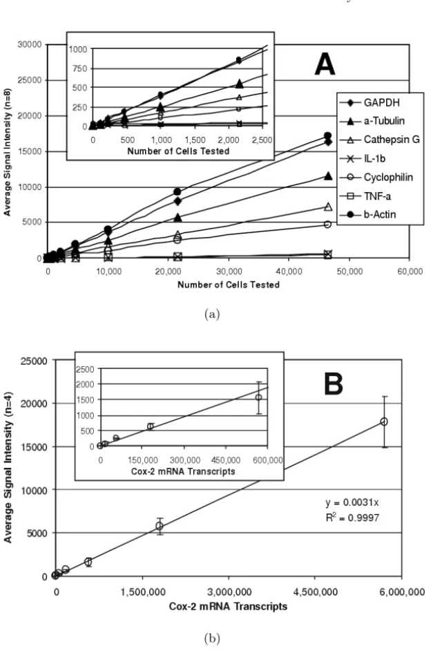
Sensitivity was determined by examining serial dilutions of a bulk lysate of LPS-stimulated THP–1 monocytes. The assay was linear for all expressed tar-get genes over a broad range of sample sizes (Fig. 1.3a) and, more importantly, expression ratios between genes remained constant. Useful gene expression data could be obtained from samples of 1,000 cells or fewer. However, the assay was most robust for samples ranging from 25,000 to 50,000 cells.
To determine the absolute sensitivity of the assay, quantified cox–2 mRNA obtained by in vitro transcription was tested (Fig. 1.3b). Here too, assay re-sponse was linear over the entire range that was tested (up to nearly 6,000,000 molecules) with the best fit linear regression showing a coefficient of correla-tion greater than 0.99. As few as 150,000 cox–2 mRNA molecules were de-tectable. Similar sensitivities were observed with in vitro transcripts of other genes (data not shown). The reproducibility of the mRNA assay was deter-mined for each target using 30,000 cells/well samples of untreated THP–1 cells (n=48) and cells treated with PMA and LPS (n=48). The data for each well were normalized to GAPDH (the housekeeping gene for these experiments) and the coefficient of variability (CV, i.e. standard deviation as a percentage of the average) was determined for each gene (Table 1.2). The average CV was 6.4% for untreated cells and 7.6% for treated cells, ranging from a low of 3% for cathepsin G in untreated cells to a high of 13% for GST Pi–1 and cyclophilin in treated cells.
Antibody Array
(a)
(b)
Fig. 1.3. Sensitivity of the mRNA Assay. (a) Serial dilutions of LPS-stimulated cells were analyzed. The linear response for seven of the target genes is shown with
the low range enlarged in the insert.(b)Serial dilutions of cox–2 mRNA obtained
Table 1.2. Reproducibility of the mRNA Assay
GENE UNTREATED CELLS TREATED CELLS
Average Average
Name Accession Signal %CV Signal %CV
Number (n=48) (n=48)
GAPDH M17851 1000 6% 1000 9%
IL–1β M15840 – – 1778 5%
TNF–α M10988 – – 1416 4%
Tubulin AF141347 224 7% 80 10%
Cathepsin G M16117 510 3% – –
Cox 2 M90100 – – 791 6%
G–CSF E01219 – – 103 8%
GM–CSF E02975 – – 77 10%
GST Pi–1 X06547 79 10% 35 13%
HMG–17 M12623 541 6% – –
Cyclophilin X52851 333 10% 251 13%
β–Thromboglobulin M17017 – – 895 6%
LDH X02152 228 5% 268 7%
TIMP–1 X03124 – – 833 6%
MMP–9 J05070 – – 1117 4%
Actin M10277 1231 4% 1000 5%
AVERAGE: 6.4% 7.6%
Performance Characteristics
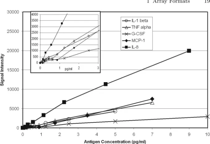
The recombinant standards were used to establish the specificity of each of the five antibody sets in the array and to determine the sensitivity and re-producibility of the assay. Figure 1.4 shows the five sensitivity curves that were obtained. For each of the five antigens, the sensitivity of the multiplexed assay was approximately the same as reported by the antibody supplier for the corresponding traditional ELISA and ranged from less than 0.5 pg/ml for IL–8 to approximately 2 pg/ml for G–CSF. To determine the reproducibility of the multiplexed ELISA, a solution that contained each of the five antigens at 5 pg/ml was analyzed in 36 replicate wells. Data were normalized to 10,000 luminescence counts per well and assigned to each of the five elements accord-ing to their relative intensities. CV values ranged from 7% for IL–8 to 15% for MCP–1 (Table 1.3).
Examples
Fig. 1.4. Sensitivity of the Multiplexed ELISA. Serial dilutions of recombinant antigen standards were tested. The sensitivity curves are shown
Table 1.3. Reproducibility of the multiplexed ELISA
AVERAGE
ANTIGEN SIGNAL S.D. %C.V.
(Normalized)
IL–1β 1,646 192 12%
TNF–α 1,685 129 8%
G–CSF 973 102 10%
MCP–1 1,415 214 15%
IL–8 4,281 280 7%
while mRNA and intracellular proteins were measured in cell lysate. Eight replicates (one column in a 96– well plate) were examined at each of six time points. Results for IL–1βare shown in Fig. 1.5. The induction of IL–1βmRNA, the intracellular accumulation IL–1βand the secretion of protein could all be measured for samples derived from individual wells. Additionally, similar data were obtained for four other proteins and 15 additional genes.
1.6 Conclusion
Fig. 1.5.mRNA and Protein Levels Following Treatment. THP–1 monocytes were examined at different intervals following treatment with PMA. mRNA and intracel-lular proteins were measured in cell lysate while secreted proteins were measured in
the culture media. The results obtained for IL–1βare shown. The error bars show
the standard deviations for eight replicates at each time point
drug discovery process. The ArrayPlateTM mRNA assay is an automation-compatible method for quantifying 16 genes simultaneously with a sensitivity of 150,000 molecules and reproducibility of <10% average CV. The use of reagent-modifiable arrays and of whole–plate imaging of chemiluminescent read-out signals are features that will allow this multiplexed format to be applied to a variety of high throughput screening assays.
References
1. Lockhart DJ, Dong H, Byrne MC, Follettie MT, Gallo MV, Chee MS, Mittmann
M, Wang C, Kobayashi M, Horton H, Brown EL (1996)Expression monitoring
by hybridization to high–density oligonucleotide arrays. Nat Biotechnol 14:1675– 80
2. Wodicka L, Dong H, Mittmann M, Ho MH, Lockhart DJ (1997)Genome–wide
expression monitoring in Saccharomyces cerevisiae. Nat Biotechnol 15:1359–67
3. Schweitzer B, Kingsmore SF (2002) Measuring proteins on microarrays. Curr
Opin Biotechnol 13:14–9
4. Love KR, Seeberger PH (2002) Carbohydrate arrays as tools for glycomics.
Angew Chem Int Ed Engl 41:3583–6, 3513
5. Lam KS, Renil M (2002)From combinatorial chemistry to chemical microarray.
6. Wu RZ, Bailey SN, Sabatini DM (2002) Cell–biological applications of transfected–cell microarrays. Trends Cell Biol 12:485–8
7. Fejzo MS, Slamon DJ (2001) Frozen tumor tissue microarray technology for
analysis of tumor RNA, DNA, and proteins. Am J Pathol 159:1645–50
8. Yang L, Tran DK, Wang X (2001)BADGE, Beads Array for the Detection of
Gene Expression, a high–throughput diagnostic bioassay. Genome Res 11:1888– 98
9. Tung WS, Lee JK, Thompson RW (2001) Simultaneous analysis of 1176 gene
products in normal human aorta and abdominal aortic aneurysms using a membrane–based complementary DNA expression array. J Vasc Surg 34:143– 50
10. Golub TR, Slonim DK, Tamayo P, Huard C, Gaasenbeek M, Mesirov JP, Coller H, Loh ML, Downing JR, Caligiuri MA, Bloomfield CD, Lander ES (1999)
Molecular classification of cancer: class discovery and class prediction by gene expression monitoring. Science 286:531–7
11. Hedenfalk I, Duggan D, Chen Y, Radmacher M, Bittner M, Simon R, Meltzer P, Gusterson B, Esteller M, Kallioniemi OP, Wilfond B, Borg A, Trent J (2001)
Gene–expression profiles in hereditary breast cancer. N Engl J Med 344:539–48 12. Heller RA, Schena M , Chai A, Shalon D, Bedilion T, Gilmore J, Woolley DE,
Davis RW (1997)Discovery and analysis of inflammatory disease–related genes
using cDNA microarrays. Proc Natl Acad Sci USA 94:2150–5
13. Blackwell HE, Perez L, Stavenger RA, Tallarico JA, Cope Eatough E, Foley
MA, Schreiber SL (2001)A one–bead, one–stock solution approach to chemical
genetics: part 1. Chem Biol 8:1167–82
14. LeProust E, Pellois JP, Yu P, Zhang H, Gao X, Srivannavit O, Gulari E, Zhou X
(2000)Digital light–directed synthesis. A microarray platform that permits rapid
reaction optimization on a combinatorial basis. J Comb Chem 2:349–54
15. Fodor SP, Read JL, Pirrung MC, Stryer L, Lu AT, Solas D (1991)Light–directed,
spatially addressable parallel chemical synthesis. Science 251:767–773
16. David CA, Middleton T, Montgomery D, Lim HB, Kati W, Molla A, Xuei
X, Warrior U, Kofron JL, Burns DJ (2002) Microarray compound screening
(microARCS) to identify inhibitors of HIV integrase. J Biomol Screen 7:259–66
17. Heller JH (2002)DNA microarray technology: devices, systems, and applications.
Annu Rev Biomed Eng 4:129–53
18. Shoemaker DD, Linsley PS (2002) Recent developments in DNA microarrays.
Curr Opin Microbiol 5:334–337
19. Oliphant A, Barker DL, Stuelpnagel JR, Chee MS (2002)BeadArray
technol-ogy: enabling an accurate, cost–effective approach to high–throughput genotyping. Biotechniques Suppl 32:56–61
20. Umek RM, Lin SW, Vielmetter J, Terbrueggen RH, Irvine B, Yu CJ, Kayyem
JF, Yowanto H, Blackburn GF, Farkas DH, Chen YP (2001)Electronic detection
of nucleic acids: a versatile platform for molecular diagnostics. J Mol Diagn 3:74–84
21. Velculescu VE, Zhang L, Vogelstein B, Kinzler KW (1995) Serial Analysis of
Gene Expression. Science 270: 484–487
22. Bertelson AH, Velculescu VE (1998)High–throughput Gene Expression Analysis
Using SAGE. Drug Discovery Today 3:152–159
23. Haab BB, Dunham MJ, Brown PO (2001)Protein microarrays for highly
24. Kodadek T (2002) Development of protein–detecting microarrays and related devices. Trends Biochem Sci 27:295–300
25. Martel RR, Botros IW, Rounseville MP, Hinton JP, Staples RR, Morales DA,
Farmer JB, Seligmann BE (2002)Multiplexed screening assay for mRNA
com-bining nuclease protection with luminescent array detection. Assay Drug Dev Tech 1:61–71
26. Berk AJ, Sharp PA (1977)Sizing and mapping of early adenovirus mRNAs by
gel electrophoresis of S1 endonuclease–digested hybrids. Cell 12:721–32
27. Maxwell IH, Van Ness J, Hahn WE (1978)Assay of DNA–RNA hybrids by S1
nuclease digestion and adsorption to DEAE–cellulose filters. Nucleic Acids Res 5:2033–8
28. Wittelsberger SC, Hansen JN (1977)The specificity of S1 nuclease toward RNA–
Biomolecules and Cells on Surfaces –
Fundamental Concepts
Kristi L. Hanson, Luisa Filipponi, and Dan V. Nicolau
2.1 Introduction
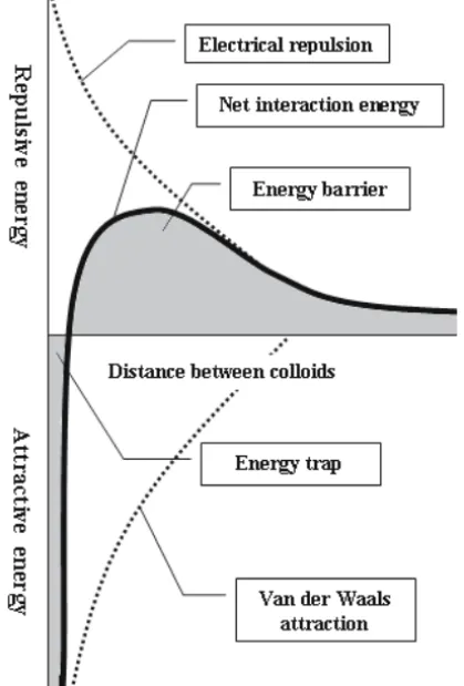
In microarray technology, surfaces must be designed and prepared to opti-mize the immobilization of probe biomolecules and/or cells, but also to resist non-specific binding of target species. Further, the surface and type of im-mobilization technique selected will affect the concentration, bioactivity and target–binding ability of bound species. For any given probe molecule, there is likely to be an optimal surface and/or technique which will allow for attach-ment at the highest possible concentration and with preservation of required activity. However, for multi–probe array formats requiring a variety of probe molecules to be bound to the same type of surface, difficulties are encoun-tered selecting a surface and immobilization method able to generate sufficient probe concentration, resolution and bioactivity for all probes. The resulting variability in probe concentration and activity within the array also leads to signal variability, causing difficulty in data interpretation. Thus, appropriate attachment methods are critical to the success of any array technology.
The aim of this chapter is to summarize the general knowledge and fun-damental concepts underlying DNA, protein, small biomolecule and cell at-tachment to surfaces, and to highlight issues arising in the field of microarray fabrication. The section will provide background knowledge for the reader not familiar with general biomolecule immobilization techniques, while more specific protocols used in microarray technology will be discussed further in Chap. 3.
![Fig. 2.5. Possible conformations of the DNA/oligonucleotide–surface complex on hydrophobic and cationic surfaces (Reprinted with permission from [7]](https://thumb-us.123doks.com/thumbv2/123dok_es/5680467.133986/50.892.121.784.671.927/possible-conformations-oligonucleotide-hydrophobic-cationic-surfaces-reprinted-permission.webp)
![Fig. 2.6. Concentration of DNA molecules as a function of surface chemistry (Adapted from [7]](https://thumb-us.123doks.com/thumbv2/123dok_es/5680467.133986/52.892.116.787.148.947/fig-concentration-dna-molecules-function-surface-chemistry-adapted.webp)