Genetic Variation and
Clinical Heterogeneity in
Cystic Fibrosis
Mitchell L. Drumm,
1,2Assem G. Ziady,
1,3and Pamela B. Davis
1,3,4Departments of1Pediatrics,2Genetics,3Physiology and Biophysics, and4Molecular Biology
and Microbiology, Case Western Reserve University School of Medicine, Cleveland, Ohio 44106; email: [email protected]
Annu. Rev. Pathol. Mech. Dis. 2012. 7:267–82 First published online as a Review in Advance on October 17, 2011
TheAnnual Review of Pathology: Mechanisms of Diseaseis online at pathol.annualreviews.org This article’s doi:
10.1146/annurev-pathol-011811-120900 Copyright c2012 by Annual Reviews. All rights reserved
1553-4006/12/0228-0267$20.00
Keywords
polymorphism, ion channel, genetic and protein profiling
Abstract
Cystic fibrosis (CF), a lethal genetic disease, is characterized by substan-tial clinical heterogeneity. Work over the past decade has established that much of the variation is genetically conferred, and recent stud-ies have begun to identify chromosomal locations that identify specific genes as contributing to this variation. Transcriptomic and proteomic data, sampling hundreds and thousands of genes and their products, point to pathways that are altered in the cells and tissues of CF patients. Genetic studies have examined more than half a million polymorphic sites and have identified regions, and probably genes, that contribute to the clinical heterogeneity. The combination of these approaches has great potential because genetic profiling identifies putative disease-modifying processes, and transcript and protein profiling is shedding light on the biology involved. Such studies are providing new insights into the disease, such as altered apoptotic responses, oxidative stress dysregulation, and neuronal involvement, all of which may open new therapeutic avenues to exploration.
Annu. Rev. Pathol. Mech. Dis. 2012.7:267-282. Downloaded from www.annualreviews.org
by Auburn University on 07/20/13. For personal use only.
Click here for quick links to Annual Reviews content online, including:
• Other articles in this volume • Top cited articles • Top downloaded articles • Our comprehensive search
Further
ANNUALCFTR: cystic fibrosis transmembrane conductance regulator
CYSTIC FIBROSIS
When cystic fibrosis (CF) was first described in 1938, it was clearly recognized as an inherited disorder. Although the parents of the affected children were not affected, siblings or cousins often were. Even before the responsible gene was identified, the inheritance pattern was de-termined to be autosomal recessive (1). In 1989, the gene causing CF was identified (2–4), and all or nearly all patients with CF have errors in both copies of a single gene that encodes a cyclic AMP (cAMP)-regulated chloride chan-nel known as the cystic fibrosis transmembrane conductance regulator (CFTR). The gene that encodes CFTR resides on the long arm of chro-mosome 7 at q31.2, consists of 27 exons, is ap-proximately 190 kb in size, and shows some level of conservation across species from frogs, pufferfish, and evenCaenorhabditis elegans(5) to humans. This gene encodes a cAMP-activated anion channel, which conducts chloride (6) and other anions such as bicarbonate (7). Although the protein contains two membrane-spanning domains, two ATP-binding domains, and an in-tracellular regulatory (R) domain, and although ATP binding and hydrolysis must occur for the channel to open, CFTR only transports the chloride ion down the electrochemical gradi-ent (2–4, 8). Thus, in the sweat duct, CFTR conducts chloride into the epithelial cell (and ultimately into the interstitium) from the duct, but in the gut, CFTR secretes chloride from the epithelial cell into the lumen, and in the airway, CFTR can play either a secretory or absorptive role, depending on the direction of the gradi-ent. Marked reduction of CFTR function gives rise to disease that is ultimately fatal.
When CF was first described, malnutrition due to pancreatic insufficiency (which arises from plugging of the pancreatic ducts and fail-ure to secrete pancreatic enzymes into the gut) was a major cause of death, but currently, more than 90% of the deaths due to CF are due to lung disease (1). Such lung disease arises from the failure to transport chloride ions and, therefore, water into the bronchi. The periciliary fluid layer is too shallow, and cilia
cannot clear the mucus and the environmental agents trapped by it. Thus, inhaled bacteria are not cleared properly and establish chronic in-fection with consequent inflammation, which leads to bronchiectasis and eventual respira-tory failure (1). Nearly 2,000 disease-causing mutations have been observed in the CFTR gene; these mutations have a range of effects on the protein product (seehttp://www.genet. sickkids.on.ca/Home.html). The number of mutations, coupled with CFTR expression in respiratory epithelia (sinuses and airways), pan-creatic duct epithelia, intestinal epithelium, the epithelium lining the vas deferens and the cervix, the liver, the sweat ducts, and nonepithe-lial cells, probably explains the varying severity of protean clinical manifestations of CF in dif-ferent organ systems in difdif-ferent patients.
VARIATION INCFTRAND ITS EFFECT ON CLINICAL
PHENOTYPE
The population and molecular genetics of CF are somewhat unique. There are nearly 2,000 disease-associated mutations documented for CFTR(seehttp://www.genet.sickkids.on.ca/ Home.html). Among them is a single com-mon mutation, a three-nucleotide deletion that causes the absence of amino acid 508 of the normally 1,480–amino acid protein. This mutation, which lacks a single phenylalanine codon and is commonly referred to asF508 (c.1521_1523delCTT for the DNA mutation and F508del for the mutant protein, by current standard nomenclature), accounts for approx-imately 70% of the CF alleles in the popula-tion. The remaining 30% of alleles comprise the other 2,000 different mutations, each of which, individually, has a very low frequency. More recent molecular investigations suggest that some disease-causing alleles do not carry simply a single functional change but rather are complex alleles that contain multiple changes with additive effects that reduce function to a disease-causing level (9).
There is substantial clinical variation among CF patients with the sameCFTRgenotype (10),
Annu. Rev. Pathol. Mech. Dis. 2012.7:267-282. Downloaded from www.annualreviews.org
which indicates non-CFTR sources of varia-tions, but variation in CFTR itself is a strong contributor to clinical heterogeneity. With so many different mutations, as well as combi-nations of changes in many cases, classifica-tion is a challenging task. The most commonly used scheme is to classify disease-causing mu-tations by their molecular effects on CFTR, incorporating mutations that produce trun-cated proteins, misprocessed proteins, chan-nels that fail to open, mutations that conduct ions poorly, and splice mutants that fail to pro-duce functionalCFTR. In this classification sys-tem, class 1 mutations result in no functional CFTRproduced, class 2 mutations cause mis-processed/misfolded proteins, class 3 mutations exhibit impaired activation or channel gating, class 4 mutations show reduced ion conduction through the channel pore, and class 5 mutations are quantitative ones that reduce the amount of CFTR in the cell (11).Figure 1 demon-strates the effects of different classes of muta-tions ofCFTR, exemplified by the predominant mutation of each class.
Class 1 mutations are defined as loss of function, and the disease phenotypes associated with them are relatively severe and consistent. However, mutations in the other groups display a continuum of effects on CFTR both quantitatively and qualitatively, which reduces the clarity of the relationship between mutation classification and clinical outcome. For example, patients who display exocrine pancreatic sufficiency are unlikely to carry two class 1 mutations; rather, they have at least one copy of a class 2, 3, 4, or 5 mutation, as these alleles may exhibit some residual CFTR function and preserve some exocrine pancreatic function. The converse, however, is not always true. That is, mutations in any of the other classes display a range of effects, so one cannot infer the clinical presentation of a patient simply on the basis of the class of mutation they carry. This point can be demonstrated by numerous examples. Take, for instance, codon 551, which normally encodes a glycine (G551). Two different amino acid–substitution muta-tions have been identified in this codon: one
for aspartic acid (G551D) and one for serine (G551S). The clinical consequences are quite different for these two changes, as the G551D variant has virtually no detectable activity, and consequently a classic, severe phenotype is associated. G551S, however, has reduced but clearly detectable function and is associated with a much milder presentation of CF (13).
Another example involves the amino acid substitution R117H, in which an arginine is re-placed with a histidine. This mutant CFTR has a reduced ability to conduct chloride (class 4), but it clearly has function. The R117H muta-tion, however, can be found on two different “backgrounds” of CFTR with respect to se-quence variation in intron 8 (14). This variant, a string of thymidines (T), can be found in three varieties, 5T, 7T, and 9T, and it affects the abil-ity of the CFTR messenger RNA (mRNA) to be spliced correctly. The 5T variant makes the smallest proportion of normally spliced mRNA, 7T an intermediate proportion, and 9T the largest proportion. The R117H mutation exists on both the 5T and 7T backgrounds and, when on the 7T background, often has minimal asso-ciated clinical features. The 5T background, in contrast, reduces the quantity of partially func-tional CFTR below a threshold that results in CF, usually of a pancreatic-sufficient form.
In a further demonstration of the effects of CFTR heterogeneity, these same intron 8 T-tract variants, in the absence of other known functional changes, can impart some physiolog-ical effects. Nearly all male patients with CF, of whatever severity or genotype, have congenital bilateral absence of the vas deferens (CBAVD) (15). Homozygosity of the 5T allele in males can be sufficient to cause CBAVD, but without evident clinical consequences in females.
CFTR-RELATED DISORDERS
Although mortality is usually due to pulmonary complications, similar variations in effects oc-cur in other tissues as well. Nearly all CF patients have either sinusitis or maldevelop-ment of sinuses (16). In contrast, severe loss of function of CFTR causes early manifestation of
Annu. Rev. Pathol. Mech. Dis. 2012.7:267-282. Downloaded from www.annualreviews.org
Exon 10 F508del
5T, 7T, 9T Intron 8
Exon 11 G551D
Exon 11 G542X Exon 4
R117H
ATP Cl–
Cl–
Cl– ADP
R117H
Fewer CFTRs 5T
G551D
5
3
F508del
4
AAAAAA
AAAAAA AAAAAA
1
2
G542X
Figure 1
Effect ofCFTRgene mutations on the cystic fibrosis transmembrane conductance regulator (CFTR) protein and function. (Top) TheCFTRgene with examples of each of the mutation classes and their positions in the gene. The horizontal black arrow represents the transcriptional promoter, and the vertical lines represent exons. The 5and 3untranslated regions are designated by gray shading, and the mutations are designated by vertical arrows. (Bottom) The effects of the mutations on function; the mutation class (12) is circled. G542X, a class 1 mutation, creates a stop codon and causes no full-length, functional protein to be produced. F508del, a class 2 mutation, misfolds and is mostly degraded. If able to reach the plasma membrane, F508del does not activate as readily as normal CFTR; therefore, this mutation is a class 3 example. G551D, another class 3 example, is completely nonresponsive to activation. R117H, a class 4 mutation, resides in a transmembrane region of the protein and impairs chloride transit (conduction) through the channel pore. The T-tract variants in intron 8 represent class 5 mutations in that they alter splicing efficiency. These variants do not eliminate functional CFTR, which differentiates them from class 1 mutants. Note that the original description ofCFTRcontained only 24 exons; later, exons 6, 14, and 17 were found to contain small introns. These exons were renumbered 6a and 6b, 14a and 14b, and 17a and 17b. Thus, what is referred to as exon 10, for example, is actually exon 11.
pancreatic insufficiency, but patients with so-called mild mutations (that are, in general, thought to be those associated with small residual CFTR function; these cases constitute as much as 15% of the CF population) develop
pancreatic failure later in life (17), which is sometimes marked by recurrent episodes of pancreatitis. However, other clinical manifes-tations that occur in a minority of patients, such as hepatic failure and cirrhosis (severe in up
Annu. Rev. Pathol. Mech. Dis. 2012.7:267-282. Downloaded from www.annualreviews.org
to 10% of patients), meconium ileus (neonatal intestinal obstruction, which announces the disease in 15% to 30% of CF newborns), and distal intestinal obstruction syndrome (a blockage, usually near the ileocecal junction, caused by accumulated solid or semisolid feces), cannot be unambiguously linked to the CF genotype per se.
Sweat chloride concentration is markedly elevated in patients with CF compared with healthy controls, and this concentration differs enough between CF and non-CF individuals to serve as the diagnostic test for the dis-ease. However, patients with mutations that allow some residual CFTR function often have lower (although still abnormal) sweat chloride concentrations than do those with severe mutations; rarely, the concentration may be normal (18). Lung disease appears to be less severe in patients with mutations that either allow some CFTR to arrive at the membrane or permit a small amount of normal CFTR to be produced—that is, patients who have some residual CFTR function (19, 20)—but it is difficult to be certain whether improved pul-monary outcome arises from residual CFTR function in the airway itself or from better nutrition due to retained pancreatic function. Nevertheless, CF genotype is associated with pulmonary disease phenotype.
Some studies indicate that even a single ab-normal copy of CFTR can pose a risk factor for disease. Abnormalities in CFTR are highly prevalent in patients with CBAVD, even in the absence of CF (21). The vas deferens appears to be exquisitely sensitive to reduced function of CFTR. Abnormalities in CFTR have also been associated with recurrent or chronic pancreati-tis, including pancreas divisus (22, 23). Abnor-malities in CFTR probably predispose patients to sinusitis (24, 25). Some evidence suggests that abnormalities in a single copy of CFTR may predispose patients to non-CF bronchiec-tasis (26), chronic obstructive pulmonary dis-ease (COPD), (27), or asthma (28), but such associations have been difficult to confirm and may occur in some populations but not in others.
However, even for siblings or unrelated pa-tients with the same CF genotype, severity of the lung disease varies widely. Other factors, including environmental factors and adventi-tious infections, may influence the outcome, but other, non-CFTR genetic variations, termed modifier genes, probably affect the severity of lung disease as well as other aspects of CF.
MOLECULAR PHENOTYPES THAT VARY WITH DISEASE OR DISEASE SEVERITY
Technological advancements have enabled the simultaneous measurement of hundreds and thousands of analytes from individual samples. Such measurements have resulted in large data sets of mRNA, proteins, and metabolites and are referred to as transcriptomics, proteomics, and metabolomics, respectively.
Numerous studies have reported that CF tissues, both mouse and human, exhibit differ-ential expression of many mRNA transcripts and protein (29–36). Xu and colleagues (30) measured significant differences in the expres-sion of numerous genes in the lungs of murine Cftr knockout mice compared with those of non-CF controls. The study employed gut-corrected mice that expressed human CFTR under the control of the fatty acid–binding protein promoter. Significant differences (29 increased and 25 decreased) were identified in the expression of genes involved in signal transduction, transcriptional activation, gly-colysis, cAMP biosynthesis, and inflammatory response in the lung. These changes were observed even in the absence of gross disease pathology, which suggests that there are innate differences in CF cells and tissues and that these changes are not simply due to immune responses, remodeling, and other secondary consequences of tissue injury. These authors (30) concluded that some of the observed dif-ferences, such as increases in the transcription factor CCAAT enhancer–binding protein and in interleukin (IL)-1β (both of which regu-late CFTR expression), indicate an adaptive response to the loss ofCftr(30). Interestingly,
Annu. Rev. Pathol. Mech. Dis. 2012.7:267-282. Downloaded from www.annualreviews.org
even in the absence of lung infection, elevations in genes encoding calgranulin-S100 family members, IL-1β, and IL-4 were observed, which indicates a predisposition to inflamma-tory signaling in CF lungs. In a follow-up study by Xu et al. (29), transcriptome analyses were carried out on murine respiratory epithelia of Cftr knockout mice carrying transgenes that express either normal or F508 human CFTR. The expression ofF508 in respiratory epithelia resulted in the altered expression of numerous genes in specific pathways, including a significant increase in the expression of pro-oxidant genes and a decrease in antipro-oxidant genes in mice that express F508 (29). This finding may indicate that CF lungs are unable to properly regulate redox balance.
To study the effects in human cells, Wright and colleagues (34) examined gene expression in samples collected by nasal curettage. These authors collected airway epithelial cells of the nose, well away from the diseased area of the lung, from homozygous F508 CF patients and compared them with cells from age-matched non-CF control subjects. The authors stratified their data by severity of CF lung disease on the basis of the lowest (severe) or highest (mild) twentieth percentile of predicted forced expiratory volume in 1 s (FEV1)
cor-rected for age (FEV1% predicted). This study
identified significant differences in the expres-sion of genes between CF patients and non-CF controls as well as between severity groups of CF patients (34). In results similar to those obtained in studies conducted with F508 mice (29), these authors reported the highest number of significant increases in genes that influence oxidoreductase activity, followed by those that influence lipid metabolism, protein processing, and ubiquitin conjugation activity in severe CF patients. Genes with increased expression in individuals with mild CF were the most numerous for pathways that regulate lipid metabolism, ion transport, and cell signaling. Significant decreases in gene expression in CF patients versus non-CF controls occurred in pathways that control growth, apoptosis, and immune tolerance. Although most differences
in specific gene-expression profiles vary between animal models and patients, the cellular pathways affected by differential gene expression in CF, as a whole, are strikingly similar. This finding indicates that in addition to the effects of disease pathology, both the loss of CFTR function and the presence of mutant proteins (especially F508) elicit significant gene-expression differences that are related to secondary defects associated with the disease, such as increased inflammatory and redox signaling and decreased growth.
Transcriptome analyses identify changes in gene expression that reflect perturbations in regulatory networks (transcription factors, epigenetic modifications, etc.) but do not nec-essarily predict protein levels or biochemical effects on the cells involved. Thus, an important additional assessment is how protein quantity or function changes. Accordingly, numerous studies have demonstrated that differential protein expression plays a significant role in CF pathophysiology. Much of the current understanding of the inflammatory processes altered in CF has come from measuring protein levels in the airway surface liquid of CF patients via bronchoscopy and bronchoalveolar lavage. Differences in protein expression contribute to the inflammatory response in CF on many lev-els. Inflammatory mediators such as IL-8 (37) and IL-9 (38) are overexpressed in the airways of CF patients. Significant increases in proin-flammatory cytokine levels have been detected in CF airways, even in the absence of detectable infection (39). In animal models and cultured cells, anti-inflammatory cytokines such as IL-10 (40) are decreased in CF. Furthermore, numerous transcriptional activators such as signal transducer and activator of transcription 1 (STAT1) (41) and nuclear factorκB (NF-κB) (42, 43), as well as the proteins that regulate them, are differentially expressed or activated in such a way as to enhance the inflammatory response or diminish host defenses. In the case of STAT1, although its overexpression is observed in CF, increased activation of protein inhibitor of activated STAT1 interferes with STAT1 activity, resulting in a significant
Annu. Rev. Pathol. Mech. Dis. 2012.7:267-282. Downloaded from www.annualreviews.org
decrease in the expression of various proteins regulated by STAT1, including nitric oxide synthase 2. This decrease reduces nitric oxide production, thereby reducing host defense against bacterial infection. The expression of antioxidant proteins in CF, including thioredoxin 1; peroxiredoxins 1, 3, and 6; and glutathione-S-transferase 1 is decreased in CF, whereas levels of superoxide dismutase 2 are elevated compared with those in non-CF controls (32). This alteration in the expression of redox-related protein hinges on the dys-function of an important transcription factor, nuclear factor E2–related factor 2 (32).
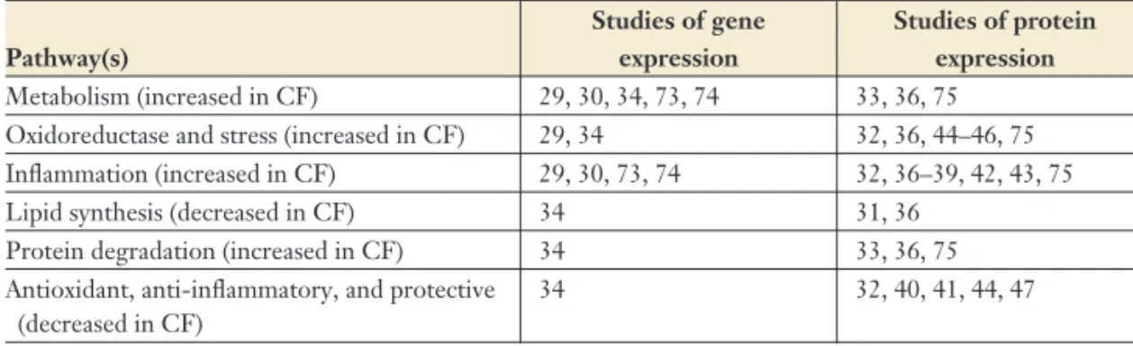
The advent of mass spectrometric tech-niques for the high-throughput study of differential protein expression has allowed in-vestigators to conduct wide-scale comparisons between CF and non-CF samples and to identify cellular signaling pathways whose components differ quantitatively or qualita-tively. Importantly, these analyses have been confined mostly to cell culture models, both immortalized and primary, and to animal models; therefore, the degree to which they reflect changes in humans in vivo has yet to be determined. Proteomic analyses have identified significant increases in proteins that induce or respond to oxidative stress, proinflammatory signaling, and increased metabolism (e.g., gly-colysis) (8, 9). Significant decreases in protein expression occurred in antioxidant proteins (32) and in numerous transcription factors (35). Table 1 summarizes studies in humans and
model systems of differential gene and protein expression in CF versus non-CF controls. Taken together, proteomic studies suggest alterations in pathways that are similar to those observed in gene-array studies. However, whereas some specific differences match well with those observed in gene-expression studies, most protein differences are not identified as such at the message level. Pathways that are heavily affected in transcriptomic and proteomic screens predict further alterations in the homeostasis of CF cells. For example, on the basis of data gathered in laboratory cell and animal models and in severe-CF patients, increased levels of, and a decreased ability to resolve, oxidative stress is predicted for CF cells. This prediction is supported by findings that levels of glutathione are deceased (44), whereas levels of nitrates and nitrotyrosine (oxidized protein) (45) are elevated in the air-ways of CF patients. In the lung, the influx of inflammatory cells, which produce oxidants to kill bacteria, introduces a heavy oxidative chal-lenge, which would not be well resolved on the basis of transcriptomic and proteomic data and may therefore exacerbate activation of redox-sensitive cascades, such as the NF-κB pathway (46). Furthermore, effects of protein oxidation, including altered function, reduce enzymatic activity and enhance susceptibility to prote-olytic degradation (47), a pathway that is ele-vated in CF at both the gene- (2, 7) and protein-expression levels (35). Similar inferences, based on screening studies, can be made for other
Table 1 Messenger RNA and protein-expression differences in cellular pathways
Pathway(s)
Studies of gene expression
Studies of protein expression
Metabolism (increased in CF) 29, 30, 34, 73, 74 33, 36, 75 Oxidoreductase and stress (increased in CF) 29, 34 32, 36, 44–46, 75 Inflammation (increased in CF) 29, 30, 73, 74 32, 36–39, 42, 43, 75 Lipid synthesis (decreased in CF) 34 31, 36
Protein degradation (increased in CF) 34 33, 36, 75 Antioxidant, anti-inflammatory, and protective
(decreased in CF)
34 32, 40, 41, 44, 47
Abbreviation: CF, cystic fibrosis.
Annu. Rev. Pathol. Mech. Dis. 2012.7:267-282. Downloaded from www.annualreviews.org
SNP: single nucleotide polymorphism
secondary defects that contribute to CF pathol-ogy, such as inflammation and metabolism problems.
In addition to yielding useful information about altered pathways in disease, transcrip-tomic and proteomic studies can in themselves be an indicator of modifier genes of CF disease, discussed in more detail below. Comparisons such as the one undertaken by Wright et al. (34) can reveal novel genes associated with the sever-ity of disease. For example, this study found that the expression of the genes for statherin, a protein that protects against infection, and for adiponectin, a protein that can act as an in-hibitor of inflammation, were elevated in mild-CF patients. Similar findings were observed for genes that modulate inflammatory signal-ing, metabolism, oxidative stress, and protein degradation. However, it is difficult to discern whether these differences contribute to the phe-notypes of mild or severe CF or whether un-derlying differences in disease pathology cause these disparities. Therefore, for such explo-ration of modifier genes, extensive validation and examination of the individual genes’ se-quence for single nucleotide polymorphisms (SNPs) must be performed. Nevertheless, sig-nificant differences observed at the gene- and protein-expression levels shed light on both the pathways that are influenced and implicated in the disease and the functional outcomes of modulation of such pathways. In the context of modifier genes, information about the pathways that are associated with and produce the vari-ously severe forms of CF provides a focus for investigators on the potential role of identified modifier genes (discussed in the following sec-tion), allowing for the characterization of their impact on molecular phenotypes that define and affect the progression of disease in patients. The relationship between modifier genes and broad changes in genes and proteins is not yet clear, but it is reasonable to expect one to exist. The exploration of modifiers of CF disease is relatively new, and a full understand-ing of candidates that significantly contribute to CF disease severity has yet to be realized. Nevertheless, alterations in modifiers that are
associated with poorer prognoses in CF patients may be related to the differential expression of genes and proteins (molecular phenotypes) that contribute to disease pathology. Interactions between modifier genes and molecular pheno-types may be direct, such as would be the case if the modifier gene were a transcription factor or a modulator of transcription factor activity. Indirect interactions may include the effects of products of modifier genes (e.g., metabolites) on the expression of molecular phenotypes.
Such studies provide important correla-tive data that suggest relationships between molecules and processes and are useful for hy-pothesis generation. In some cases, a correla-tion may be so strong that one is tempted to infer causal relationships, but descriptive anal-yses do not provide causal relationships. That is, when multiple phenotypes track together, one cannot conclude that one causes another; rather, they may be independent consequences of the same or different causes. To determine causation, one must employ other approaches. These methods typically involve prospective studies in which a variable (e.g., a drug) can be manipulated to determine its effect on outcome, or genetic studies, which identify outcomes that track with genetic variation.
GENETIC HETEROGENEITY AND MODIFIER GENES
Among individuals with CF, the heterogeneity of clinical outcomes is broad. Even in patients with the same mutantCFTRgenotype, varia-tion in features such as lung funcvaria-tion can be dramatic (48, 49). The human genome contains a large amount of variation; there are more than 3 million SNPs (50), along and hundreds, per-haps thousands, of copy number variants (51) in which a region of DNA is duplicated or deleted. Although the functional consequences of these variants is largely unknown, virtually all of the ∼25,000 genes in our genome have variants. In situations where the effects of such variants have been examined, they can be neutral, but many influence gene products quantitatively or qual-itatively. Thus, they provide a mechanism to
Annu. Rev. Pathol. Mech. Dis. 2012.7:267-282. Downloaded from www.annualreviews.org
explain much of the interindividual phenotypic variation observed in the human population.
From a clinical perspective, identification of genomic variants that cause differences in disease outcome has significant implications. The products of these genes, or the pathways in which they act, may become potential therapeutic targets. That is, nature has pro-vided evidence that a particular target can modify disease, so drugs that can mimic the effects of the advantageous version of the gene are likely to be effective therapeutically. For example, enzymes or receptors with deleterious functions could be targeted with inhibitors or antagonists, respectively. Conversely, enzymes or receptors with beneficial or protective effects could be activated or stimulated. An extension of this concept is that the gene product conferring the phenotypic effect may not be a readily “druggable” target, but understanding the pathway in which it works would probably present more feasible targets.
GENES THAT MODIFY CYSTIC FIBROSIS LUNG DISEASE
The search for genetic causes of clinical varia-tion has been approached in various ways. The generic recipe for such studies is to examine the relationship between variants in a gene or position in the genome (known as a locus) and a clinical trait that varies among patients. The trait may be continuous, such as pulmonary function indices, or discrete, such as intestinal obstruction. Because mortality is closely associ-ated with lung disease, pulmonary phenotypes have been the most intensely studied in this regard.
Early modifier studies revolved around one or a few candidate genes, focusing on those genes that fit into a pathway or process believed to contribute to CF disease pathophysiology (Figure 2); such pathways include ion nels (particularly chloride and sodium chan-nels, etc.) that could compensate for CFTR’s absence (52, 53), innate defense (54–57) and in-flammatory pathway components (58, 59) that could modulate the response to pulmonary
IL-1β IL-8 TNF-α
Na+, Cl– H2O
Macrophage Epithelial cell
Mucus
Neutrophil
Pseudomonas aeruginosa
ell
N
β 8 α a++,
O
α a 2O
1β -8 F-N H2
IL-TNF
Mucus
udomonas uginosa
Epithelial ce
Figure 2
Selection of candidate modifier genes. Cystic fibrosis (CF) pulmonary and airway disease has been modeled as a consequence of insufficient luminal hydration (including mucus), an inability to clear bacteria such asPseudomonas aeruginosa, and an excessive inflammatory burden, either as a response to infection or inherent to CF epithelia. Candidate modifying genes were chosen for their potential to modulate these processes. Genes encoding epithelial ion channels were examined because they can influence airway luminal hydration. Genes encoding chemotactic signaling molecules, such as interleukin (IL)-1β, IL-8, and tumor necrosis factor (TNF)-α, were examined because they can influence the level of immune cell recruitment, and genes involved in
antimicrobial processes or immune cell function were investigated because they may exert modifying effects on airway disease by influencing the ability to clear or suppress infection.
pathogens, and so on. For the most part, these studies were carried out on relatively small numbers of patients and were dictated by the small number of variants known for most genes, the genotyping technologies available, and the cost of using the technology. As genome in-formation and technology have progressed, and as multicenter approaches have provided larger patient and family cohorts, large-scale geno-typing has been made more technologically and economically feasible and possible. Con-sequently, increased sample sizes are providing better statistical power to detect associations, and scans encompassing the whole genome are
Annu. Rev. Pathol. Mech. Dis. 2012.7:267-282. Downloaded from www.annualreviews.org
allowing one to probe nearly every gene and region of the genome, which may permit the discovery of effects of loci not previously con-sidered in CF.
Although this approach has resulted in many published associations, few of these have been replicated. Replication is important for the validation of modifiers, as the small sample size and the testing of multiple genes and polymor-phisms make these studies prone to spurious associations. The first CF modifier study to use large patient cohorts and replication identified SNPs in the 5 region of the TGFB1 gene that are associated with lung function in CF patients. The initial association was found in CF subjects who were homozygous for the
F508 mutation and in the upper and lower quartiles for lung function; the replicating population included other CF genotypes (48). TGFB1 encodes transforming growth factor β1, a signaling molecule made by many cell types involved in multiple processes, such as inflammation and wound healing. The same region ofTGFB1also associates similarly with COPD in smokers (60), whereas the opposite association was found for asthma (61).
Using a different approach, in which pa-tient samples with similar disease severity were pooled for genotyping, Gu et al. (62) identified allele frequency differences in the interferon-related developmental regulator 1 (IFRD1) gene between severely and mildly affected CF patients. TheIFRD1SNPs were examined in a second cohort of patients and revealed skewed frequencies that tracked with pulmonary dis-ease severity; they were also examined in a mouse model in which the gene was expressed predominantly in neutrophils.
The neutrophil chemoattractant IL-8 is elevated in the airways of CF patients and is believed to be largely responsible for the neutrophilia that is characteristic of CF lungs. Variants in IL-8 that associate with other pulmonary conditions (63) were examined and found to associate with pulmonary function in two independent cohorts of CF subjects. Molecular studies indicated that the adverse
allele expressed nearly three times more IL-8 mRNA than did the protective allele (58).
From a list of genes proposed to be involved in various aspects of airway physiology, alleles of SNPs in the endothelin receptor A gene (EDNRA) were associated with pulmonary function in the same cohort used to assess TGFB1, and these findings were replicated in cohorts originating from the Seattle, Wash-ington, area as well as from Ireland (64). Endothelin stimulation of this receptor causes contraction and proliferation of smooth mus-cle. The variants associated with pulmonary function were also associated with mRNA-expression differences; the adverse variant showed greaterEDNRAmRNA levels in airway smooth muscle than did the protective variant. The ability to replicate associations adds confidence to their validity and supports the idea that such associations are the consequences of biologically based mechanisms. These asso-ciations and molecular investigations support models such as that depicted in Figure 3, in which inflammation is deleterious to the air-ways. However, they also suggest that we may have underestimated the role of tissue remodel-ing (TGFB1andEDNRA) and airway mechan-ics (EDNRA) in the disease process.
MODIFIERS OF OTHER CYSTIC FIBROSIS TRAITS
Although the most attention has been paid to pulmonary disease in the hunt for modifiers, it is by no means the only relevant pheno-type to the disease. For example, meconium ileus and other gastrointestinal manifestations affect a large proportion of CF patients. Genetic influences on intestinal obstruction were iden-tified in mouse studies (65), and those observa-tions appear to hold true in human CF patients as well (66, 67).
CF-related diabetes (CFRD) is a prominent comorbidity of CF that affects nearly half of CF patients at some point in their lifetime. The etiology of CFRD is unclear, and it has features that are similar to those of type 1 and
Annu. Rev. Pathol. Mech. Dis. 2012.7:267-282. Downloaded from www.annualreviews.org
type 2 diabetes. The type 1 similarities appear to revolve around the loss of insulin-producing islet cells, but there is no evidence for an autoimmune mechanism. However, having a relative with type 2 diabetes increases the risk of CFRD, which suggests that there are mech-anistic similarities between the two. To explore this hypothesis, Blackman et al. (68) examined SNPs in genes reported to be susceptibility genes for type 2 diabetes for their association with CFRD; they found that one of the most significant type 2 diabetes genes, an islet cell transcription factor named transcription factor 7–like 2, also associated with CFRD and did so reproducibly in several cohorts of subjects.
These studies provide important principles and insight into CF pathogenesis. The genet-ics of intestinal obstruction suggest that mouse models may be powerful tools to examine in vivo mechanisms of obstruction and perhaps a resource for testing therapeutics designed to alleviate or prevent obstruction.
Regarding CFRD, the loss of endocrine cells was historically thought to be a bystander ef-fect of the disease in which the destruction of exocrine tissue spreads to the endocrine cells. The parallels with type 2 diabetes bring those models into question and will probably re-quire revisiting those models as the pathogen-esis of type 2 diabetes becomes clearer. As CF patients appear to be a diabetes-prone popu-lation, our understanding of type 2 diabetes may incorporate mechanistic information from CFRD.
WHOLE-GENOME STUDIES
As a consequence of the large number of small studies, consortia have evolved to pro-vide the ability to replicate associations and to increase the power to detect them. The Gene Modifier Study (48), the CF Twin and Sib Study (69), and the Canadian Consortium for Genetic Studies (48, 56) formed a North Amer-ican CF Gene Modifier Consortium. Com-bined, the groups surveyed 3,467 CF patients, and because of the various study designs used
CAT
ELF
EHF APIP CD44
PDHX Chromosome 11
200 kb
CBLN4 MC3R
AURKA CSTF1 CASS4 8
6
4
2
0
8
6
4
2
0
Chromosome 20
–log
10
(
P
value)
–log
10
(P
value)
Figure 3
Location of single nucleotide polymorphisms (SNPs) identified by genome-wide association studies as associated with lung function. Clusters of associating SNPs are shown as gray boxes. The height of the box corresponds to the most significant SNP in the cluster. For chromosome 11, the most significant association lies between the genesAPIPandEHF; less significant associations overlap both genes. The chromosome 20 association is most significant with SNPs 3toCBLN4, but there is also a peak overMC3R.
by each group, they were able to examine these subjects by case/control designs, as well as by linkage analysis, thereby capitalizing on the strengths of each approach.
The consolidation of cohorts presents logistical problems, particularly with regard to phenotype. Studies investigating related but different phenotypes, or phenotypes arrived at by different methods, may generate different results. To reduce the chances of such occur-rences, investigators developed a phenotypic definition based on lung function, but cor-rected for survival and referenced to individuals with CF rather than the non-CF population, to characterize all the subjects in the study (70).
Another potential confounding feature for genetic studies is the effect of ancestry on asso-ciations. Individuals of common ancestry, such as racial and ethnic groups, are genetically more
Annu. Rev. Pathol. Mech. Dis. 2012.7:267-282. Downloaded from www.annualreviews.org
similar than are individuals from different back-grounds. As a consequence, phenotypes that are more common in one related group than in an-other may display apparent genetic associations that are due to population structure rather than the gene or genes in question. To explore this possibility, investigators studied the structure of the population (71) and found it to be of pre-dominantly Caucasian descent.
With a sample size of nearly 3,500 CF sub-jects to examine, more than 500,000 SNPs spread across the genome were genotyped and assessed for association with pulmonary func-tion. Two genomic intervals surpassed the strict statistical criteria for multiple testing. One re-gion, on chromosome 11 and residing between the Apaf-1-associated protein gene (APIP) and the Ets-homologous factor gene (EHF), showed significant association (P = 9.4 × 10⁻⁹). A
second region, on chromosome 20, was de-tected by linkage analysis [logarithm (base 10) of odds score of 5.033] in families, and when the region was examined for association in unrelated CF individuals, SNPs in the genes encoding melancortin-3 receptor (MC3R) and cerebellin-like 4 (CBLN4) showed the most significant association (P = 1.34 × 10⁻⁴);
combining linkage and association this region achieved a significance level ofP<10−8(74).
Figure 3shows a schematic of these two regions and the genes known to reside in those regions. In addition to the associations depicted in Figure 3, there are other tantalizing loci that nearly meet the rigorous statistical thresholds of these types of associations. Such loci include the major histocompatibility complex genes on chromosome 6 and SNPs included in the an-giotensin 2 type 2 receptor gene (AGTR2) on the X chromosome.
THE UTILITY OF VARIANTS AND VARIATION
Studying variation is a standard approach for understanding biology and, in this case, the
biology underlying a disease. By understanding the variation that occurs between individuals affected by CF compared with those not affected, we hope to obtain insight into the pathogenesis. By examining variation between CF patients and comparing it with their clinical outcome, we hope to identify variation that plays a causal role in modifying disease severity and thereby become enlightened about therapeutic approaches. To this end, the massive amounts of data that continue to be collected must be integrated in recognition of the complementary attributes of the data. The molecular and clinical data that have been collected in various venues for decades have provided us with extensive view of systemic effects of CF, but they do not typically inform us about causal relationships and where one might intervene. Historically, that has been left to the clinician to predict. Although this process has been effective by dramatically extending the life span of CF patients dramat-ically, we now have the opportunity to directly identify genetic factors that cause differences in outcome, as outlined above for modifier genes.
Thus, one of the next key challenges will be to integrate the massive amounts of molec-ular, genetic, and clinical data for these pur-poses. Genetic analyses will allow us to focus attention on a finite number of processes, whit-tling down the 20,000 to 30,000 genes of the genome to a handful that influence pathogen-esis. The molecular characterizations should allow us to place those modifying genes into pathways, and comparison with clinical infor-mation should permit us to determine how to alter the disease itself. However, we are still in the early stages of tapping into this potential; the effects of genetic variation on the plethora of relevant traits have yet to be realized. Two fascinating questions that remain are: How does gender affect modifying influences? What are the genetics of drug metabolism in cystic fibrosis?
Annu. Rev. Pathol. Mech. Dis. 2012.7:267-282. Downloaded from www.annualreviews.org
SUMMARY POINTS
1. Much of the clinical variation among individuals with CF is conferred genetically. 2. Molecular technology has allowed massive amounts of information to be generated in
the form of transcriptomic, proteomic, and genetic data.
3. Analyses of these data have begun to identify pathways not previously considered im-portant to CF and to point out key players in pathways that were previously considered important.
4. The identification of these processes and their key components will provide novel ther-apeutic targets and insight into how to improve existing therapies.
FUTURE ISSUES
1. The identification of putative disease-modifying genes must be followed by biological studies to verify the modifying effects of these genes.
2. The proteins encoded by these modifying genes, as well as the pathways in which they act, may become potential therapeutic targets for cystic fibrosis. Thus, future work should aim to evaluate the modulation of these pathways for therapy.
3. Additional genotype/phenotype studies for associations with pharmacologic responses (pharmacogenetics) will be possible and may allow the optimization of CF pharmacology based on an individual’s genetic profile.
4. The biology of CF certainly overlaps with that of other disorders; therefore, the mod-ifying effects of genes identified in CF studies probably have effects in other disorders and will need to be examined.
DISCLOSURE STATEMENT
The authors are not aware of any affiliations, memberships, funding, or financial holdings that might be perceived as affecting the objectivity of this review.
LITERATURE CITED
1. Davis PB. 2006. Cystic fibrosis since 1938.Am. J. Respir. Crit. Care Med.173:475–82
2. Kerem B, Rommens JM, Buchanan JA, Markiewicz D, Cox TK, et al. 1989. Identification of the cystic fibrosis gene: genetic analysis.Science245:1073–80
3. Riordan JR, Rommens JM, Kerem B, Alon N, Rozmahel R, et al. 1989. Identification of the cystic fibrosis gene: cloning and characterization of complementary.Science245:1066–73
4. Rommens JM, Iannuzzi MC, Kerem B, Drumm ML, Melmer G, et al. 1989. Identification of the cystic fibrosis gene: chromosome walking and jumping.Science245:1059–65
5. McCombie WR, Adams MD, Kelley JM, FitzGerald MG, Utterback TR, et al. 1992.Caenorhabditis elegansexpressed sequence tags identify gene families and potential disease gene homologues.Nat. Genet.
1:124–31
6. Bear CE, Li CH, Kartner N, Bridges RJ, Jensen TJ, et al. 1992. Purification and functional reconstitution of the cystic fibrosis transmembrane conductance regulator (CFTR).Cell68:809–18
7. Quinton PM. 2001. The neglected ion: HCO3.Nat. Med.7:292–93
Annu. Rev. Pathol. Mech. Dis. 2012.7:267-282. Downloaded from www.annualreviews.org
8. Gadsby DC, Vergani P, Csanady L. 2006. The ABC protein turned chloride channel whose failure causes cystic fibrosis.Nature440:477–83
9. McGinniss MJ, Chen C, Redman JB, Buller A, Quan F, et al. 2005. Extensive sequencing of theCFTR
gene: lessons learned from the first 157 patient samples.Hum. Genet.118:331–38
10. Kerem E, Kerem B. 1995. The relationship between genotype and phenotype in cystic fibrosis.Curr. Opin. Pulm. Med.1:450–56
11. MacDonald KD, McKenzie KR, Zeitlin PL. 2007. Cystic fibrosis transmembrane regulator protein mu-tations: ‘class’ opportunity for novel drug innovation.Paediatr. Drugs9:1–10
12. Tsui LC. 1992. The spectrum of cystic fibrosis mutations.Trends Genet.8:392–98
13. Drumm ML, Wilkinson DJ, Smit LS, Worrell RT, Strong TV, et al. 1991. Chloride conductance ex-pressed byF508 and other mutant CFTRs inXenopusoocytes.Science254:1797–99
14. Kiesewetter S, Macek M Jr, Davis C, Curristin SM, Chu CS, et al. 1993. A mutation in CFTR produces different phenotypes depending on chromosomal background.Nat. Genet.5:274–78
15. Jarzabek K, Zbucka M, Pepinski W, Szamatowicz J, Domitrz J, et al. 2004. Cystic fibrosis as a cause of infertility.Reprod. Biol.4:119–29
16. Watelet JB, Van Cauwenberge P, Bachert C. 2000. Rhinological aspects of cystic fibrosis.Monaldi Arch. Chest Dis.55:475–77
17. Walkowiak J, Lisowska A, Blaszczynski M. 2008. The changing face of the exocrine pancreas in cystic fibrosis: pancreatic sufficiency, pancreatitis and genotype.Eur. J. Gastroenterol. Hepatol.20:157–60 18. Davis PB, Schluchter MD, Konstan MW. 2004. Relation of sweat chloride concentration to severity of
lung disease in cystic fibrosis.Pediatr. Pulmonol.38:204–9
19. McKone EF, Goss CH, Aitken ML. 2006. CFTR genotype as a predictor of prognosis in cystic fibrosis.
Chest130:1441–47
20. de Gracia J, Mata F, Alvarez A, Casals T, Gatner S, et al. 2005. Genotype-phenotype correlation for pulmonary function in cystic fibrosis.Thorax60:558–63
21. Cuppens H, Cassiman JJ. 2004. CFTR mutations and polymorphisms in male infertility.Int. J. Androl.
27:251–56
22. de Cid R, Ramos MD, Aparisi L, Garcia C, Mora J, et al. 2010. Independent contribution of common CFTR variants to chronic pancreatitis.Pancreas39:209–15
23. DiMagno MJ, DiMagno EP. 2005. Chronic pancreatitis.Curr. Opin. Gastroenterol.21:544–54 24. Wang X, Cutting GR. 2011. Chronic rhinosinusitis.Adv. Otorhinolaryngol.70:114–21
25. Wang X, Kim J, McWilliams R, Cutting GR. 2005. Increased prevalence of chronic rhinosinusitis in carriers of a cystic fibrosis mutation.Arch. Otolaryngol.131:237–40
26. Rowntree RK, Harris A. 2003. The phenotypic consequences of CFTR mutations.Ann. Hum. Genet.
67:471–85
27. Sandford AJ, Joos L, Pare PD. 2002. Genetic risk factors for chronic obstructive pulmonary disease.Curr. Opin. Pulm. Med.8:87–94
28. Noone PG, Knowles MR. 2001. ‘CFTR-opathies’: disease phenotypes associated with cystic fibrosis transmembrane regulator gene mutations.Respir. Res.2:328–32
29. Xu Y, Liu C, Clark JC, Whitsett JA. 2006. Functional genomic responses to cystic fibrosis transmembrane conductance regulator (CFTR) and CFTR508in the lung.J. Biol. Chem.281:11279–91
30. Xu Y, Clark JC, Aronow BJ, Dey CR, Liu C, et al. 2003. Transcriptional adaptation to cystic fibrosis transmembrane conductance regulator deficiency.J. Biol. Chem.278:7674–82
31. Karp CL, Flick LM, Park KW, Softic S, Greer TM, et al. 2004. Defective lipoxin-mediated anti-inflammatory activity in the cystic fibrosis airway.Nat. Immunol.5:388–92
32. Chen J, Kinter M, Shank S, Cotton C, Kelley TJ, Ziady AG. 2008. Dysfunction of Nrf-2 in CF epithelia leads to excess intracellular H2O2and inflammatory cytokine production.PLoS ONE3:e3367
33. Srivastava M, Eidelman O, Jozwik C, Paweletz C, Huang W, et al. 2006. Serum proteomic signature for cystic fibrosis using an antibody microarray platform.Mol. Genet. Metab.87:303–10
34. Wright JM, Merlo CA, Reynolds JB, Zeitlin PL, Garcia JG, et al. 2006. Respiratory epithelial gene expression in patients with mild and severe cystic fibrosis lung disease.Am. J. Respir. Cell Mol. Biol.
35:327–36
Annu. Rev. Pathol. Mech. Dis. 2012.7:267-282. Downloaded from www.annualreviews.org
35. Pollard HB, Eidelman O, Jozwik C, Huang W, Srivastava M, et al. 2006. De novo biosynthetic profiling of high abundance proteins in cystic fibrosis lung epithelial cells.Mol. Cell Proteomics5:1628–37 36. Pollard HB, Ji XD, Jozwik C, Jacobowitz DM. 2005. High abundance protein profiling of cystic fibrosis
lung epithelial cells.Proteomics5:2210–26
37. Muhlebach MS, Reed W, Noah TL. 2004. Quantitative cytokine gene expression in CF airway.Pediatr. Pulmonol.37:393–99
38. Hauber HP, Manoukian JJ, Nguyen LH, Sobol SE, Levitt RC, et al. 2003. Increased expression of interleukin-9, interleukin-9 receptor, and the calcium-activated chloride channel hCLCA1 in the upper airways of patients with cystic fibrosis.Laryngoscope113:1037–42
39. Rosenfeld M, Gibson RL, McNamara S, Emerson J, Burns JL, et al. 2001. Early pulmonary infection, inflammation, and clinical outcomes in infants with cystic fibrosis.Pediatr. Pulmonol.32:356–66 40. Soltys J, Bonfield T, Chmiel J, Berger M. 2002. Functional IL-10 deficiency in the lung of cystic fibrosis
(Ctfr−/−) and IL-10 knockout mice causes increased expression and function of B7 costimulatory molecules on alveolar macrophages.J. Immunol.168:1903–10
41. Kelley TJ, Elmer HL. 2000. In vivo alterations of IFN regulatory factor 1 and PIAS1 protein levels in cystic fibrosis epithelium.J. Clin. Investig.106:403–10
42. Blackwell TS, Stecenko AA, Christman JW. 2001. Dysregulated NF-κB activation in cystic fibrosis: evidence for a primary inflammatory disorder.Am. J. Physiol. Lung Cell. Mol. Physiol.281:69–70 43. Stecenko AA, King G, Torii K, Breyer RM, Dworski R, et al. 2001. Dysregulated cytokine production in
human cystic fibrosis bronchial epithelial cells.Inflammation25:145–55
44. Roum JH, Buhl R, McElvaney NG, Borok Z, Crystal RG. 1993. Systemic deficiency of glutathione in cystic fibrosis.J. Appl. Physiol.75:2419–24
45. Jones KL, Hegab AH, Hillman BC, Simpson KL, Jinkins PA, et al. 2000. Elevation of nitrotyrosine and nitrate concentrations in cystic fibrosis sputum.Pediatr. Pulmonol.30:79–85
46. Hayashi T, Ueno Y, Okamoto T. 1993. Oxidoreductive regulation of nuclear factorκB. Involvement of a cellular reducing catalyst thioredoxin.J. Biol. Chem.268:11380–88
47. Kraynack NC, Corey DA, Elmer HL, Kelley TJ. 2002. Mechanisms of NOS2 regulation by Rho GTPase signaling in airway epithelial cells.Am. J. Physiol. Lung Cell. Mol. Physiol.283:604–11
48. Drumm ML, Konstan MW, Schluchter MD, Handler A, Pace R, et al. 2005. Genetic modifiers of lung disease in cystic fibrosis.N. Engl. J. Med.353:1443–53
49. Kerem E, Reisman J, Corey M, Canny GJ, Levison H. 1992. Prediction of mortality in patients with cystic fibrosis.N. Engl. J. Med.326:1187–91
50. Frazer KA, Ballinger DG, Cox DR, Hinds DA, Stuve LL, et al. 2007. A second generation human haplotype map of over 3.1 million SNPs.Nature449:851–61
51. Redon R, Ishikawa S, Fitch KR, Feuk L, Perry GH, et al. 2006. Global variation in copy number in the human genome.Nature444:444–54
52. Viel M, Leroy C, Hubert D, Fajac I, Bienvenu T. 2008. ENaCβandγgenes as modifier genes in cystic fibrosis.J. Cyst. Fibros.7:23–29
53. Blaisdell CJ, Howard TD, Stern A, Bamford P, Bleecker ER, Stine OC. 2004. CLC-2 single nucleotide polymorphisms (SNPs) as potential modifiers of cystic fibrosis disease severity.BMC Med. Genet.5:26 54. Davies JC, Turner MW, Klein N. 2004. Impaired pulmonary status in cystic fibrosis adults with two
mutatedMBL-2alleles.Eur. Respir. J.24:798–804
55. Garred P, Pressler T, Madsen HO, Frederiksen B, Svejgaard A, et al. 1999. Association of mannose-binding lectin gene heterogeneity with severity of lung disease and survival in cystic fibrosis.J. Clin. Investig.104:431–37
56. Dorfman R, Sandford A, Taylor C, Huang B, Frangolias D, et al. 2008. Complex two-gene modulation of lung disease severity in children with cystic fibrosis.J. Clin. Investig.118:1040–49
57. Stanke F, Becker T, Kumar V, Hedtfeld S, Becker C, et al. 2011. Genes that determine immunology and inflammation modify the basic defect of impaired ion conductance in cystic fibrosis epithelia.J. Med. Genet.48:24–31
58. Hillian AD, Londono D, Dunn JM, Goddard KA, Pace RG, et al. 2008. Modulation of cystic fibrosis lung disease by variants in interleukin-8.Genes Immun.9:501–8
Annu. Rev. Pathol. Mech. Dis. 2012.7:267-282. Downloaded from www.annualreviews.org
59. Hull J, Thomson AH. 1998. Contribution of genetic factors other thanCFTRto disease severity in cystic fibrosis.Thorax53:1018–21
60. Celedon JC, Lange C, Raby BA, Litonjua AA, Palmer LJ, et al. 2004. The transforming growth factor
β1 (TGFB1) gene is associated with chronic obstructive pulmonary disease (COPD).Hum. Mol. Genet.
13:1649–56
61. Silverman ES, Palmer LJ, Subramaniam V, Hallock A, Mathew S, et al. 2004. Transforming growth factor
β1 promoter polymorphism C-509T is associated with asthma.Am. J. Respir. Crit. Care Med.169:214–19 62. Gu Y, Harley IT, Henderson LB, Aronow BJ, Vietor I, et al. 2009. Identification ofIFRD1as a modifier
gene for cystic fibrosis lung disease.Nature458:1039–42
63. Hull J, Ackerman H, Isles K, Usen S, Pinder M, et al. 2001. Unusual haplotypic structure of IL8, a susceptibility locus for a common respiratory virus.Am. J. Hum. Genet.69:413–19
64. Darrah R, McKone E, O’Connor C, Rodgers C, Genatossio A, et al. 2010.EDNRAvariants associate with smooth muscle mRNA levels, cell proliferation rates, and cystic fibrosis pulmonary disease severity.
Physiol. Genomics41:71–77
65. Rozmahel R, Wilschanski M, Matin A, Plyte S, Oliver M, et al. 1996. Modulation of disease severity in cystic fibrosis transmembrane conductance regulator deficient mice by a secondary genetic factor.Nat. Genet.12:280–87
66. Zielenski J, Corey M, Rozmahel R, Markiewicz D, Aznarez I, et al. 1999. Detection of a cystic fibrosis modifier locus for meconium ileus on human chromosome 19q13.Nat. Genet.22:128–29
67. Blackman SM, Deering-Brose R, McWilliams R, Naughton K, Coleman B, et al. 2006. Relative con-tribution of genetic and nongenetic modifiers to intestinal obstruction in cystic fibrosis.Gastroenterology
131:1030–39
68. Blackman SM, Hsu S, Ritter SE, Naughton KM, Wright FA, et al. 2009. A susceptibility gene for type 2 diabetes confers substantial risk for diabetes complicating cystic fibrosis.Diabetologia52:1858–65 69. Vanscoy LL, Blackman SM, Collaco JM, Bowers A, Lai T, et al. 2007. Heritability of lung disease severity
in cystic fibrosis.Am. J. Respir. Crit. Care Med.175:1036–43
70. Taylor C, Commander CW, Collaco JM, Strug LJ, Li W, et al. 2011. A novel lung disease phenotype adjusted for mortality attrition for cystic fibrosis genetic modifier studies.Pediatr. Pulmonol.In press 71. Li W, Sun L, Corey M, Zou F, Lee S, et al. 2011. Understanding the population structure of North
American patients with cystic fibrosis.Clin. Genet.79:136–46
72. Wright FA, Strug LJ, Doshi VK, Commander CW, Blackman SM, et al. 2011. Genome-wide association and linkage identify modifier loci of lung disease severity in cystic fibrosis at 11p13 and 20q13.2.Nat. Genet.43:539–46
73. Perez A, Davis PB. 2004. Gene profile changes afterPseudomonas aeruginosaexposure in immortalized airway epithelial cells.J. Struct. Funct. Genomics5:179–94
74. Perez A, Issler AC, Cotton CU, Kelley TJ, Verkman AS, Davis PB. 2007. CFTR inhibition mimics the cystic fibrosis inflammatory profile.Am. J. Physiol. Lung Cell. Mol. Physiol.292:383–95
75. Pollard HB, Eidelman O, Jozwik C, Huang W, Srivastava M, et al. 2006. De novo biosynthetic profiling of high abundance proteins in cystic fibrosis lung epithelial cells.Mol. Cell. Proteomics5:162837
Annu. Rev. Pathol. Mech. Dis. 2012.7:267-282. Downloaded from www.annualreviews.org
Annual Review of Pathology: Mechanisms of Disease
Volume 7, 2012
Contents
Instantiating a Vision: Creating the New Pathology Department at Stanford Medical School
David Korn p p p p p p p p p p p p p p p p p p p p p p p p p p p p p p p p p p p p p p p p p p p p p p p p p p p p p p p p p p p p p p p p p p p p p p p p p p p p p p p p p p p p1
The Life and Death of Epithelia During Inflammation: Lessons Learned from the Gut
Stefan Koch and Asma Nusrat p p p p p p p p p p p p p p p p p p p p p p p p p p p p p p p p p p p p p p p p p p p p p p p p p p p p p p p p p p p p p p p35
The Cell Biology of Phagocytosis
Ronald S. Flannagan, Valentin Jaumouill´e, and Sergio Grinstein p p p p p p p p p p p p p p p p p p p p p p p p61
Human Microbiome in Health and Disease
Kathryn J. Pflughoeft and James Versalovic p p p p p p p p p p p p p p p p p p p p p p p p p p p p p p p p p p p p p p p p p p p p p p p p99
Merkel Cell Carcinoma: A Virus-Induced Human Cancer
Yuan Chang and Patrick S. Moore p p p p p p p p p p p p p p p p p p p p p p p p p p p p p p p p p p p p p p p p p p p p p p p p p p p p p p p p123
Molecular Pathogenesis of Ewing Sarcoma: New Therapeutic and Transcriptional Targets
Stephen L. Lessnick and Marc Ladanyi p p p p p p p p p p p p p p p p p p p p p p p p p p p p p p p p p p p p p p p p p p p p p p p p p p p p145
Mechanisms of Function and Disease of Natural and Replacement Heart Valves
Frederick J. Schoen p p p p p p p p p p p p p p p p p p p p p p p p p p p p p p p p p p p p p p p p p p p p p p p p p p p p p p p p p p p p p p p p p p p p p p p p p161
Pathology of Demyelinating Diseases
Bogdan F.Gh. Popescu and Claudia F. Lucchinetti p p p p p p p p p p p p p p p p p p p p p p p p p p p p p p p p p p p p p p p185
Molecular Mechanisms of Fragile X Syndrome: A Twenty-Year Perspective
Michael R. Santoro, Steven M. Bray, and Stephen T. Warren p p p p p p p p p p p p p p p p p p p p p p p p p p219
Pathogenesis of NUT Midline Carcinoma
Christopher A. French p p p p p p p p p p p p p p p p p p p p p p p p p p p p p p p p p p p p p p p p p p p p p p p p p p p p p p p p p p p p p p p p p p p p p p247
Genetic Variation and Clinical Heterogeneity in Cystic Fibrosis
Mitchell L. Drumm, Assem G. Ziady, and Pamela B. Davis p p p p p p p p p p p p p p p p p p p p p p p p p p p p267
Annu. Rev. Pathol. Mech. Dis. 2012.7:267-282. Downloaded from www.annualreviews.org
The Pathogenesis of Mixed-Lineage Leukemia
Andrew G. Muntean and Jay L. Hess p p p p p p p p p p p p p p p p p p p p p p p p p p p p p p p p p p p p p p p p p p p p p p p p p p p p p283
ATM and the Molecular Pathogenesis of Ataxia Telangiectasia
Peter J. McKinnon p p p p p p p p p p p p p p p p p p p p p p p p p p p p p p p p p p p p p p p p p p p p p p p p p p p p p p p p p p p p p p p p p p p p p p p p p p303
RNA Dysregulation in Diseases of Motor Neurons
Fadia Ibrahim, Tadashi Nakaya, and Zissimos Mourelatos p p p p p p p p p p p p p p p p p p p p p p p p p p p p p p323
Tuberculosis Pathogenesis and Immunity
Jennifer A. Philips and Joel D. Ernst p p p p p p p p p p p p p p p p p p p p p p p p p p p p p p p p p p p p p p p p p p p p p p p p p p p p p353
Psoriasis
Gayathri K. Perera, Paola Di Meglio, and Frank O. Nestle p p p p p p p p p p p p p p p p p p p p p p p p p p p p p385
Caveolin-1 and Cancer Metabolism in the Tumor Microenvironment: Markers, Models, and Mechanisms
Federica Sotgia, Ubaldo E. Martinez-Outschoorn, Anthony Howell,
Richard G. Pestell, Stephanos Pavlides, and Michael P. Lisanti p p p p p p p p p p p p p p p p p p p p p p p423
Pathogenesis of Plexiform Neurofibroma:
Tumor-Stromal/Hematopoietic Interactions in Tumor Progression
Karl Staser, Feng-Chun Yang, and D. Wade Clapp p p p p p p p p p p p p p p p p p p p p p p p p p p p p p p p p p p p p p469
Indexes
Cumulative Index of Contributing Authors, Volumes 1–7p p p p p p p p p p p p p p p p p p p p p p p p p p p p p p497
Cumulative Index of Chapter Titles, Volumes 1–7 p p p p p p p p p p p p p p p p p p p p p p p p p p p p p p p p p p p p p p500
Errata
An online log of corrections toAnnual Review of Pathology: Mechanisms of Diseasearticles may be found at http://pathol.annualreviews.org
Annu. Rev. Pathol. Mech. Dis. 2012.7:267-282. Downloaded from www.annualreviews.org