Environmental cues controlling the pathogenicity
of Ralstonia solanacearum on plants
Señales ambientales que determinan la patogenicidad de
Ralstonia solanacearum en plantas
Freddy Miguel de Oliveira Monteiro
Aquesta tesi doctoral està subjecta a la llicència Reconeixement- NoComercial –
SenseObraDerivada 3.0. Espanya de Creative Commons.
Esta tesis doctoral está sujeta a la licencia Reconocimiento - NoComercial – SinObraDerivada 3.0. España de Creative Commons.
TESI DOCTORAL
UNIVERSITAT DE BARCELONA
PROGRAMA DE DOCTORAT DE GENÈTICA
FACULTAT DE BIOLOGIA
DEPARTAMENT DE GENÈTICA
Environmental cues
controlling the pathogenicity of
Ralstonia solanacearum
on plants
Señales ambientales que determinan la
patogenicidad de
Ralstonia solanacearum
en
plantas
Memòria presentada per Freddy Miguel de Oliveira Monteiro per optar al títol
de Doctor per la Universitat de Barcelona.
El director,
_________________________
El doctorand,
_________________________
Marc Valls i Matheu
Freddy Miguel de Oliveira Monteiro
Je
“Re
a
efferson’s Co
eading m
confer
nd writ
Sir
ongress Lib
PR
maketh
ence a r
ing an e
Francis Bac
rary, Washi
REFAC
a full m
ready m
exact ma
con, “Of Stu
hington DC,
CE
man;
man;
an”.
udies”
USA.
|
vii
This dissertation is the result of a Ph.D. project developed from October 1st 2008 to May 31st 2013, in the Department of Genetics of the Universitat de Barcelona and the
Centre for Research in Agricultural Genomics, under the supervision Dr. Marc Valls. During this time I also completed the M.Sc. degree in Developmental Biology and Genetics (M0Q03), with the final mark of 8.8 from the Universitat de Barcelona. The development of this thesis was possible thanks to economic support of the Universitat de Barcelona, which allowed me the fellowship “Beca de col·laboració en Projectes de Recerca” from October 1st 2008 to May 31st 2009 and the Fundação para a Ciência e a
Tecnologia (FCT) that provided me with a PhD grant RFRH/BD/45850/2008 from June 1st 2009 to May 31st 2013.
This document is divided in 6 main sections. First, I provide a general introduction to the molecular mechanisms employed by the phytopathogen Ralstonia solanacearum to successfully colonize plants; I introduce the generated publications and provide the framework in which the work was developed. Second, I present the objectives of the research developed. Third, I provide two research articles published in international peer-review journals, along with additional results not included in the final version of the publications; I also include two drafts that summarize the current progress and perspectives of two different projects I’ve been developing during the last months. Forth, I draw a general discussion, in which I highlight the scientific relevance of our findings and I correlate them with the available literature. Fifth, I present the conclusions of the work developed and lastly a summary of the document is provided to readers and evaluators in Spanish.
Freddy Miguel de Oliveira Monteiro.
ACK
KNOW
WLED
DGEM
MENT
TS
|
xi
In the first place I’d like to thank my parents and family for their efforts and support throughout my education. They were the first to nurture inquiry, to provide fresh perspectives and to teach me errors were learning experiences one never forgets, nor repeats in life.
I am thankful to Amanda Denuc’s support and cheerful company; for providing the best insights and opinions at the most critical moments. You are the light in my path.
I thank my advisor Marc Valls for giving me the opportunity to apply my efforts in his research group and for teaching me so much about science and academia during the last years.
Christian Boucher and Caitilyn Allen for their inspiring words, advices and encouragement, but also… for providing me with shelter when I was away from home; Yumiko Sakuragi for her support and wisdom; Laure Plener and Alejandra Huerta for their friendship. Stephane Genin for the “intellectual exchanges and trans-frontier friendship” as he once said, but more importantly for his mentorship and inspiration.
I am also thankful to all past and present members of the BIO-BACT group, currently known as Bacterial Pathogens and Plant Cell Death, and to the amazing colleagues at the Department of Genetics and CRAG for their friendship and for the unforgetable times spent together.
|
xiii
Dedicated to my father,
Prefa
Ackn
Tabl
Intro
Th
W
W
In
M
face
...nowledgem
le of Conte
oduction
...he origin o
What is
Ral
Why is
R. so
Geograph
Economi
nfection cy
Saprophy
Plant infe
Molecular m
Transpos
The Type
determin
The
Phc
n
signals
...EPS
......
ments
...ents
... ...of bacterial
lstonia sola
olanacearu
hic distribu
c impact o
ycle
...ytic phase
.ection
...mechanism
son mutage
e III Secret
nant in
R. s
network: A
... ...T
... ... ... ...l plant dise
anacearum
um
import
ution
...f bacterial
... ... ...ms governin
enesis: The
ion System
solanacear
A global reg
... ...
TABLE
... ... ... ...eases
...m
?
...tant?
... ...wilt
... ......
...
ng
R. solan
e first step
m (T3SS): T
rum
...gulatory ne
... ...E OF
... ... ... ... ... ... ... ... ... ... ... ...nacearum
p
in the mak
The main p
...
etwork inte
... ...CON
... ... ... ... ... ... ... ... ... ... ... ...pathogenic
king of a m
pathogenic
...egrating en
... ...NTENT
... ... ... ... ... ... ... ... ... ... ... ...city
...model organ
city
...nvironmen
... ...|
TS
... v ... ix ... xv ... 1 ... 3 ... 4 ...8 ...8 ...8 ... 10 ... 10 ... 12 ... 14nism
... 14Twitching and Swimming motilities
... 22R. solanacearum
genome sequence: A door towards a complete perspective
of pathogenicity
... 23Other virulence traits
... 24Secretion systems:
... 24Attachment may be mediated by lectins or type IV pili
... 26Relevance of the developed research
... 29Objectives
... 33Publications
... 37Informe del director de tesi del factor d’impacte dels articles publicats
... 39Publication 1
... 41Resumen de la publicación 1
... 43Additional results to publication 1
... 61Publication 2
... 71Resumen de la publicación 2
... 73Additional results not included in publication 2
... 87Draft 1
... 91Draft 2
... 103Discussion
... 117Conclusions
... 131Summary in Spanish
/Resumen en Castellano
... 137References
... 161Eu
d No t crop
“R. sola uropean Un
United Stat eveloping n rth America this pathoge p losses on a
nacearum i nion and is s tes. This leg nations whe an and Euro en must rec a local scale
is a content subject to st gislation has ere millions
opean mark cognize that , it also play
(APS [
I
tious topic i trict quaran s had unfor of ornamen kets. (...) Mo t although b ys a comple
S Press. The [http://www
INTR
in agricultur ntine and er reseen econo
ntal plant cu ore than eve bacterial wil ex and signi
e American w.shopapsp
RODU
ral trade ne radication re
omic impac uttings are p er, scientist lt certainly ficant role i agric Phytopatho press.org/ba
UCTIO
egotiations i egulations i cts on laborproduced fo ts who work can cause s in the world
cultural ma ological Soc awidiisoco.h
ON
in the in the ers in or the k with evere dwide atrix”. ciety). html]|
3
For millions of people across the world, who still depend on self or local plant produce for survival, sporadic outbreaks of plant diseases may bring along hunger. In the mid 1840’s the potato late blight caused by the oomycete Phytophthora infestans lead to the well-known Irish famine. The epidemic destroyed completely the production of potato during two consecutive years (1845 and 1846), causing the death of an estimated 1.5 million Irish people and the migration of another 1.5 million to the U.S.A. Other pathogens, such as the ergot-producing fungus Claviceps purpurea, have evolved with mankind throughout centuries and have been responsible for causing serious health conditions on people throughout centuries, like hallucinations, dementia, gangrene, loss of limbs and even death. Besides causing direct health problems, phytopathogen outbreaks can also affect the economy on a regional or even national level as in the case of France, where consecutives outbreaks of powdery mildew and phylloxera aphids reduced by 80 % the national wine production between 1840s and the early 1860s. Last but not least important are the consequences of plant diseases to the environment. Nowadays a very destructive ascomycete fungus (Hymenoscyphus pseudoalbidus) is radically changing England woods landscape, swiping away thousand-year-old ash trees in the Norfolk forest with a disease called ash dieback.
Although developed countries might be immune to the impact of agricultural losses, the destruction of food and feed products by pathogens in remote parts of the world can cause the increase of prices to consumers. As a consequence, people with less economic resources will not be able to afford the higher prices and will go hunger. Moreover, it is worth recalling that a handful of local crops are still of extreme importance to developed countries, because of the geographical and climate particularities of the regions, cultural value, or the economic impact on the markets. In countries such as the United States that seriously care about economy, financial resources and national statistics, annual crop losses due to plant diseases ascend to approximately 9.1 billion U.S. dollars and estimate a worldwide crop loss per year of 221 billion US dollars (Agrios, 2005).
The origin of bacterial plant diseases
first description of a plant disease caused by bacteria was the fire blight of pomaceous fruit trees, caused by Erwinia amylovora (Agrios, 2005; Glawe, 1992), bringing along a great deal of controversy at the end of the XIXth century. Many European scientists, led by the German botanist Alfred Fisher, were reluctant to accept that bacteria were the causal agents of plant diseases, but rather they were secondary invaders exploiting the lesions provoked by fungi pathogens (Agrios, 2005; Paulin et al, 2001). Soon after, many other plant diseases have been described to be caused by bacteria. The most famous bacterial disease, and probably the best characterized plant-bacteria interaction is the crown gall caused by Agrobacterium tumefaciens (Smith & Townsend, 1907).
Only a small fraction of the speculated 6,400 to 38,000 species per gram of soil are plant pathogenic bacteria (Bonas & Ackerveken, 1996; Curtis et al, 2002). Bacterial diseases are predominant in warm and moist regions, like the tropics, but when environmental conditions are favourable, the rapid multiplication of bacteria ensures the proliferation of those diseases in any other region of the globe. Pathogenic bacteria are able to invade plants through wounds or natural cracks and openings like stomata, and to multiply extensively at the metabolic expenses of the host. Colonization is often accompanied by the development of a disease condition, which impairs normal growth and development of the plant. Disease symptoms may be caused by the dislocation of nutrients to the pathogen, but may also be due to the secretion of bacterial compounds that cause physiological damage with irreversible consequences on the plant. Based on the type of symptoms plant pathogenic bacteria induce on plants, we can distinguish: 1) necrotrophs, bacteria that can survive on dead organic material, but opportunistically can attack living plants and proliferate on them, while inducing the death and degradation of plant tissues to use them as source of nutrients; 2) hemibiotrophs, bacteria that live most of the time as a parasite, but complete part of the disease cycle as a saprophyte, being the most extensive group of plant pathogenic bacteria; and 3) biotrophs, those that can only live and multiply as parasites and for that reason establish an intimate relation with their host, constantly subverting plant defences and canalizing nutrients to their own benefit (Agrios, 2005).
What is
Ralstonia solanacearum
?
|
5
extensively to the elucidation of the molecular mechanisms governing plant diseases (Mansfield et al, 2012). Among many other names, the most relevant basonyms for this organism are listed in “Bergey's Manual of Systematic Bacteriology” (Staley et al, 2005): Bacillus solanacearum (Smith, 1896), Pseudomonas solanacearum (Smith, 1914) and Burkholderia solanacearum (Yabuuchi et al, 1992).
Ralstonia solanacearum, as it is known today, is a soil borne beta-proteobacterium that affects more than 200 plant species worldwide. It is the causal agent of bacterial wilt of economically relevant solanaceous vegetables like tomato, eggplant and pepper; leguminous plants like peanut, bean and Medicago sp.; causes the brown rot of potato; wilts ornamental varieties like Geranium spp., Strelitzia sp., Anthurium spp. and Heliconia spp.; affects medical and pharmaceutically-relevant plants such as clove, davana (Prasannakumar et al, 2011) and coleus (Chandrashekara et al, 2011); infects monocotyledonous such as ginger; causes the moko disease of banana trees and even infects trees like eucalyptus. Last, but not least important, some R. solanacearum strains are able to naturally infect Nicotiana species (Li et al, 2011), and some Arabidopsis accessions are susceptible to the pathogen (Deslandes et al, 1998). This incredibly wide host range is the result of the use of an extensive arsenal of pathogenicity factors that will be summarized in this introduction.
Table 1 – Host range and distribution of strains of Ralstonia solanacearum. Adapted from (Persley et al, 1985) and (EPPO, 2004).
Races Host Range Geographical distribution Tests
Race 1 Wide host range. Affects tobacco, tomatoes, potatoes, aubergines, diploid bananas and many other (solanaceous) crops and weeds.
Distributed throughout the lowlands of the tropics and subtropics. Namely Asia, Australia, Americas.
high optimal temperature 35 ºC
Wilts tomato and eggplant
Wilts tobacco cv. white burley (stem inoculation)
Necrosis (48 h) and wilting (7-8 days) in tobacco cv. white burley (leaf infiltration)
No reaction in banana
Race 2 Causes Moko disease on triploid bananas and affects Heliconia spp.
Initially limited to Central and South America and Caribbean. Now spreading to Asia (Philippines, Indonesia and Viet Nam).
high optimal temperature 35 ºC
No reaction in tomato, eggplant or tobacco cv. white burley (stem inoculation)
HR (12-24 h) in tobacco cv. white burley (leaf infiltration)
Wilts banana
Race 3 Affects potatoes and tomatoes without a high virulence on other solanaceous crops. Possible latent infections of
S. dulcamara, S. nigrum, S. cinereum,
Melampodium
perfoliatum and
Pelargonium hortorum
Present at higher altitudes in the tropics and in subtropical and temperate areas.
Human activity provided means for
worldwide distribution, mostly due to importation of contaminated plant material.
Under eradication in the EU and the USA. Unspecified races on potato in the EPPO region are treated as probable records of race 3
low optimal temperature 27°C
Wilts tomato and eggplant
No reaction in tobacco cv. white burley (stem inoculation)
Chlorosis (2-8 days) in Tobacco cv. white burley (leaf infiltration)
No reaction in Banana
Race 4 Restricted to Ginger
(Zingiber officinale) Philippines
high optimal temperature 35 ºC
Wilts ginger and few other plants
Race 5 Mulberry (Morus spp.) China
high optimal temperature 35 ºC
Only wilts mulberry
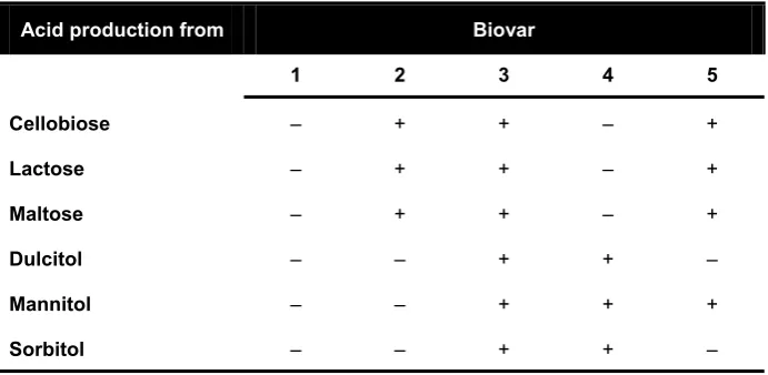
Table 2 – Biovar classification of R. solanacearum. Adapted from (EPPO, 2004).
Acid production from Biovar
1 2 3 4 5 Cellobiose – + + – +
Lactose – + + – +
Maltose – + + – +
Dulcitol – – + + –
Mannitol – – + + +
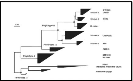
[image:24.595.126.472.587.756.2]reflec Afric nove solan on th hrpB class being Nucl 70% and c (Gen Figur joinin solana
cting the A can contine el and quit
nacearum s he genetic s B and endog sification sy The phyl g assessed leotide Iden identity in could provi nin & Denny
re 1 – Phylog
ng tree based
acearum spec
American or ent was iden
te discrimi strains into similarities glucanase ( stem strain logenetic di by taxonom ntity (ANI). DNA-DNA ide a confid y, 2012).
genetic tree
d on the par cies complex. E
r Asian orig ntified (Coo inatory cla
four differ of the
16S-eglI) gene s ns can be arr
ifferences a mists and e . ANI value hybridizati dence thresh
e of the R. so rtial endogluc Extracted from
gin. Later o ok et al, 19 assification
rent genetic -to-23S int sequences ranged in a
among diffe experts in es over 95% ion (Remen hold for a n
olanacearum canase (egl) m (Genin, 201
on, a third 989; Poussi system wa c groups or
ernal trans (Fegan & P phylogenet
rent strains the field us % have bee nant et al, 2 new classific
m species co
gene sequenc 0).
clade corre ier et al, 20
as propose phylotypes cribed spac Prior, 2005) tic tree (see
s in the spec sing genom n considere 010; Remen cation syste
omplex. Phylo ces from 771
esponding t 000). In 20 ed, groupin s (I to IV), b cer region ( ). Using the Figure 1).
cies comple me-wide Av
ed equivale nant et al, 2 em of the sp
ogenetic neigh 1 strains of t
to the 005 a ng R.
based (ITS), e new ex are verage ent to 2011), pecies hbour-the R.
[image:25.595.88.526.391.657.2]Why is
R. solanacearum
important?
Geographic distribution
Bacterial wilt caused by R. solanacearum is devastating and endemic in the tropics and subtropical regions of the globe, where natural hosts are predominant. The optimal infection temperature was determined to be 32 ºC in tomato lines with different susceptibilities (Krausz & Thurston, 1975). Wilting has been reported in crops planted for the first time in virgin soils in Indonesia, Central America and Florida (Buddenhagen & Kelman, 1964). Figure 2 summarizes the current worldwide distribution of R. solanacearum. Unfortunately, strains belonging to phylotype II B1, most commonly referred to as race 3 biovar 2, are able to infect tomato and potato plants at much lower temperatures than other R. solanacearum (see Table 1), and their distribution is spreading to temperate areas of the United States and Europe (Figure 2 top panel). The dispersal of the pathogen is mainly due to the importation of contaminated material (Champoiseau et al, 2009; Elphinstone, 1996). With the accidental introduction of R. solanacearum in Sweden in the mid 70’s, intensive studies were carried out to determine the survival of the pathogen in the environment (Ciampi et al, 1980; Champoiseau et al, 2009; Elphinstone, 1996; Graham et al, 1979; Graham & Lloyd, 1979; Grey & Steck, 2001; Milling et al, 2009; Persson, 1998). Both, the U.S.A. and the E.U. have implemented strict legislation (EC, 1998; EC, 2011)and classified the pathogen as a quarantine pest (EPPO/CABI, 1997) or even a select agent in the U.S.A. territory, cited on the list of bioterrorism organisms (Animal and Plant Health Inspection Service, 2002). At the moment when this thesis was written a few publications detailed information on the vast array of strains, their geographical distribution and original host (Castillo & Greenberg, 2007; Cellier et al, 2012; Coupat et al, 2008b; Elphinstone, 2005) .
Economic impact of bacterial wilt
Figur Intern compl dots f (cross inform
the u 2002 litera 10% respe for a work inocu preve regar
re 2 – Ral national world
lex (bottom p for the period ses) records; mation for eac
use of non-2). Some inf ature, as the and 2-5% o ectively, and agriculture (
k developed ulum in the ent the pa rding new lstonia sola dwide detectio anel). Official 1999-2012. L Detection on ch detection ar
-host plant formation o e one repor of the total t
d up to 50% (Persley et a d by Luis Se
e tropical v thogen dis outbreaks
anacearum ons of R. sola
l detections, al Legend: Patho n a single spo
re available in
species th on historica ted in the P tomato, egg % of the loss al, 1985; Ze equeira in P virgin soils persal (Seq of bacteria
geographic
anacearum ra along with rep gen present a oradic event the PQR pack
he only alte al R. solana Philipinnes f
gplant, pepp ses took pla ehr, 1969). A Peru and Co
and develo queira, 198 al wilt is no
c distributio
ce 3 (top pan orted cases in according to a (triangles). T kage from EPP
ernatives av acearum epi from 1966 t per and tob ace in virgin Another exa osta Rica, tr
oping imme 88; Sequeir
ot yet centr
on. Confirme nel) and R. sol
n the literature national (circ These maps, PO (EPPO, 201
vailable (Ge idemics can to 1968, in w
acco produ n soils used f
ample is the racking dow
ediate cont a, 1998). T ralised, alth
ed EPPO and
lanacearum s e are represen cles), or subna along with s 12).
enin & Bou n be found i which 15%, uctions were
for the first e inspiring wn the sourc
trol strategi The inform hough step d CAB species nted by ational specific ucher, in the 10%, e lost, t time field-ces of ies to mation ps are
[image:27.595.89.519.71.443.2]being made towards the integration of epidemics information, like the EPPO Plant Quarantine data Retrieval system (PQR) (EPPO, 2012), an outstanding tool retrieving updated information on global plant disease epidemics. The lack of information makes it hard to quantify the current losses due to bacterial wilt. However, an estimated 1 billion U.S. dollars is thought to be lost yearly (Champoiseau et al, 2009). Especially important is the impact in staple crops of small-scale producers in developing countries (Elphinstone, 2005), originating meaningful economic and social concerns. The most detailed document regarding the economic importance of wilting disease produced up to date was compiled by John Elphinstone (Elphinstone, 2005).
Infection cycle
Taking into account the definition adopted in this text, R. solanacearum can be regarded as a hemibiotroph and its infection cycle is depicted in Figure 3.
Saprophytic phase
R. solanacearum-loaded soils are one of the possible sources of inoculum. Bacterial cells, deposited along with wilted plant debris have been reported to migrate naturally down to 75 cm of depth in the soil maintaining their full pathogenicity towards susceptible plants (Graham & Lloyd, 1979). In most cases, hydrated sediments, typical in the warm and moist climate of the tropics, are able to sustain R. solanacearum for long periods of time (van Elsas et al, 2001). A second source of
inoculum is the presence of natural reservoirs for the bacteria. Many weeds are alternative hosts and become latently infected with R. solanacearum without showing any symptom. The growth of Solanum dulcamara, one of those asymptomatic species along rivers provides the pathogen with a means of multiplication and release into the environment (Persson, 1998). The bacteria can reach and persist for long periods of time in water streams or ponds, which are often used for field irrigation and represent the most common and dangerous sources of inoculum (Hong et al, 2008). Researchers noticed that i) R. solanacearum isolates can be stored in sterile distilled water stocks for years in the laboratory (Buddenhagen & Kelman, 1964); ii) when stored in those conditions R. solanacearum mantains its pathogenicity up to 4 years, and maybe more (Alvarez et al, 2008); and iii) the bacterium is able to go through several replication cycles in distilled water and in irrigation waters (van Elsas et al, 2001). Such long-term persistence in the environment seems to be due to physiological survival mechanisms the bacterium possesses to overcome starvation (Alvarez et al, 2008), such as the viable but not culturable state. R. solanacearum can enter in this state during the saprophytic phase of the disease cycle, when the temperature drops, or after completing the disease cycle in a plant. However, this is a reversible state from which bacteria can recover and efficiently colonize plants (Grey & Steck, 2001). Moreover, persistency in the environment can be justified by the formation of a biofilm, which may protect the bacterium from desiccation (Yao & Allen, 2007).
Plant infection
|
13
vascular parenchyma (Digonnet et al, 2012; Vasse et al, 1995). During these first stages of disease, the metabolism of the bacterium is probably directed towards the suppression of plant defences thanks to the action of type III secretion effectors; but also to the secretion of specific cell-wall-degrading enzymes (see below), in order to promote multiplication in the intercellular spaces (Digonnet et al, 2012; Genin et al, 2005; Genin & Denny, 2012; Schell, 2000). Wallis and Truter observed that bacteria in the root parenchyma multiply preferentially around small diameter cells close to the xylem vessels (Wallis & Truter, 1978). A few cells adjacent to the xylem vessel form tyloses, balloon-like structures through the pit of the vessel not observed in non-infected plants. These structures are speculated to be either a mechanism of protection against infection, or a structure induced by the release of Indole-3-acetic acid from the pathogen. Interestingly, 24 to 48 hours post-inoculation the rupture of tyloses occur, with subsequent release of the cell content and the bacteria in the vicinity to the xylem vessel (Wallis & Truter, 1978). This mechanism of xylem colonization was not visualized by Vasse and collaborators, who advanced that R. solanacearum enters the vascular tissues on lateral root emergence sites, where the endodermis is re-oriented (Vasse et al, 1995). In either case, no bacteria were ever visualized in the endodermis, probably because the polarly-suberized cell walls avoid the pathogens passage. Both publications agree that after colonization of the xylem vessels, bacteria multiply heavily and rapidly spread to aerial parts of the plant, with an associated degradation of the vessel cell walls (Vasse et al, 1995; Wallis & Truter, 1978). The formation of compressed pockets of bacterial cells in the plant root and stem xylem vessels reorients the metabolism of the bacterium and it starts to produce massive amounts of extracellular polysaccharide (EPS), a highly heterogeneous and hydrated extracellular matrix (Orgambide et al, 1991). The production of EPS and the extensive bacterial multiplication (up to 109 cells/g of tissue) interferes with the upward water movement from the roots and leads to wilting symptoms (see Figure 4). At first, the mechanical plug affects a few leaves, but later symptoms become widespread causing complete and irreversible wilt of the plant, eventually leading to plant collapse and death.
Fi on su wi
Mo
pat
scien toma deter UW5 Wisc temp epide virul solanTran
orga
solan destr to cigure 4 – Sym
n the left show uspention of b ith a healthy p
olecular
thogenic
In recen ntific comm ato in Frenc
rminants o 551, a phylo consin, U.S peratures th
emiologies, ence deter nacearum c
nsposon
anism
For abou nacearum w ructive effec
orrelate pa
mptom of b
ws completely w bacteria at 108
plant of the sam
mech
city
nt years two munity. GMI
ch Guyana of pathogen
otype II (ra S.A. (Swans
han GMI10 have bee rminants a clades with
mutagen
ut three de was such an cts on a wid athogenicity
acterial wilt
wilted and col CFU/ml. On me age (5 wee
anisms
o R. solana I1000, a ph (Boucher e nicity are b ace 3 biova son et al, 2 000 (Millin
en useful f and have great impac
nesis: Th
ecades (50’s n extremely de-range of y with thet of tomato c
llapsed plants the right a co ek-old).
gover
acearum st hylotype I et al, 1985) best charac ar 2) strain 2005), able ng et al, 20 for the ch become re ct on agricu
e first st
s-80’s) phy y efficient v f plants. Du e biochemi
caused by R 10 days after ollapsed
stem-rning
R
trains recei strain (race ), is the stra cterized to n isolated fr
e to efficien 009). These
haracterizat epresentativ ulture.
tep in the
ytopatholog vascular pa uring that p ical and m
. solanacear soil-drench in -inoculated pl
R. sola
ived most e 1 biovar 3 ain in whic
date. The rom import ntly infect e two strain
ion of pat ves of two
e making
ists tried to athogen, wit
period, scien morphologic
rum.The ima noculation wit
lant is compar
anacear
attention in 3) isolated ch the mole other stra ted geraniu plants at l ns, with dis
thogenicity o importan
g of a m
o unveil wh th extraord ntists attem cal/physiolo
age th a red
rum
n the from ecular ain is um in lower stinct y and nt R.odel
|
15
characteristics of the bacterium. Professor Arthur Kelman described the loss of pathogenicity linked to a colony-morphology change from smooth to rough (Kelman, 1954). Kelman also observed that the same morphological switch was responsible for altered motility and chemotaxis, two important virulence determinants (Kelman & Hruschka, 1973). In addition, the colony morphological mutants had different lipopolysaccharide composition (Whatley et al, 1980), failed to synthesize normal extracellular polysaccharide (Dudman, 1959), and produced about ten times more indole-3-acetic acid than wild-type smooth isolates (Buddenhagen & Kelman, 1964). Taken together, these changes associated with the non-mucoid avirulent mutants seemed too pleiotropic, affecting many biochemical traits and raised doubts on the causality of this shift to virulence or normal physiology of the bacterium. Later on, the molecular basis of the colony morphology switch was explained by mutations in phcA (Brumbley & Denny, 1990), a gene encoding a global transcriptional regulator at the centre of a complex regulatory network coordinating multiple virulence determinants (Brumbley et al, 1993; Huang et al, 1995; Poussier et al, 2003).
Figur from II) an frame grey, syring
was o
syring of bo and 1992 sourc mini 1992 when indu the r below expre hrp c striki of ex Yersi secre Gijse whic inter resis
re 5 – Genet m Pseudomon
nd Xanthom es (arrow shap
those shared
gae appear in obtained after
gae pairs usin
The regu oth host- an 6, which ar 2), units 1, 2 ces (such a
mal mediu 2). Moreove n bacteria ction is also right-hand w. Whilst
ession, an i cluster from ing similari xtracellular
inia enteroc etion injecti egem et al, ch hrp gen ractions is tant plants
tic organiza
nas syringa monas camp ped boxes) are
by R. solana
n magenta, R.
merging syn ng the MaGe (M
ulation of th nd environm re constituti 2, 3, 4 and 7 as casamino um is clearl er, transcrip are co-cult o mediated end of the scientists incredible e m R. solana
ities (from 4 virulence d
colitica and isome in an
1993). Th ne products
the secreti leading to v
tion of the a
ae pv. tomato pestris pv. v e joined by bla
cearum and X solanacearum
nteny maps o Magnifying Ge
he hrp clust mentally-der ively expres 7 are catabo o acids) (Ge
ly induced ptional units
tivated with by HrpB a e cluster (M
were unco effort was m
acearum. T 40-50% to determinant
d Yersinia p nimal pathog
us, it was s control a
on of aviru visible HR s
archetypical
o DC3000(t
vesicatoria ack lines. ORF
X. campestris m in yellow a of R. solanace
enomes) interf
ter exempli rived signal ssed in any olically repr enin et al, 2 through th s 1, 2, 3 and h Arabidop and depende Marenda et
overing the made to obt The sequen 70%) with p ts in the ma pestis Yop a
gens (Fense possible to and determ ulence facto symptoms (
l type I and type I), R. so
85-10 (type
Fs shared by al
s in green, wh and X. campes earum-X. cam
face (Vallenet
ifies exquisi lling. Excep y growth co
ressed in th 2005). In co he hrpB re d 4 are high psis or tom
ent on the p al, 1998), e regulatory
ain the full ce of the g proteins req ammalian p and Ipa secr elau et al, 19 o hypothesi mine the o ors, elicitor (Gough et al
type II hrp olanacearum
e II). Ortholo ll three organi hile hrp genes
stris in blue.
mpestris and R
et al, 2006).
itely the coo pt for transc
ndition test he presence ontrast, the gulatory ge hly and spe mato cell su
presence of which func y circuitry nucleotide enes in the quired for t pathogens S retion system
992; Gough ze that the utcome of rs or toxin
l, 1992).
clusters of
m GMI1000
ogous open r isms are depic s only presen This represen
R. solanacear
ordinated a criptional un
ted (Genin e of rich nitr
eir expressi ene (Arlat ecifically ind uspensions. f prhA, a ge ction is det governing e sequence o
e cluster sh the transloc Shigella flex
m, a well-kn h et al, 1992
e mechanis plant-path ns recognize genes (type eading cted in nt in P.
ntation rum-P. action nits 5 et al, rogen on in et al, duced This ene in tailed g hrp
on th hrp mult descr were usefu was a patho
The
dete
The b (Aldo putat depe not mini comp transFigure 6 –
The orientat thick arrow a, p and c GMI1000 da
The gene he role of h mutants s tiply in pla ribed until e induced in
ul for the de a link betwe ogenicity de
e Type II
erminant
No ident bacteriumr on et al, 200
tive outer-endent sider involved in mal mediu ponent (Al smembrane
– Genetic org
tions of the se under the OR
respectively. atabase (Salan
eration of a hrp genes a creened sh nta, a featu then, ii) th n planta. Th efinition of
een avirulen eterminant
II Secret
t in
R. sol
tified plant responds, in 00; Marend -membrane rophore rec n iron star um (Marend
ldon et al, e sensor pro
ganization o
even transcrip RFs. prh, hpa, Color-code of noubat et al, 20
a mutant co as molecula hared three ure not con
ey were no he genetic a
new concep nce and the in plant pa
tion Syst
lanacearu
-derived di n a contact-da et al, 199 e receptor ceptors (Ma rvation sen da et al, 19
2000), an otein PrhR,
of the R. sol ptional units p
hrp and hrc g f genes is in 002).
ollection us ar mediator e main cha nfirmed in ot affected in
approach an pts in bacter e presence o athogenic ba
tem (T3S
um
iffusible sig -dependent 98). This rec
protein P arenda et al, sing and d 998). PrhA
nd integra which is in
lanacearum present in the gene designat accordance w
sing transpo rs of plant-aracteristics any other
n housekee nd the deri rial pathoge of a type III acteria.
SS): The
gnal is perc t manner, to cognition ta
rhA, homo , 1998). Des does not co A recognizes
tes the sig n turn respo
m GMI1000 h e cluster are d ions were rep with the R. so
osable elem pathogen in s: i) they
pathogenic eping activit ved mutant enicity. From
secretion s
e main p
eived by R. o the presen akes place vi ologous to spite this sim ontrol hrpB s a still-unk gnal via ac onsible for t
hrp cluster.
depicted by a placed with h,
olanacearum
ments threw nteractions were unab city determ ties and iii) t collection m then on, system, the
pathogen
. solanacea nce of plant via activation
several T milarity, Pr B expressio
the ECF sigma factor PrhI (see Figure 7 for a schematic representation) (Brito et al, 2002). The homologues of these two sequential proteins (PrhR and PrhI) in other bacterial systems are known to act together with FecA/PupB proteins, which are in turn homologous to PrhA (Brito et al, 2002). The activated form of PrhI is then able to induce prhJ expression. PrhJ is a transcriptional activator of the LuxR/UhpA family, driving HrpG expression (Brito et al, 1999). Until this point of the cascade all the transcriptional factors involved are named Prh, after plant regulatory hrp. All prh-deffective mutants are only mildly affected on their pathogenicity towards tomato plants (Brito et al, 1999; Genin et al, 2005). The next intermediary of the signalling cascade is HrpG, a response regulator related to the OmpR subfamily of two-component signal transduction systems. This transcription factor induces the ultimate expression of hrpB, an AraC family transcription factor (Genin et al, 1992). HrpB will finally trigger the expression of most operons in the hrp cluster, putatively binding to the so-called hrpIIbox found in several hrp promoters (Cunnac et al, 2004a). HrpB is
considered the main regulator of hrp promoters and, together with HrpG, is responsible for the induction of hrp genes both in minimal media and through plant cell contact, whereas PrhA, PrhR, PrhI and PrhJ are required specifically for the induction by plant cells. There is growing evidence that HrpG is a key regulator through which the two different signalling pathways (nutritional/metabolic state and plant-cell contact) are integrated (Brito et al, 1999; Yoshimochi et al, 2009b). Recently, a hrpG paralog – prhG –, was characterized and shown to be sufficient to activate hrpB expression in minimal medium, probably integrating metabolic signals necessary for expression of the T3SS (Plener et al, 2010). Besides its important role as a convergence point, HrpG has also been shown to play a role in the activation of other genes required for efficient plant colonization (Valls et al, 2006; Vasse et al, 2000). Finally, other environmental conditions such as pH, temperature, osmolarity and carbon sources also affect hrp gene expression in still-unknown ways (Genin et al, 1992). All the information on the regulation of hrp and prh genes, as well as the downstream activities that follow the activation of this pathway, have been obtained in vitro. Minimal medium and co-culture with Arabidopsis and tomato cells were the two conditions used in those studies. At the beginning of this work scarce information was available regarding gene/promoter activities during plant infection.
Figur
solan and a protei of ma To da indica indica exten plant 2012 likely whic trans injec cytos 2002 discu
re 7 – Schem
nacearum. I activates an in
ins (part of th any virulence a ate, the nature ate transcripti ate post-transc
nsively in th ts.
The T3S 2), with an i y evolved fr ch probably sfer (Abby ctisome is re
sol directly 2). These ef ussion held
me of the re
n the presenc ntracellular ca he hrpB regulo
activities in R.
e of the metab ional activatio criptional mod
he xylem an
SS is an ex intricate bio rom a very a y became w & Rocha, 2 esponsible f into the ho ffector prote on Twitter
egulatory net
e of plant cell ascade leading
on) (Cunnac e
solanacearum
bolic signal af on; lines endin
difications.
nd are also u
xtremely co osynthesis a ancient adap widespread
2012; Goph for the tran ost cytoplas eins were d r as “molecu
etwork contr
ls PrhA recogn g to the expre et al, 2004a).
m (Valls et al, ffecting hrp ge ng with a cro
unable to tr
omplex sup and mounti ptation of t
among pat hna et al, 2 nslocation o sm (Arlat e defined rece
ules release
rolling the e
nizes a still un ession of T3SS
HrpG, a main 2006), is a ke ene expression ossbar transcr rigger aviru pramolecula ing process he bacterial thogenic ba 2003; Van G of effector p et al, 1994; ntly in 140
ed by an or
expression o
known plant c S genes and t n regulator con ey point for sig
n is not yet kn iptional repre
ulence react
ar structure s (Buttner, 2 l flagellum acteria via Gijsegem et proteins from Hueck, 199 characters ganism tha
of hrp genes
cell-wall comp the cognate e ntrolling expr gnalling integ known. Black a
ession and red
tions in resi
e (Loquet 2012). The secretin act
horizontal t al, 1995). m the bacte 98; Szurek after a scie
at act direc
|
s in R. [image:37.595.85.518.69.391.2]|
21
virulence activities that could play a role in pathogen adaptation to the environment and to different hosts (Genin & Boucher, 2004) (see below).
The
Phc
network: A global regulatory network integrating
environmental signals
Besides employing a canonical autoinduction system to monitor its own population density, by production of acyl-homoserine lactones, R. solanacearum possesses an unorthodox and very particular quorum-sensing mechanism. This mechanism for high cell density sensing is mediated by 3-hydroxypalmitic acid methyl ester (3-OH PAME) (Clough et al, 1997b; Flavier et al, 1997a). At the centre of this network is PhcA, a member of the LysR family of transcriptional regulators (Brumbley et al, 1993; Brumbley & Denny, 1990). At low cell densities there is a low concentration of 3-OH-PAME, and phcA is repressed by the PhcS/PhcR two-component system (see Figure 8 for a schematic representation). As a result, genes involved in twitching and swimming motility will be expressed (Liu et al, 2001; Tans-Kersten et al, 2001) as well as genes mediating attachment to surfaces (Kang et al, 2002). As bacterial density increases from about 106 to 108 CFU/ml, local concentration of the autoinduction signal molecule 3-OH PAME fires up and the repression on PhcA is released. The active form of PhcA activates the production of extracellular polysaccharide (EPS) and the secretion of endoglucanases (egl) (Schell, 2000). At the same time this system represses both i) PehSR, the two component regulator responsible for the expression of polygalacturonase (pglA), swimming and twitching motilities (Allen et al, 1997; Kang et al, 2002; Schell, 2000; Tans-Kersten et al, 2001), and ii) T3SSexpression by either a hypothetical post-transcriptional modification of HrpG, most likely a phosphorylation (Genin et al, 2005; Yoshimochi et al, 2009b), or by repression of prhIR expression (Yoshimochi et al, 2009a) (see Figure 7). A model was proposed in which the hrp regulatory cascade is responsible for the early stages of infection, while late stages arise from the incremented activity of the PhcA regulatory network (Genin et al, 2005). Nonetheless, the studies that lead to this global view over the regulation of T3SS expression are based on artificial situations, like gene activity assays performed in artificial culture media or infiltrations of plant leaves.
EPS
main reason why those mutants failed to infect plants, a thorough analysis revealed that EPS-defficiency was a consequence of mutation in the phenotypic conversion network regulator PhcA, governing EPS expression (Brumbley et al, 1993; Huang et al, 1995). In fact, mutants in the cluster of genes responsible for the synthesis of EPS evidenced delayed development of wilting symptoms on tomato plants, but were still pathogenic either after soil drench or petiole inoculations (Kao et al, 1992a; Saile et al, 1997).
Twitching and Swimming motilities
The Phc regulatory system indirectly controls the flagellum-driven motility called swarming (on solid agar surfaces), or swimming (in liquid). This control is exerted via PehSR, active only at low cell densities, which in turn controls the heterotetrameric regulatory protein FlhDC. Mutation of the flagellin gene fliC, or in the flagellar motor gene fliM produced mutants that showed delayed wilting of tomato plants after soil drenching inoculations. The effect was not observed when bacteria were directly delivered in the xylem through petiole inoculations (Tans-Kersten et al, 2001). A particular flagellum independent motility called twitching motility is provided by the type IV pili. The term derives from the erratic movements of the bacteria in suspension (Mattick, 2002). Twitching motility was first described in R. solanacearum upon the following observations: i) small (microscopic) colonies in a plate exhibited reticulate appearance at the margins due to the movement of cells in layers rather than from concentric growth; ii) phcA mutants showed uncontrolled twitching motility, even when colonies became as big as 17,2 mm diameter; and iii) identification of 6 putative pilin-codifying genes from the GMI1000 genome sequence (Liu et al, 2001).
Figur plant compl simpli plant twitc camp posse envir comp
R.
com
bene solanre 8 – The Ph
cell wall deg lex network (S icity.
ts after soi ching motil
pestris viru esses a ve ronment. Fu plemented t
solanac
mplete p
By the efited from nacearum Ghc regulator
grading enzym Schell, 2000).
il inoculati lity in viru ulence (Su e ery differen
urthermore to exclude a
cearum
perspect
end of the m the strik
GMI1000 g
ry circuit. Ph mes, exopolysa . The negative
on and dir lence is th et al, 1999), nt route o e, the defect a loss of fitn
genom
tive of pa
e 90’s, gen king advan genome seq
hcA is a centra accharide pro e influence on
rect deliver he fact that , although i of coloniza ts of pilA in ness in the p
me sequ
athogen
neticists wo nces in DN quence was
al regulatory el oduction and
T3SS express
ry in the x t is not im t can be arg tion and n pathogen pathogen (K
uence: A
nicity
orking on NA sequen published lement contro attachment-re sion is represexylem. Agai mportant for gued that R
occupies a icity were n Kang et al, 2
A door
plant path cing autom in 2002 (S
olling motility elated protein ented in Figur
ainst the ro r Xanthom R. solanacea a different never genet 2002).
toward
hogenic bac mation. Th Salanoubat|
genes, ns in a re 7 for
ole of monas arum host tically
ds a
cteria he R. et al, [image:41.595.88.518.69.412.2]2002). It was the second plant pathogenic bacterial genome to be unveiled, after Xylella fastidiosa (Simpson et al, 2000) and revealed a vast array of virulence factors (Genin & Denny, 2012). The total genome size is 5,81 Mb, divided in two replicons of 3.72 and 2.09 Mb, with 67% G+C content. The nucleotide sequence, together with a thorough annotation, provided a new tool to define R. solanacearum as a model organism in plant-bacterium interactions. The description of this genome opened doors to many new studies, particularly on evolutionary history of the species complex and on the different traits determining the pathogen virulence. The second R. solanacearum genome to see the light was that of strain UW551, a strain adapted to infect plants at cooler temperatures (Gabriel et al, 2006; Milling et al, 2009). Up to date, the genomes of CFBP2957, PSI07, CMR15 (Remenant et al, 2010), K60 (Remenant et al, 2012), Po82 (Xu et al, 2011) and Y45 (Li et al, 2011), together with the close relatives R. syzygii R24 and R. celebensis R229 (Remenant et al, 2011) are available.
Other virulence traits
The GMI1000 genome revealed many genetic evidences justifying R. solanacearum abilities to adapt to different natural habitats during its life-cycle, to persist for long periods of time in a harsh and competitive soil environment and to interact, colonize and effectively infect its hosts (Genin & Boucher, 2002; Genin & Boucher, 2004). Functional redundancy is one of the common features of the genomic information determining pathogenicity and virulence in this pathogen. I will shortly review below other features of the genome that could have an influence in virulence.
Secretion systems
The R. solanacearum genome contains information for expressing all six major secretion systems (Figure 9), although only a limited number of them have been characterized.
Type I Secretion System
adhe exop 2006 Type (Gen 12 g form cano their secre PehC endo pecti cano degra type secre Geni alter date
Figure 9 – have been membrane; protein.[Ima esins and polysacchari 6).
e II Secret
R. solan nin & Bouch enes in th med by the G
onical type I r ability to p ete at least C) (Gonzale oglucanase ( in methyle onical type ading enzym II secretion eted protein in, 2009). T
native GspD (Zuleta, 20
– Schematic shown to m
OM: outer age source (Ts
glycanases ide and bio
tion System
nacearum G her, 2004; Z e chromos GspD secret II secretion produce wilt 36 proteins ez & Allen (Egl) (Robe sterase (Pm II secretio mes mentio n apparatus ns may play The other ty
D proteins, 007).
representat mediate prote
membrane; seng et al, 200
s (Delepela ofilm forma
m and Cel GMI1000 p Zuleta, 200 ome (RSc3 in (also ide n systems sh
ting disease s (Zuleta, 2 n, 2003; H erts et al, 19
me) (Tans-on system oned above
s mutant. T an importa ype II secre but no can
tion of the s ein export in
IM: inner 09)].
aire, 2004 ation in Rhi
ll Wall Deg possesses u 07). A first a 3105-RSc311 entified as S
howed redu e on tomato 2007). Thre Huang & A 988; Saile e
-Kersten et in R. sola do not show This fact de ant role in v etion system ndidate sec
six distinct n Gram neg
membrane;
). It is a izobium leg
grading E up to three
and orthod 16) and em SdpD1 in the uced virulen plants (Kan ee polygalac
Allen, 1997 et al, 1997; S
t al, 1998) anacearum. w the same emonstrates virulence (L
ms are uno reted prote secretion sy ative bacter OMP: outer also requir guminosaru nzymes type II se ox system i mploys a p e literature) nce as they ng et al, 199 cturonases ( 7; Schell et Schell, 1987 ) are all s
. Mutants reduction i s that other iu et al, 200 orthodox sy eins have be
ystems that ria. HM: host r membrane
red for no um (Russo
ecretion sys is made up periplasmic ). Mutants i are impaire 94), and fail
(PglA, PehB t al, 1988 7) and a put secreted via in all cell in virulence r, still unkn 05; Poueym ystems, feat een identifi
|
ormal et al, stems from pore in the ed on led to B and ), an tative a the wall e as a nown, miro & uring ied to
Type IV Secretion System
The clustering of all the genes required for the biosynthesis of a functional type IV secretion system (Rsc2574-RSc2588) inside a transposon element is a proof of the extraordinary genome flexibility of R. solanacearum. Most likely, the type IV secretion system was acquired via horizontal gene transfer, a strong evolutionary force driving evolution and facilitated by the natural transformability of the bacterium (Bertolla et al, 1999; Guidot et al, 2009). Similarly to the T3SS, type IV are complex molecular structures spanning the bacterial inner and outer membranes, responsible for the translocation of genetic material with other bacterial cells or effector proteins into the host cytosol (Angot et al, 2007; Burns, 2003)
Type V Secretion System
Up to 14 genes for TpsA proteins are described in the GMI1000 genome (Genin & Boucher, 2004; Salanoubat et al, 2002). TpsA proteins are associated with virulence traits in a number of pathogens (Jacob-Dubuisson et al, 2001), and are transported in a sec-dependent manner.
Type VI Secretion System
Type VI secretion apparatus are widely spread among Gram negative bacteria (Records, 2011). An approx. 42 kb region of the R. solanacearum GMI1000 megaplasmid contains 15 genes hypothetically required for synthesis of this secretion system (RSp0572, RSp0672, RSp0732, RSp0739-41, RSp0743-49, RSp0759, RSp0761 -63 and RSp0768). The macromolecular structure of this secretion system is very similar to bacteriophage tails, and is thought to mediate protein and DNA translocation (Leiman et al, 2009). In animal pathogens, type VI secretion systems have been reported contribute to pathogenicity (Pukatzki et al, 2006; Shalom et al, 2007), but so far there are no evidences for such a role in R. solanacearum pathogenicity (Poueymiro & Genin, 2009).
Attachment may be mediated by lectins or type IV pili
Type IV pili
|
27
competence have been reported in a number of animal pathogens (Giltner et al, 2012). This filamentous appendage is composed of one subunit, the pilin, or PilA in R. solanacearum (Kang et al, 2002). The molecular mechanism by which the adherence occurs is still unclear, but it is currently accepted that expression of type IV pili promotes virulence on tomato plants and adherence to various surfaces. The main role of these appendices seems to be linked to the polar attachment of bacteria to plant cells during root invasion and multiplication inside xylem vessels (Kang et al, 2002; Vasse et al, 2000).
Lectins
Table 3 – Binding preferences of the three lectins present in R. solanacearum. [Adapted from (Sudakevitz et al, 2002a; Sudakevitz et al, 2004; Šulák et al, 2007)].
Name in literature
Proposed ORF in GMI1000
ORF in UW551
MW (kD)
Binding prefrences
RS20L lecX Rsp0569 RRSL_03943 20 L-fucose D-mannose D-xylose *
RS-IIL lecM Rsc3288 RRSL_02788 11,6 D-Fructose ≈ D-Mannose >> L-Fucose > L-Galactose ≈ D-Arabinose
RSL lecF Rsc2107 NP 9,9 L-Fucose > L-Galactose >> D-Arabinose > D-manose > D-fructose
NP – not present * – unpublished data
RSL: R. solanacearum fucose-binding lectin
The RSL protein is similar to the AAL lectin from the mushroom Aleuria aurantia (Wimmerova et al, 2003). It is a 90 amino acid protein purified from R. solanacearum GMI1000 extracts with a high preference for L-fucose, in a similar fashion to the Pseudomonas aeruginosa lectin PA-IIL (Sudakevitz et al, 2002b). The monosaccharide concentrations needed to inhibit agglutination of erythrocytes was much higher for RSL than for PA-IIL, revealing that the R. solanacearum lectin has a much lower affinity for fucose than PA-IIL. In 2005 the crystal structure of RSL was solved and the fucose affinity values were considered higher than previously described (Kostlanova et al, 2005). Apparently, during hemagglutination inhibition assays the alpha-Fuc1–2Gal epitope of the human erythrocyte H antigen competed for the preferential binding to the lectin, shifting the dissociation constant.
|
29
RS-IIL: The mannose-binding lectin
RS-IIL is a lectin present in both R. solanacearum GMI1000 and UW551 strains. It shares 70% amino acid identity with the Pseudomonas aeruginosa lectin PA-IIL (Sudakevitz et al, 2002b). A similar protein is found in Chromobacterium violaceum, a Gram negative saprophytic bacterium from soil and water (Sudakevitz et al, 2004). The preferential binding affinity of this lectin is not in accordance with the structural similarities to PA-IIL, as it preferentially binds D-Fructose and D-Mannose. The authors hypothesize that the difference in affinity correlates with differences in the “specificity loop” region of the protein (Adam et al, 2007; Sudakevitz et al, 2004). Mannose is incorporated in plant cell walls in the form of mannans. For example, glucomannan is a mannose containing polysaccharide present in angiosperm secondary walls (Piro et al, 1993), but no information is yet available on the affinity of any biologically relevant mannose-containing oligosaccharides to RS-IIL.
RS20L: The xylose-binding lectin
The second lectin present in both the genomes GMI1000 and UW551 is RS20L, a 19.9 kDa lectin with L-fucose, D-mannose and D-xylose binding capacities. This lectin presents no similarity to other known lectins. Its crystal structure was solved and affinity studies are being conducted (Šulák et al, 2007).
Relevance of the developed research
fusions. We noticed that new molecular tools for functional genetic studies adapted to R. solanacearum were needed. The Tn5-B20 and other transposons had been useful to create mutants and genetic fusions for gene expression studies. However, the creation of gene disruptions leads to virulence and pathogenicity defects rendering, in some cases, bacteria unable to multiply inside susceptible plants. In addition, a common issue in R. solanacearum studies was the difficulty to trans-complement gene disruptions. The only alternative available was the use of plasmids, which provided a means of overexpression rather than stoichiometrical complementation, due to their copy number. Another problem associated to the use of plasmids in this pathogen was the need of continuous selective pressure. During plant infection the use of antibiotics is not an option due to the complexity of the system.
Our long-term aim was the determination of the genetic program used by R. solanacearum during host colonization and at the different stages of disease. To this end, we developed a novel system – pRC, after Ralstonia chromosome –, based in targeted and stable insertions in a precise and permissive location of the bacterial chromosome. We proposed the use of our versatile set of suicide plasmids for the study of transcriptional output (promoter probing) during plant infection, effector overexpression and purification, and monocopy gene complementation in any R. solanacearum strain. The development of these molecular tools were described in the first publication included in this thesis (Monteiro et al, 2012b). Currently the pRC system is being used by a number of research groups and collaborators. The use of the pRC system in any strain will allow the standardization of the genetic studies made in the field.
|
31
OBJEC
CTIVE
|
ES
|
35
The long-term goal of the research topic developed in this thesis is the determination the genetic program used by R. solanacearum during host colonization at the different stages of disease. In this work, the fundamental aim, was improving the molecular tools, strategies and approaches to study the expression of pathogenicity genes in biologically relevant situations, like plant colonization. The specific research objectives are:
1. Develop a versatile molecular toolbox for assessment of native gene expression and in vivo
functional analyses.
1.1. Construct a set of suicide plasmids directed to a specific position in the R. solanacearum genome.
1.2. Determine the genetic stability of the integrated constructs.
1.3. Construct a molecular adaptor allowing the use of all generated plasmids on different
R. solanacearum strains.
1.4. Explore the capacities of the molecular tools for mutant complementation assays. 1.5. Explore the possibility of native protein production in R. solanacearum employing
strong promoters.
1.6. Validate the promoter activity of pathogenicity-related genes using promoter::lacZ
fusions.
1.7. Visualize promoter activity of pathogenicity-related genes during plant infection using promoter fusions to a strong fluorescent reporter gene.
2. Analyse the regulatory circuitry governing T3SS expression in planta. 2.1. Generate a luxCDABE reporter.
2.2. Determine the half-life of the luxCDABE reporter.
2.3. Generate hrpB and eps promoter fusions to the luxCDABE reporter in order to measure and visualize their epression in planta.
2.4. Validate hrpB expression at advanced stages of infection by an independent method.
3. Investigate the molecular basis of the hrpB repression observed in co-culture with plant cells. 3.1. Determine if hrpB or hrcC are responsible for the regulatory negative feedback on gene
expression.
3.2. Determine at which level of the PrhA-mediated T3SS signalling casacade is integrated the negative feedback.
4. Ascertain the role of a fucose-mannose binding lectin (RS-IIL) on the interaction between R. solanacearum and its host plants.


![Figure 9 –have been membrane; protein.[Ima– Schematic shown to m OM: outer age source (Ts representatmediate prote membrane; seng et al, 200tion of the sein export in IM: inner 09)]](https://thumb-us.123doks.com/thumbv2/123dok_es/5267789.96822/43.595.104.497.71.211/figure-membrane-protein-schematic-source-representatmediate-membrane-export.webp)
![Table 3 – Binding preferences of the three lectins present in R. solanacearum. [Adapted from (Sudakevitz et al, 2002a; Sudakevitz et al, 2004; Šulák et al, 2007)]](https://thumb-us.123doks.com/thumbv2/123dok_es/5267789.96822/46.595.69.524.97.217/binding-preferences-lectins-solanacearum-adapted-sudakevitz-sudakevitz-sulak.webp)


