Sociedad Cubana de Cardiología
____________________________Artículo Original
Características de los pacientes con disección aórtica aguda en
Villa Clara: Estudio multicéntrico
Dr. Daniel A. Vera Rivero
1,2, Dr. Yamir Santos Monzón
2, Dra. Marli Gamito González
3y
MSc. Dr. Carlos M. Aguiar Mota
41 Universidad de Ciencias Médicas de Villa Clara, Filial de Sagua La Grande. Villa Clara, Cuba.
2 Servicio de Cardiología, Hospital Universitario Mártires del 9 de Abril. Sagua La Grande, Villa Clara, Cuba.
3 Servicio de Angiología y Cirugía Vascular, Hospital Universitario Arnaldo Milián Castro. Santa Clara, Villa Clara, Cuba. 4 Servicio de Pediatría, Hospital Universitario Mártires del 9 de Abril. Sagua La Grande, Villa Clara, Cuba.
Full English text of this article is also available
INFORMACIÓN DEL ARTÍCULO
Recibido: 23 de octubre de 2018 Aceptado: 6 de diciembre de 2019
Conflictos de intereses
Los autores declaran que no existen conflictos de intereses
Abreviaturas
DAo: disección aórtica
DA Vera Rivero
Hospital Universitario Mártires del 9 de Abril. Carretera Circuito Norte a Quemado de Güines, km 2½. Sagua La Grande 52300. Villa Clara, Cuba. Correo electrónico:
RESUMEN
Introducción: La disección aórtica aguda es considerada como una de las
enfer-medades cardiovasculares más catastróficas que ocurren en el ser humano, tiene una alta mortalidad que obliga a un diagnóstico y tratamiento precoces.
Objetivo: Describir las características de los pacientes con disección aórtica
atendidos en 4 instituciones hospitalarias de la provincia de Villa Clara.
Método: Se realizó un estudio observacional, descriptivo, de corte transversal,
multicéntrico, en 25 pacientes que fueron atendidos con el diagnóstico de disec-ción aórtica en el período comprendido entre enero de 2012 y diciembre de 2017, en 4 centros hospitalarios de nivel secundario de la provincia de Villa Clara, Cuba.
Resultados: La media de la edad de los pacientes fue 60,48 ± 13,99 años, 21 fueron
del sexo masculino lo que representó el 84,0%. Según la clasificación de Stanford, predominó el tipo A, en 17 pacientes (68,0%). El síntoma más referido fue el dolor torácico anterior y el taponamiento cardíaco fue la complicación más frecuente (28,0%).
Conclusiones: Las características de los pacientes con disección aórtica en Villa
Clara fueron similares a lo que acontece en el ámbito nacional e internacional, con una elevada mortalidad y una mayor incidencia en hombres, hipertensos y mayo-res de 65 años de edad. El dolor torácico fue el síntoma cardinal y el taponamiento cardíaco la complicación más temida. La disección aórtica requiere un alto nivel de sospecha por parte del médico para un diagnóstico y un tratamiento tempra-nos.
Palabras clave: Enfermedades de la aorta, Disección aórtica, Síndrome aórtico
agudo
Characteristics of patients with acute aortic dissection in Villa
Clara: Multicentric study
ABSTRACT
Introduction: Acute aortic dissection is considered one of the most tragic
cardio-vascular diseases that occur in humans; with high mortality which requires early diagnosis and treatment.
Objectives: To describe the characteristics of patients with aortic dissection
treated in 4 hospital institutions in the province of Villa Clara.
Method: An observational, descriptive, cross-sectional, multicenter study was
Características de los pacientes con disección aórtica aguda en Villa Clara
CorSalud 2019 Abr-Jun;11(2):97-103
98
the period between January 2012 and December 2017, in 4 secondary-level hospi-tal centers in Villa Clara province, Cuba.
Results: The mean age of the patients was 60.48 ± 13.99 years, 21 were male, which
represented 84.0%. According to the Stanford classification, type A predominated in 17 patients (68.0%). The most common symptom was anterior chest pain while the most frequent complication was cardiac tamponade (28.0%).
Conclusions: The characteristics of patients with aortic dissection in the Villa
Clara setting manifested in a similar way to those in the national and international sphere. A high level of suspicion is required by the doctor to achieve a timely diagnosis and treatment.
Keywords: Aortic diseases, Aortic dissection, Acute aortic syndrome
INTRODUCCIÓN
Mucho se ha escrito acerca del desafío que repre-senta el diagnóstico y tratamiento de la disección aórtica (DAo) aguda y las fatales consecuencias de-rivadas de fallar en su intento. La historia de las DAo está marcada por el personaje que sufrió la primera descrita en la literatura médica, el rey Jorge II de
Inglaterra1. Existen indicios de que Galeno describió
la disección arterial en el siglo II y de que Vesalio aportó otros conocimientos de la enfermedad en el
siglo XVI1. Las primeras descripciones clínicas
deta-lladas las realiza Maunoir en 1802 y fue definida co-mo aneurisma disecante por el médico francés René
Théophile Hyacinthe Laenec, en 18191,2.
La DAo se define como la rotura de la capa media causada por una hemorragia intramural que resulta en la separación de las capas de la pared aórtica y la posterior formación de una luz verdadera y otra
falsa, con o sin comunicación entre ellas3. Este
des-garro acontece como resultado de repetidas fuerzas de torsión aplicadas a la arteria durante los ciclos cardíacos, así como a cifras elevadas de tensión arterial mantenidas, entre otras causas. Puede ocu-rrir también DAo en ausencia de hipertensión arte-rial, tal es el caso de anormalidades del músculo li-so, tejido elástico, colágeno, embarazo, válvula
aór-tica bicúspide y coartación aóraór-tica4,5.
Existen pocos datos actualizados sobre la epide-miología de esta enfermedad, pero a pesar de su baja incidencia se considera el episodio más catas-trófico que afecta a la aorta, con una incidencia es-timada –según el estudio Oxford Vascular– en 6/
100000 personas/año6, tiene una alta mortalidad que
obliga a un diagnóstico y a un tratamiento precoces. En una serie de necropsias, la prevalencia de la DAo
osciló entre el 0,2 y el 0,8%7. Por otra parte, en un
análisis de 464 pacientes del IRAD (International
Registry of Acute Aortic Dissection), dos tercios de
los pacientes eran varones, con una media de edad
de 63 años5; aunque las mujeres suelen resultar
afec-tadas con menor frecuencia por la DAo aguda, su edad es significativamente mayor que la de los
va-rones, con una media de 67 años8.
La DAo se clasifica en función de su duración y localización. Será aguda si las manifestaciones clíni-cas han durado 14 días o menos (período de mayor morbilidad y mortalidad); subaguda, entre 2 y 6
se-manas; y crónica, más allá de 6 semanas9. Desde el
punto de vista de su localización existen 2 clasifica-ciones: el grupo de Stanford habla de los tipos A y B, según esté afectada o no la aorta ascendente; y el de De Bakey, diferencia entre el tipo I cuando están afectadas la aorta ascendente y descendente, más el arco aórtico; tipo II, cuando sólo interesa la aorta ascendente y al arco aórtico; y tipo III cuando sólo
se afecta la descendente3,5,9-11.
La DAo representa un reto diagnóstico debido al amplio espectro de manifestaciones y presentacio-nes atípicas de esta enfermedad, lo que obliga a diferenciarla de muchas otras enfermedades. Según
el IRAD5, el diagnóstico no es acertado en el 38% de
los pacientes, y en Cuba existen estudios que tam-bién han reflejado dicha problemática, pues en la provincia de Cienfuegos el diagnóstico de DAo al
ingreso solo se planteó en un 12,9% de los casos13.
Por tal motivo se realizó el presente estudio con el objetivo de describir las características de los pa-cientes con DAo atendidos en 4 instituciones hospi-talarias de la provincia de Villa Clara, para así pro-porcionar un material que permita conocer la reali-dad de esta enfermereali-dad en el ámbito villaclareño.
MÉTODO
atendidos con el diagnóstico de DAo en el período comprendido entre enero de 2012 y diciembre de 2017 en los siguientes centros hospitalarios de nivel secundario de la provincia de Villa Clara: Hospitales Universitarios Mártires del 9 de Abril, Arnaldo Mi-lián Castro, Celestino Hernández Robau y Hospital Militar Comandante Manuel Fajardo Rivero.
Se estudiaron las variables año de ocurrencia del suceso, edad, sexo, antecedentes patológicos perso-nales, impresión diagnóstica al ingreso y clasifica-ción; así como las principales manifestaciones clíni-cas, imagenológiclíni-cas, y las complicaciones presenta-das durante la estadía hospitalaria. La información fue extraída de las historias clínicas individuales.
Para la organización y registro de la información recopilada se creó una base de datos en Microsoft Excel con todas las variables, a fin de viabilizar los cálculos correspondientes para el análisis descripti-vo de los resultados y la confección de tablas y grá-ficos. Además se empleó la estadística inferencial calculada con el paquete estadístico Epidat 3.1 con
la prueba no paramétrica Chi cuadrado (χ2), para
determinar las diferencias encontradas en las distri-buciones de las variables cualitativas. Se aceptó un nivel de significación del 95 % (p<0,05), de ahí que se consideraran los resultados según valor asociado de p en: no significativos (p>0,05), significativos (p<0,05) y muy significativos (p<0,01).
RESULTADOS
La media de la edad de la población fue de 65,48 ± 13,991 años, 21 pacientes fueron del sexo masculino lo que representó el 84,0% para una relación mascu-lino: femenino de 5,25 a 1. Los pacientes con piel de color blanca constituyeron el 80,0% de los casos.
La presencia de enfermedades asociadas (Tabla
1) se hizo patente en 18 pacientes (72,0%) que eran
hipertensos y le siguen en orden decreciente el há-bito de fumar (56,0%) y la dislipidemia (36,0%).
La tabla 2 muestra la impresión diagnóstica de estos pacientes al ingreso, la cual fue interpretada correctamente como DAo en 8 pacientes (32,0%), si-guiéndole en orden de frecuencia el infarto agudo de miocardio, que fue la enfermedad con que más se confundió el diagnóstico en 6 pacientes (24,0%).
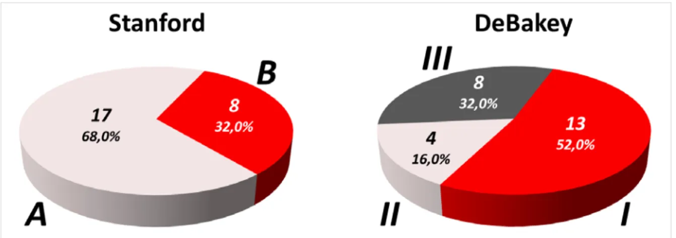
Según la clasificación de Stanford de la DAo, predo-minó el tipo A en 17 pacientes (68,0%) de los cuales
13 (52,0%) se correspondieron con el tipo I y 4 (16%), con el tipo II de De Bakey. Las disecciones
ti-poBdeStanfordytipoIIIde DeBakeyse
encontra-Tabla 1. Distribución de pacientes diagnosticados con
disec-ción aórtica, según variables demográficas y antecedentes patológicos personales.
Variables Nº %
Edad (años, χ̅±DE) 65,48 ± 13,99
Sexo
Femenino 4 16,0
Masculino 21 84,0
Color de piel
Blanca 20 80,0
No blanca 5 20,0
Antecedentes patológicos personales
Hipertensión arterial 18 72,0
Hábito de fumar 14 56,0
Dislipidemia 9 36,0
Ingestión de alcohol 8 32,0
Diabetes mellitus 7 28,0
Obesidad 4 16,0
Cardiopatía isquémica 6 24,0
Insuficiencia renal 2 8,0
Insuficiencia cardíaca 2 8,0
EPOC 5 20,0
Gastritis crónica 2 8,0
Lupus eritematoso sistémico 1 4,0 Aneurisma aórtico previo 4 16,0 EPOC, enfermedad pulmonar obstructiva crónica; χ̅±DE, media ± desviación estándar.
Tabla 2. Distribución de pacientes diagnosticados con
disec-ción aórtica según impresión diagnóstica al ingreso.
Impresión diagnóstica al
ingreso Nº %
Disección aórtica 8 32,0
Urgencia hipertensiva 4 16,0
Infarto agudo de miocardio 6 24,0 Enfermedad cerebrovascular 3 12,0
Muerte súbita 1 4,0
Edema agudo del pulmón 1 4,0
Abdomen agudo 1 4,0
Enfermedad tromboembólica 1 4,0
ron en 8 (32,0%) pacientes (Figura).
Características de los pacientes con disección aórtica aguda en Villa Clara
CorSalud 2019 Abr-Jun;11(2):97-103
100
más frecuente (Tabla 3) y fue más referido en la
parte anterior del tórax en la DAo tipo A respecto a
las de tipo B (94,1 vs. 50%; p=0,010). En estas últimas,
el dolor se localizó con mayor frecuencia en la re-gión dorsal (11,8 vs. 87,5%; p<0,0001) o en el
abdo-men (5,9 vs. 75,0%; p<0,0001). Tanto el síncope como
el shock/hipotensión fueron más frecuentes en las
disecciones tipo A (35,3 vs. 25,0% y 35,3 vs. 12,5%;
respectivamente) con relación a las tipo B. Por su
parte los trastornos gastrointestinales fueron más frecuentes en las de tipo B (62,5%; p=0,002).
Se puede observar que la complicación más fre-cuentemente encontrada fue el taponamiento car-díaco en 7 pacientes (28,0) con relación
estadística-mente significativa (p=0,032) a favor del tipo A (
Ta-bla 4). En cambio, la insuficiencia renal aguda fue la que tuvo una presentación significativamente mayor en las disecciones tipo B (p=0,002).
Figura. Distribución de pacientes según las clasificaciones de Stanford y De Bakey.
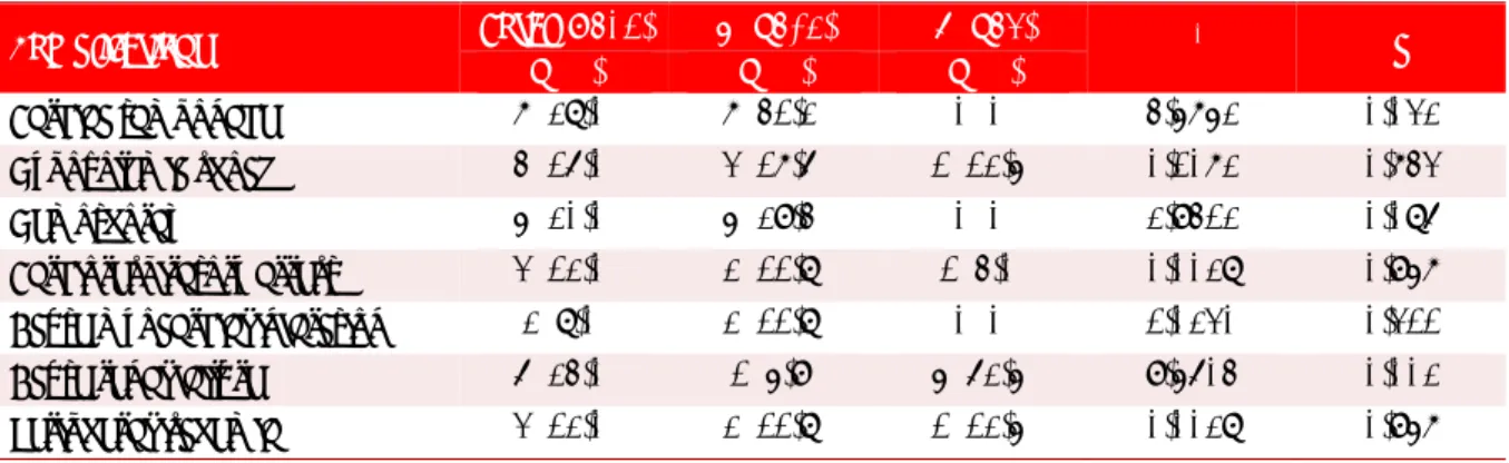
Tabla 3. Distribución de pacientes diagnosticados con disección aórtica, según la clasificación de Stanford y
manifestaciones clínicas.
Manifestaciones clínicas Total (N=25) A (n=17) B (n=8) χ2 p
n (%) n (%) n (%)
Dolor torácico anterior 20 (80,0) 16 (94,1) 4 (50,0) 6,6176 0,010
Dolor abdominal 7 (28,0) 1 (5,9) 6 (75,0) 12,8910 <0,0001
Dolor dorsal 9 (36,0) 2 (11,8) 7 (87,5) 13,5429 <0,0001
Soplo 9 (36,0) 8 (47,1) 1 (12,5) 2,8199 0,093
Síncope 8 (32,0) 6 (35,3) 2 (25,0) 1,7236 0,189
Palidez y sudoración 9 (36,0) 7 (41,2) 2 (25,0) 0,6179 0,431 Síntomas neurológicos 6 (24,0) 5 (29,4) 1 (12,5) 0,8530 0,355
Shock/hipotensión arterial 7 (28,0) 6 (35,3) 1 (12,5) 1,4020 0.236
Trastornos gastrointestinales 6 (24,0) 1 (5,9) 5 (62,5) 9,5604 0,002
Disnea 8 (32,0) 6 (35,3) 2 (25,0) 0,2649 0,606
Cianosis 5 (20,0) 4 (23,5) 1 (12,5) 0,4136 0,520
Hemoptisis 3 (12,0) 3 (17,6) 0 (0) 1,6043 0.205
Ausencia de pulso 10 (40,0) 7 (41,2) 3 (37,5) 0,0306 0,861
DISCUSIÓN
Los estudios imagenológicos son de elección para diagnosticar una DAo, principalmente la tomografía axial computarizada, además de la ecocardiografía. Existen informes que muestran la utilidad del ultra-sonido a la cabecera del paciente en el diagnóstico
de la DAo con presentación clínica atípica12.
Nuestros resultados son similares a los
encontra-dos en prestigiosos estudios como el IRAD5,
RA-DAR14 y el RESA15, donde predominan los pacientes
mayores de 60 años y del sexo masculino. La edad avanzada, muy asociada a alta prevalencia de hiper-tensión arterial y la enfermedad arterioesclerótica se describen como factores de riesgo o condicionantes para la aparición de DAo. Aunque las mujeres sue-len resultar afectadas con menor frecuencia por la DAo aguda, el sexo femenino es más propenso a complicaciones clínico-quirúrgicas durante esta
en-fermedad7.
El factor de riesgo más frecuente fue la hiperten-sión arterial, lo que coincide con el 72,1% de los
casos del estudio del IRAD5, además ha sido descrita
en el 65-75% de los pacientes con DAo, la mayoría
mal controlados10. Esta enfermedad origina un
en-grosamiento de la íntima aórtica, con calcificación y fibrosis de la adventicia, lo que aumenta la rigidez de la pared arterial y la predisposición a los aneu-rismas y disecciones. Otros factores de riesgo se incluyen enfermedades aórticas preexistentes o enfermedad de la válvula aórtica, antecedentes fami-liares de este tipo de enfermedades, antecedentes de cirugía cardíaca, tabaquismo, traumatismo toráci-co directo y el uso intravenoso de toráci-cocaína y
anfeta-minas5,11.
A pesar de tener una presentación clínica tan aparatosa, suele haber considerables retrasos en su
diagnóstico, debido a que la DAo es poco común y suele confundirse con mucha frecuencia con el sín-drome coronario agudo. Esto explica que el diagnós-tico al ingreso solo se planteará en un 32% de los pacientes, cifra superior a la encontrada por
Rome-ro-Cabrera et al13 en la provincia de Cienfuegos,
don-de el diagnóstico clínico acertado al ingreso se plan-teó solo en un 12,9% de los casos. Por su parte,
Mon-zó Blasco et al9 encontraron que de los casos que
fallecieron por DAo, el 40% había acudido al hospital un día antes y recibió diagnósticos médicos erró-neos.
Según la bibliografía, la disección puede simular muchos otros trastornos más frecuentes, como pleu-ritis, pericarditis, embolia pulmonar, isquemia
coro-naria e incluso accidente cerebrovascular11,16; y
pue-de presentar una amplia variedad pue-de síntomas y sig-nos, estos en presencia, a la vez, de numerosas com-plicaciones. Las principales manifestaciones inclu-yen dolor torácico intenso, irradiado a la espalda, abdomen, cuello o extremidades superiores e infe-riores; puede acompañarse de síncope, sudoración profusa, manifestaciones neurológicas, isquemia in-testinal e insuficiencia renal; además, puede auscul-tarse soplo por afectación retrógrada de los velos valvulares aórticos, provocar insuficiencia cardíaca
y taponamiento cardíaco11. Por todas estas razones,
es muy importante un alto índice de sospecha para hacer el diagnóstico de esta enfermedad.
La clasificación de De Bakey es muy importante desde el punto de vista fisiopatológico y la de Stan-ford se ha ganado el favor de la mayoría de los auto-res porque es más útil para determinar la conducta a
seguir5.
Los resultados del presente estudio coinciden
con los encontrados por Valdés Dupeyrón et al17,
con un predominio del tipo A de Stanford en 543
fa-Tabla 4. Distribución de pacientes, según la clasificación de Stanford y complicaciones intrahospitalarias.
Complicaciones Total (N=25) A (n=17) B (n=8) χ2 p
n (%) n (%) n (%)
Taponamiento cardíaco 7 (28,0) 7 (41,2) 0 (0) 4,5752 0,032
Shock hipovolémico 4 (16,0) 3 (17,6) 1 (12,5) 0,1072 0,743
Hemotórax 5 (20,0) 5 (29,4) 0 (0) 2,9412 0,086
Características de los pacientes con disección aórtica aguda en Villa Clara
CorSalud 2019 Abr-Jun;11(2):97-103
102
llecidos (61,1%). Por su parte, en cuanto a la clasifi-cación de De Bakey, nuestros resultados discrepan con los encontrados por dicho autor y otros de la
provincia de Cienfuegos13, donde predominaron los
de tipo II. Algunas series, sin embargo, informan una
mayor frecuencia de DAo tipo B18.
Aunque se conoce que los pacientes usualmente presentan dolor torácico, que se describe como agu-do, punzante o desgarrante, la sintomatología puede ser diversa, y alrededor de un 5% pueden no
aque-jar dolor11. Nuestros resultados coinciden con
publi-caciones previas3,5,11 que plantean que el dolor
torá-cico anterior suele estar asociado a la DAo tipo A, si bien los pacientes con disecciones tipo B suelen presentarse con dolor de espalda o abdomen. Las presentaciones clínicas de los dos tipos de DAo sue-len confundirse. El dolor puede migrar de su punto de origen a otras localizaciones, al seguir por la
ex-tensión de la disección. En el IRAD5 se observó
do-lor migratorio en menos del 15% de los pacientes con DAo aguda tipo A y en aproximadamente el 20% de aquellos con DAo tipo B.
La hipertensión arterial en la fase aguda fue más frecuente en la DAo tipo B; por el contrario, la hipo-tensión y el shock lo fue en los pacientes con disec-ción tipo A, resultados que concuerdan con los
en-contrados por Hagan et al5, Evangelista et al15 y Pape
et al19.
Los signos físicos más característicamente aso-ciados con una DAo (ausencia de algún pulso, soplo de insuficiencia aórtica y manifestaciones neurológi-cas) son más típicos de las disecciones de la aorta
ascendente11. Y las causas de muerte más frecuentes
son la ruptura aórtica con taponamiento cardíaco
(41,6%) y la isquemia visceral (13,9%)5. En el
preste estudio la complicación más frecuenprestemenpreste en-contrada en la evolución de los pacientes fue el ta-ponamiento cardíaco, el cual fue significativamente mayor en las DAo tipo A, lo cual coincide con los
resultados derivados del estudio de Evangelista et
al15. Por su parte, Valdés Dupeyrón et al17,
encontra-ron como causa directa de muerte más frecuente, el hemopericardio (390 fallecidos, 43,9%), lo cual habla de la alta incidencia de dicha complicación y su relación directa con las tasas de mortalidad. Resul-tados similares también han sido descritos por Gilon
et al20.
La DAo debe considerarse siempre en el diagnós-tico diferencial de pacientes con un infarto agudo de
miocardio, en especial de topografía inferior11, sobre
todo si sus factores de riesgo, síntomas y explora-ción física son compatibles con este diagnóstico.
Además porque ambas enfermedades pueden co-existir en un mismo paciente como muestran este y
otros estudios5,11. Debido a que la isquemia
miocár-dica y el infarto agudos son mucho más frecuentes que la DAo, si un infarto agudo complica la disec-ción aguda, su diagnóstico puede pasar
inadverti-do11.
Como se ha señalado, la DAo aguda no
constitu-ye una rareza en nuestro medio13,17, a pesar de su
baja incidencia en la población es una situación crítica que requiere una respuesta clínica inmediata y una intervención quirúrgica emergente en la ma-yoría de los casos. Las limitaciones del presente estudio derivan del reducido número de pacientes debido a la baja incidencia de esta enfermedad, lo cual limita el poder estadístico de los hallazgos y la imposibilidad matemática de calcular predictores necesarios. No obstante, sirve de referencia para comparar dicha afección respecto a otras áreas geo-gráficas y ofrecer las características clínico epide-miológicas, hasta ahora desconocidas, en el contex-to villaclareño.
CONCLUSIONES
Las características de los pacientes con disección aórtica en el contexto villaclareño fueron similares a lo que acontece en el ámbito nacional e internacio-nal, con una elevada mortalidad y una mayor inci-dencia en hombres, hipertensos y mayores de 65 años de edad. El dolor torácico fue el síntoma cardi-nal y el taponamiento cardíaco la complicación más temida. La disección aórtica requiere un alto nivel de sospecha por parte del médico, para un diagnós-tico y un tratamiento tempranos.
BIBLIOGRAFÍA
1. Ros-Díe E, Fernández-Quesada F, Ros-Vidal R,
Sal-merón-Febres LM, Linares-Palomino JP, Sellés-Ga-liana F. Historia natural de la diseccion aórtica. Angiología. 2006;58(Supl 1):59-67.
2. Carbonell Cantí C. Historia de la cirugía de la
aor-ta torácica. En: Vaquero C, ed. Cirugía de la aoraor-ta torácica. Valladolid: Gráficas Andrés Martín SL; 2010. p. 15-32.
3. Erbel R, Aboyans V, Boileau C, Bossone E, Di
Bar-tolomeo R, Eggebrecht H, et al. Guía ESC 2014
4. Davies RR, Goldstein LJ, Coady MA, Tittle SL,
Riz-zo JA, Kopf GS, et al. Yearly rupture or dissection
rates for thoracic aortic aneurysms: Simple pre-diction based on size. Ann Thorac Surg. 2002; 73(1):17-27.
5. Hagan PG, Nienaber CA, Isselbacher EM,
Bruck-man D, Karavite DJ, RussBruck-man PL, et al. The
Inter-national Registry of Acute Aortic Dissection (IRAD): new insights into an old disease. JAMA. 2000;283(7):897-903.
6. Howard DP, Banerjee A, Fairhead JF, Perkins J,
Silver LE, Rothwell PM, et al. Population-based
study of incidence and outcome of acute aortic dissection and premorbid risk factor control: 10-year results from the Oxford Vascular Study. Cir-culation. 2013;127(20):2031-7.
7. Tsai TT, Trimarchi S, Neinaber CA. Acute aortic
dissection: Perspectives from the International Registry of Acute Aortic Dissection (IRAD). Eur J Vasc Endovasc Surg. 2009;37(2):149-59.
8. Nienaber CA, Fattori R, Mehta RH, Richartz BM,
Evangelista A, Petzsch M, et al. Gender-related
dif-ferences in acute aortic dissection. Circulation. 2004;109(24):3014-21.
9. Monzó Blasco A, Alpañez Carrascosa N, Salvador
Martínez MC, Sancho Jiménez J, Amorós Comes
D, Casado de Amezúa AC, et al. Muerte súbita por
disección aórtica. CorSalud [Internet]. 2017 [cita-do 16 Oct 2018];9(4):229-35. Disponible en:
http://www.revcorsalud.sld.cu/index.php/cors/ar ticle/view/265/549
10.Di Eusanio M, Trimarchi S, Patel HJ, Hutchison S,
Suzuki T, Peterson MD, et al. Clinical
presenta-tion, management, and short-term outcome of pa-tients with type A acute dissection complicated by mesenteric malperfusion: observations from the International Registry of Acute Aortic Dissec-tion. J Thorac Cardiovasc Surg. 2013;145(2):385-90.
11.Braverman AC. Diseases of the aorta. En: Mann
DL, Zipes DP, Libby P, Bonow RO, Braunwald E, eds. Braunwald's Heart Disease: A Textbook of Cardiovascular Medicine. 10 ed. Philadelphia: El-sevier Saunders; 2015. p. 1210-69.
12.Sparks SE, Kurz M, Franzen D. Early identification
of an atypical case of type A dissection by trans-thoracic echocardiography by the emergency
physician. Am J Emerg Med. 2015;33(7):985.e1-3.
13.Romero Cabrera ÁJ, Olivert Cruz M, Bermúdez
López J. Disección aórtica aguda: serie cronológi-ca (1987-2007). Finlay [Internet]. 2011 [citado 20 Oct 2018];1(2): 75-80. Disponible en:
http://www.revfinlay.sld.cu/index.php/finlay/arti cle/view/36/1348
14.Higa C, Guetta J, Borracci RA, Meribilhaa R,
Mar-turano MP, Marenchino R, et al. Registro
multi-céntrico de disección aórtica aguda. Estudio RA-DAR. Resultados preliminares. Rev Argent Car-diol. 2009;77(5):354-60.
15.Evangelista A, Padilla F, López-Ayerbe J, Calvo F,
López-Pérez JM, Sánchez V, et al. Registro
Espa-ñol del Síndrome Aórtico Agudo (RESA). La me-jora en el diagnostico no se refleja en la
reduc-cióndelamortalidad.RevEspCardiol.2009;62(3):
255-62.
16.Valencia Guadalajara MC, Hernández González A,
Jiménez Aragón F, Quintana de la Cruz RM. Ictus como presentación tardía de extensa disección aórtica. Neurol Arg. 2018;10(1):63-4.
17.Valdés Dupeyrón O, Hurtado de Mendoza Amat
J, Montero González TJ, Álvarez Santana R, Ara-zoza Hernández A, Chao García JL. Comporta-miento de la mortalidad por disección aórtica en Cuba. CorSalud [Internet]. 2014 [citado 21 Oct 2018];6(2):140-7. Disponible en:
http://www.revcorsalud.sld.cu/index.php/cors/ar ticle/view/153/395
18.Hu G, Jin B, Zheng H, Lai C, Ouyang C, Xia Y, et
al. Analysis of 287 patients with aortic dissection:
General characteristics, outcomes and risk factors in a single center. J Huazhong Univ Sci Technolog Med Sci. 2011;31(1):107-13.
19.Pape LA, Awais M, Woznicki EM, Suzuki T,
Tri-marchi S, Evangelista A, et al. Presentation,
diag-nosis, and outcomes of acute aortic dissection: 17-year trends from the International Registry of Acute Aortic Dissection. J Am Coll Cardiol. 2015; 66(4):350-8.
20.Gilon D, Mehta RH, Oh JK, Januzzi JL, Bossone E,
Cooper JV, et al: Characteristics and in-hospital
CorSalud 2019 Apr-Jun;11(2):97-103
RNPS 2235-145 © 2009-2019 Cardiocentro Ernesto Che Guevara, Villa Clara, Cuba.
Article licensed under a Creative Commons Attribution – CC BY-NC-ND 4.0 97
Cuban Society of Cardiology
____________________________Original Article
Characteristics of patients with acute aortic dissection in Villa Clara:
Multicentric study
Daniel A. Vera Rivero
1,2, MD; Yamir Santos Monzón
2, MD; Marli Gamito González
3, MD; and
Carlos M. Aguiar Mota
4, MD, MSc
1 Universidad de Ciencias Médicas de Villa Clara, Sagua La Grande’s Affiliated. Villa Clara, Cuba.
2 Department of Cardiology, Hospital Universitario Mártires del 9 de Abril. Sagua La Grande, Villa Clara, Cuba.
3 Department of Angiology and Vascular Surgery, Hospital Universitario Arnaldo Milián Castro. Santa Clara, Villa Clara,
Cuba.
4 Department of Pediatrics, Hospital Universitario Mártires del 9 de Abril. Sagua La Grande, Villa Clara, Cuba.
Este artículo también está disponible en español
ARTICLE INFORMATION
Received: October 23, 2018 Accepted: December 6, 2018
Competing interests
The authors declare no competing interests
Acronyms
AD: aortic dissection
DA Vera Rivero
Hospital Universitario Mártires del 9 de Abril. Carretera Circuito Norte a Quemado de Güines, km 2½. Sagua La Grande 52300. Villa Clara, Cuba.
E-mail address: [email protected]
ABSTRACT
Introduction: Acute aortic dissection is considered one of the most tragic
cardio-vascular diseases that occur in humans, with high mortality which requires early diagnosis and treatment.
Objectives: To describe the characteristics of patients with aortic dissection
treated in 4 hospital institutions in the province of Villa Clara.
Method: An observational, descriptive, cross-sectional, multicenter study was
con-ducted in 25 patients who were treated under the diagnosis of aortic dissection in the period between January 2012 and December 2017, in 4 secondary-level hospi-tal centers in Villa Clara province, Cuba.
Results: The mean age of the patients was 60.48 ± 13.99 years, 21 were male, which
represented 84.0%. According to the Stanford classification, type A predominated in 17 patients (68.0%). The most common symptom was anterior chest pain while the most frequent complication was cardiac tamponade (28.0%).
Conclusions: The characteristics of patients with aortic dissection in the Villa
Clara context manifested in a similar way to those in the national and international sphere. A high level of suspicion is required by the doctor to achieve a timely diagnosis and treatment.
Keywords: Aortic diseases, Aortic dissection, Acute aortic syndrome
Características de los pacientes con disección aórtica aguda en
Villa Clara: Estudio multicéntrico
RESUMEN
Introducción: La disección aórtica aguda es considerada como una de las
enfer-medades cardiovasculares más catastróficas que ocurren en el ser humano, tiene una alta mortalidad que obliga a un diagnóstico y tratamiento precoces.
Objetivo: Describir las características de los pacientes con disección aórtica
atendidos en 4 instituciones hospitalarias de la provincia de Villa Clara.
Método: Se realizó un estudio observacional, descriptivo, de corte transversal,
multicéntrico, en 25 pacientes que fueron atendidos con el diagnóstico de disec-ción aórtica en el período comprendido entre enero de 2012 y diciembre de 2017, en 4 centros hospitalarios de nivel secundario de la provincia de Villa Clara, Cuba.
Resultados: La media de la edad de los pacientes fue 60,48 ± 13,99 años, 21 fueron
predominó el tipo A, en 17 pacientes (68,0%). El síntoma más referido fue el dolor torácico anterior y el taponamiento cardíaco fue la complicación más frecuente (28,0%).
Conclusiones: Las características de los pacientes con disección aórtica en Villa
Clara fueron similares a lo que acontece en el ámbito nacional e internacional, con una elevada mortalidad y una mayor incidencia en hombres, hipertensos y mayo-res de 65 años de edad. El dolor torácico fue el síntoma cardinal y el taponamiento cardíaco la complicación más temida. La disección aórtica requiere un alto nivel de sospecha por parte del médico para un diagnóstico y un tratamiento tempra-nos.
Palabras clave: Enfermedades de la aorta, Disección aórtica, Síndrome aórtico
agudo
INTRODUCTION
A lot has been written about the challenge posed by the diagnosis and treatment of the acute aortic dis-section (AD) and the fatal consequences of failing its attempt. The history of the AD is marked by the per-son who suffered the first one described in the
med-ical literature, King George II of England1. There are
indications that Galen described the arterial dissec-tion in the II century and that Vesalius contributed with other knowledge of the disease in the XVI
cen-tury1. The first detailed clinical descriptions were
made by Maunoir in 1802, and it was defined as a dissecting aneurysm by the French physician René
Théophile Hyacinthe Laenec, in 18191,2.
The AD is defined as the rupture of the middle layer caused by intramural hemorrhage that results in the separation of the layers of the aortic wall and the subsequent formation of a true light and a false light, with or without communication between
them3. This tear takes place as a result of repeated
torsional forces applied to the artery during the car-diac cycles, as well as the sustained high blood pressure levels, among other causes. The AD may also occur in the absence of high blood pressure, as in the case of abnormalities as: smooth muscle, elas-tic tissue, collagen, pregnancy, bicuspid aorelas-tic valve
and aortic coarctation4,5.
There are few updated data on the epidemiology of this disease, but despite its low incidence, it is considered the most catastrophic event that affects to the aorta, with an estimated incidence –according to the Oxford Vascular Study– in 6/100000
peo-ple/year6; it has a high mortality that requires an
early diagnosis and treatment. In a series of necrop-sies, the prevalence of AD ranged between 0.2 and
0.8%7. On the other hand, in an analysis of 464
pa-tients from the IRAD (International Registry of Acute
Aortic Dissection), two thirds of the patients were
male, with a mean age of 63 years5; although women
tend to be less frequently affected by the acute AD, their age is significantly higher than that of men, with
an average of 67 years8.
The AD is classified according to its duration and location. It will be acute if the clinical manifestations have lasted 14 days or less (period of increased morbidity and mortality); subacute, between 2 and 6
weeks; and chronic, beyond 6 weeks9. From the
point of view of its location there are two classifica-tions: the Stanford group refers to types A and B, according to whether or not the ascending aorta is affected; and that of De Bakey differences between type I when the ascending and descending aorta are affected, and also the aortic arch; type II, when only the ascending aorta and the aortic arch are of inter-est; and type III when only the descending aorta is
affected3,5,9-11.
The AD represents a diagnostic challenge be-cause of the broad spectrum of manifestations and atypical presentations of this disease, forcing to dif-ferentiate it from many other diseases. According to
IRAD5, the diagnosis is not correct in 38% of patients,
and in Cuba there are studies that have also reflect-ed this problem, since in the province of Cienfuegos the diagnosis of AD at admission was only raised in
12.9% of cases13. For this reason, the present study
was carried out with the aim of describing the char-acteristics of the patients with AD treated in four hospital institutions in the province of Villa Clara, in order to provide a material that allows to know the reality of this disease in this province.
METHOD
multi-Vera Rivero DA, et al.
CorSalud 2019 Apr-Jun;11(2):97-103 99
center study was conducted in 25 patients who were treated under the diagnosis of AD in the period be-tween January 2012 and December 2017, in the fol-lowing secondary-level hospital centers in the
prov-ince of Villa Clara: Hospitales Universitarios Mártires
del 9 de Abril, Arnaldo Milián Castro, Celestino
Her-nández Robau and Hospital Militar Comandante
Ma-nuel Fajardo Rivero.
The variables studied were: year of occurrence of the event, age, sex, personal pathological records, diagnostic impression at admission and classifica-tion; as well as the main clinical manifestations, im-aging, and the complications presented during the hospital stay. The information was extracted from individual medical records.
For the organization and records of the informa-tion collected, a database was created in Microsoft Excel with all the variables, in order to enable the corresponding calculations for the descriptive anal-ysis of the results and the preparation of tables and graphs. In addition, the inferential statistics were calculated with the statistical package Epidat 3.1 with
the non-parametric Chi-square test (χ2), were used
to determine the differences found in the distribu-tions of the qualitative variables. A significance level of 95% (p<0.05) was accepted, thus, the results were considered depending on the associated p value in: not significant (p>0.05), significant (p<0.05) and high-ly significant (p<0.01).
RESULTS
The average age of the population was of 65.48±13.99 years, 21 patients were males representing 84.0%. for a male:female ratio of 5.25 to 1. Patients with white skin color represented 80.0% of the cases.
There were associated diseases (Table 1)
pres-ent in 18 patipres-ents (72.0%), who were hypertensive, followed, in decreasing order, by the ones with smoking habit (56.0%) and dyslipidemia (36.0%).
In table 2 is shown the clinical impression of these patients on admission, which was interpreted correctly as AD in 8 patients (32.0%), followed in order of frequency, by the acute myocardial infarc-tion, which was the disease that confused the most the diagnosis in 6 patients (24.0%).
According to the Stanford classification of AD, the type A prevailed in 17 patients (68.0%), of which 13 (52.0%) corresponded with type I and 4 (16%) with type II of De Bakey. The Stanford’s type B and De Bakey’s type III dissections were found in 8 (32.0%)
Table 1. Distribution of patients diagnosed with aortic
dissection, according to demographic variables and personal pathological background.
Variables Nº %
Age (years, χ̅±SD) 65.48 ± 13.99
Sex
Female 4 16.0
Male 21 84.0
Skin color
White 20 80.0
Not white 5 20.0
Personal pathological records
High blood pressure 18 72.0 Smoking habit 14 56.0 Dyslipidemia 9 36.0 Alcohol consumption 8 32.0 Diabetes mellitus 7 28.0
Obesity 4 16.0
Ischemic heart disease 6 24.0 Renal failure 2 8.0 Heart failure 2 8.0
COPD 5 20.0
Chronic gastritis 2 8.0 Systemic erythematosus lupus 1 4.0 Previous aortic aneurysm 4 16.0 COPD, chronic obstructive pulmonary disease; χ̅±SD, mean ± standard deviation.
Table 2. Distribution of patients diagnosed with aortic
dissection according to diagnostic impression at admission.
Diagnostic impression at
admission Nº %
Aortic dissection 8 32.0 Hypertensive emergency 4 16.0 Acute myocardial infarction 6 24.0 Cerebrovascular disease 3 12.0 Sudden death 1 4.0 Acute pulmonary edema 1 4.0 Acute abdomen 1 4.0 Thromboembolic disease 1 4.0
patients (Figure).
of presentation (Table 3) and it was further referred in the anterior part of the chest in the type A AD
with respect to the type B AD (94.1 vs. 50%; p=0.010).
In these last ones, the pain was most often located in
the dorsal region (11.8 vs. 87.5%; p<0.0001) or in the
abdomen (5.9 vs. 75.0%; p<0.0001). Both, syncope
and shock/hypotension were more frequent in type
A dissections (35.3 vs. 25.0% and 35.3 vs.12.5%;
re-spectively) in relation to type B. Moreover, the
gas-trointestinal disorders were more frequent in type B (62.5%; p=0.002).
There can be observed, that the most common complication was the cardiac tamponade in 7 pa-tients (28.0) with significant statistical ratio (p=0.032)
in favor of type A (Table 4). In contrast, the acute
renal failure was the one that had a significantly higher presentation in type B dissections (p=0.002).
Figure. Distribución de pacientes según las clasificaciones de Stanford y De Bakey.
Table 3. Distribution of patients diagnosed with aortic dissection, according to the Stanford classification and
clinical manifestations.
Clinical manifestations Total (N=25) A (n=17) B (n=8) χ2 p
n (%) n (%) n (%)
Vera Rivero DA, et al.
CorSalud 2019 Apr-Jun;11(2):97-103 101
DISCUSSION
The imaging studies are one choice for diagnosing AD, mainly the computed axial tomography, in addi-tion to the echocardiography. There are reports that show the usefulness of ultrasound at the patient's bedside in the diagnosis of AD with atypical clinical
presentation12.
Our results are similar to those found in
prestig-ious studies such as IRAD5, RADAR14 and RESA15,
where male patients over 60 years old predominat-ed. Advanced age, closely associated with high prevalence of high blood pressure, and atheroscle-rotic disease are described as risk factors or condi-tions for the occurrence of AD. Although women tend to be less frequently affected by the acute AD, the female sex is more prone to clinical-surgical
complications during this disease7.
The most common risk factor was high blood pressure, which coincides with 72.1% of cases in the
IRAD study5, besides, it has also been described in
65-75% of patients with AD, most of them poorly
con-trolled10. This disease causes a thickening of the
aortic intima, with calcification and fibrosis of the adventitia, which increases the stiffness of the arte-rial wall and the predisposition to aneurysms and dissections. Other risk factors included are the pre-existing aortic diseases or aortic valve disease, in-cluding family history of such disease, previous car-diac surgery, smoking, direct chest trauma and
in-travenous use of cocaine and amphetamines5,11.
Despite having such a complicated clinical presentation, there are often considerable delays in its diagnosis, because the AD is rare and it is fre-quently confused with the acute coronary syn-drome. This explains that the diagnosis at admission
will only be considered in 32% of the patients, a
fig-ure higher than that found by Romero-Cabrera et al13
in the province of Cienfuegos, where the correct clinical diagnosis at admission was only raised in
12.9% of cases. Meanwhile, Monzó Blasco et al 9
found that, from the cases who died due to AD, 40% had gone to the hospital a day earlier and received erroneous medical diagnoses.
According to the bibliography, the dissection can simulate many other more frequent disorders, such as pleurisy, pericarditis, pulmonary embolism,
cor-onary ischemia and even stroke11,16; and it can
pre-sent a wide variety of symptoms and signs, these in the presence, at the same time, of numerous compli-cations. The main manifestations include: severe chest pain, irradiated to the back, abdomen, neck or upper and lower extremities; it can be accompanied by syncope, profuse sweating, neurological manifes-tations, intestinal ischemia and renal failure; also, a murmur due to retrograde involvement of aortic valve leaflets can be auscultated; it can also cause
cardiac failure to and cardiac tamponade11. For all
these reasons, a high index of suspicion is very im-portant in order to diagnose this disease.
The De Bakey classification is very important from the physiological point of view and the Stan-ford’s has won the favor of most authors because it is more useful for determining the practice to
fol-low5. The results of the present study coincide with
those found by Valdés Dupeyrón et al17, with a
prev-alence of the Stanford’s type A in 543 deceased pa-tients (61.1%). On the other hand, regarding the De Bakey classification, our results disagree with those found by that author and others from the province
of Cienfuegos13, where those patients with type II
predominated. Some series, however, inform an
Table 4. Distribution of patients according to the Stanford classification and in-hospital complications.
Complications Total (N=25) A (n=17) B (n=8) χ2 p
n (%) n (%) n (%)
increased frequency of type B AD18.
Although it is known that patients usually have chest pain, which is described as acute, stabbing or tearing, the symptoms may be diverse, and about 5%
may not experience pain11. Our results agree with
previous publications3,5,11 which pose that the
ante-rior chest pain is usually associated with type A AD, although patients with type B dissections usually present abdominal or back pain. The clinical presen-tations of the two types of AD are often confused. The pain may migrate from its point of origin to oth-er locations, by continuing through the dissection’s
extension. In the IRAD5, a migratory pain was
ob-served in less than 15% of patients with acute type A AD and in approximately 20% of those with type B AD.
The high blood pressure in the acute phase was more frequent in type B AD; conversely, low blood pressure and shock was present in patients with type A AD, results that agree with those found by
Hagan et al5, Evangelista et al15 and Pape et al19.
The physical signs most characteristically associ-ated with an AD (absence of pulse, murmur of aortic failure and neurological manifestations) are more
typical of dissections of the ascending aorta11. The
most frequent causes of death are: the aortic rupture with cardiac tamponade (41.6%) and the visceral
ischemia (13.9%)5. In the present study, the most
common complication found in the evolution of the patients was the cardiac tamponade, which was sig-nificantly greater in the type A AD, which agrees
with the results of the study Evangelista et al 15.
Meanwhile, Valdés Dupeyrón et al17 found, as the
most frequent direct cause of death, the hemoperi-cardium (390 deceased patients, 43.9%), which rep-resents the high incidence of such complication and its direct relation to the mortality rates. Similar
re-sults have also been described by Gilon et al20.
The AD should always be considered in the dif-ferential diagnosis of patients with acute myocardial infarction, especially in those of inferior
topogra-phy11, also if the risk factors, symptoms and physical
examination are compatible with this diagnosis. In addition, because both diseases can coexist in the
same patient, as shown in this and other studies5,11.
Because the acute myocardial ischemia and infarc-tion are much more frequent than AD, if an acute infarction complicates the acute dissection, its
diag-nosis may go unnoticed11.
As it was pointed out, the acute AD is not rare in
our context13,17, despite its low incidence in the
pop-ulation, it is a critical situation that requires
immedi-ate clinical response and an emergent surgical inter-vention, in most cases. The limitations of this study were derived from the small number of patients be-cause of the low incidence of this disease, which limits the scope of the findings and the mathematical impossibility of calculating necessary predictors. However, it works as a reference for comparing this condition with respect to other geographical areas and provide the clinical and epidemiological charac-teristics, until now unknown in the Villa Clara’s con-text.
CONCLUSIONS
The characteristics of patients with aortic dissection in Villa Clara were similar to those of the national and international contexts, with high mortality and a higher incidence in hypertensive males over 65 years old. The chest pain was the cardinal symptom, and cardiac tamponade the most feared complica-tion. The aortic dissection requires a high level of suspicion by the physician, for early diagnosis and treatment.
REFERENCES
1. Ros-Díe E, Fernández-Quesada F, Ros-Vidal R,
Sal-merón-Febres LM, Linares-Palomino JP, Sellés-Ga-liana F. Historia natural de la diseccion aórtica. Angiología. 2006;58(Supl 1):59-67.
2. Carbonell Cantí C. Historia de la cirugía de la
aor-ta torácica. En: Vaquero C, ed. Cirugía de la aoraor-ta torácica. Valladolid: Gráficas Andrés Martín SL; 2010. p. 15-32.
3. Erbel R, Aboyans V, Boileau C, Bossone E, Di
Bar-tolomeo R, Eggebrecht H, et al. Guía ESC 2014
so-bre diagnóstico y tratamiento de la patología de la aorta. Rev Esp Cardiol. 2015;68(3):242.e1-e69.
4. Davies RR, Goldstein LJ, Coady MA, Tittle SL,
Riz-zo JA, Kopf GS, et al. Yearly rupture or dissection
rates for thoracic aortic aneurysms: Simple pre-diction based on size. Ann Thorac Surg. 2002; 73(1):17-27.
5. Hagan PG, Nienaber CA, Isselbacher EM,
Bruck-man D, Karavite DJ, RussBruck-man PL, et al. The
Inter-national Registry of Acute Aortic Dissection (IRAD): new insights into an old disease. JAMA. 2000;283(7):897-903.
6. Howard DP, Banerjee A, Fairhead JF, Perkins J,
Vera Rivero DA, et al.
CorSalud 2019 Apr-Jun;11(2):97-103 103
study of incidence and outcome of acute aortic dissection and premorbid risk factor control: 10-year results from the Oxford Vascular Study. Cir-culation. 2013;127(20):2031-7.
7. Tsai TT, Trimarchi S, Neinaber CA. Acute aortic
dissection: Perspectives from the International Registry of Acute Aortic Dissection (IRAD). Eur J Vasc Endovasc Surg. 2009;37(2):149-59.
8. Nienaber CA, Fattori R, Mehta RH, Richartz BM,
Evangelista A, Petzsch M, et al. Gender-related
dif-ferences in acute aortic dissection. Circulation. 2004;109(24):3014-21.
9. Monzó Blasco A, Alpañez Carrascosa N, Salvador
Martínez MC, Sancho Jiménez J, Amorós Comes
D, Casado de Amezúa AC, et al. Muerte súbita por
disección aórtica. CorSalud [Internet]. 2017 [cita-do 16 Oct 2018];9(4):229-35. Disponible en:
http://www.revcorsalud.sld.cu/index.php/cors/ar ticle/view/265/549
10.Di Eusanio M, Trimarchi S, Patel HJ, Hutchison S,
Suzuki T, Peterson MD, et al. Clinical
presenta-tion, management, and short-term outcome of pa-tients with type A acute dissection complicated by mesenteric malperfusion: observations from the International Registry of Acute Aortic Dissec-tion. J Thorac Cardiovasc Surg. 2013;145(2):385-90.
11.Braverman AC. Diseases of the aorta. En: Mann
DL, Zipes DP, Libby P, Bonow RO, Braunwald E, eds. Braunwald's Heart Disease: A Textbook of Cardiovascular Medicine. 10 ed. Philadelphia: El-sevier Saunders; 2015. p. 1210-69.
12.Sparks SE, Kurz M, Franzen D. Early identification
of an atypical case of type A dissection by trans-thoracic echocardiography by the emergency physician. Am J Emerg Med. 2015;33(7):985.e1-3.
13.Romero Cabrera ÁJ, Olivert Cruz M, Bermúdez
López J. Disección aórtica aguda: serie cronológi-ca (1987-2007). Finlay [Internet]. 2011 [citado 20 Oct 2018];1(2): 75-80. Disponible en:
http://www.revfinlay.sld.cu/index.php/finlay/arti
cle/view/36/1348
14.Higa C, Guetta J, Borracci RA, Meribilhaa R,
Mar-turano MP, Marenchino R, et al. Registro
multi-céntrico de disección aórtica aguda. Estudio RA-DAR. Resultados preliminares. Rev Argent Car-diol. 2009;77(5):354-60.
15.Evangelista A, Padilla F, López-Ayerbe J, Calvo F,
López-Pérez JM, Sánchez V, et al. Registro
Espa-ñol del Síndrome Aórtico Agudo (RESA). La me-jora en el diagnostico no se refleja en la
reduc-cióndelamortalidad.RevEspCardiol.2009;62(3):
255-62.
16.Valencia Guadalajara MC, Hernández González A,
Jiménez Aragón F, Quintana de la Cruz RM. Ictus como presentación tardía de extensa disección aórtica. Neurol Arg. 2018;10(1):63-4.
17.Valdés Dupeyrón O, Hurtado de Mendoza Amat
J, Montero González TJ, Álvarez Santana R, Ara-zoza Hernández A, Chao García JL. Comporta-miento de la mortalidad por disección aórtica en Cuba. CorSalud [Internet]. 2014 [citado 21 Oct 2018];6(2):140-7. Disponible en:
http://www.revcorsalud.sld.cu/index.php/cors/ar ticle/view/153/395
18.Hu G, Jin B, Zheng H, Lai C, Ouyang C, Xia Y, et
al. Analysis of 287 patients with aortic dissection:
General characteristics, outcomes and risk factors in a single center. J Huazhong Univ Sci Technolog Med Sci. 2011;31(1):107-13.
19.Pape LA, Awais M, Woznicki EM, Suzuki T,
Tri-marchi S, Evangelista A, et al. Presentation,
diag-nosis, and outcomes of acute aortic dissection: 17-year trends from the International Registry of Acute Aortic Dissection. J Am Coll Cardiol. 2015; 66(4):350-8.
20.Gilon D, Mehta RH, Oh JK, Januzzi JL, Bossone E,
Cooper JV, et al: Characteristics and in-hospital