Fontan-Associated Liver Disease:
A Review
Luis Téllez, Enrique Rodríguez-Santiago, Agustín Albillos
Servicio de Gastroenterología y Hepatología, Hospital Universitario Ramón y Cajal. Universidad de Alcalá. Instituto Ramón y Cajal de Investigación Sanitaria (IRYCIS). Centro de Investigación Biomédica en Red Área de Enfermedades Digestivas (CIBERehd), Madrid, Spain.
March-April, Vol. 17 No. 2, 2018: 192-204
Fontan surgery includes several techniques that divert systemic venous return to the pulmonary arterial system, usually without an intervening ventricle. This form of sur-gery is the palliative procedure of choice for many patients with a single functional ventricle. Fontan procedure survi-vors almost invariably experience long-term complications involving the heart, lungs, kidneys, brain, gut and liver. Fontan-associated liver disease (FALD) comprises a wide spectrum of structural and functional alterations of the liver caused by hemodynamic disturbances following Fontan surgery. As in all forms of chronic liver disease, FALD progresses through several stages before reaching a final stage, when the main complications of portal hypertension and hepatocellular carcinoma occur. Although liver damage in Fontan patients is universal, it likely starts before surgery and its progression differs in each patient. The extent of this damage mainly depends on heart disease progression, local complications in the Fontan circulation and acute cardiop-ulmonary events such as cardiac arrhythmias or pcardiop-ulmonary thromboembolism. As an ever increasing number of Fon-tan patients reach adulthood, the expertise of the hepatolo-gist is essential. This review examines the pathophysiology
Manuscript received: Manuscript received: Manuscript received: Manuscript received:
Manuscript received: December 25, 2017. Manuscript accepted:Manuscript accepted:Manuscript accepted: December 25, 2017.Manuscript accepted:Manuscript accepted: DOI:10.5604/01.3001.0010.8634
A B S T R A C T A B S T R A C T A B S T R A C T A B S T R A C T A B S T R A C T
Fontan-associated liver disease is a hepatic disorder arising from hemodynamic changes and systemic venous congestion following Fontan surgery. The histological changes produced in the liver are similar but not equivalent to those seen in other forms of cardiac liver disease. While the natural history of this form of liver disease is not well established, over time many Fontan patients develop portal hypertension-related complications such as ascites, variceal hemorrhage or encephalopathy. Fontan survivors also show an in-creased risk of hepatocellular carcinoma. Early diagnosis of advanced liver disease is mandatory for the prevention and treatment of complications such as hepatocellular carcinoma, esophageal varices and malnutrition. This review updates current knowledge of the pathophysiology and management of Fontan-associated liver disease including new diagnostic methods and treatments.
Key words. Key words.Key words. Key words.
Key words. Fontan surgery. Liver disease. Hepatocarcinoma. Ascites. Fontan failure.
and management of FALD and addresses issues related to heart and liver transplantation.
FONTAN SURGERY: DEFINITION AND TYPES
The Fontan procedure is used in patients who have a single functioning ventricle due to a complex congenital heart disease and when biventricular repair is not feasible. In these patients, intracardiac mixing of venous and arteri-al blood leads to severe hypoxemia. The Fontan procedure separates the pulmonary and systemic circulation and al-lows systemic venous return to the pulmonary arteries, avoiding the right ventricle. In simple terms, the Fontan technique creates a connection between systemic venous return from both the inferior and superior venae cavae and the pulmonary arteries, which will passively transmit blood to the single ventricular chamber. Surgery serves to preserve adequate oxygenation of the blood, maintaining cardiac output slightly below normal and ultimately creat-ing new circulatory dynamics.1
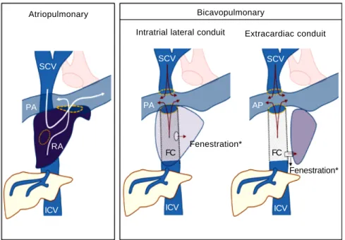
There are two major variants of the technique: atriopul-monary and bi-cavopulatriopul-monary (Figure 1). The original The Official Journal of the Mexican Association of Hepatology,
Table 1. Systemic consequences of the “failure” of the Fontan circulation.
Organ/system Complication Mechanism Clinical Findings
Lungs Veno-venous/atrial Gradient-dependent Cyanosis, dyspnea,
shunts passive circulation hypoxia,
exercise intolerance Plastic bronchitis ↓ Lymphatic return
Chylothorax ↓ Lymphatic return
Thromboembolism Hypercoagulability
Pulmonary Vascular
hypertension hyperreactivity
Kidneys Proteinuria Hyperfiltration due to Edema, ascites
venous hypertension
Kidney injury Ischemia due to ↓CO Dyspnea, oliguria
(acute/chronic)
Bowel Protein-losing ↓ Lymphatic return Malnutrition,
enteropathy Splanchnic venous edema, ascites,
congestion diarrhea
Systemic inflammation Hormonal activation
Liver Chronic liver disease Liver congestion Ascites, varices,
Ischemia due to encephalopathy,
↓ CO hepatocarcinoma
Brain Cerebrovascular disease Cardioembolic Decreased
Ischemia due to executive skills
↓ CO
Heart Bradi and Atrial and ventricular Hemodynamic
Tachyarrhythmias remodeling instability
Ventricular Activation of Dyspnea, exercise
dysfunction neurohormonal intolerance
systems
Vascular Varicosities Venous hypertension Edema, varicose
system ↓ Lymphatic return veins
CO: Cardiac output.
Figure 1. Figure 1.Figure 1.
Figure 1.Figure 1. Variants of Fontan surgery. IVC: in-ferior vena cava. FC: Fontan conduit. PA: pul-monary artery. RA: right atrium. SVC: superior vena cava. (*) The creation of a fenestration communicating FC and RA is a variant used only in selected cases.
Atriopulmonary Bicavopulmonary
Intratrial lateral conduit Extracardiac conduit
SCV SCV
PA
RA
ICV
PA
ICV ICV
AP SCV
FC FC
Fontan procedure (atriopulmonary anastomosis) converts the atrium into a canal that connects blood from both cava veins to the pulmonary artery. An anastomosis is created be-tween the atrium and the pulmonary artery and both the tri-cuspid valve and atrial septal defect are closed. Although initially it was thought that a hypertrophied right atrium would serve as a functional pump, this was later found to in-crease the risk of tachyarrhythmia and atrial thrombi.2,3 The
more recent bi-cavopulmonary procedure is carried out in two stages: in the first, the superior vena cava is connected to the pulmonary arteries (bidirectional Glenn procedure); and in the second, or Fontan completion stage, the inferior vena cava is also connected through an artificial conduit to the pul-monary arteries, thus completing the system.
The Fontan technique was first described in 1968, though it was not until the 1980s that it was widely adopt-ed.4 The procedure is indeed one of the major advances in
Pediatric Cardiology of our time as it guarantees survival rates of around 80% at 20 years and should be viewed as a great success considering the severity of the underlying anatomical cardiac defects.5,6 However, follow up over
decades of the growing Fontan population has revealed the
emergence of multiple and varied consequences on sever-al organ systems. These include arrhythmias and single ventricle dysfunction, aortic valve disease, exercise intol-erance, poor somatic growth, delayed puberty, plastic bronchitis, thromboembolic events, kidney disease due to glomerular and tubular injury, delayed neurocognitive de-velopment, peripheral venous insufficiency, protein-los-ing enteropathy and liver disease.7,8
The term used to describe the multisystemic clinical consequences of the Fontan circulation is “Fontan Fail-ure” (Table 1). This failure may be the final outcome of el-evated pulmonary resistance, pulmonary thrombi, narrowing and scarring of the Fontan pathway or pulmo-nary arteries, arrhythmias and failure of the systemic ven-tricle. Such alterations can damage target organs through two different mechanisms:9-12
• Venous passive congestion. Chronic elevation of sys-temic venous pressure promotes congestion in the splanchnic venous circulation (lacking self-regulating flow capacity) and reduces lymphatic return through the thoracic duct.
Figure 2. Figure 2. Figure 2. Figure 2.
Figure 2. Pathophysiology of FALD. LSEC: Liver Sinusoidal endothelial cell. SC: Stellate cell. TGF-β. Transforming growth factor beta. Systemic venous hy-pertension secondary to FS results in a decrease in venous drainage, which increases pressure and dilates the sinusoid. There will be phenomena of hyperfiltra-tion towards the space of Disse and the mechanical stress will induce a LSEC phenotypic change. The secrehyperfiltra-tion of some molecules will activate autocrine SC, promoting fibrogenesis. Hypoxia and perisinusoidal fibrosis will eventually lead to hepatocyte parenchymal necrosis, more evident in zone 3 (near the centrolob-ular vein).
Thrombosis
Portal space Sinusoid Centro lobular
[O2]
Zone 1 Zone 2 Zone 3
Ischemia
TGF-β
Congestion
Hepatocytes
Hepatic artery
Portal vein LSEC LMC
Mechanical tension
Venous return Sinusoidal dilation
Hyperfiltration
Perinusoidal edema
• Arterial ischemia. Reduced cardiac output from the func-tioning single ventricle, secondary to both diastolic and systolic dysfunction, causes ischemia in target organs.
PATHOPHYSIOLOGY OF
FONTAN-ASSOCIATED LIVER DISEASE
The pathogenesis of liver fibrosis in the Fontan popu-lation is not well-documented. Fibrogenesis is thought to be driven by a non-inflammatory mechanism, as the in-flammatory infiltrate in biopsy and autopsy samples is minimal or absent.13,14
The key point in the pathophysiology of FALD is a distur-bance in the liver's vascular supply and drainage (Figure 2). Elevated systemic venous pressure leads to inefficient blood drainage of the liver, determining a state of chronic passive congestion. This state promotes sinusoidal dilation and blood hyperfiltration causing perisinusoidal edema and hypoxia in centrilobular hepatocytes.15 In addition,
mechanical tension also likely plays an important role by
inducing a phenotypic change in the endothelial cell and enabling the activation of hepatic stellate cells and fibrob-lasts. As in other forms of liver fibrosis, TGF-β seems to be the central profibrogenic molecule driving the proc-ess.16 Finally, in a murine model, Simonetto, et al. recently
observed that sinusoidal thrombosis and mechanical strain secondary to blood stasis were the main fibrogenic pro-moters. These authors also confirmed minimal inflamma-tory activity, supporting the hypothesis that inflammation is not a common feature of cardiac liver disease.17
Wan-less, et al. have also argued that intrahepatic thrombosis is the main cause of cardiac cirrhosis.18 In line with data
sug-gesting that Fontan patients show an inherent prothrom-botic state,17 the hypothesis of intrahepatic thrombosis
becomes more important and opens the way for future therapeutic strategies such as anticoagulation (Figure 3).
Finally, it should be highlighted that Fontan patients feature additional risk factors for chronic liver disease which are unrelated to this unique vascular system. These include a higher prevalence of hepatitis C virus infection
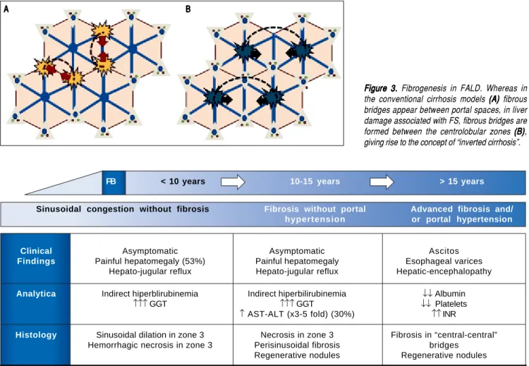
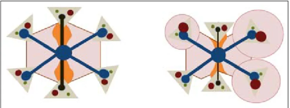
Figure 3. Figure 3.Figure 3.
Figure 3.Figure 3. Fibrogenesis in FALD. Whereas in the conventional cirrhosis models (A) (A) (A) (A) (A) fibrous bridges appear between portal spaces, in liver damage associated with FS, fibrous bridges are formed between the centrolobular zones (B)(B)(B)(B)(B), giving rise to the concept of “inverted cirrhosis”.
Figure 4. Figure 4.Figure 4.
Figure 4.Figure 4. Natural history of FALD. The temporal sequence is approximate since chronology will depend on the evolution of the heart disease. ALT: alanine transaminase. AST: aspartate aminotransferase. GGT: gamma-glutamyl transpeptidase. INR: International normalized ratio.
FB < 10 years 10-15 years > 15 years
Sinusoidal congestion without fibrosis Fibrosis without portal Advanced fibrosis and/
hypertension or portal hypertension
Clinical Asymptomatic Asymptomatic Ascitos
Findings Painful hepatomegaly (53%) Painful hepatomegaly Esophageal varices
Hepato-jugular reflux Hepato-jugular reflux Hepatic-encephalopathy
Analytica Indirect hiperblirubinemia Indirect hiperbilirubinemia ↓↓ Albumin
↑↑↑ GGT ↑↑↑ GGT ↓↓ Platelets
↑ AST-ALT (x3-5 fold) (30%) ↑↑ INR
Histology Sinusoidal dilation in zone 3 Necrosis in zone 3 Fibrosis in “central-central” Hemorrhagic necrosis in zone 3 Perisinusoidal fibrosis bridges
Regenerative nodules Regenerative nodules
A A A A
increased, though not well quantified, risk of develop-ing hepatocarcinoma and portal hypertension-related complications such as ascites, bleeding from esoph-agogastric varices or hepatic encephalopathy.25
In any of these stages, a clinical picture of its own entity may occur: ischemic hepatitis, characterized by markedly elevated AST, ALT and LDH in the context of an acute low cardiac output. This situation is usually reversible once hemodynamic stability has been reestablished.
As we are not dealing with primary liver disease, the chronology of FALD is difficult to establish and its pro-gression depends on the cardiological and hemodynamic situation. Table 2 lists the variables associated with an in-creased risk of liver damage.26-28 However, the main risk
factor for Fontan liver disease is the time since surgery. This means that compared to a post-Fontan time under 5 years, the odds of having a hepatic complications for post-Fontan durations of 11-15 years or 16-20 years are 4.4-fold (confidence interval [CI]: 1.1-17.2) or 9.0-fold (CI: 2.2-36.2), respectively.29
DIAGNOSIS OF ADVANCED
FONTAN-ASSOCIATED LIVER DISEASE
Liver damage in Fontan patients is universal. However, as not all patients develop liver-related complications, in-vasive and non-inin-vasive diagnostic methods are needed to identify who may require close and targeted monitoring.30
Serological tests
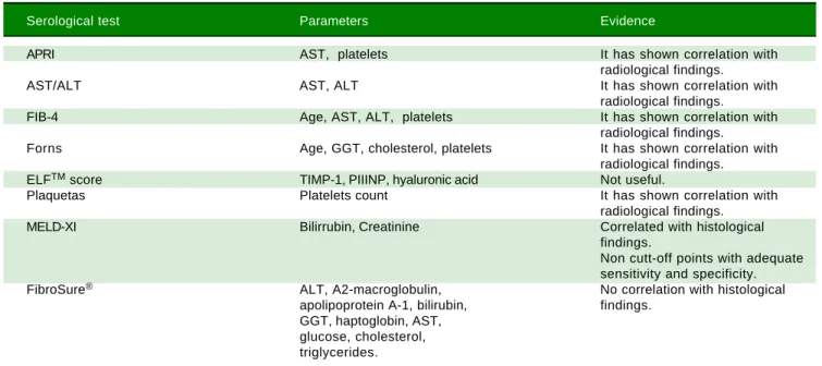
A basic blood panel, including liver function test, blood cell count and basic coagulation parameters may be suffi-cient as an initial assessment of liver disease in Fontan pa-tients. A platelet count < 150,000/μL is the main indicator of hypersplenism, which is notoriously associated with portal hypertension. Serum albumin is a classic marker of liver dysfunction and, although protein-losing enteropathy can facilitate its loss, low albumin levels usually indicate some extent of liver damage. Multiple simple methods that combine analytical and radiological findings have been recently developed. Although designed to diagnose and stratify patients with liver fibrosis other than Fontan patients, many of these methods have been used in the lat-ter patients (Table 3). However, these methods have not been validated against the gold standard, so we lack well-defined cut-off values. In a cohort of 204 patients (26% with hepatic decompensation), Baek, et al. compared sev-eral serological tests and identified Forns index as the best predictor of advanced liver damage with an area under the ROC curve of 0.786.29 In patients with cirrhosis, MELD
score is the main prognostic tool. However, as many Table 2. Risk factors for liver damage in FS patients.
Related to the hemodynamic situation
↓ Cardiac output.
↑ Pulmonary capillary pressure.
↑ Central venous pressure.
Related to the surgery
Pulmonary atresia as surgery precipitant. Classical technique (atriopulmonary variant). Fontan conduit stenosis / thrombosis. Time since surgery.
Related to cardiological events Cardiac arrhythmias.
Systolic ventricular dysfunction.
Others
Hepatotrope virus infection.
Exposure to hepatotoxic drugs (e.g. amiodarone).
or the use of hepatotoxic antiarrythmia drugs (eg, amiodar-one).19,20
NATURAL HISTORY OF
FONTAN-ASSOCIATED LIVER DISEASE
Symptomatic FALD is frequently the first manifesta-tion of Fontan failure and may coincide with other sys-temic manifestations such as protein-losing enteropathy or a progressive decline in functional capacity. However, liv-er damage begins before symptoms appear. Although its natural history is still poorly understood, 3 main stages have been described (Figure 4):
• Liver congestion and sinusoidal dilation. This stage starts even before Fontan surgery and continues into the following few years.21,22 Although many patients are
asymptomatic, 53% develop painful hepatomegaly and/ or hepatojugular reflux. Analytically, this phase is char-acterized by mild indirect hyperbilirubinemia and in-creased GGT, in relation to perisinusoidal edema. Biopsy usually reveals sinusoidal dilation and hepato-cellular necrosis in zone 3 of the lobule.23
• Fibrosis without portal hypertension. Around 5-10 years after surgery, patients show perisinusoidal fibro-sis, regenerative nodules and hepatocellular necrosis. In this stage, necrosis can be aggravated if low cardiac output further decreases, so slight elevations in AST, ALT and LDH are frequent. At this stage fibrosis is po-tentially reversible if the patient undergoes heart trans-plantation.24
Fontan patients are under anticoagulation therapy, MELD is artificially increased. To overcome this limitation, a new score MELD-XI has been designed.31 In Fontan
pa-tients, this new score has shown good correlation with the extent of liver fibrosis but is unable to define a cut-off for FALD.32
Doppler ultrasonography
Doppler ultrasonography is today the most useful radi-ological tool for the assessment of liver disease. By identi-fying a few simple signs detailed in table 4, a diagnosis of advanced liver disease can be made. The most specific ul-trasound finding for this purpose is the irregular surface of
the parenchyma as determined by a high frequency trans-ducer.33,34 Inverted portal flow offers a specificity of 100%
for identifying portal hypertension.35 The most frequent
echographic findings in Fontan patients are heterogeneous echogenicity, a nodular surface and small-sized hypere-choic nodules.36 In a controlled study in 106 individuals,
Kutty, et al. found a higher resistance and pulsatility index in the celiac trunk and mesenteric artery with a significant decrease in portal velocity in their Fontan patients.37
While the loss of the three-phase Doppler pattern in the hepatic veins is universal following bi-cavopulmonary surgery (as there is no atrial beat), observation of a monophasic pattern indicates advanced liver injury.26
Elastography
Elastography is the main non-invasive tool for the as-sessment of liver fibrosis in the majority of liver diseases.38
There are two ways to measure liver stiffness: transient elastography (Fibroscan®), which is easier and wide-spread in Europe, and sonoelastography, which needs to be conducted and interpreted by an expert operator and is more popular in the US. Transient elastography serves to stratify patients into four fibrosis stages and has been vali-dated for use in many liver diseases. This method thus avoids liver biopsy in a large number of situations. Its major drawback is its rate of false positives including hepatic congestion.39 Consequently, in Fontan patients
Table 3. Serological methods for diagnosis FALD y FS patients.
Serological test Parameters Evidence
APRI AST, platelets It has shown correlation with
radiological findings.
AST/ALT AST, ALT It has shown correlation with
radiological findings.
FIB-4 Age, AST, ALT, platelets It has shown correlation with
radiological findings.
Forns Age, GGT, cholesterol, platelets It has shown correlation with
radiological findings.
ELFTM score TIMP-1, PIIINP, hyaluronic acid Not useful.
Plaquetas Platelets count It has shown correlation with
radiological findings.
MELD-XI Bilirrubin, Creatinine Correlated with histological
findings.
Non cutt-off points with adequate sensitivity and specificity.
FibroSure® ALT, A2-macroglobulin, No correlation with histological
apolipoprotein A-1, bilirubin, findings. GGT, haptoglobin, AST,
glucose, cholesterol, triglycerides.
ALT: alanine transaminase. APRI: aspartate transaminase to platelet ratio index. AST: aspartate aminotransferase. FIB-4: fibrosis index based on four fac-tors. GGT: gamma-glutamyl transpeptidase. MELD-XI: model for end stage liver disease-XI. PIIINP: N-terminal propeptide of procollagen type III. TIMP-1: Tissue inhibitor-1 of metalloproteinase.
Table 4. Suggestive sonographic findings of advanced liver damage and portal hypertension in FALD.
Mode B
Nodular liver surface.
Heterogenicity of hepatic parenchyma. Intrahepatic venous-venous fistulas. Diameter of the portal vein > 12 mm. Splenomegaly (Area > 50 cm2).
Doppler
Decreased portal velocity (< 16 cm/s). Inverted portal flow
Resistance index > 0.71 and pulsatility index > 1.3 of principal hepatic artery.
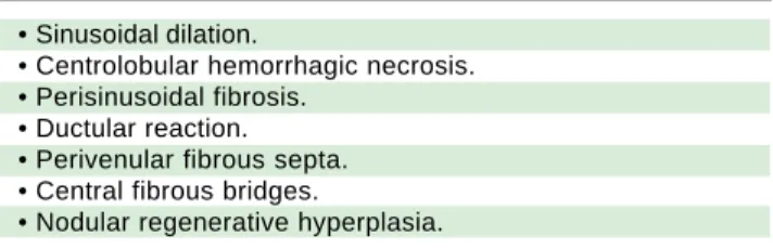
Table 5. Histological findings in FALD.
• Sinusoidal dilation.
• Centrolobular hemorrhagic necrosis. • Perisinusoidal fibrosis.
• Ductular reaction. • Perivenular fibrous septa. • Central fibrous bridges.
• Nodular regenerative hyperplasia.
elastography may overestimate liver stiffness. Fontan sur-gery induces an immediately increased Liver stiffness (LS) due only to hepatic congestion.40 It has been well
de-scribed that over time, signs of Fontan failure will appear. LS values will then increase, sometimes exceeding 15 kPa, and fibrosis progression may be to blame.41,42 A recent
study in which liver fibrosis was assessed through Shear-wave sonoelastography has shown good correlation be-tween liver stiffness and histology.37 Although cut-offs still
remain to be established, elastography is an accurate non-invasive method of measuring fibrosis.
Magnetic resonance imaging
The usefulness of MRI in FALD patients stems from its accuracy in detecting and characterizing liver nodules.43
Further, since cardiac resonance is now one of the main tools used in the follow up of the Fontan population, si-multaneous dynamic hepatic resonance reduces costs and the number of medical visits. MRI-elastography is a novel and useful technique for the estimation of hepatic fibrosis, but its high cost and scarce availability limits its use in clinical practice. In Fontan patients, MRI-elastography has shown positive correlation between estimated stiffness and APRI index, MELD, pressures in the Fontan conduit and even histological damage.44
Hepatic hemodynamics
In patients with chronic liver disease, hepatic venous pressure gradient (HVPG) is the best prognostic factor and has also been proposed as a key tool for the differential di-agnosis of ascites in patients with FALD. Measuring HVPG is a quick, simple and minimally invasive procedure that does not require sedation.45 The procedure consists of
jug-ular puncture to insert a balloon catheter through the Fon-tan connection into one of the hepatic veins. Hepatic pressure readings facilitate the differential diagnosis of por-tal hypertension such that high hepatic pressures (free and wedged) and a normal HVPG suggest a post-hepatic origin, and this is the most frequent finding after Fontan surgery.27
In patients with advanced parenchymal damage, HVPG may exceed 6 mmHg, or even 10 mmHg, which indicates a high risk of decompensation in most liver diseases.46
Neverthe-less, there are several situations in which HVPG may be un-derestimated such as vascular fistulas between the hepatic veins themselves and the portal branches. In our experi-ence, the development of such communications is very fre-quent in Fontan patients, which makes the HVPG reading difficult to interpret. We strongly recommend that HVPG measurements in these patients are carried out by expert staff and interpreted with caution.
Liver biopsy
Liver biopsy is the gold standard diagnostic procedure and, pending the proper validation of non-invasive diag-nostic methods, is still necessary to determine the extent of liver damage. Typical histological features of FALD are provided in table 5. Sinusoidal dilation is present in 90-97% of Fontan patients and is the earliest parenchymal change. Typically, it is more pronounced than in other causes of cardiogenic hepatopathy. The distribution of fi-brosis in the early stages is typically perisinusoidal (in the space of Disse); this is not the case in other forms of car-diac-derived liver disease. Extensive bridges of centrolob-ular fibrosis associated with regenerative nodules are a late finding in patients with advanced liver disease. Periportal inflammation is usually minimal or absent, allowing the differential diagnosis with other liver disease etiologies.47
Some authors recommend a liver biopsy in all patients 10 years after Fontan surgery.25 In a cohort of 67 patients, this
strategy revealed that all patients developed hepatic fibro-sis and that its extent increased with time. However, the authors found no correlation between the degree of fibro-sis and clinically relevant events, hemodynamic or analyti-cal parameters,48 thus questioning its utility in clinical
practice. Hepatic biopsy is currently recommended when the etiology of liver disease is not clear and in candidates for heart and/or liver transplantation.37,39,49
HEPATIC COMPLICATIONS OF FONTAN SURGERY
As in other forms of hepatic cirrhosis, in advanced FALD stages esophageal varices, ascites, hepatic encepha-lopathy, hepatocarcinoma and splenomegaly with throm-bocytopenia may occur. The characteristics and natural history of these complications in FALD remain largely unknown, probably because no attention had been paid to FALD until the last five years.
Hepatic nodules and hepatocellular carcinoma
nodules in the hepatic parenchyma is a frequent finding in FALD.15,50 These nodules are usually multiple,
hyper-vascular, hyperechoic, < 3 cm and located in the outer margins of the liver. Histologically, they usually corre-spond to nodules of regeneration, focal nodular hyper-plasia or adenomas.28,36,51,52 Although the pathophysiology
behind benign liver nodules in patients with FALD is unknown, their peripheral location and radiological be-havior indicate a vascular origin (Figure 5).53 In recent
years, some cases and small case series of hepatocarcino-ma have been reported in young Fontan patients.53,66
These are often patients with advanced liver disease and a post-Fontan duration longer than 5-10 years. Malignant nodules are usually hypervascular, show washout and al-pha-fetoprotein is typically elevated. However, these fea-tures are not pathognomonic and their diagnostic accuracy in FALD has not been addressed, making the differential diagnosis of nodules a main clinical chal-lenge. Hence, until more data becomes available, the di-agnosis of hepatocarcinoma in patients with FALD always requires histological confirmation.44
Screening every 4-12 months with radiological tech-niques has proven to be cost-effective and to increase sur-vival in patients with cirrosis. Nandwana, et al. retrospectively analyzed a cohort of 145 Fontan patients subjected to periodical liver imaging: in the initial assess-ment one case of hepatocarcinoma was present and four incident cases were detected during a median follow up of 3.05 years.67 Most experts recommend periodic screening,
although the optimal radiologic technique and interval are unknown.25,51 Treatment of hepatocarcinoma should
fol-low clinical practice guidelines used in other forms of cir-rhosis.68
Esophageal varices
Following Fontan surgery, the prevalence of esopha-geal varices has been estimated at between 2 and
43%.25,30,69 In the VAST study on a retrospective cohort of
73 Fontan patients, the presence of varices along with other manifestations of portal hypertension was
associat-ed with an increasassociat-ed risk of death, heart transplantation and hepatocarcinoma.70 Case reports exist of variceal
bleeding, some with a fatal outcome, highlighting the need for screening and proper prevention performed in a systematized manner when other signs of portal hyper-tension are present.25 In other forms of cirrhosis, the
cornerstone of variceal bleeding prophylaxis is non-car-dioselective beta-blockers. However, the effects of this form of prophylaxis have not yet been addressed in FALD.71 Unlike in all other forms of cirrhosis, the portal
hypertension model in patients with FALD is character-istically hypodynamic such that the efficacy of these drugs is debatable. Further, if we also consider that beta-blockers may have deleterious effects on the Fontan cir-culation, as prophylaxis, it is advisable to consider rubber band ligation. An acute episode of gastrointestinal bleed-ing should be managed with vasoactive drugs (somatosta-tin, terlipressin or octreotide) and endoscopic therapy (band ligation). In cases of bleeding refractory to stand-ard treatment, a transjugular intrahepatic portosystemic shunt (TIPS) has been shown to improve survival in oth-er types of hepatopathy.71 Notwithstanding, the
result-ant increase in cardiac preload may precipitate pulmonary hypertension and cardiac failure. To the best of our knowledge, there is only one reported case of a 42-year-old man with cirrhosis after Fontan surgery in whom variceal hemorrhage was controlled by TIPS. Hence, its use should be restricted to highly selected cases not responding to conventional treatment when cardiac function is within the normal range or mini-mally impaired.72
Ascites
Ascites is a late manifestation of cirrhosis and is associ-ated with reduced survival and a poor quality of life.73 It is
the most frequent sign of clinical hepatic decompensation and its prevalence in the Fontan population ranges from 2 to 17%.51 In chronic liver disease with intrahepatic portal
hypertension, ascites appears when HVPG ≥ 10 mmHg, which also has prognostic capacity.46 However, in the
Figure 5. Figure 5.Figure 5.
Fontan patient, HVPG is usually normal and ascites may appear in the absence of cirrhosis. This means its value as a prognostic marker and its pathophysiological mechanisms differ from those related to other forms of liver dis-ease.28,74 There are several causes of ascites in Fontan
pa-tients (Table 6).74
Ascites is usually manageable by optimizing cardiac function, and nutrition along with the use of loop diuret-ics and anti-aldosterone drugs since the renin-angiotensin aldosterone system is hyperactivated in portal hyperten-sion and heart failure.46,75 Large-volume paracentesis could
Table 7. Follow-up recommendations related liver disease after Fontan surgery at Hospital Universitario Ramón y Cajal.
Diagnosis of FALD
0-10 after Fontan surgery HAV (IgG), HBV and HCV Baseline
antibodies* and screening for autoimmune and metabolic liver disease.
Liver function parameters. Annual
Doppler ultrasound. Every 5 years
Elastography. Every 5 years
≥10 after Fontan Liver function parameters. Every 6 months
surgery or “Fontan Alpha-fetoprotein. Every 6 months
failure” Doppler ultrasound. Every 6 months
Elastography. Baseline y annual
Dynamic MRI/CT. Baseline
Liver biopsy Uncertain diagnosis and candidates for liver and/or cardiac transplantation.
Screening and diagnosis of hepatocellular carcinoma
Doppler ultrasound Every 6 months since the 10th year from surgery ** Dynamic MRI/CT • ≥ 10 year after surgery (baseline).
• If “benign” nodules are present in the basal test (hypercapiting in the arterial phase, without venous or late phase clearance, multiple, peripheral and with normal AFP) repeat at 3 months. If there is no suspicion of hepatocarcinoma, continue with semiannual ultrasound screening.
• If one or more nodules develop during follow-up.
• Hepatic MRI is recommended when cardiac MRI is performed. Biopsy/FNAB Any nodule suggestive of hepatocellular carcinoma
(venous phase clearance, growth or elevated AFP) require histologic confirmation.
Esophagogastric varices screening
Analytical, clinical, Basal upper digestive endoscopy.
radiological or If no varices veins or these are small, watch every 1-3 years. elastographic data of
FALD
(*) Perform HAV, HCV and HBV ELISA (HBsAg, HBcAb and HBsAb) in all patients with Fontan surgery. If not immunized, vaccination against HAV and HBV should be indicated and its efficacy tested with new serologies. 10 years after effective vaccination against HBV, levels of HBsAg should be determined and a new dose should be indicated if levels are <100 IU/L. (**) It will be advanced in those patients with “Fontan failure”, Fontan's duct thrombosis or transitional elastography ≥ 15 KPa. AFP: Alpha-fetoprotein. CT: Computed tomography. HAV: Hepatitis A virus. HBsAg: Hepatitis B surface antigen. HBcAb: Hepatitis B core antibody. HBsAb: Hepatiti B surface antibody. HBV: Hepatitis B virus. HVC: Hepatitis C virus. MRI: Magnetic resonance imaging.
Table 6. Differential diagnosis of ascites in Fontan patients.
• Systemic venous hypertension due to failure of the Fontan circulation (arrhythmias, thrombosis/stenosis of Fontan conduit, ventricular dysfunction, pulmonary hypertension).
• Post-hepatic portal hypertension.
• Sinusoidal portal hypertension (advanced liver fibrosis).
be a rescue treatment, although it is rarely required. TIPS does not seem a suitable option in Fontan anatomy and no case reports of this exist.
Hepatic encephalopathy
It is estimated that 30-40% of patients with liver cirrho-sis will present this condition at some point,76 although in
FALD this event is poorly documented. Only 3 cases of hepatic encephalopathy following Fontan surgery exist in the literatura.25,30,69 However, it seems likely that its real
incidence and prevalence are underestimated because of the retrospective nature of most studies and the possibility that hepatic encephalopathy was not considered in the dif-ferential diagnosis.
Liver transplantation
In the twentieth century, cirrhosis was conceived as an irreversible disease. However, current knowledge arising from studies on viral and alcoholic cirrhosis indicate that fibrosis is effectively reversible.76,77 In cardiac cirrhosis,
experimental models and small case series point to the idea that if cardiac function is restored, liver disease may also improve and even fully normalize.73,78,79 Considering
that the severity of heart disease is directly linked to liver damage and that liver damage is practically universal in Fontan patients, the key question arises as to which sub-groups of patients will require an isolated heart transplant or a double cardiohepatic transplant.
To shed light on this question, we have data available from two studies which are nevertheless retrospective, have a small sample size, and suffer from methodological flaws. Simpson, et al. analyzed 20 abdominal CT scans of Fontan patients before heart transplantation: 7 had mor-phological changes compatible with liver cirrhosis, 5 had liver changes not meeting cirrhosis criteria and the re-maining were normal. Cirrhotic patients were older (17.6 vs. 9.6 years, p = 0.002) and showed a longer time since surgery (180 vs. 50 months, p < 0.05); without significant differences in laboratory parameters. One-year survival was similar between cirrhotic and non-cirrhotic patients (86% vs. 77%, p = 0.681). In the group of transplanted cir-rhotic patients, two patients died, none of them of a hepat-ic cause.58 The absence of biopsy and lack of a detailed
description of cirrhosis stage in some subjects limit the validity of this research. In 2016, D'Souza, et al. described 7 Fontan patients who received a double transplant. The cri-terion for liver transplantation was the presence of bridge fibrosis or cirrhosis in a pre-transplantation biopsy. Three patients had portal hypertension and ascites, while the oth-er four had no complications related to FALD. All pa-tients survived a median follow up of 4.6 years.39,47 From
our perspective, this approach is too aggressive since liver damage in patients with compensated disease will likely be insufficient to justify liver transplantation; especially considering the current shortage of organs and the poten-tial improvement of liver function after heart transplanta-tion. In addition, no study has shown that compensated liver disease is a perioperative risk factor or a long-term poor prognostic marker after cardiac transplantation in FALD. At present, there are no consensus guidelines re-garding indications for double transplantation. The insti-tutions with more experience in this field recommend an individual analysis of each case by a multidisciplinary committee.
CONCLUSIONS
Liver damage following Fontan surgery is universal. The main risk factors are time since surgery and poor car-diac function. Although the natural history of FALD is not well established, patients operated on more than 10 years ago should be closely followed to check for the onset of portal hypertension-related complications or hepatocellu-lar carcinoma. Its peculiar pathophysiology and clinical behavior make FALD a unique entity of liver disease that requires an individualized management program designed by a multidisciplinary committee (Table 7). It is essential to create multicenter research groups to gather scientific evidence on which to base the optimal follow up and treat-ment of the growing adult Fontan population.
ABBREVIATIONS
• ALT: alanine transaminase. • APRI: AST to platelet Ratio Index. • AST: aspartate aminotransferase. • CT: computed tomography.
• FALD: Fontan-associated liver disease. • GGT: gamma-glutamyl transpeptidase. • HVPG: hepatic vein pressure gradient. • LDH: lactate dehydrogenase.
• LS: liver stiffness.
• MELD: model for end stage liver disease. • MR: nuclear magnetic resonance.
• TGF- : transforming growth factor beta.
• TIPS: transjugular intrahepatic portosystemic shunt.
FINANCIAL SUPPORT
This work was financed by grants from the Spanish Ministry of Health, Instituto de Salud Carlos III (no. PI14/00876 and PI051871, CIBERehd) awarded to Agustín Albillos. Ciberhed is funded by the Instituto de Salud Carlos III.
CONFLICTS OF INTEREST
All authors have nothing to disclosure.
ACKNOWLEDGEMENT
We thank the Department of Pediatric Cardiology of the Hospital Universitario Ramón y Cajal, in particular María Jesús del Cerro and Elvira Garrido-Lestache, for their enthusiastic dedication to Fontan-associated liver disease.
REFERENCES
1. Gewillig M, Brown SC. The Fontan circulation after 45 years: update in physiology. Heart Br Card Soc 2016; 102: 1081-6. 2. Quinton E, Nightingale P, Hudsmith L, Thorne S, Marshall H,
Clift P, de Bono J, et al. Prevalence of atrial tachyarrhythmia in adults after Fontan operation. Heart Br Card Soc 2015; 101: 1672-7.
3. Balaji S, Gewillig M, Bull C, de Leval MR, Deanfield JE. Ar-rhythmias after the Fontan procedure. Comparison of total cavopulmonary connection and atriopulmonary connection.
Circulation 1991; 84: III162-7.
4. Fontan F, Baudet E. Surgical repair of tricuspid atresia. Tho-rax 1971; 26: 240-8.
5. Allen KY, Downing TE, Glatz AC, Rogers LS, Ravishankar C, Rychik J, Fuller S, et al. Long-term survival after the Fontan operation: Twenty years of experience at a single center. J Thorac Cardiovasc Surg 2017; 154: 243-53.e2.
6. Pundi KN, Johnson JN, Dearani JA, Pundi KN, Li Z, Hinck CA, Dahl SH, et al. 40-Year Follow-Up After the Fontan Opera-tion: Long-Term Outcomes of 1,052 Patients. J Am Coll Car-diol 2015; 66: 1700-10.
7. Ohuchi H. Adult patients with Fontan circulation: What we know and how to manage adults with Fontan circulation? J Cardiol 2016; 68: 181-9.
8. Menon S, Chennapragada M, Ugaki S, Sholler GF, Ayer J, Winlaw DS. The Lymphatic Circulation in Adaptations to the Fontan Circulation. Pediatr Cardiol 2017; 38: 886-92. 9. Ostrow AM, Freeze H, Rychik J. Protein-losing enteropathy
af-ter Fontan operation: investigations into possible pathophysio-logic mechanisms. Ann Thorac Surg 2006; 82: 695-700. 10. Heinemann M, Breuer J, Steger V, Steil E, Sieverding L,
Zie-mer G. Incidence and impact of systemic venous collateral development after Glenn and Fontan procedures. Thorac Cardiovasc Surg 2001; 49: 172-8.
11. Anne P, Du W, Mattoo TK, Zilberman MV. Nephropathy in pa-tients after Fontan palliation. Int J Cardiol 2009; 132: 244-7. 12. Egbe AC, Connolly HM, Khan AR, Niaz T, Said SS, Dearani
JA, Warnes CA, et al. Outcomes in adult Fontan patients with atrial tachyarrhythmias. Am Heart J 2017; 186: 12-20. 13. Fernández-Iglesias A, Gracia-Sancho J. How to Face
Chron-ic Liver Disease: The Sinusoidal Perspective. Front Med
2017; 4: 7.
14. Tsochatzis EA, Bosch J, Burroughs AK. Liver cirrhosis. Lan-cet Lond Engl 2014; 383: 1749-61.
15. Asrani SK, Asrani NS, Freese DK, Phillips SD, Warnes CA, Heimbach J, et al. Congenital heart disease and the liver.
Hepatol Baltim Md 2012; 56: 1160-9.
16. Greuter T, Shah VH. Hepatic sinusoids in liver injury, inflam-mation, and fibrosis: new pathophysiological insights. J Gastroenterol 2016; 51: 511-9.
17. Tomkiewicz-Pajak L, Hoffman P, Trojnarska O, Lipczynska M, Podolec P, Undas A. Abnormalities in blood coagulation, fibri-nolysis, and platelet activation in adult patients after the Fontan procedure. J Thorac Cardiovasc Surg 2014; 147: 1284-90. 18. Simonetto DA, Yang H, Yin M, de Assuncao TM, Kwon JH,
Hilscher M, Pan S, et al. Chronic passive venous congestion drives hepatic fibrogenesis via sinusoidal thrombosis and mechanical forces. Hepatol Baltim Md 2015; 61: 648-59. 19. Sung PS, Yoon SK. Amiodarone hepatotoxicity. Hepatol
Bal-tim Md 2012; 55: 325-6.
20. Wang A, Book WM, McConnell M, Lyle T, Rodby K, Mahle WT. Prevalence of hepatitis C infection in adult patients who underwent congenital heart surgery prior to screening in 1992. Am J Cardiol 2007; 100: 1307-9.
21. Schwartz MC, Sullivan L, Cohen MS, Russo P, John AS, Guo R, Guttenberg M, et al. Hepatic pathology may develop be-fore the Fontan operation in children with functional single ventricle: an autopsy study. J Thorac Cardiovasc Surg
2012; 143: 904-9.
22. Johnson JA, Cetta F, Graham RP, Smyrk TC, Driscoll DJ, Phil-lips SD, John AS. Identifying predictors of hepatic disease in patients after the Fontan operation: a postmortem analysis. J Thorac Cardiovasc Surg 2013; 146: 140-5.
23. Wu FM, Jonas MM, Opotowsky AR, Harmon A, Raza R, Uko-madu C, Landzberg MJ, et al. Portal and centrilobular hepatic fibrosis in Fontan circulation and clinical outcomes. J Heart Lung Transplant Off Publ Int Soc Heart Transplant 2015; 34: 883-91.
24. Dichtl W, Vogel W, Dunst KM, Grander W, Alber HF, Frick M, Antretter H, et al. Cardiac hepatopathy before and after heart transplantation. Transpl Int Off J Eur Soc Organ Transplant 2005; 18: 697-702.
25. Rychik J, Veldtman G, Rand E, Russo P, Rome JJ, Krok K, Goldberg DJ, et al. The precarious state of the liver after a Fontan operation: summary of a multidisciplinary symposium.
Pediatr Cardiol 2012; 33: 1001-12.
26. Camposilvan S, Milanesi O, Stellin G, Pettenazzo A, Zancan L, D'Antiga L. Liver and cardiac function in the long term af-ter Fontan operation. Ann Thorac Surg 2008; 86: 177-82. 27. Mori M, Hebson C, Shioda K, Elder RW, Kogon BE, Rodriguez
FH, Jokhadar M, et al. Catheter-measured Hemodynamics of Adult Fontan Circulation: Associations with Adverse Event and End-organ Dysfunctions. Congenit Heart Dis 2016; 11: 589-97.
28. Kiesewetter CH, Sheron N, Vettukattill JJ, Hacking N, Sted-man B, Millward-Sadler H, Haw M, et al. Hepatic changes in the failing Fontan circulation. Heart Br Card Soc 2007; 93: 579-84.
29. Baek JS, Bae EJ, Ko JS, Kim GB, Kwon BS, Lee SY, Noh CI, et al. Late hepatic complications after Fontan operation; non-invasive markers of hepatic fibrosis and risk factors. Heart Br Card Soc 2010; 96: 1750-5.
30. Pundi K, Pundi KN, Kamath PS, Cetta F, Li Z, Poterucha JT, Driscoll DJ, et al. Liver Disease in Patients After the Fontan Operation. Am J Cardiol 2016; 117: 456-60.
31. Heuman DM, Mihas AA, Habib A, Gilles HS, Stravitz RT, San-yal AJ, Fischer RA. MELD-XI: a rational approach to "sickest first" liver transplantation in cirrhotic patients requiring antico-agulant therapy. Liver Transplant Off Publ Am Assoc Study Liver Dis Int Liver Transplant Soc 2007; 13: 30-7.
32. Evans WN, Acherman RJ, Ciccolo ML, Carrillo SA, Galindo A, Rothman A, Mayman GA, et al. MELD-XI Scores Correlate with Post-Fontan Hepatic Biopsy Fibrosis Scores. Pediatr Cardiol 2016; 37: 1274-7.
de-tection--analysis of 300 cases. Radiology 2003; 227: 89-94. 34. Berzigotti A, Ashkenazi E, Reverter E, Abraldes JG, Bosch J.
Non-invasive diagnostic and prognostic evaluation of liver cirrhosis and portal hypertension. Dis Markers 2011; 31: 129-38.
35. Giannini E, Botta F, Borro P, Risso D, Romagnoli P, Fasoli A, Mele MR, et al.. Platelet count/spleen diameter ratio: proposal and validation of a non-invasive parameter to predict the presence of oesophageal varices in patients with liver cir-rhosis. Gut 2003; 52: 1200-5.
36. Bae JM, Jeon TY, Kim JS, Kim S, Hwang SM, Yoo SY, Kim JH. Fontan-associated liver disease: Spectrum of US find-ings. Eur J Radiol 2016; 85: 850-6.
37. Kutty SS, Peng Q, Danford DA, Fletcher SE, Perry D, Talmon GA, Scott C, et al. Increased hepatic stiffness as conse-quence of high hepatic afterload in the Fontan circulation: a vascular Doppler and elastography study. Hepatol Baltim Md 2014; 59: 251-60.
38. Sandrin L, Fourquet B, Hasquenoph JM, Yon S, Fournier C, Mal F, Christidis C, et al. Transient elastography: a new non-invasive method for assessment of hepatic fibrosis. Ultra-sound Med Biol 2003; 29: 1705-13.
39. Caste’ra L, Foucher J, Bernard P-H, Carvalho F, Allaix D, Merrouche W, Couzigou P, et al. Pitfalls of liver stiffness measurement: a 5-year prospective study of 13,369 exami-nations. Hepatol Baltim Md 2010; 51: 828-35.
40. Deorsola L, Aidala E, Cascarano MT, Valori A, Agnoletti G, Pace Napoleone C. Liver stiffness modifications shortly after total cavopulmonary connection. Interact. Cardiovasc Tho-rac Surg 2016; 23: 513-8.
41. Agnoletti G, Ferraro G, Bordese R, Marini D, Gala S, Berga-masco L, Ferroni F, et al. Fontan circulation causes early, severe liver damage. Should we offer patients a tailored strategy? Int J Cardiol 2016; 209: 60-5.
42. Chen B, Schreiber RA, Human DG, Potts JE, Guttman OR. Assessment of Liver Stiffness in Pediatric Fontan Patients Using Transient Elastography. Can J Gastroenterol Hepatol
2016; 2016: 7125193.
43. Bulut OP, Romero R, Mahle WT, McConnell M, Braithwaite K, Shehata BM, Gupta NA, et al. Magnetic resonance imaging identifies unsuspected liver abnormalities in patients after the Fontan procedure. J Pediatr 2013; 163: 201-6.
44. Poterucha JT, Johnson JN, Qureshi MY, O’Leary PW, Kamath PS, Lennon RJ, Bonnichsen CR, et al. Magnetic Res-onance Elastography: A Novel Technique for the Detection of Hepatic Fibrosis and Hepatocellular Carcinoma After the Fontan Operation. Mayo Clin Proc 2015; 90: 882-94. 45. Bosch J, Abraldes JG, Berzigotti A, García-Pagan JC. The
clinical use of HVPG measurements in chronic liver disease.
Nat Rev Gastroenterol Hepatol 2009; 6: 573-82.
46. Bosch J, Abraldes JG, Albillos A, Aracil C, Bañares R, Ber-zigotti A, Calleja JL, et al. Hipertensión portal: recomenda-ciones para su evaluación y tratamiento. Gastroenterol Hepatol 2012; 35: 421-50.
47. Kendall TJ, Stedman B, Hacking N, Haw M, Vettukattill JJ, Salmon AP, Cope R, et al. Hepatic fibrosis and cirrhosis in the Fontan circulation: a detailed morphological study. J Clin Pathol 2008; 61: 504-8.
48. Goldberg DJ, Surrey LF, Glatz AC, Dodds K, O’Byrne ML, Lin HC, Fogel M, et al. Hepatic Fibrosis Is Universal Following Fontan Operation, and Severity is Associated With Time From Surgery: A Liver Biopsy and Hemodynamic Study. J Am Heart Assoc 2017; 6. J Am Heart Assoc. 2017 Apr 26;6(5). pii: e004809. doi: 10.1161/JAHA.116.004809. 49. D’Souza BA, Fuller S, Gleason LP, Hornsby N, Wald J, Krok
K, Shanked A, et al. Single-center outcomes of combined
heart and liver transplantation in the failing Fontan. Clin Transplant 2017; 31. doi: 10.1111/ctr.12892. Epub 2017 Feb 4.
50. Ford RM, Book W, Spivey JR. Liver disease related to the heart. Transplant Rev Orlando Fla 2015; 29: 33-7.
51. Wu FM, Kogon B, Earing MG, Aboulhosn JA, Broberg CS, John AS Harmon A, et al. Liver health in adults with Fontan circulation: A multicenter cross-sectional study. J Thorac Cardiovasc Surg 2017; 153: 656-64.
52. Wallihan DB, Podberesky DJ. Hepatic pathology after Fontan palliation: spectrum of imaging findings. Pediatr Radiol 2013; 43: 330-8.
53. Shimamatsu K, Wanless IR. Role of ischemia in causing ap-optosis, atrophy, and nodular hyperplasia in human liver.
Hepatol Baltim Md 1997; 26: 343-50.
54. Ghaferi AA, Hutchins GM. Progression of liver pathology in patients undergoing the Fontan procedure: Chronic passive congestion, cardiac cirrhosis, hepatic adenoma, and hepato-cellular carcinoma. J Thorac Cardiovasc Surg 2005; 129: 1348-52.
55. Saliba T, Dorkhom S, O'Reilly EM, Ludwig E, Gansukh B, Abou-Alfa GK. Hepatocellular carcinoma in two patients with cardiac cirrhosis. Eur J Gastroenterol Hepatol 2010; 22: 889-91.
56. Asrani SK, Warnes CA, Kamath PS. Hepatocellular carcino-ma after the Fontan procedure. N Engl J Med 2013; 368: 1756-7.
57. Elder RW, Parekh S, Book WM. More on hepatocellular carcino-ma after the Fontan procedure. N Engl J Med 2013; 369: 490. 58. Takuma Y, Fukada Y, Iwadou S, Miyatake H, Uematsu S,
Okamoto R, Sato D, et al. Surgical Resection for Hepatocel-lular Carcinoma with Cardiac Cirrhosis after the Fontan Pro-cedure. Intern. Med Tokyo Jpn 2016; 55: 3265-72.
59. Rajoriya N, Clift P, Thorne S, Hirschfield GM, Ferguson JW. A liver mass post-Fontan operation. QJM Mon J Assoc Physi-cians 2014; 107: 571-2.
60. Weyker PD, Allen-John Webb C, Emond JC, Brentjens TE, Johnston TA. Anesthetic implications of extended right hepatectomy in a patient with Fontan physiology. Case Rep
2014; 2: 99-101.
61. Kwon S, Scovel L, Yeh M, Dorsey D, Dembo G, Krieger EV, Bakthavatsalam R, et al. Surgical management of hepatocel-lular carcinoma after Fontan procedure. J Gastrointest On-col 2015; 6: E55-60.
62. Yamada K, Shinmoto H, Kawamura Y, Wakamatsu H, Ka-wauchi T, Soga S, Soga S, et al. Transarterial embolization for pediatric hepatocellular carcinoma with cardiac cirrhosis. Pediatr. Int Off J Jpn Pediatr Soc 2015; 57: 766-70.
63. Oh C, Youn JK, Han J-W, Kim GB, Kim H-Y, Jung S-E. Hepa-tocellular carcinoma after the Fontan procedure in a 16-year-old girl: A case report. Medicine (Baltimore) 2016; 95: e4823.
64. Conroy MR, Moe TG. Hepatocellular carcinoma in the adult Fontan patient. Cardiol Young 2017; 27: 407-9.
65. Josephus Jitta D, Wagenaar LJ, Mulder BJM, Guichelaar M, Bouman D, van Melle JP. Three cases of hepatocellular car-cinoma in Fontan patients: Review of the literature and sug-gestions for hepatic screening. Int J Cardiol 2016; 206: 21-6.
66. Martínez-Quintana E, Monescillo A, Rodríguez-González F. Hepatocellular carcinoma in a non-failing Fontan circulation.
Rev Española Enfermedades Dig Organo Of Soc Española Patol Dig 2017; 109: 375.
Carcinoma. Curr Probl Diagn Radiol 2017; 2018 Jan -Feb;47(1):19-22..
68. Forner A, Reig M, Varela M, Burrel M, Feliu J, Bricen~o J, Sastre J, et al. [Diagnosis and treatment of hepatocellular carcinoma. Update consensus document from the AEEH, SEOM, SERAM, SERVEI and SETH]. Med Clin (Barc) 2016; 146: 511.e1-511.e22.
69. Koteda Y, Suda K, Kishimoto S, Iemura M. Portal-systemic encephalopathy after Fontan-type operation in patient with polysplenia syndrome. Eur J Cardio-Thorac Surg Off J Eur Assoc Cardio-Thorac Surg 2009; 35: 1083-5.
70. Elder RW, McCabe NM, Hebson C, Veledar E, Romero R, Ford RM, (Missing author) et al. Features of portal hyperten-sion are associated with major adverse events in Fontan pa-tients: the VAST study. Int J Cardiol 2013; 168: 3764-9. 71. de Franchis R, Baveno VI Faculty. Expanding consensus in
portal hypertension: Report of the Baveno VI Consensus Workshop: Stratifying risk and individualizing care for portal hypertension. J Hepatol 2015; 63: 743-52.
72. Velpula M, Sheron N, Guha N, Salmon T, Hacking N, Veldt-man GR. Direct measurement of porto-systemic gradient in a failing Fontan circulation. Congenit Heart Dis 2011; 6: 175-8. 73. Pericleous M, Sarnowski A, Moore A, Fijten R, Zaman M. The clinical management of abdominal ascites, spontaneous bac-terial peritonitis and hepatorenal syndrome: a review of cur-rent guidelines and recommendations. Eur J Gastroenterol Hepatol 2016; 28: e10-18.
74. Myers RP, Cerini R, Sayegh R, Moreau R, Degott C, Lebrec D, Lee SS. Cardiac hepatopathy: Clinical, hemodynamic, and histologic characteristics and correlations. Hepatology 2003; 37: 393-400.
75. Hilscher MB, Johnson JN, Cetta F, Driscoll DJ, Poterucha JJ, Sanchez W, Connolly HM, et al. Surveillance for liver
compli-cations after the Fontan procedure. Congenit Heart Dis
2017; 12: 124-32.
76. American Association for the Study of Liver Diseases, Euro-pean Association for the Study of the Liver. Hepatic en-cephalopathy in chronic liver disease: 2014 practice guideline by the European Association for the Study of the Liver and the American Association for the Study of Liver Diseases. J Hepatol 2014; 61: 642-59.
77. Zoubek ME, Trautwein C, Strnad P. Reversal of liver fibrosis: From fiction to reality. Best Pract Res Clin Gastroenterol
2017; 31: 129-41.
78. Ellis EL, Mann DA. Clinical evidence for the regression of liv-er fibrosis. J Hepatol 2012; 56: 1171-80.
79. Dichtl W, Vogel W, Dunst KM, Grander W, Alber HF, Frick M, Antretter H, et al. Cardiac hepatopathy before and after heart transplantation. Transpl Int Off J Eur Soc Organ Transplant 2005; 18: 697-702.
80. Crespo-Leiro MG, Robles O, Paniagua MJ, Marzoa R, Naya C, Flores X, Naya C, et al. Reversal of cardiac cirrhosis fol-lowing orthotopic heart transplantation. Am J Transplant Off J Am Soc Transplant Am Soc Transpl Surg 2008; 8: 1336-9.
Correspondence and reprint request:
Agustín Albillos, M.D. Ph.D.
Departamento de Medicina y Especialidades Médicas, Facultad de Medicina y Ciencias de la Salud-Campus Universitario, Universidad de Alcalá, Carretera Madrid-Barcelona km. 33.600,
28871 Alcalá de Henares, Madrid, Spain. Tel.: +34918854870. Fax: +34918854526.