SUSTAINABLE FOREST MANAGEMENT RESEARCH INSTITUTE
Department of Plant Production and Forest Resources
IDENTIFICATION, CHARACTERIZATION AND PATHOGENICITY OF
Phytophthora spp. ASSOCIATED WITH THE MORTALITY OF
Alnus glutinosa IN SPAIN
The present thesis fulfils the necessary requisites to obtain the
Doctorate Mention through the University of Valladolid
Mohammed Masum Ul Haque
Dr. Julio Javier Diez Casero Dr. Jorge Martín-García
It is my great pleasure to express deepest sense of gratitude to my PhD supervisor Dr. Julio Javier Diez Casero, Chair Professor, Department of Plant Production and Forest Resources, Universidad de Valladolid, Spain for his indispensible guidance, generous help, valuable suggestion, encouragement and cooperation during the course of my doctoral study.
I express my sincere gratitude to my PhD co-supervisor Dr. Jorge Martín-García, Researcher, Department of Plant Production and Forest Resources, Universidad de Valladolid, Spain for his guidance, generous help and co-operation, and valuable suggestion during my PhD research.
I convey my sincere gratitude to all academic members of the Department of Plant Production and Forest Resources, Universidad de Valladolid, Spain for their cooperation during my PhD study.
I thank Dr. Thomas Jung (Universidade do Algarve, Portugal), Dr. Alejandro Solla (Universidad de Extremadura, Spain) and Dr. Ana Pérez-Sierra (Universitat Politecnica de Valencia, Spain) for their co-operation and technical assistance.
A big thank to all of my co-workers and PhD candidates Pablo, Diana, Carmen, Cristina, Estela, Leticia, Temesgen, Mehari, Raul, José María, Gonzalo, Iñaki, Luis and others of the Department of Plant Production and Forest Resources, Universidad de Valladolid, Spain. I appreciate their moral support, help, encouragement and friendship.
I pass on my thankfulness to Juan Carlos and other staffs of the Centro de Sanidad Forestal de Calabazanos, Junta de Castilla y León, Spain for their technical assistance during fieldwork and sampling.
process.
I am grateful to many anonymous staffs of the Department of Forest and Environment, Junta de Castilla y León, Spain, who helped me directly and indirectly during my field
visit.
Abstract in English……… 1
Abstract in Spanish……… 2
List of original articles………4
Introduction……… 5
Aims of the study………..14
Materials and Methods………. 15
Results and discussion………..18
Conclusions in English………. 25
Conclusions in Spanish……… 27
References……… 29
Articles………. 37
Article I………... 39
Article II……….. 47
Article III……… 77
Article IV………101
Resumen en inglés………1
Resumen en castellano………..2
Lista de artículos originales………..4
Introducción………..5
Objetivos……….14
Materiales y Métodos………. 15
Resultados y discusión………18
Conclusiones en inglés………25
Conclusiones en castellano………. 27
Referencias………. 29
Artículos………. 37
Artículo I………39
Artículo II………..47
Artículo III……… 77
Artículo IV………101
ABSTRACT
Haque MM. 2014. Identification, characterization and pathogenicity of Phytophthora spp. associated with the mortality of Alnus glutinosa in Spain.
Mortality of alders (Alnus spp.) has emerged as an important problem in the natural ecosystems of many European countries including Spain. The main objective of the study was to identify and characterize the pathogens involved in riparian alder mortality in Spain. For this purpose, disease affected riparian alder stands in several provinces of the country were surveyed and sampled. The pathogens recovered were characterised by analysing their morphology, physiology and genetic traits. The frequently isolated pathogen was homothallic in nature and had terminal and ornamented oogonia mostly with two-celled amphigynous antheridia. Colony patterns developed on several growth media were usually uniform, radial or irregular with appressed and woolly morphology. rDNA sequences from the internal transcribed spacer region (ITS), comparison with GenBank and subspecies specific primers amplification confirmed identity of the pathogen as Phytophthora alni ssp. alni, reported earlier in several European countries. In addition to that, another homothallic Phytophthora sp. was also isolated from diseased Alnus glutinosa. Morphological, physiological and molecular studies identified the pathogen as P. plurivora, previously reported on diseased alders in few countries of Europe. Seeds and seedlings of A. glutinosa were inoculated with zoospore suspensions and mycelial agar discs of P. alni ssp. alni, P. cinnamomi, P. citrophthora, P. nicotianae and P. palmivora. The study was done to assess the susceptibility of these
two reproductive materials to the Phytophthora species tested under controlled environment. Results have suggested that the common alder and its seeds and seedlings are at risk to be infected by them. Pathogenicity of the three subspecies of P. alni (P. alni ssp. alni, P. alni ssp. uniformis and P. alni ssp. multiformis) on detached leaves, twigs and branches of A. glutinosa were examined under artificial conditions. Results have demonstrated that wounds and temperatures have significantly influenced virulence of the isolates of three subspecies. This is one important finding concerning pathogenicity of P. alni as plant parts of A. glutinosa could act as potential sources of inoculums, which may prompt spreading of the pathogen to the natural ecosystems and hamper alder regeneration.
RESUMEN
Haque MM. 2014. Identificación, caracterización y patogenicidad de las especies de Phytophthora asociadas a la mortalidad de Alnus glutinosa en España.
La mortalidad de los alisos (Alnus spp.) es un problema reciente en los bosques de muchos países europeos, incluido España. El principal objetivo de este trabajo fue identificar y caracterizar los patógenos implicados en la mortalidad de los alisos en los bosques de ribera españoles. Para ello, varias riberas con presencia de alisos con claros
síntomas de decaimiento de varias provincias españolas fueron muestreadas. Las características morfológicas, fisiológicas y genéticas de los patógenos aislados fueron utilizadas para su caracterización. El patógeno más frecuentemente aislado fue homotálico, mostrando oogonios terminales y ornamentados por lo general con anteridios bicelulares posicionados en forma anfigina. Las colonias desarrolladas en distintos medios de cultivo fueron generalmente uniformes, radiales o irregulares de poco grosor y de aspecto lanoso. La comparación de las secuencias del ADN obtenidas de la región ITS con las secuencias depositadas en el GenBank y la amplificación de los cebadores específicos de cada subespecie confirmaron que el patógeno estudiado era Phytophthora alni ssp. alni, previamente encontrado en varios países europeos. Por otro lado, otra especie homotálica del género Phytophthora fue posteriormente aislada desde otros alisos enfermos. El estudio de sus características morfológicas, fisiológicas y genéticas sirvió para identificar al patógeno como P. plurivora, apenas aislada previamente en alisos enfermos de Europa. Semillas y plántulas de A. glutinosa fueron inoculadas con una suspensión de esporas y mediante micelio de P. alni ssp. alni, P. cinnamomi, P. citrophthora, P. nicotianae and P. palmivora. El ensayo fue llevado a cabo para evaluar la susceptibilidad de estos dos materiales forestales de reproducción bajo condiciones ambientales controladas. Los resultados han sugerido que el aliso común, tanto semillas como plántulas, son susceptibles de ser infectadas por las especies inoculadas. A su vez, un estudio de la patogenicidad de las tres subespecies de
plantas de A. glutinosa podrían actuar como fuente potencial de inóculo, el cual podría
dispersar rápidamente el patógeno a ecosistemas naturales y dificultar la regeneración de los alisos.
LIST OF ORIGINAL ARTICLES
The thesis is based on the following manuscripts, which in the text will be referred to by their Roman numerals (I-V).
I. Solla A, Pérez-Sierra A, Corcobado T, Haque MM, Diez JJ, Jung T. 2010. Phytophthora alni on Alnus glutinosa reported for the first time in Spain. Plant Pathology59, 798.
II. Haque MM, Hidalgo E, Martín-García J, De-Lucas AI, Diez JJ. 2014. Morphological, physiological and molecular characterization of Phytophthora alni isolates from western Spain. (Recommended for publication after minor revision in ‘European Journal of Plant Pathology’, Ref. EJPP-D-14-00378R1).
III. Haque MM, Diez JJ. 2012. Susceptibility of common alder (Alnus glutinosa) seeds and seedlings to Phytophthora alni and other Phytophthora species. Forest Systems 21(2), 313-322.
IV. Haque MM, Martín-García J, Diez JJ. 2014. Variation in pathogenicity among the three subspecies of Phytophthora alni on excised leaves, twigs and branches of Alnus glutinosa. Preliminary manuscript.
V. Haque MM, Martínez-Álvarez P, Lomba JM, Martín-García J, Diez JJ. 2014. First report of Phytophthora plurivora causing collar rot on common alder in
Spain. Plant Disease 98(3), 425.
INTRODUCTION
The genus, Phytophthora
The word Phytophthora derived from Greek words ‘Phyton’ and ‘pthora’ literally means ‘plant destroyer’, is a genus of Oomycetes which mainly differ from fungi by
having primarily diploid hyphae and cell walls composed of cellulose and β-glucans instead of chitin (Erwin and Ribeiro 1996). They also differ in terms of zoosporic dispersal and oogamy, characteristic features that are not owned by true fungi (Brasier and Hansen 1992). Earlier Phytophthora was considered as fungi because of its morphological and physiological similarities to true fungi. Nevertheless, because of evolutionary phylogeny and structures of zoospores, Phytophthora has recently been grouped into the kingdom Chromalveolata, which also includes brown algae and other protists, and is considered as one of the six major groups within the Eukaryota (Adl et al. 2005). Most of the Phytophthora species are pathogenic to herbaceous and woody plant species and have been usually associated with root and crown rot and stem necroses of woody plants (Erwin and Ribeiro 1996). The number of Phytophthora species increased linearly over the first half of the last century, however an exponential increase has been taken place since then, as a result of intensive sampling campaigns and the developments on molecular techniques that have enhanced the differentiation of species genetically close (Figure 1). Until 1999, 55 species of Phytophthora have been described (Erwin and Ribeiro 1996; Jung et al. 1999). However, since 2000 and
onward, more than 50 new Phytophthora species (morphologically and molecularly identified but not formally described) have been added to the list (Brasier 2007).
The infective units of Phytophthora species include zoospores, oospores,
chlamydospores, sporangia, and hyphal fragments. However, zoospores constitute the dominant infection units (Thomson 1972). In the presence of water, chlamydospores or oospores germinate to produce sporangia. When sporangia get maturity, they release motile biflagellate zoospores which move passively or swim actively in water to
potential infection sites (Duniway 1976). Zoospores can swim short distances in water and chemo-tactically attracted to roots of potential hosts where they settle and encyst (Ho and Zentmeyer 1977; Hardham 2001).
Figure 2. Generalized life cycle of Phytophthora (adapted from Adrienne Hardham, The Australian National University, Australia)
Source: http://www.environment.gov.au/system/files/resources/23925ac2-8fda-4036-aa56-5451f5d8b06d/files/appendix1.pdf
The zoospore cyst germinates to form microscopic thread like structures called hyphae which allow the pathogen to grow into plant cells to obtain nutrients (Hardham 2001). Once Phytophthora infect plants, it produces chlamydospores in cortical cells and
zoospores move or swim for new infection sites (Deacon and Donaldson 1993). Thus
the infection cycle is repeated.
The alder pathogen: Phytophthora alni
Origin and taxonomic features of P. alni
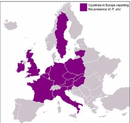
Phytophthora alni (Brasier and S.A. Kirk) involved in alder mortality was first detected in southern Britain in 1993 (Gibbs 1995). Since then, it has been reported from several European countries: Austria, Belgium, Czech Republic, France, Germany, Hungary, Ireland, Italy, Lithuania, the Netherlands, Poland, Slovakia, Slovenia, Sweden and Spain (Figure 3) (Gibbs et al. 1999; Brasier and Kirk 2001; Santini et al. 2001; Brasier 2003; Gibbs et al. 2003; Santini et al. 2003; Jung and Blaschke 2004; Ioos et al. 2005; Cerný and Strnadová 2010; Solla et al. 2010; Varela et al. 2010, 2012). In Europe, the pathogen has been reported to cause diseases on alders growing mainly along riverbanks, in orchard shelterbelts and forest plantations (Gibbs 1995; Gibbs et al. 1999, 2003; Jung and Blaschke 2004).
Figure 3. Distribution map of P. alni in Europe (the map has been published before 2010. So,
presence of P. alni in Spain has not been indicated).
Source:http://www.forestry.gov.uk/website/forestresearch.nsf/ByUnique/INFD737J2S
homothalism, colony growth patterns, optimum temperature for growth, high level of
zygotic abortion and poorly developed oogonia (Brasier et al. 1995, 1999). Later, it was hypothesized that the Phytophthora engaged in alder mortality evolved as a result of hybridization between heterothallic P. cambivora and another unknown taxon of Phytophthora closely related to homothallic P. fragariae Hickman (Brasier et al. 1999).
Further investigation by them demonstrated that the alder Phytophthora consisted of a variety of heteroploid hybrid species and later they divided it into a ‘standard’ type and several variants. Analysis of the internal transcribed spacer (ITS) region revealed that the isolates of ‘standard’ type displayed an unusual ITS polymorphism i.e., dimorphic sites within the ITS sequences while the variants were monomorphic for the same genome region or monomorphic at some sites and dimorphic at other sites (Brasier et al. 1999, 2004). Brasier et al. (2004) formally named alder Phytophthora as P. alni and based on morphological studies, cytological evidences and genetic data, and they divided P. alni into three subspecies: P. alni ssp. alni (Brasier and S.A. Kirk) corresponding to former ‘standard’ type, P. alni ssp. uniformis (Brasier and S.A. Kirk) corresponding to Swedish variant and P. alni ssp. multiformis (Brasier and S.A. Kirk) corresponding to the Dutch, German and UK variants. More recent genetic studies have hypothesized that Paa has occurred from a single or multiple hybridization event between Pau and Pam, although the origin and genetic diversity of these taxa are still under discussion (Ioos et al. 2006, 2010). All three sub-species of P. alni showed distinctive colony morphologies and different growth-temperature relations on culture
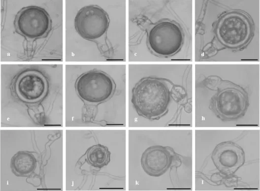
media. Sporangia produced by the sub-species were non-caducous and non-papillate and formed conspicuous basal plugs in empty sporangia. Sporangial shape varied from ovoid to ellipsoid with a rounded or occasionally tapered base (Figure 4a, 4b). P. alni ssp. alni produced ornamented oogonia and elongated two-celled antheridia whereas both P. alni ssp. uniformis and P. alni ssp. multiformis produced unique unusual
Figure 4. Reproductive structure (asexual and sexual) of P. alni. a, b. Non-papillate and
ovoid sporangia; c. ornamented oogonia with two-celled amphigynous antheridia; d.
ornamented oogonia with aborted oospore.
The genus ‘Alnus’
‘Alnus’ belongs to family Betulaceae and is characterized by their ability to colonize
A. glutinosa is the most widespread species occurring spontaneously in almost every
region of Spain, except in arid southeast part. This species grows in small groups along riverbanks, streams and in wet places. The distribution map of common alder in Spain is given in Figure 5.
Figure 5. Distribution map (highlighted in green) of A. glutinosa in Spain
Source: http://www.inia.es/gcontrec/pub/alnus_glutinosa_publi_1186051443234.pdf
A. glutinosa grows well in moist soils and can reach up to a height of 20-30 meters and exceptionally up to 37 metres with a normal diameter ranged 0.6-0.7 metres. The root system of the tree is shallow, strong and well branched, especially in wet and shallow soils. Young trees have a habit of grow upright with a main axial stem where as the older develop an arched crown with crooked branches. The trunk is straight, cylindrical and overall pretty clean. The bark is smooth, shiny and greenish-brown in the young while in the older trees it is dark grey in colour. The buds are thick, ovate or oblong, hairless, slimy, coated by two or three scales. The tree is characterized by its short-stalked green coloured rounded leaves becoming wedge-shaped at the base and with a slightly toothed margin. The flowers are monoecious. The catkins appear in summer and developed at the end of next winter. The catkins of both sexes appear together at the apex of the twigs of the same year, in clusters of 3-6 (Cela et al. 1998).
forestland or abandoned farmland and other problematic soil that do not support
vegetation easily. It has excellent ability to ameliorate soil due to a root symbiosis with actinomycetes (Frankia) which are able to fix atmospheric nitrogen. It has traditionally been used in furniture and veneer industry as well as for window and door production. Besides, its leaves and bark have medicinal properties.
The genus, Alnus also has high ecological value because of its conservation role (Claessens 2003; Thoirain et al. 2007). However, despite its numerous benefits, riparian zones, and in particular alder forests, have been disturbed by human activities over the past century (Kauffman et al. 1997; Naiman and Décamps 1997). In fact, virgin vegetation of these riparian zones was almost totally lost when stream flow was regulated by storage reservoir and canalizations in the middle of the 20th century (Schnitzler 1994; González and García 2007).
The disease
Decline of the riparian alder population has recently become an important issue in Europe, and particularly in Spain, because of the rapid spreading of P. alni. Establishment of plantations on former agricultural land and riverbanks to stabilize slopes with infested alders and use of water for irrigation from rivers contaminated by diseased alders contributing a lot to the spreading of alder Phytophthora to natural ecosystems (Jung and Blaschke 2004). Production of zoospores in the presence of water significantly contributes to the dispersal of Phytophthora via irrigation (Yamak et al. 2002; Hong et al. 2006). Importation of nursery stock for afforestation purposes and use of contaminated river water to irrigate nurseries may have resulted rapid increase of P. alni in Europe. Besides, out-planting with Phytophthora infected nursery stocks have contributed to the dissemination process of Phytophthora to natural ecosystems (Gibbs et al. 2003; Jung and Blashchke 2004). Falling of seeds, young shoot or leaves onto the contaminated water or soil and later disseminating far distances through water ways.In
Great Britain, spreading of alder Phytophthora has occurred through watercourses (Gibbs et al. 1999) whereas in Bavaria (Germany) P. alni has been introduced into many places either by planting infected nursery stock or by irrigation water (Jung and Blaschke 2004).
foliage (Figure 6c). Tarry spots (6d) and rusty exudates (6e) on the surface of the bark
or in its cracks at collar and lower stem with excessive fructification are the common characteristics. In case of severely affected trees, leaves often fall prematurely, leaving branches bare. Sometimes, the tarry spots can occur up to 2-3 meters from ground level (Figure 4f). Tarry spots indicate the infections and necroses persist in the inner bark and
cambium (Gibbs et al. 1999, 2003; Jung and Blaschke 2004).
Figure 6. Phytophthora decline of common alder. a. Tree stem with bleeding canker; b.
typical die-back of A. glutinosa; c. trees with small, sparse and yellowish leaves; d. tarry spots
at lower stems; e. rusty and dark coloured exudates coming out from the point of infection; f.
necroses moving towards upward direction.
a
a
bbcc
d
d
Management of alder disease
Alders play an important role in stabilizing banks of rivers and is considered as the key element of riparian ecosystems in Europe. Spreading of P. alni, may lead drastic changes in the riparian ecosystems and may cause serious economic losses at the same time. Attempts to reduce the impact of the disease for short term period by coppicing in
the natural riparian alder stands of UK have been found useful by Gibbs (2003) that showed low infection a couple of years after coppicing. However, coppicing was not recommended for plantations infected with P. alni by other authors (Jung and Blashchke 2006). Provenance trial could be an option to mitigate alder mortality. However, field and laboratory experiments conducted with alder seedlings collected from different provenances in Europe rejected the idea of resistance of alders against P. alni at provenance level (Gibbs 2003). For long term control, another possible strategy could be the genetic selection of a variety of clones in order to maintain healthy alder as it was possible for Eucalyptus marginata against P. cinnamomi Rands in Australia (Hüberli et al. 2003; Jung and Blaschke 2006).
There are currently no known methods to completely eradicate P. alni from an infested site. But, there is a need of good integrated management of nursery and irrigation system to stop dissemination of the pathogen. Molecular-based identification system should be initiated for rapid and effective detection. It is also important to ensure safer environment for A. glutinosa to grow in nurseries and in plantations. Finally, a common strategy and co-ordinated programme for limiting the transportation of infected plants
from country to country is needed in Europe. In order to mitigate spread of P. alni in nurseries and plantations including riparian ecosystems, the following measures could be adapted: (a) production of alder seedlings from disease-free areas, (b) avoidance of frequent seedling transportation with soils from nurseries to plantations, (c) examination of seedlings before planting to check if they have any disease symptoms and elimination
AIMS OF THE STUDY
The main objective of the study was to identify and characterize the pathogens involved in common alder mortality in Spain. Furthermore, studies were conducted to obtain information on the potential risks that may pose by the pathogens to A. glutinosa and its propagating materials.
More specifically, the aims of the individual study were:
1. To isolate and identify Phytophthora species involved in common alder (Alnus glutinosa) mortality in Spain (I & V)
2. To characterized the Phytophthora species by analysis their morphology, physiology and molecular traits (II)
3. To examine susceptibility of seeds and seedlings of A. glutinosa to Phytophthora species (III)
MATERIALS AND METHODS
A brief description of materials and methods is given below, but for detailed information, please refer to the original articles (Roman numerals).
Materials
Sampling sites
Common alder (Alnus glutinosa) growing stands with disease symptoms (I, II & V).
Phytophthora isolates
Cultured Spanish isolates of Phytophthora alni (I, II, III & IV). Cultures French isolates of P. alni (II & IV).
Cultured Spanish isolates of P. cinnamomi Rands, P. citrophthora (R.E. Sm. and
E.H. Sm.) Leonian, P. nicotianae Breda de Haan (= Phytophthora parasitica Dastur) and P. palmivora (E.J. Butl.) (III).
Cultured Spanish isolates of P. plurivora sp. nov. (IV).
Plant materials
Seeds of A. glutinosa were obtained from the National Centre for Forest
Breeding, Guadalajara, Spain (III).
Seedlings were obtained from Viveros Fuenteamarga S.L., Valladolid, Spain
(V).
Vegetative materials of A. glutinosa (leaves, branches and twigs) were collected
from the province of Salamanca, Spain (IV).
Methods
Sampling of diseased and healthy trees
Trees selected for sampling having symptoms of die-back, small, sparse and
yellowish leaves, tarry spots and rusty exudations at collar and lower stems (I, II, IV & V).
Bark samples were taken with the help of a hammer and chisel, placed in
Fully expanded fresh leaves were collected from mature healthy A. glutinosa
and kept in poly bags moistened with distilled water and transferred to cool boxes to avoid desiccation (IV).
Branches and twigs were collected from both disease-free and disease
affected areas, cut into small segments, wrapped in moist tissue during
transport and placed in cool boxes (IV).
Isolation method
Small pieces were cut from the active lesions of inner bark, dried on filter paper
and plated on selective media V8-PARPH agar. Isolations were made according to a method previously described (Jung and Blashchke 2004) (I, II & V).
Identification method
Morphological study
Colony morphologies were produced on V8 juice agar (V8A) (I, II & V), carrot juice agar (CA) (II), corn-meal agar (CMA) (II) and potato-dextrose agar (PDA) (II).
Sporangia and oogonia were produced according to protocol as described by
Jung et al. (1999) (I, II & V) Physiological study
Cardinal and radial growth rates response to temperatures (I, II, V), pHs (II) and osmotic potentials according to methodology described by Lira-Méndez and Mayek-Pérez (2006) (II) were examined on V8A.
Molecular analyses
Molecular identification protocol adapted from Vilgalys and Hester (1990) (II); White et al. (1990) (II & V); Gardes and Bruns (1993) (II); Vainio et al. (1998) (II); Cooke et al. (2000) (I & V); Jung and Burgess 2009 (V).
Identification up to subspecies was done following protocols as described by
Ioos et al. (2005, 2006) (II).
Inoculation
In case of zoospore inoculation (III & IV), zoospore suspensions were prepared following the method adapted from Denman et al. (2005).
Data recording
Seed germination and seedling mortality percentage were started to record 7
days after inoculation. Data on germination and mortality were taken at every 5 days interval until 42 days and 67 days respectively (III).
Leaves, branches and twigs were examined for lesions 6 days after inoculation
and lesion lengths were measured (IV) and seedlings were examined 3 months after inoculation for lesions (V).
Reisolation
To confirm the infections caused by the Phytophthora spp., small segments from
the inoculated plant materials were surface disinfected and placed directly on V8-PARPH agar in order to re-isolate the pathogens (IV & V).
Statistical analyses
Analysis of variance (ANOVA) and Tukey’s HSD post-hoc tests (α = 0.05) were
used to test the effects of pH and osmotic potential (II).
Repeated measures analysis of variance (ANOVA) were performed according to
Tukey multiple range test (III).
Three-way fixed factors effects ANOVAs were performed under non-normality
RESULTS AND DISCUSSION
A brief description of results and discussion is given below. But for details, please refer to the original articles (Roman numerals)
Phytophthora species involved in the mortality of Alnus glutinosa (I, II & V)
Phytophthora alni ssp. alni and itscharacteristic features (I & II)
P. alni was recovered from necrotic bark at collar regions and lower stems of diseased
A. glutinosa in both studies (I & II). In culture media, the isolates produced abundant terminal, spherical oogonia which were ornamented with two-celled amphigynous antheridia. Rarely comma-shaped smaller oogonia and very often oogonia with aborted oospores were also observed. Sporangia were non-caducous and nonpapillate, and shape
of the sporangia were ovoid to ellipsoid. The length: breadth ratios of sporangia ranged from 1.3 to 1.6. Sporangiophores were simple or sympodial with terminal sporangia proliferating internally, often nested, and extended internal proliferation and wide exit pores. Colony patterns developed on CA were usually appressed and irregular in growth with very limited aerial mycelium, whereas on V8A, colonies had slightly woolly morphology. Besides, fluffy and considerably woolly growth patterns were observed on CMA and PDA. rDNA sequences from internal transcribed spacer (ITS) were compared with the sequences available in the GenBank showed that the sequences of all isolates were nearly identical. On the basis of the best hits and comparison with GenBank another time, the pathogen was identified as P. alni ssp. alni (I & II). As a whole, morphological, physiological and genetical features of the pathogen described in this study corresponded to those of P. alni ssp. alni, previously known as the ‘standard’ type (Brasier et al. 2004).
Discriminating among the three subspecies of P. alni (II)
Analysis of internal transcribed spacer (ITS) region revealed that isolates of P. alni ssp. alni (I & II) showed an unusual ITS polymorphism i.e., dimorphic sites within the ITS sequences. This type of dimorphism has been described as typical of P. alni ssp. alni, whereas P. alni ssp. multiformis and P. alni ssp. uniformis tend to be monomorphic (Ioos et al. 2006). Ambiguities that have been related to the recent hybrid condition of P. alni ssp. alni, believed to be the result of a unique or multiple hybridization events of
Isolates of P. alni ssp. alni from (II) showed a unique and common pattern of amplifications with a series of species specific primers previously described (Ioos et al. 2005, 2006). Amplification with PA primers indicated that the isolates belonged to P. alni complex, but failed to differentiate between P. alni ssp. alni, P. alni ssp. uniformis or P. alni ssp. multiformis; PAM amplification excluded P. alni ssp. uniformis; and TRP
and RAS primers amplifications excluded P. alni ssp. multiformis and P. alni ssp. uniformis, respectively.
P. plurivoraand its characteristic features (V)
During conducting surveys (V) for alder Phytophthora, another homothallic species of Phytophthora was isolated from active fresh lesions at collar of declining A. glutinosa. On culture media, isolates produced smooth-walled spherical oogonia having paragynous antheridia with both plerotic and aplerotic oospores. Sporagia were non-caducous, semipapillate, mainly ovoid and obpyriform, obovoid to limoniform but sometimes distorted or bilobed. Colonies developed on V8 juice agar (V8A) displayed radiate and slightly chrysanthemum-like growth pattern with limited aerial mycelium at the centre. These morphological and physiological features of the pathogen (V) corresponded to the characteristics features of P. plurivora as described previously by (Jung and burgess 2009). For molecular identification, the internal transcribed spacer (ITS) region of the rDNA was amplified using the protocol as described by (White et al. 1990; Cooke et al. 2000). Sequences obtained were compared with the reference
sequences which appeared in Jung and Burgess (2009).
Impact of pH, osmotic potential and temperature on mycelial growth of P. alni ssp.
alni (II)
Mycelial growth in response to pH
Mycelial growth of P. alni ssp. alni isolates (II) was observed on a wide range of pH. Although, the isolates grew at higher pH, they demonstrated a tendency of low and slow growth with increasing pH value which indicated that they are less tolerant to higher
pH. Optimum growth of the isolates of P. alni ssp. alni (II) was recorded at pH 7, which in turn corresponded to the best pH for growth and sporangia formation of P. alni as previously determined (Schumacher et al. 2006; Kong et al. 2012). Phytophthora species are water molds in terms of their zoospore activity. Irrigation reservoirs in
impacts on the survival of many Phytophthora (Kong et al. 2009). However, studies are
necessary to know more about the survival of the pathogens in different aquatic environment under pH stress. In soil environment, Phytophthora species are considered to be tolerant to a wide range of pH and increasing pH generally favours growth (Weste 1983; Andrivon 1994). In this view, our findings are very important as P. alni could
remain active over a wide pH range, being well adapted to both aquatic and soil environment and pose threat to new areas through dispersal.
Mycelial growth in response to osmotic potential
Most favourable growth of the isolates of P. alni ssp. alni (II) was recorded at higher osmotic potential and declined with decreasing osmotic potential. Studies carried out with other Phytophthora species also revealed a similar trend for mycelial growth concerning higher and lower osmotic potentials (Sommers et al. 1970; Turco et al. 2005). The studies showed the importance of a minimum amount of water in the environment for development of Phytophthora. Osmotic potential has been identified as an important factor in the growth and ecology of pathogens. Free water in soils or in the environment is essential to all stages of their life cycle (Davis et al. 2000). However, water needs may differ among the pathogens throughout their reproduction time and tolerance to dry conditions. For example, P. quercina sp. nov. and P. citricola Sawada recovered from dry upland sites in Turkey where they were able to survive over extended dry periods in the absence of suitable host (Balci and Halmschlager 2003).
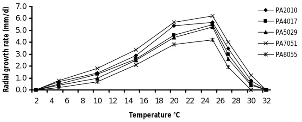
Mycelial growth in response to temperature
Isolates of P. alni ssp. alni (I & II) showed optimum mycelial growth at between 20 to 25°C. This range of temperature for optimum growth is in accordance with previous studies (Brasier et al. 1995, 2004) and is the most ideal temperature range at which many Phytophthora were reported to be very active and virulent (Harris and Tobutt 1986; Brasier et al. 1995; Hardham 2001). Susceptibility of plant species to invasion by Phytophthora is strongly temperature dependent (Matheron and Mateika 1993).
environmental factors such as site temperature along with several other risk factors have
been accounted which influence the occurrence of disease by P. alni (Jung and Blaschke 2004; Schumacher et al. 2006; Elegbede et al. 2010). Isolates of P. alni ssp. alni recovered during the study (II) did not show any grow at 2 and 32°C. Alder Phytophthora has been reported to have less probability to survive in cold winters or
under extreme frosts and poor oospores viability (Schumacher et al. 2006; Cerny and Strnadova 2012). On the other hand, an increase in the temperature of river water again increases the probability of disease incidence (Thoirain et al. 2007). However, Chandelier et al. (2006) reported that the sporangia production of P. alni was affected by microbial communities present in the river water together with high temperature. Being a thermophilous pathogen with relatively high optimum growth temperatures (22-25°C), temperature might influence the survival of the pathogen and disease development.
Inoculum effects on seed germination and seedling mortality (III)
Seed germination (III)
Isolates of P. alni ssp. alni (I & III), P. cinnamomi, P. citrophthora, P. nicotianae and P. palmivora (III) reduced significantly seed germination when zoospore suspension was used to inoculate. Forty two days after inoculation, all the isolates of the Phytophthora species tested hampered significantly germination regardless of the method used for inoculation. When zoospore suspension was applied at the centre of the
plate (method CE), no differences were found among the tested isolates, but when suspension was applied in each seed (method IS), P. cinnamomi significantly reduced the germination percentage than that of the others. On average, seed germination percentage when inoculation was made for each seed, was 26.04%; and when zoospore suspension was applied at the centre, the percentage was 36.97%. In controls, it was
Seedling mortality (III)
Seedling mortality was significantly influenced by the isolates of P. alni ssp. alni (I and III), P. cinnamomi, P. citrophthora, P. nicotianae and P. palmivora (III), inoculation methods and time period of the experiment. Inoculated seedlings started to die after 7 days of inoculation with a progressive manner reaching the maximum number after
15-20 days. Such maximum mortality was achieved by both isolates of P. alni and P. citrophthora. Between inoculation methods, differences were found mainly in the first 2-5 measurements depending of the isolate. In those first records, inoculation with the mycelial agar plugs at each seedling (method IT) caused greater seedling mortality than the inoculation in the centre of the plate (method CE). At the end of the experiment all the isolates caused a seedling mortality rate higher than 90%, regardless of the inoculation method. But, P. cinnamomi produced a mortality which was 46.9% when inoculation method (method CE) was applied, whereas it was 78.1% for inoculation method (method IT). Controls did not result any seedling mortality. Ability of P. alni and P. citrophthora to cause seedling mortality has also been revealed from inoculation studies made by Santini et al. (2003, 2006). No information is available so far on the pathogenicity of P. cinnamomi, P. nicotianae and P. palmivora on seedlings of Alnus but, in our present inoculation test (III), these pathogen species have revealed their capacity to cause seedlings mortality. So, it can be assumed that container grown seedlings of Alnus are vulnerable to the Phytophthora species as they may exist with other contaminated plants in the same nurseries. Furthermore, as most infected
seedlings may not attain emergence and, therefore, this kind of damage might otherwise be attributed to other causes. These results also suggest that seedlings of common alder are at risk to be infected by them and showed relative non-host-specificity of this genus. However, this experiment does not provide reliable findings on host and pathogen interaction at the natural level.
Influence of damage, temperature and sampling location on the pathogenicity of P.
alni (IV)
Effect of damage (non-wound vs. wound) on lesion development
incubation on inoculated wounded leaves. This study (IV) has proved that wounding was an influential factor to cause infection by the subspecies of P. alni and they might have entered into the tissues through wounds. Such wounding effect is also consistence with other previous findings (Erwin and Ribeiro 1996; Thomidis 2003; Kaminski and Wagner 2008), who demonstrated that growth of Phytophthora was associated with
wounds and wounded plant species were more vulnerable to infection than the non-wounded ones. At the end of experiment, when the isolates of three subspecies of P. alni were ranked according to their virulence, two isolates of subspecies P. alni ssp. alni were the most virulent based on length of lesion, although no significant differences in lengths were noted for remaining isolates of P. alni ssp. alni and the other two subspecies. This means, all three subspecies were equally pathogenic on the wounded leaves of A. glutinosa. Pathogenic ability of Phytophthora species on leaves of their woody hosts have been well documented where artificial wounding was necessary in most incidences to cause infections and necroses. In vitro leaf inoculation studies on leaves of broad-leaved and coniferous trees using zoospores of P. ramorum Werres, DeCook & Man in’t Veld by Denman et al. (2005) showed that necroses and disease incidence increased significantly when wound inoculations were done. Ability of P. alni to cause lesions on leaves following artificial wounding and failure to produce any lesions on non-wounded leaves suggest that the pathogen would unlikely to be a foliar invader for alders.
Effect of temperature and sampling location on lesion and girdling formation
Temperature has been reported to influence on growth, reproduction and pathogenicity of Phytophthora species (Sujkowski 1987; Sing and Chauhan 1988; Matheron and Matejka 1992). In the current tests (IV), temperature has also been considered as a key factor that significantly influenced the pathogenicity of the three subspecies of P. alni
logs of A. glutinosa. Largest lesions occurred on excised twigs and branches at 25ºC
followed by 20ºC, in case of all isolates of three subspecies. Temperatures range (20ºC-25ºC), was considered as the most suitable range at which P. alni, P. cactorum var. applanata Chester and P. cryptogea Pethybr. and Laff. were reported to be very active and pathogenic (Harris and Tobutt 1986; Hardham 2001; Brasier et al. 2004). A similar
CONCLUSIONS
1.
Phytophthora alni, causal agent of alder (Alnus spp.) mortality in Europe, is alsoinvolved in the mortality and decline process of common alder (Alnus glutinosa)
in the north and central-western part of Spain. Although P. alni has been reported as the primary agent of riparian common alder mortality in Spain, P. plurivora has also been isolated from declining A. glutinosa. Further investigations are necessary in order to examine the severity and damage caused by this Phytophthora species on A. glutinosa. In addition, further surveys are crucial in order to locate the presence of P. plurivora in different alder growing areas of Spain.
2. P. alni can easily be distinguished from other homothallic Phytophthora species by its unique morphological, physiological and genetic traits. Features like two-celled ornamented oogonia with amphigynous antheridia, nonpapillate and non-caducous sporangia, growth patterns on culture media, optimum and maximum temperatures for growth have given the distinctiveness to alder Phytophthora. Considering its morphological, physiological features, and ITS sequences, the isolates recovered from common alder clearly belong to P. alni spp. alni which is considered to be the most aggressive one among the three subspecies of P. alni. Additional surveys are essential in order to trace out the existence of remaining two subspecies of P. alni (P. alni ssp. multiformis and P. alni ssp. uniformis) as they might convey equal threat to the existence of riparian alder populations along the rivers and other waterways in Spain.
natural ecosystems. Since plantation establishment with infected alder seedlings
is one of the primary pathways by which P. alni spread, so careful management and selection of forest reproductive material is important.
4.
Pathogenicity tests performed on the detached plant materials (leaves, twigs andbranches) of A. glutinosa concerning damage (wound vs. non-wound),
temperature (15, 20, 25 and 30°C) and sampling location (disease-free areas vs. diseased areas) has confirmed variation in the virulence among the isolates of three subspecies of P. alni. Wounds and temperatures have appeared as the most important influential factors in the process of lesion development. Results have
CONCLUSIONES
1.
Phytophthora alni, causante de la mortalidad de alisos (Alnus spp.) en Europa,está también implicado en la mortalidad y decaimiento del aliso común (Alnus
glutinosa) en el norte y centro occidental de España. Aunque P. alni ha sido identificado como el agente principal de la mortalidad de los alisos en España, P. plurivora ha sido también aislado en alisos enfermos. Más investigación es necesaria para conocer la distribución de P. plurivora en España y examinar su virulencia y daños causados en A. glutinosa.
2. P. alni puede ser fácilmente distinguible de otras especies homotálicas del género Phytophthora por sus características morfológicas, fisiológicas y genéticas. Sus oogonios ornamentados con anteridios bicelulares y posicionados de forma anfigina, sus esporangios no caducos y no papilados, junto con la morfología de sus colonias en medio de cultivo, temperaturas óptimas y máximas de crecimiento diferencian a P. alni. A tenor de las características morfológicas, fisiológicas y de las secuencias de la región ITS, los aislamientos obtenidos corresponden claramente a P. alni spp. alni, la cual es considerada la más agresiva de las tres subespecies de P. alni. Otros muestreos de campo serían
fundamentales para determinar la existencia de las otras dos subespecies de P. alni (P. alni ssp. multiformis y P. alni ssp. uniformis) en otros ríos y cursos fluviales de España.
aliso infectadas es una de las vías principales para la dispersión de P. alni, un
correcto manejo de los viveros y una selección adecuada del material forestal de reproducción a utilizar es fundamental.
4. Los ensayos de patogenicidad llevados a cabo en material vegetal (hojas,
REFERENCES
Adl SM, Simpson AGB, Farmer MA, Andersen RA, Anderson OR, Barta JR, Bowser SS, Brugerolle G, Fensome RA, Fredericq S, James TY, Karpov S, Kugrens P, Krug J, Lane CE, Lewis LA, Lodge J, Lynn DH, Mann DG, McCourt RM,
Mendoza L, Moestrup O, Mozley-Standridge SE, Nerad TA, Shearer CA, Smirnov AV, Spiegel FW, Taylor MFJR (2005) The new higher level classification of eukaryotes with emphasis on the taxonomy of protists. Journal of Eukaryotic Microbiology 52: 399–451.
Aguayo J, Adams GC, Halkett F, Catal M, Husson C, Nagy ZÁ, Hansen EM, Marçais B, Frey P (2013) Strong genetic differentiation between North American and European populations of Phytophthora alni subsp. uniformis. Phytopathology 103 (2): 190–199.
Andrivon D (1994) Fate of Phytophthora infestans in a suppressive soil in relation to pH. Soil Biology and Biochemistry 26:953–956.
Balci Y, Halmschlager E (2003) Phytophthora species in oak ecosystems in Turkey and their association with declining oak tress. Plant Pathology 52: 694–702.
Brasier CM, Hansen EM (1992) Evolutionary biology of Phytophthora, Part II: Phylogeny, speciation, and population structure. Pages 173–200, in: Annual Review of Phytopathology, vol. 30. APS, St. Paul, MN.
Brasier CM, Rose J, Giggs JN (1995) An Unusual Phythophothora associated with
widespread alder mortality in Britain. PlantPathology 44: 999–207.
Brasier CM, Cooke DEL, Duncan JM (1999) Origin of a new Phytophthora pathogen through interspecific hybridization. Proceedings of the National Academy of Sciences, USA 96: 5878–5883.
Brasier CM, Kirk SA (2001) Comparative aggressiveness of standard and variant hybrid
alder Phytophthora, Phytophthora cambivora and other Phytophthora species on the bark of Alnus, Quercus and other woody hosts. Plant Pathology 50: 218– 229.
Brasier CM, Kirk SA, Delcan J, Cooke D, Jung T, Man In’T Veld WA (2004)
Phytophthora alni sp. nov. and its variants: designation of emerging heteroploid hybrid pathogens spreading on Alnus trees. Mycological research 108: 1172– 1184.
Brasier C (2007) Phytophthora Biodiversity: How Many Phytophthora Species Are
There? In: Goheen EM, Frankel SJ. (eds.), Phytophthoras in Forests and Natural Ecosystems. Proceedings of the Fourth Meeting of the International Union of Forest Research Organizations (IUFRO) Working Party S07.02.09. August 26–31, 2007, Monterey, California.
Cela PG, Gamarra RG, Viñas JIG (1998) Árboles y arbustos de la Península Ibérica e Islas Baleares. Ediciones Jaguar S.A., Madrid, 768pp.
Cerný K, Strnadová V (2010) Phytophthora alder decline: disease symptoms, causal agent and its distribution in the Czech Republic. Plant Protection Science 46 (1): 12–18.
Cerný K, Strnadová V (2012) Winter survival of Phytophthora alni subsp. alni in aerial tissues of black alder. Journal of Forest Science 58: 328–336.
Chandelier A, Abras S, Laurent F, Debruxelles N, Cavelier M (2006) Effect of temperature and bacteria on sporulation of Phytophthora alni in river water. Communications in agricultural and applied biological sciences 71: 873–880. Claessens H (2003) The alder populations of Europe. In: Phytophthora disease of Alder
in Europe, Gibbs J, Van Dijk C, Webber J. Edinburgh, p. 82.
Cooke DEL, Drenth A, Duncan JM, Wagels G, Brasier CM (2000) A molecular phylogeny of Phytophthora and related oomycetes. Fungal Genetics and Biology 30: 17–32.
Davis DJ, Burlak C, Money NP (2000) Osmotic pressure of fungal compatible osmolytes. Mycological Research 104 (7): 800–804.
Deacon JW, Donaldson SP (1993) Molecular recognition in the homing responses of zoosporic fungi, with special reference to Pythium and Phytophthora. Mycological Research 97: 1153–1171.
Denman S, Kirk SA, Brasier CM, Webber JF (2005) In vitro leaf inoculation studies as an indication of tree foliage susceptibility to Phytophthora ramorum in the UK. Plant Pathology 54: 512–521.
Duniway JM (1976) Movement of zoospores of Phytophthora cryptogea in soils of
various textures and matric potentials. Phytopathology 66: 877–882.
Elegbede CF, Pierrat J-C, Aguayo J, Husson C, Halkett F, Marçais B (2010) A statistical model to detect asymptomatic infectious individuals with an application in the Phytophthora alni induced Alder decline. Phytopathology 100:
1262–1269.
Erwin DC, Ribeiro OK (1996) Phytophthora Disease Worldwide. APS Press, St Paul, Minnesota, USA, 562 pp.
Gardes M, Bruns TD (1993) ITS primers with enhanced specificity for basidiomycetes: application to the identification of mycorrhiza and rusts. Molecular Ecology 2 (2): 113–118.
Gibbs JN (1995) Phytophthora root disease of alder in Britain. Bulletin OEPP/EPPO Bulletin 25, 661–664.
Gibbs JN, Lipscombe MA, Peace AJ (1999) The impact of Phytophthora disease on riparian populations of common alder (Alnus glutinosa) in southern Britain. European Journal of Forest Pathology 29: 39–50.
Gibbs JN (2003) Phytophthora disease of alder: management and control. In: Gibbs JN, Van Dijk C, Webber JF. (eds.), Phytophthora Disease of Alder in Europe. Edinburgh, UK: Forestry Commission Bulletin No. 126, 73–78.
Gibbs JN, Van Dijk C, Webber JF (2003) Phytophthora Disease of Alder in Europe. Edinburgh, UK: ForestryCommission Bulletin No. 126. 82p.
González M, García D (2007) Restauración de ríos. Guía metodológica para la elaboración de proyectos. Ed. Secretaria General Técnica. Centro de publicaciones. Ministerio de Medio Ambiente. Madrid. ISBN: 978-84-8320-413-9. 318 pp
Haque MM, Martínez-Álvarez P, Lomba JM, Martín-García J, Diez JJ (2014) First
report of Phytophthora plurivora causing collar rot on common alder in Spain. Plant Disease 98(3): 425.
Hardham AR (2001) The cell biology behind Phytophthora pathogenicity. Australasian Plant Pathology 30: 91-98.
Ho HH, Zentmeyer GA (1977) Infection of avocado and other species of Persea by
Phytophthora cinnamomi. Phytopathology 67: 1085–1089.
Hong CX, Richardson PA, Kong P (2006) Phytophthora tropicalis isolated from diseased leaves of Pieris japonica and Rhododendron catawbiense and found in irrigation water and soil in Virginia. Plant Disease 90: 525.
Horner IJ, Wilcox WF (1996) Temporal changes in activity and dormant spore populations of Phytophthora cactorum in New York apple orchard soils. Phytopathology 86: 1133–1139.
Hüberli D, Tommerup IC, Colquhoun I, Hardy GE St J (2003) Measuring resistance in Jarrah, Eucalyptus marginata, to Phytophthora cinnamomi: what factors change disease expression? In: McComb JA, Hardy GE St J, Tommerup I. (eds.), Phytophthora in Forests and Natural Ecosystems. Proceedings of the Second International Meeting of IUFRO Working Party 7·02·09. Albany, Western Australia. Perth, Australia: Murdoch University, 259.
Ioos R, Husson C, Andrieux A, Frey P (2005) SCAR-based PCR primers to detect the hybrid pathogen Phytophthora alni and its subspecies causing alder disease in Europe. European Journal of Plant Pathology 112: 323–335.
Ioos R, Andrieux A, Marçais B, Frey P (2006) Genetic characterization of the natural hybrid species Phytophthora alni as inferred from nuclear and mitochondrial DNA analyses. Fungal Genetics and Biology 43: 511–529.
Ioos R, Barrès B, Andrieux A, Frey P (2007) Characterization of microsatellite markers
in the interspecific hybrid Phytophthora alni ssp. alni, and cross-amplification with related taxa. Molecular Ecology Notes 7: 133–137.
Ioos R, Fabre B, Saurat C, Fourrier C, Frey P, Marçais B (2010) Development, comparison, and validation of real-time and conventional PCR tools for the detection of the fungal pathogens causing brown spot and red band needle
blights of pine. Phytopathology 100: 105–114.
Jung T, Blaschke H (1996) Phytophthora root rot in declining forest trees. Phyton 36: 95–102.
Jung T, Blaschke M (2004) Phytophthora root and collar rot of alders in Bavaria:
Distribution, modes of spread and possible management strategies. Plant pathology 53: 1497–208.
Jung T, Blaschke M (2006) Phytophthora dieback of alders in Bavaria: distribution, pathways, and management strategies. In: Brasier CM, Jung T, Obwald W.
(eds.), Progress in research on Phyophthora dieases of forest trees. Proceedings of the third International Union of Forest Research Organizations, Working party 7.02.09. Farnham Surrey, United Kingdom: Forest Research: 61–66. Jung T, Nechwatal J (2008) Phytophthora gallica sp. nov., a new species from
rhizosphere soil of declining oak and reed stands in France and Germany. Mycological Research 112: 1195–1205.
Jung T, Burgess TI (2009) Re-evaluation of Phytophthora citricola isolates from multiple woody hosts in Europe and North America reveals a new species, Phytophthora plurivora sp. nov. Persoonia 22, 95–110.
Kaminski K, Wagner S (2008) In vitro Inoculation Studies for Estimating the Susceptibility of Ornamental Plants to Phytophthora ramorum. Journal of Phytopathology 156: 480–486.
Kauffman JB, Beschta RL, Otting N, Lytjen D (1997) An ecological perspective of riparian and stream restoration in the Western United States. Fisheries 22(5): 12–24.
Kong P, Moorman GW, Lea-cox JD, Ross DS, Richardson PA, Hong C (2009)
Zoosporic tolerance to pH stress and its implications for Phytophthora species in aquatic ecosystems. Applied and Environmental Microbiology 75 (13): 4307– 4314.
Kong P, Lea-cox JD, Moorman GW, Hong C (2012) Survival of Phytophthora alni, Phytophthora kernoviae, and Phytophthora ramorum in a simulated aquatic
environment at different levels of pH. FEMS Microbiology Letters 332 (1): 54– 60.
Lira-Méndez K, Mayek-Pérez N (2006) Potencial osmótico variable en el crecimiento in vitro y la patogenicidad en frijol (Phaseolus vulgaris L.) de Fusarium spp. Revista Mexicana de Fitopatologia, 24 (2): 88–97.
Matheron ME, Matejka JC (1992) Effects of temperature on sporulation and growth of
Phytophthora citrophthora and P. parasitica and development of foot and root rot on citrus. Plant Disease 76: 1103–1109.
Naiman RJ, Décamps H (1997) The ecology of interfaces: Riparian zones. Annual Review of Ecology, Evolution and Systematic 28: 621–658.
Oszako T (2010) Contribution of Phytophthora spp. in the phenomenon of alder decline in Poland. Phytopathologia, 57: 53–62.
Santini A, Barzanti GP, Capretti P (2001) A new Phytophthora root disease of alder in Italy. Plant Disease 5: 560.
Santini A, Barzanti GP, Capretti P (2003) Susceptibility of some Mesophilic hardwoods to alder Phytophthora. Journal of Phytophthora 151: 406–410.
Santini A, Biancalani F, Barzanti GP, Capretti P (2006) Pathogenicity of four Phytophthora species on wild Cherry and Italian alder seedlings. Journal of Phytopathology 154: 163–167.
Schnitzler A (1994) Conservation of biodiversity in alluvial hardwood forests of the temperate zone. The example of the Rhine valley. Forest Ecology and Management 68: 385–398.
Schumacher J, Leonhard S, Grundmann BM, Roloff A (2006) New alder disease in Spreewald biosphere reserve: causes and incidental factors of an epidemic. Nachrichtenbl. Deut. Pflanzenschutzd 58 (6): 141–147.
Sing UP, Chauhan VB (1988) Effect of temperature on germination of zoospores of
Phytophthora drechsleri f.sp. cajani. Indian Phytopathology 41: 80–85.
Solla A, Pérez-Sierra A, Corcobado T, Haque MM, Diez JJ, Jung T (2010) Phytophthora alni on Alnus glutinosa reported for the first time in Spain. Plant Pathology 59: 798.
Sommers LE, Harris RF, Dalton FN, Gardner WR (1970) Water potential relations of
three root-infecting Phytophthora species. Phytopathology 60: 932–934.
Sujkowski LS (1987) Seasonal variation in sporulation of Phytophthora infestans. Journal of Phytopapology 117: 357–361.
Thoirain B, Husson C, Marçais B (2007) Risk factors for the Phytophthora-induced decline of Alder in Northeastern France. Phytopathology 97: 99–105.
Thomson SV (1972) Occurrence and Biology of Phytophthora parasitica and other
plant pathogenic fungi in irrigation water. PhD. Thesis. University of Arizona, Tucson. 120p.
Turco E, Barzanti GP, Capretti P, Ragazzi A (2005) Effect of polyethylene glycol and composition of basal medium on the mycelial growth of Phytophthora spp.
Journal of Plant Diseases and Protection 112 (5): 426–436.
Vainio EJ, Korhonen K, Hantula J (1998) Genetic variation in Phlebiopsis gigantea as detected with random amplified microsatellite (RAMS) markers. Mycological Research 102: 187–192.
Varela CP, Martínez CR, Casal OA, Vázquez, JPM, Yebra AA (2012) First Report of Phytophthora alni subsp. uniformis on Black Alder in Spain. Plant Disease 96 (4): 589.
Varela CP, Martinez CR, Vázquez JPM, Casal OA (2010) First Report of Phytophthora rot on Alders Caused by Phytophthora alni subsp. alni in Spain. Plant Disease 94 (2): 273.
Vilgalys R, Hester M (1990) Rapid genetic identification and mapping of enzymatically amplified ribosomal DNA from several Cryptococcus species. Journal of Bacteriology 172: 4238–4246.
Webber J, Gibbs J, Hendry S (2004) Phytophthora disease of alder. Forestry Commission, Edinburgh, UK.
Weste G (1983) Population dynamics and survival of Phytophthora, p. 237–258. In
Erwin DC, Bartnicki-Garcia, S and Tsao PH. (eds.), Phytophthora: Its Biology, Taxonomy, Ecology and Pathology. APS Press, St. Paul, MN.
White TJ, Bruns T, Lee S, Taylor J (1990) Amplification and direct sequencing of fungal ribosomal RNA genes for phylogenetics. In: Innis MA, Gelfand DH, Sninsky JJ, White TJ (eds), PCR protocols: a guide to methods and
applications: 315–322. Academic Press, San Diego, California, USA.
ARTICLE I
Phytophthora alni ON Alnus glutinosa REPORTED FOR THE FIRST TIME
IN SPAIN
Alejandro Sollaa , Ana Pérez-Sierrab , Tamara Corcobadoa , Mohammed
Masum Ul Haquec , Julio Javier Diezc and Thomas Jungde*.
aIngeniería Técnica Forestal, Universidad de Extremadura, Avenida
Virgen del Puerto 2, 10600 Plasencia
bInstituto Agroforestal Mediterráneo, Universidad Politécnica de
Valencia, Camino de Vera s⁄n, 46022 Valencia
cProducción Vegetal y Recursos Forestales, Universidad de Valladolid,
Avenida de Madrid 44, 34071 Palencia, Spain
dPhytophthora Research and Consultancy, Thomastrasse 75, 83098
Brannenburg, Germany and eCentre for Phytophthora Science and
Management, School of Biological Sciences and Biotechnology, Murdoch University, 90 South Street, Murdoch, WA 6150, Australia.
*Corresponding E-mail: [email protected]
ABSTRACT
Surveys were carried out for alder Phytophthora along the river Miño, Galicia, Spain. Bark samples including cambium were taken from the active fresh lesions at collar of diseased Alnus glutinosa and isolates were obtained by placing small segments on a
selective agar medium. The isolates were homothallic mainly with two-celled amphigynous antheridia, occasionally comma-shaped oogonia having ornamentation. In soil-extract they produced nonpapillate, ellipsoid to ovoid sporangia. Colony developed on V8 agar, had slightly woolly morphology and showed radial growth. DNA of the ITS region was amplified and sequences obtained were compared with the GenBank showed 99% identity with Phytophthora alni ssp. alni. This is the first report of P. alni in Spain.
RESUMEN
Muestreos de campo para la detección de Phytophthora alni fueron llevados a cabo en el curso del río Miño (Galicia, España). Muestras de corteza con cambium fueron tomadas del cuello de la raíz y las partes bajas del tronco de alisos enfermos con daños
frescos. Los aislamientos fueron obtenidos tras cultivar pequeños fragmentos de dicho material vegetal en un medio de cultivo selectivo. Los aislamientos fueron homotálicos, generalmente con anteridios bicelulares posicionados de forma anfigina y oogonios ornamentados. Produciendo en los extractos de suelo esporangios no papilados, con formas desde elipsoides a ovoideas. Las colonias desarrolladas en el medio de cultivo V8 agar mostraron un aspecto ligeramente lanoso con un crecimiento radial. El ADN de la región ITS fue amplificada y sus secuencias confrontadas con las depositadas previamente en el GenBank obteniéndose una homología del 99% con las identificadas como P. alni ssp. alni. Este es la primera cita de P. alni en España.