Breast cancer epidemiology: mammographic screening and molecular subtypes
148
0
0
Texto completo
(2)
(3) Doctoral Thesis. Breast cancer epidemiology: Mammograhpic screening and molecular subtypes. Montserrat Puig i Vives 2014. PhD Programme in Experimental Sciences and Sustainability. Directed by: Dr. Marc Saez Zafra and Dr. Rafael Marcos Gragera Thesis delivered to obtain the doctoral degree by the Universitat de Girona.
(4)
(5) El Dr. Marc Saez Zafra, de la Universitat de Girona i membre del Grup de Recerca en Estadística, Econometria i Salut (GRECS) i el Dr. Rafael Marcos Gragera, de la Universitat de Girona i epidemiòleg de la Unitat d‟Epidemiologia i Registre de Càncer de Girona (UERCG) de l‟Institut Català d‟Oncologia (ICO),. CERTIFICA:. Que aquest treball, titulat “Breast cancer epidemiology: Mammographic screening and molecular subtypes”, presentat per Montserrat Puig i Vives per optar al títol de Doctora per la Universitat de Girona, ha estat realitzat sota la seva direcció.. I, perquè així consti i tingui els efectes oportuns, signem aquest document.. Dr. Marc Saez Zafra. Dr. Rafael Marcos Gragera.
(6)
(7) Al pare, de qui tant he après i a qui dedico aquesta tesi.
(8)
(9) Acknowledgments.
(10)
(11) Acknowledgments. Aquesta tesi ha estat possible gràcies al suport professional i personal que he rebut de moltes persones. Voldria aprofitar aquest espai per dir-los moltes gràcies a totes elles. Em resulta difícil mencionar-les a totes, però si que voldria fer menció especial a algunes d‟elles: En primer lloc a en Rafa, per donar-me la oportunitat de realitzar la tesi doctoral al Registre de Càncer de Girona. Gràcies a ell he disposat de les eines necessàries per anar construint el que finalment ha estat la meva tesi i ha despertat el meu interès pel món de l‟epidemiologia i la prevenció del càncer. Moltes gràcies! També a la resta de l‟equip del registre, a l’Àngel, la Loreto, la Maria i la Joana. Per la paciència en codificar, entrar dades, validar-les, recolzar-me quan ho he necessitat i compartir bons moments (carnaval ja és una tradició al registre!). Joana, en tu he conegut més que una companya de feina, una molt bona amiga. Hem compartit experiències intenses, de doloroses però també de felicitat i comprensió. Mil gràcies pel teu suport! No voldria deixar de mencionar tot el personal que ha passat aquest temps pel registre: la Marian, la Patri, la Irene, la Carla, la Carme, l‟Anna... La feina de tots plegats permet tirar endavant projectes com aquest. Sempre dic el mateix, sense unes dades de qualitat no hi ha investigació, i sense investigació no hi ha raó de pes que justifiqui l‟enorme esforç que suposa l‟obtenció de les dades. No em voldria pas oblidar de la Gemma, la meva gran companya de viatge d‟aquests anys! Què puc dir de tu que no ens hàgim dit fins ara? Hem fet la tesi plegades, ho hem compartit tot, discussions d‟articles i anàlisis, nervis de congressos, frustracions en veure que hem de tornar a enviar un article, paciència, alegria, males notícies, bones notícies... Gràcies per tots aquests moments, per estar sempre a punt per donar-me un cop de mà i també per estar disposada a anar a fer un cafè sempre que l‟he necessitat. Sort dels cafès! (i no, no hi ha “poyata”...) A en Marc Sáez del Grup de Recerca en Estadística, Econometria i Salut per obrir-me les portes a la Universitat de Girona. I a la Gemma Renart, per l‟ajuda en temes d‟estadística i en la identificació dels càncers d‟interval. A Marina Pollán por transmitirme su experiencia en epidemiología del cáncer de mama y echarme una mano con el primer proyecto de la tesis, juntamente con Montse Rué. Aprendí mucho con vosotras. Montse, moltes gràcies pel suport estadístic. A Maria José Sánchez, Julia Sánchez-Cantalejo y el resto del Registro de Cáncer de Granada por las lecciones de estadística, el empeño en llevar adelante el proyecto de patrones asistencial y enseñarme donde tomar las mejores tapas en Granada! También a los registros de España que colaboraron en este estudio, por su paciencia en la recogida y validación de los datos. The stage in Oslo has been one of the best professional and personal experience during my PhD. In office, in meetings, eating rakfisk, at the cinema, and walking in wonderful forests. I am deeply grateful to Solveig Hofvind for providing me with the opportunity to work in her team and for her guidance in the last article and in this thesis. I would also like to express my III.
(12) Acknowledgments. gratitude to all members of the Mammografiprogrammet. Takk! Marta, gràcies pel suport estadístic, per ensenyar-me com conviure amb la fred i la foscor d‟Oslo i per rebre‟m amb els braços oberts a aquesta ciutat. Durant aquest camí he estat molt ben acompanyada per amics que m‟han fet desconnectar, m‟escolten, em donen suport i em fan sentir bé. Els de tota la vida, els de la carrera, els que vaig conèixer a Barcelona, els que vaig coneixent a Girona, amb els que xerrem, ballem, caminem... Moltes gràcies a tots ells! Finalment i el més important, els de casa, gràcies pel suport incondicional rebut. El pare i a la mare, per donar-me el millor d‟ells, per fer-me créixer amb esperit crític, per ajudar-me sempre a tirar endavant i per haver-me ensenyat el valor de l‟esforç, a veure el got mig ple i a valorar els bons moments. La mare, per la ferma determinació a seguir endavant els últims anys i continuar ajudant-me quan ho necessito. El pare, per la gran petja que ha deixat en mi. La Núria, per ser al meu costat des de sempre i ser la meva millor amiga. En Martí per encomanar-me de la seva felicitat i innocència i l‟Albert per estar sempre a punt en el que calgui. Els avis, per ajudar-me i recolzar-me. La Ini, per acollir-me a la família des del primer dia. I en David, per escoltar-me, aconsellar-me, fer-me sentir especial i sobretot per fer-me entendre què és el més important a la vida. Ah! I gràcies per la portada!. GRÀCIES, GRACIAS, THANKS, TAKK!!!. Montserrat Puig i Vives Girona, setembre 2014. IV.
(13) This doctoral thesis was supported by:. FPU fellowship (Ayudas de posgrado para la formación de profesorado universitario), AP2009-4789, from the Spanish Ministry of Education.. CIBERESP (Consorcio de Investigación Biomédica de Epidemiología y Salud Pública) fellowship from 2013, PhD stage in the Norwegian Cancer Registry during 3 months: “Ayudas para estancias breves en el extranjero con el fin de obtener la mención europea del doctorado”.. V.
(14)
(15) List of publications.
(16)
(17) List of publications. This thesis is presented as a compendium of articles. ARTICLE 1 Title: Rapid increase in incidence of breast ductal carcinoma in situ in Girona, Spain 1983-2007 Authors: Puig-Vives M, Pollan M, Rue M, Osca-Gelis G, Saez M, Izquierdo A, MarcosGragera R. Journal: The Breast. 2012 Oct;21(5):646-51 Impact factor (2011): 2.491 (Q1 Obstetrics & Gynecology, position 16 of 79) DOI: 10.1016/j.breast.2012.01.014 ARTICLE 2 Title: Proportion of breast cancer in women aged 50 to 69 years from Girona according to detection method Authors: Puig-Vives M, Osca-Gelis G, Camprubí-Font C, Vilardell ML, Izquierdo A, MarcosGragera R Journal: Med Clin (Barc). 2014 Oct 7;143(7):300-2. Epub 2013 Dec 28 Impact factor (2012): 1.399 (Q2 Medicine, General & Internal, position 65 of 155) DOI: 10.1016/j.medcli.2013.09.042 ARTICLE 3 Title: Distribution and prognosis of molecular breast cancer subtypes defined by immunohistochemical biomarkers in a Spanish population-based study Authors: Puig-Vives M, Sánchez MJ, Sánchez-Cantalejo J, Torrella-Ramos A, Martos C, Ardanaz E, Chirlaque MD, Perucha J, Díaz JM, Mateos A, Machón M, Marcos-Gragera R Journal: Gynecol Oncol. 2013 Sep; 130(3):609-14. Impact factor (2012): 3.929 (Q1 Obstetrics & Gynecology, position 5 of 78) DOI: 10.1016/j.ygyno.2013.05.039 ARTICLE 4 Title: Molecular subtypes and survival of breast cancer diagnosed within and outside a national mammographic screening program Authors: M Puig-Vives, LA Akslen, Å Holen, L Solhaug, R Marcos-Gragera, G Ursin, S Hofvind Journal: Submitted in The Breast Impact factor (2013): 2.581 (Q1 Obstetrics & Gynecology, position 17 of 78) DOI:. IX.
(18) List of publications. The article titled “Molecular subtypes and survival of breast cancer diagnosed within and outside a national mammographic screening program” has been submitted in The breast. This is The Breast application to submit articles.. X.
(19) Abbreviations.
(20)
(21) Abbreviations. Abbreviation. Meaning. ANOVA. One-way analysis of variance. ASRW. Age-standardized to the world standard population. ASRE. Age-standardized to the European standard population. BRCA1/2. Breast cancer gene 1 and 2. CI. Confidence interval. CK. Cytokeratin. COX-2. Cyclooxygenase-2. CR. Crude rate. DCIS. Ductal carcinoma in situ of the breast. DCO. Death certificate only. EAPC. Estimated annual percentage change. EGF. Epidermal growth factor. EGFR. Epidermal growth factor receptor. EMA. European Medicines Agency. ENCR. European Network of Cancer Registries. ER. Estrogen receptor. FDA. Food and Drug Administration. FISH. Fluorescence in situ hybridization. HER1. Human epidermal growth factor receptor 1. HER2. Human epidermal growth factor receptor 2. HER3. Human epidermal growth factor receptor 3. HER4. Human epidermal growth factor receptor 4. HR. Hazard ratio. HRT. Hormone replacement therapy. IACR. International Association of Cancer Registries. IARC. International Agency for Research on Cancer. ICD-O-2. International Classification of Diseases for Oncology, second edition. ICD-O-3. International Classification of Diseases for Oncology, third edition. ICD-10. International Statistical Classification of Diseases, tenth edition. IDESCAT. Institut d’Estadística de Catalunya XIII.
(22) Abbreviations. IHC. Immunohistochemistry. LCIS. Lobulillar in situ carcinoma. M/I. Ratio of mortality and incidence. mTOR. Mammalian target of rapamycin. MV. Microscopic verification. NBCSP. Norwegian Breast Cancer Screening Programme. NPI. Nottingham Prognostic Index. PARP. Poly (adenosine disphosphate-ribose) polymerase. PDPCM. Programa de Detecció Precoç del Càncer de Mama. PR. Progesterone receptor. RER. Relative excess risk of death. SBR. Scarff, Bloom and Richardson. SD. Standard deviation. SERM. Selective estrogen receptor modulator. TNBC. Triple-negative breast cancers. TP53. Tumour protein p53. US. United States. VEGF. Vascular endothelial growth factor. VEGFR. Vascular endothelial growth factor receptor. WHO. World Health Organization‟s. XIV.
(23) List of Figures and Tables.
(24)
(25) List of Figures and Tables. List of Figures Figure 1. Anatomy of the human mammary gland....................................................................... 3 Figure 2. Linear model of breast carcinogenesis. ......................................................................... 4 Figure 3. Estimated age-standardized incidence and mortality rates of cancer in women worldwide .............................................................................................................................. 8 Figure 4. Estimated age-standardized incidence and mortality rates for female breast cancer in different countries ................................................................................................................. 9 Figure 5. Overview of disease progression with the intervention of an early-detection screening test ....................................................................................................................................... 16 Figure 6. Overview of rapidly and slowly progressive tumours in relation to breast cancer screening with 12 patient examples .................................................................................... 16 Figure 7. Varying screen detection capability in relation to tumour growth rate. ...................... 18 Figure 8. HER receptors 1, 2, 3 and 4, their ligands and the formation of homodimers and heterodimers ........................................................................................................................ 25 Figure 9. Distribution of ER+/HER2+, ER+/HER2-, ER-/HER2+ and ER-/HER2- clinical groups within each intrinsic subtype of breast cancer ......................................................... 29 Figure 10. Trastuzumab binds domain IV of HER2................................................................... 33 Figure 11. The ten Spanish Cancer Registries participating in the “Spanish High Resolution Breast Cancer Study”. ......................................................................................................... 48 Figure 12. Proportion of invasive breast cancer and breast ductal carcinoma in situ (DCIS) in Girona province from 1983 to 2010 .................................................................................. 104 Figure 13. Trends in age-adjusted incidence of breast ductal carcinoma in situ in Girona, 1983 – 2010 ................................................................................................................................... 105. XVII.
(26) List of Figures ant Tables. List of Tables Table 1. Risk and protective factors for breast cancer................................................................ 12 Table 2. Overview of the 7th TNM breast cancer classification system (pathological classification, pTNM).......................................................................................................... 22 Table 3. Intrinsic subtypes classification of breast cancer ......................................................... 27 Table 4. Definition of breast cancer molecular subtypes suggested in the last St Gallen recommendation, 2013 ........................................................................................................ 30 Table 5. Classification of molecular subtypes defined by ER, PR and HER2 status in the Spanish and Norwegian study ............................................................................................. 48 Table 6. Study periods and populations of each of the four articles included in the present doctoral thesis...................................................................................................................... 53 Table 7. Definition of luminal A-like and luminal B-like used in different studies ................. 110 Table 8. New proposal for surrogate definitions of intrinsic subtypes for HER2-negative endocrine responsive breast cancer ................................................................................... 112 Table 9. Definition of triple-negative breast cancer / basal-like subtype used in different studies................................................................................................................................ 113 Table 10. Some therapeutic targets and related agents under investigation in triple-negative breast cancer ...................................................................................................................... 115 Table 11. Survival multivariate analysis from seven European studies. .................................. 117. XVIII.
(27) Summary.
(28)
(29) Summary. Summary Breast cancer is the leading cancer site and the most common cause of death among women worldwide. Over recent decades, breast cancer incidence and survival rates have changed considerably in many countries due mainly to new prevention strategies, novel treatment approaches and changes in lifestyle. The aim of this thesis is to carry out an in-depth study of various aspects of breast cancer epidemiology via the analysis of different population-based datasets. It focuses on the following: incidence trends of breast ductal carcinoma in situ (DCIS) in Girona province, paying particular attention to recent changes in mammography use; identifying interval cancers, screen-detected cancers and non-screen-detected cancers in Girona in women aged 50-69; and evaluating the prognostic value of breast cancer molecular subtypes defined by immunohistochemistry (IHC) biomarkers and method of detection, through the analysis of population-based datasets including patients diagnosed in Spain and Norway. Firstly, we have confirmed that DCIS incidence in women resident in Girona province has increased over recent decades (1983–2007) in parallel with an increase in women undergoing periodical mammography. Proportions of screen-detected cancers, interval cancers and non-screendetected cancers during the start-up phase of the mammographic screening programme (2002– 2006) were found to be 42.2%, 5.8% and 52.2%, respectively. Secondly, we have found that luminal A-like was the most frequent subtype associated with the most favourable histopathological characteristics and the best survival rate, while triple-negative breast cancer was related to the most aggressive behaviour and had the lowest survival rate. These studies included women diagnosed with breast cancer in 2004-2005 in Spain and in 2005-2011 in Norway. Importantly, we have concluded that breast cancer molecular subtype defined by IHC biomarkers provides prognostic value, regardless of age, tumour size, histological grade, lymph node involvement and method of detection. And thirdly, we have demonstrated that screendetected cancers have more favourable histopathological characteristics than non-screendetected cancers. It is interesting to note that method of detection also provides prognostic value regardless of age, tumour size, histological grade, lymph node involvement and breast cancer molecular subtype defined by IHC biomarkers.. XXI.
(30)
(31) Summary. Resum A nivell mundial, el càncer de mama és el càncer més freqüent i la principal causa de mortalitat per càncer entre les dones. Durant les últimes dècades, les taxes d‟incidència i de supervivència del càncer de mama han canviat considerablement en molts països, degut principalment a noves estratègies de prevenció, nous enfocaments terapèutics i canvis en l‟estil de vida. L‟objectiu d‟aquesta tesi és realitzar un estudi per aprofundir en diversos aspectes de l'epidemiologia del càncer de mama, a través de l'anàlisi de diferents bases de dades de cobertura poblacional. Es centra en els següents temes: la tendència de la incidència del carcinoma ductal in situ de mama (DCIS) a la província de Girona, prestant especial atenció als canvis recents en l'ús de la mamografia; la identificació dels càncers d'interval, els càncers detectats mitjançant el programa de cribratge i de la resta de càncers diagnosticats a Girona en dones de 50 a 69 anys; i l‟avaluació del valor pronòstic tant dels subtipus moleculars de càncer de mama definits per biomarcadors determinats amb tècniques d‟immunohistoquímica (IHC), com del mètode de detecció del càncer, utilitzant bases de dades poblacionals que inclouen pacients diagnosticades a Espanya i Noruega. En primer lloc, hem confirmat que la incidència del DCIS de les dones residents a la província de Girona ha incrementat en les últimes dècades (1983-2007), en paral·lel a l‟augment de dones que es fan mamografies periòdicament. Les proporcions dels càncers detectats mitjançant el programa de cribatge, fora d‟aquest i els càncers d'interval diagnosticats durant els primers anys després de l‟inici del programa de cribatge (2002-2006) van ser del 42,2%, 52,2% i 5,8%, respectivament. En segon lloc, hem trobat que el subtipus més freqüent, associat a unes característiques histopatològiques més favorables i a una supervivència més elevada va ser el subtipus luminal A-like, i que el càncer de mama triple negatiu es va relacionar amb un comportament més agressiu i va tenir la supervivència més baixa. A aquests estudis s‟hi van incloure dones diagnosticades amb càncer de mama el anys 2004 i 2005 a Espanya i del 2005 al 2011 a Noruega. És important destacar que el subtipus molecular de càncer de mama definit per biomarcadors determinats amb tècniques d‟IHC proporciona valor pronòstic, independentment de l'edat, la mida del tumor, el grau histològic, l‟afectació dels ganglis limfàtics i el mètode de detecció. I en tercer lloc, hem demostrat que els càncers detectats mitjançant el cribratge tenen unes característiques histopatològiques més favorables que els càncers detectats fora del programa. És interessant observar que el mètode de detecció del càncer també proporciona valor pronòstic independentment de l'edat, la mida del tumor, el grau histològic, l'afectació dels ganglis limfàtics i el subtipus molecular definit per biomarcadors determinats amb tècniques d‟IHC.. XXIII.
(32)
(33) Summary. Resumen A nivel mundial, el cáncer de mama es el cáncer más frecuente y la principal causa de mortalidad por cáncer entre las mujeres. En las últimas décadas, las tasas de incidencia y de supervivencia del cáncer de mama han cambiado considerablemente en muchos países, debido principalmente a las nuevas estrategias de prevención, nuevos enfoques del tratamiento y cambios de estilo de vida. El objetivo de esta tesis es realizar un estudio para profundizar en diversos aspectos de la epidemiología del cáncer de mama, a través del análisis de diferentes bases de datos de cobertura poblacional. Se centra en los siguientes temas: la tendencia de la incidencia del carcinoma ductal in situ de mama (DCIS) en la provincia de Girona, prestando especial atención a los cambios recientes en el uso de la mamografía; la identificación de los cánceres de intervalo, los cánceres detectados mediante el programa de cribado y el resto de cánceres diagnosticados en Girona en mujeres de 50 a 69 años; y la evaluación del valor pronóstico de los subtipos moleculares del cáncer de mama definidos por biomarcadores determinados con técnicas de inmunohistoquímica (IHC), así como el método de detección del cáncer, utilizando bases de datos poblacionales que incluyen pacientes diagnosticadas en España y Noruega. En primer lugar, hemos confirmado que la incidencia del DCIS en las mujeres residentes en la provincia de Girona ha incrementado en las últimas décadas, en paralelo con el aumento de mujeres que se han realizado una mamografía periódicamente. Las proporciones de los cánceres detectados mediante el programa de cribado, fuera de este y los cánceres de intervalo diagnosticados durante los primeros años después del inicio del programa de cribado (2002-2006) fueron del 42,2%, 52,2% y 5,8%, respectivamente. En segundo lugar, en los dos estudios de base poblacional español y noruego, encontramos que el subtipo más frecuente, asociado a unas características histopatológicas más favorables y a una supervivencia más elevada fue el subtipo luminal A-like, y que el cáncer de mama triple negativo se relacionó con un comportamiento más agresivo y tuvo una supervivencia más baja. Estos estudios incluyen mujeres diagnosticadas con cáncer de mama los años 2004 y 2005 en España y del 2005 al 2011 en Noruega. Es importante destacar que el subtipo molecular de cáncer de mama definido por biomarcadores. determinados. con. técnicas. de. IHC. proporciona. valor. pronóstico,. independientemente de la edad, el tamaño del tumor, el grado histológico, la afectación de los ganglios linfáticos y el método de detección. Y en tercer lugar, hemos demostrado que los cánceres detectados mediante el cribado tienen unas características histopatológicas más favorables que los cánceres detectados fuera del programa. Es interesante observar que el método de detección del cáncer también proporciona valor pronóstico independientemente de la edad, el tamaño del tumor, el grado histológico, la afectación ganglionar y el subtipo molecular de cáncer de mama definido por biomarcadores determinados con técnicas de IHC.. XXV.
(34)
(35) Contents.
(36)
(37) Contents. Acknowledgments .......................................................................................................................... I List of publications ..................................................................................................................... VII Abbreviations .............................................................................................................................. XI List of Figures and Tables ......................................................................................................... XV Summary .................................................................................................................................. XIX Contents................................................................................................................................ XXVII Introduction ................................................................................................................................... 1 1. Breast cancer: natural history and histological classification ................................................ 3 1.1.. Anatomy of the breast ................................................................................................. 3. 1.2.. The natural history of breast cancer ............................................................................ 3. 1.3.. The WHO histological classification of breast tumours ............................................. 4. 1.4.. Invasive carcinoma of the breast ................................................................................. 5. 1.5.. Ductal carcinoma in situ of the breast (DCIS) ............................................................ 6. 2. Epidemiology of breast cancer .............................................................................................. 8 2.1.. Invasive carcinoma of the breast ................................................................................. 9. 2.2.. Ductal carcinoma in situ of the breast (DCIS) .......................................................... 10. 3. Breast cancer risk factors..................................................................................................... 12 4. Prevention and screening for breast cancer ......................................................................... 14 4.1.. Benefits of mammographic screening programmes .................................................. 14. 4.2.. Biases related to mammographic screening programmes ......................................... 15. 4.3.. Adverse effects of mammographic screening programmes ...................................... 17. 4.4.. Mammography use and implementation of mammographic screening programmes 19. 5. Prognostic factors of breast cancer ...................................................................................... 21 5.1.. Lymph node involvement.......................................................................................... 21. 5.2.. Tumour size ............................................................................................................... 21. 5.3.. TNM Classification System ...................................................................................... 22. 5.4.. Histological grade ..................................................................................................... 23. XXIX.
(38) Contents. 5.5.. Histology ................................................................................................................... 23. 5.6.. Hormonal receptors ................................................................................................... 23. 5.7.. Human epidermal growth factor receptor 2 (HER2) ................................................. 24. 5.8.. Proliferation rate........................................................................................................ 25. 6. Breast cancer molecular subtypes........................................................................................ 26 6.1.. Breast cancer intrinsic subtypes ................................................................................ 26. 6.2.. Breast cancer molecular subtypes defined by IHC biomarkers................................. 28. 7. Therapeutic approaches to breast cancer ............................................................................. 32 8. Population-based cancer registries ...................................................................................... 35 Hypotheses .................................................................................................................................. 37 Objectives.................................................................................................................................... 41 Data and methods ........................................................................................................................ 45 Results ......................................................................................................................................... 51 Discussion ................................................................................................................................... 64 1. DCIS incidence and mammographic screening................................................................. 103 2. The prognostic value of breast cancer molecular subtypes defined by IHC biomarkers .. 109 3. Breast cancer survival according to method of detection .................................................. 116 Conclusions ............................................................................................................................... 119 Annex ........................................................................................................................................ 123 Bibliography.............................................................................................................................. 127. XXX.
(39) “Live as if you were to die tomorrow. Learn as if you were to live forever.” Mahatma Gandhi. XXXI.
(40)
(41) “Prevention is so much better than healing because it saves the labour of being sick.” Thomas Adams, 17th century British physician. XXXIII.
(42)
(43) Introduction.
(44)
(45) Introduction. 1.. Breast cancer: natural history and histological classification 1.1. Anatomy of the breast. The breast is composed of adipose tissue and glandular tissue with a dense fibrous stroma (Figure 1) [1-3]. The glandular tissue consists of lobules that group together into 15-20 grapelike cluster lobes. These are connected with small ducts converging into larger collecting ducts that drain into the nipple. These ducts are formed by two cell layers (epithelial and myoepithelial) surrounded by fibroblast. The layer of myoepithelial cells is in contact with the basement membrane. Epithelial cells are responsible for milk synthesis and release into the lumen. Milk flows from the lobules through the ducts to the nipple. The breast also contains blood and lymphatic vessels. Most breast lymphatic drainage takes place through the axillary lymph nodes [4].. Figure 1. Anatomy of the human mammary gland. Taken from Ali et al., 2002 [1].. 1.2. The natural history of breast cancer The natural history of breast cancer is not completely well-known. Different hypotheses have been suggested regarding breast carcinogenesis, the linear model traditionally being the most accepted. This model postulates that epithelial cells progressively evolve through the following non-obligatory phases: normal healthy breast tissue, hyperplasia, atypical hyperplasia, carcinoma in situ and invasive carcinoma (Figure 2) [3, 5]. This progression can take years or decades and requires the accumulation of genetic alterations. There is growing evidence that the carcinoma in situ is the direct precursor to most invasive breast cancers, and many of these cancers are indeed accompanied by an in situ component. Besides this, the two diseases show concordance in risk factors and genetic alterations, 3.
(46) Introduction. suggesting that they are involved in the same disease process [3, 5-7]. Invasive tumour cells can penetrate through the basement membrane into stroma. Here, they have the potential to invade the vasculature and thereby reach regional lymph nodes or other sites, causing distant metastasis (see Section 5.3 of the Introduction). Normal tissue. Hyperplasia. Atypical hyperplasia. Carcinoma in situ. Invasive carcinoma. Time (years to decades) and accumulation of genetic and epigenetic changes. Figure 2. Linear model of breast carcinogenesis. Adapted from Allred, 2010 and Burstein et al., 2004 [3, 5].. 1.3. The WHO histological classification of breast tumours From a pathological point of view breast tumours are highly heterogeneous. The World Health Organization‟s (WHO) Classification of Breast Tumours divides this disease into the groups outlined below, each with different histological characteristics, prognoses and clinical manifestations [8]. This is the most recent breast cancer classification published by the WHO, from 2012. Epithelial tumours. Most breast tumours fall into this group, which is divided into: Invasive breast carcinoma: Invasive carcinoma of no special type, invasive lobular carcinoma, tubular carcinoma, cribriform carcinoma, mucinous carcinoma, carcinoma with medullary features, carcinoma with apocrine differentiation, carcinoma with signet-ring-cell differentiation, invasive micropapillary carcinoma, metaplastic carcinoma of no special type and rare types. Epithelial-myoepithelial tumours: Pleomorphic adenoma, adenomyoepithelioma and adenoid cystic carcinoma. Precursor lesions: Ductal carcinoma in situ (DCIS) and lobular neoplasia. Intraductal proliferative lesions: Usual ductal hyperplasia, columnar cell lesions including flat epithelial atypia and atypical ductal hyperplasia. Papillary. lesions:. Intraductal. papilloma,. intraductal. papillary. carcinoma,. encapsulated papillary carcinoma and solid papillary carcinoma. Benign. epithelial. proliferations:. Sclerosing. adenosis,. apocrine. adenosis,. microglandular adenosis, radial scar/complex sclerosing lesion and adenomas.. 4.
(47) Introduction. Mesenchymal fibromatosis,. tumours: inflammatory. Nodular. fasciitis,. myofibroblastic. myofibroblastoma, tumour,. benign. desmoid-type. vascular. lesions,. pseudoangiomatous stromal hyperplasia, granular cell tumour, benign peripheral nervesheath tumours, lipoma, liposarcoma, angiosarcoma, rhabdomyosarcoma, osteosarcoma, leiomyoma and leiomyosarcoma. Fibroepithelial tumours: Fibroadenoma, phyllodes tumour and hamartoma. Tumours of the nipple: Nipple adenoma, syringomatous tumour and Paget disease of the nipple. Malignant lymphoma: Diffuse large B-cell lymphoma, Burkitt lymphoma, T-cell lymphoma, extranodal marginal-zone B-cell lymphoma of MALT type and follicular lymphoma. Metastatic tumours Tumours of the male breast: Gynaecomastia and carcinoma invasive and in situ. Clinical patterns: Inflammatory carcinoma and bilateral breast carcinoma. According to the fourth edition of the WHO Classification of Tumours of the Breast, the term “infiltrating ductal carcinoma” should be replaced by “invasive carcinoma of no special type” [8]. The WHO suggests that there is no evidence that these tumours are derived exclusively from mammary ductal epithelium in distinction from lobular carcinomas. However, since “ductal” is still widely used, “invasive ductal carcinoma” is also accepted by the WHO as alternative terminology, and hence its use in the present thesis.. 1.4. Invasive carcinoma of the breast Invasive carcinoma of the breast is defined as a malignant tumour that has the ability to penetrate the basement membrane, invade adjacent tissues and regional nodes and even metastasize to distant sites. Invasive ductal carcinoma comprises the largest group of all invasive breast cancers [8]. The most common symptom of this disease is breast lumps, which can be associated with pain. Other possible signs are nipple abnormalities, such as discharge, retraction, distortion or eczema, but these are uncommon symptoms. Prior to the widespread use of mammography, most malignant carcinomas were diagnosed clinically. Nowadays, the proportion of asymptomatic cancers detected has risen considerably. Invasive carcinomas of the breast are associated with different clinical behaviour and prognosis according to histopathological characteristics such as stage and age at diagnosis, histological grade, histology, hormonal receptors status and cell proliferation rate. Risk factors associated. 5.
(48) Introduction. with invasive breast cancer development, prevention and therapeutic approach to breast cancer will all be explained in the Introduction section.. 1.5. Ductal carcinoma in situ of the breast (DCIS) Carcinoma in situ refers to breast epithelial cells that have abnormal increased growth and accumulate within the ducts and lobules without evidence of invasion beyond the basement membrane. DCIS, also known as intraductal cancer, is the most common (80%-90%) type of in situ carcinoma of the breast [3]. It can be presented as a palpable breast mass or thickening or nipple discharge or after the diagnosis of Paget‟s disease of the nipple, but is generally not associated with clinical manifestations. Calcifications represent the most common mammographic presentation of DCIS. In fact, following the widespread of mammographic screening nearly 90% of DCIS are diagnosed while they are clinically occult [5]. The biology of DCIS is heterogeneous and poorly understood. Several histopathological classifications have been proposed to distinguish between different types of DCIS [3, 5, 8]. These classifications are based on nuclear morphology, architectural pattern of tumour growth (solid, papillary, micropapillary, or cribriform), and presence/absence of comedonecrosis (comedo, non-comedo). The first classification system is the most widely used, yielding three categories of low, intermediate and high nuclear grades. Low-grade DCIS is related to a low risk of recurrence and proliferation rate. Contrarily, high-grade DCIS is associated with aggressive tumour behaviour, high proliferation rate and well-differentiated tumours. A large proportion of DCIS displays complex combinations of nuclear grades and/or growth patterns. Several biological and genomic characteristics distinguish DCIS from both normal breast tissue and benign proliferative breast lesions. These characteristics are often factors related to cell growth and differentiation, cytoskeletal function, intracellular transport of membranes and the surrounding microenvironment [5]. Contrarily, progression from DCIS to invasive breast cancer is not well-characterized. Cell behaviour, molecular pathways and gene expression profiles of DCIS and invasive breast cancer are similar [3]. In fact, intrinsic subtypes previously identified in invasive breast cancer can be also recognized in DCIS, although their prognosis value remains unclear [9, 10]. Biological differences responsible for invasion must exist, but there is a lack of effective means to distinguish which DCIS would develop into invasive breast cancer and how long they would remain latent. Thus, a high proportion of women diagnosed with DCIS receive some form of surgical treatment. Mastectomy, excision followed by radiotherapy and excision alone have all been proposed as appropriate treatment approaches for DCIS [4]. Also, some patients undergo contralateral prophylactic-mastectomy. Tamoxifen is often recommended as an effective therapy in women with in situ tumour expressing estrogen receptor (ER) and trastuzumab has been studied to treat DCIS that overexpress human epidermal growth factor receptor 2 (HER2) [6, 11]. The absence of a tool to distinguish between progressive and non-progressive DCIS may lead to overtreatment. There is a need to determine 6.
(49) Introduction. whether these women definitively need to undergo surgery or whether all they really need is repeat mammography and to be treated as individuals with an elevated risk of the disease [12]. In small studies, ER and HER2 positivity have been inconsistently pointed to as markers for a decreased and increased risk of recurrence, respectively [6, 11]. Furthermore, high expression levels of p16, cyclooxygenase-2 (COX-2) and Ki-67 has been linked with a risk of subsequent invasive cancer [13]. In addition, it has to be considered that as well as progressing to invasive breast cancer, DCIS diagnosis is a marker for an increased chance of developing invasive breast cancer elsewhere in the ipsi- or contralateral breast. It has been estimated that between 14% and 50% of DCIS would evolve into invasive breast cancer if left untreated, whereas less than 10% of patients diagnosed with DCIS would subsequently develop invasive breast cancer elsewhere if treated by excision alone [6, 12]. A deeper understanding of the molecular mechanisms of invasion could lead to the development of new personalized therapeutic approaches to treat DCIS.. 7.
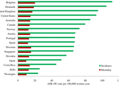
(50) Introduction. 2.. Epidemiology of breast cancer. Breast cancer is the leading cancer site and the most frequent cause of cancer death among women worldwide with an estimated 1.67 million new cancer cases diagnosed in 2012 (25% of all cancers) (Figure 3) [14]. In terms of mortality, it is estimated that around 522,000 women died from breast cancer in 2012. Female breast cancer incidence and mortality rates vary among countries (Figure 4). Estimated age-standardized incidence and mortality rates for breast cancer in Spain are similar to those in Portugal or Slovenia. In Catalonia, approximately 4841 women will be diagnosed with breast cancer and around 932 patients will die from this cancer in 2020 [15]. Breast cancer incidence rates have been slightly higher in Girona province than in the rest of Spain, with a rate ratio of 1.1 (95% CI: 1.1 to 1.2) in 2000-2004 (per 100,000 European standard population) [16]. Breast Colorectum Cervix uteri Lung Corpus uteri Stomach Thyroid Ovary Liver Non-Hodgkin lymphoma Leukaemia Pancreas Oesophagus Incidence. Brain, nervous system. Mortality. Kidney. 0. 10. 20. 30. 40. 50. ASR (W) rate per 100,000 women year. Figure 3. Estimated age-standardized incidence and mortality rates of cancer in women worldwide. ASRW: Age-standardized to the world standard population rate. Adapted from Globocan 2012 [14].. 8.
(51) Introduction. Belgium Denmark United Kingdom United States Australia Canada Norway Austria Portugal Spain Slovenia Singapore Slovakia Japan Costa Rica. Incidence. India. Mortality. Nicaragua 0. 20. 40 60 80 ASR (W) rate per 100,000 women year. 100. 120. Figure 4. Estimated age-standardized incidence and mortality rates for female breast cancer in different countries. ASRW: Age-standardized to the world standard population rate. Adapted from Globocan 2012 [14].. 2.1. Invasive carcinoma of the breast The invasive breast cancer incidence rate increased throughout the 1980s and „90s, before falling and then levelling off at the beginning of the 21st century, a trend described in many developed countries such as Australia [17], France [18], Norway [19], Spain [20] and the United States (US) [21, 22]. This pattern was mostly restricted to postmenopausal women and to tumours expressing ER. In Catalonia, breast cancer incidence rose by 2.2% (95% CI: 1.8% to 2.6%) from 1980 to 1999 [23]. This increase was more marked in women over 40, whereas incidence rates for young women remained stable. Following this period, a significant decrease of 1.5% was detected in overall breast cancer incidence (2000-2007) [24]. Similar incidence trends were observed in Spain, with the peculiarity that incidence in young women was still rising in 2004 [16]. Multiple factors might affect changes in breast cancer incidence. In many European countries and the US increasing incidence has been attributed to a rise in the use of menopausal hormone replacement therapy (HRT) among postmenopausal women in the „90s, and the decline to a drop in HRT prescription [17, 19, 21, 22]. However, in Spain the proportion of women using HRT has always been very low [25]. Prevalence of HRT use among women aged >40 years rose slightly from 0.7% in 1989 to 3.4% in 1999. According to the 2006 Spanish National Health Survey, the percentage of women using HRT was 5.3% in women aged 45-64 and 0.5% for women over 65 [26]. In the 2011-2012 Spanish survey, these rates decrease to 1.6% and 0.2%, respectively [27]. Consequently, it has been suggested that changes in HRT 9.
(52) Introduction. prescription may not be the only key factor in explaining recent trends in breast cancer incidence [16]. The implementation of mammographic screening programmes has also been posited as influencing incidence trends [22, 28, 29]. The adoption of mammographic population-based screening programmes brings the date of diagnosis forward, resulting in a transitory increment in incidence rates. Once the programme is fully established, incidence rates usually decrease before stabilizing due to the pool of prevalent undiagnosed cases being notably reduced. This change in incidence trends produced by the introduction of a screening programme is known as screening saturation. Despite the arguments presented above, the impact of HRT use and screening saturation remains disputed. Changes in lifestyle and reproductive factors may have also contributed to changes in incidence trends. These include physical inactivity, obesity in postmenopausal women, alcohol consumption, delayed childbearing, decline in fertility, early menarche and late menopause (see Section 3 of the Introduction). Regarding survival, women diagnosed with breast cancer usually present high outcomes rates. A recently published article concluded that 5-year net survival was 81% in Europe and 84% in the US [30]. However, survival differs greatly according to many prognostic factors, such as stage at diagnosis time, for example. Survival rate for women diagnosed with distant metastasis is much lower than for patients with metastatic lymph nodes or localized disease [31]. This and other prognostic factors will be explained in Section 5 of the Introduction. A steady downturn in breast cancer mortality has been described in many European countries and the US over the last 20-40 years [31, 32]. In Spain and Catalonia, a statistically significant rise in mortality trends was detected during the 1970s, „80s and the beginning of „90s, followed by an important decline [20, 24, 33, 34]. This fall has mainly been associated with increased access to more effective treatments and early detection.. 2.2. Ductal carcinoma in situ of the breast (DCIS) Women diagnosed with DCIS have about a 4-fold increased risk of developing an invasive breast cancer compared with women in the general population [35]. Given that incidence rates of DCIS are currently increasing in many developed countries, these tumours are clinically challenging and considered to be of growing importance to public health. In some countries, DCIS incidence has been seen to stabilize after a sharp increase over recent decades, with incidence trends differing by histological type and age at diagnosis [36, 37]. In the US, an increase in the incidence of non-comedo DCIS, which are not associated with subsequent breast malignancy, has been reported in recent decades, whereas rates of comedo DCIS, which are associated with subsequent invasive breast cancer, decreased or held constant throughout the 1980s and „90s [37]. Furthermore, larger or restricted upward trends have been 10.
(53) Introduction. observed in target age groups for mammographic screening [36, 38, 39]. In Norway, rates of DCIS in women aged 50-69 years, the target population for mammographic screening, steadily increased in the years prior to the start-up phase of the programme; they then peaked during its implementation, dropped, and then rose again. Since the majority of DCIS lesions do not present breast lumps but are often visible on mammography, the increase in detected cases has mainly been attributed to the widespread adoption of mammographic screening over the past decade. Whereas DCIS now accounts for around 7.4%-21% of all newly diagnosed cases of breast cancer, prior to screening DCIS diagnosis was rather rare, representing less than 5% of all breast malignancies [31, 36, 38, 40]. Nevertheless, organized screening may not completely explain the upward trend and other factors may play an important role. The number of women attending opportunistic screening, improved detection methods, improved training and skills of the radiologist and changes in risk factors have to be considered when analysing DCIS incidence [38, 41]. It has been suggested that the marked upward trend in DCIS incidence may contribute, by earlier stage detection, to declining incidence of invasive breast cancer and consequently to reduced breast cancer mortality from this disease [38, 42]. However, there are no consistent data confirming that mammography detection of DCIS directly prevents breast cancer death [43]. In fact, breast cancer mortality after 10 years of DCIS diagnosis is around 1-2%, regardless of whether mastectomy or breast-conserving surgery is applied, thus revealing that DCIS is not a life-threatening disease per se [40]. As commented previously, risk factors for DCIS and invasive breast cancer are similar, suggesting that etiologic pathways may be shared between the two diseases [3, 5-7, 43]. Family history of breast cancer, nulliparity or delayed childbearing, late age at menopause, long-term use of postmenopausal HRT, obesity in postmenopausal women and high mammography breast density all increase the risk of both DCIS and invasive breast cancer. Contrarily, early menarche, high alcohol consumption and oral contraceptive use are not consistently linked with an increased risk of DCIS development but are associated with invasive breast cancer development.. 11.
(54) Introduction. 3.. Breast cancer risk factors. The aetiology of breast cancer is multifactorial, involving hormonal and reproductive factors, dietary and lifestyle factors, and others, as described in Table 1 [4, 8, 44, 45]. It is widely accepted on the basis of epidemiological studies that endogenous and exogenous estrogen play a key role in the development of breast tumours and the risk of breast cancer development is higher with increasing estrogen levels. Breast cancer incidence is low among young women, increases sharply during the premenopausal period and then peaks at 50-69 years old before dropping again, when synthesis of estrogen ceases. This incidence trend over the course of the lifetime differs from the majority of cancers, which usually show a greater risk of tumour development with age, and suggests the involvement of reproductive hormones in breast cancer aetiology [14]. Many hormonal and reproductive factors have been considered to be risk factors for breast cancer development in women. Earlier age at menarche and later age at menopause have consistently been associated with increased risk of breast cancer by raising lifetime exposure to endogenous estrogen. Furthermore, nulliparous women over around 45 years of age are at greater risk of the development of breast cancer compared to parous women. Oral contraceptives and HRT use are both associated with breast cancer development by increasing estrogen levels. The decline in breast cancer incidence in some developed countries at the beginning of this century has mainly been attributed to a decrease in HRT use (see Section 2.1 of the Introduction) [17, 19, 21, 22, 45]. Contrarily, it has been confirmed that young age at first full-term pregnancy, high parity and lactation, preferably more than 2 years, all have a protective effect [4, 8]. Hormonal and reproductive factors. Dietary and lifestyle. Other factors. factors. Early age at menarche. High consumption of fat. Family history of breast cancer. Late age at menopause. High consumption of alcohol. History of:. Late age at first full-term birth. Obesity (postmenopausal women). Atypical hyperplasia. Nulliparity. Physical activity. DCIS. Oral contraceptives. High intake of vegetables. LCIS. HRT. Benign breast disease. High parity. Type 2 diabetes. Lactation. Hyperinsulinemia. Young age at first full-term birth. Ionizing radiation. Table 1. Risk and protective factors for breast cancer. DCIS: Ductal carcinoma in situ; HRT: Hormone replacement therapy; LCIS: Lobulillar carcinoma in situ. Red arrow: Risk factors for breast cancer; green arrow: Protective factors for breast cancer. Adapted from DeVita et al., 2008, Lakhani et al., 2012, Hamajima et al., 2002 and Boyle et al., 2003 [4, 8, 44, 45].. 12.
(55) Introduction. In terms of dietary factors, high intakes of vegetables are probably associated with a moderate protective effect for breast cancer. Epidemiologic studies have also confirmed the protective effect of physical activity with regard to breast cancer development. The influence of physical activity on breast cancer is of high interest for postmenopausal women, as in these women obesity is strongly associated with an elevated risk of breast cancer [46]. Following the menopause, adipose tissue is the major source of estrogen and obese postmenopausal women therefore have higher levels of endogenous estrogen and consequently a higher risk of breast cancer development. In addition, a high consumption of fat and alcohol are associated with an increased risk of breast cancer [4, 45]. The relationship between smoking and breast cancer has been found to be confounded by alcohol [44]. Genetic factors have to be taken into account when identifying women at high risk of breast cancer development. Risk varies with relationship to affected family members and number of affected and unaffected relatives [47]. Mutations in BRCA1 (breast cancer gene 1) and/or BRCA2 (breast cancer gene 2) are responsible for the majority of these cancers. De Sanjosé et al. described that these mutations explained about 10% of breast cancers diagnosed in Catalan women under 40 years of age [48]. Additional factors have been considered to increase the risk of breast cancer: women with a history of atypical hyperplasia, DCIS, lobulillar in situ carcinoma (LCIS) or other benign breast diseases, women with a history of ionizing radiation, women with high mammographic density and women previously diagnosed with type 2 diabetes or hyperinsulinemia [4, 45]. Risk and protective factors that determine the development of breast cancer differ according to molecular subtype [49-53]. This suggests that molecular subtype classification has to be taken into account in order to understand breast cancer aetiology. Population-based studies have found that reproductive factors such as early age at menarche, nulliparity and increasing age at first full-term birth are more strongly associated with positive than negative hormonal receptor tumours. An increase in the risk of basal-like breast cancer is described when increasing parity. Moreover, BRCA1 mutation carriers and premenopausal African American women show a high prevalence of basal-like breast cancer [50, 51, 54, 55].. 13.
(56) Introduction. 4.. Prevention and screening for breast cancer. Primary breast cancer prevention consists in avoiding or reducing exposure to risk factors mentioned in the previous section or by increasing resistance to them, thus obtaining a decrease in breast cancer incidence. The objective is to avoid the disease. Secondary prevention is the early detection and treatment of the disease, meaning it must be applied during the nondetectable phase (Figure 5). Screening is the major component of secondary prevention because it can detect disease at an early stage and so increase the probability that a cancer may be cured. Finally, tertiary prevention refers to curing cancers that have developed and preventing cancer death. It is applied during the symptomatic phase (Figure 5) through treatment and rehabilitation programmes [45, 56]. This section focuses on secondary prevention. At the end of the last century, many developed countries implemented an organized breast cancer population-based mammographic screening programme, including France [18], the Netherlands [36], Norway [57], Spain [58] and the United Kingdom [59]. Mammographic screening has been confirmed so far as the most effective method for breast screening [60]. The objective of screening for breast cancer is to reduce morbidity and mortality from the disease by detecting cancer at an early stage without adversely affecting healthy participants [61]. Screening is therefore based on the existence of an adequate treatment, which is more effective if begun earlier during the progression of the disease [59, 62]. Opportunistic breast screening coexists alongside organized population-based mammographic screening in many countries. Opportunistic screening is defined as screening that takes place outside an organized or population-based screening [61]. This type of screening may be recommended during check-ups by doctors at primary health care centres or in other health care settings. The prevalence of women attending opportunistic and organized screening varies substantially across countries and health systems. Since mammographic screening programmes were first established, there has been debate regarding their potential benefits and adverse effects. The anticipated major benefit is a reduction in breast cancer mortality and the most commonly discussed adverse effect is overdiagnosis. Both of these are discussed in the following paragraphs.. 4.1. Benefits of mammographic screening programmes First of all, implementing a mammographic screening programme reduces inequality of access to the preventive test among the target population. Access to mammography becomes homogeneous for the whole population, regardless of income or educational level [63]. It is also widely accepted that breast cancer mortality has decreased since the introduction of mammographic screening programmes. However, it is not yet fully clear whether the drop in mortality can be attributed to screening, the improved treatment available in recent years or the. 14.
(57) Introduction. interaction of both factors. Naturally, this uncertainty is no reason to interrupt mammographic screening. The effect of mammographic screening on breast cancer mortality differs between studies, although all of them observe an important reduction. In general, these studies suggest a relative risk reduction around 20% with mammography at 11 years of follow-up [60]. In particular, a meta-analysis of 11 randomized trials with 13 years of follow-up also estimated a 20% (95% CI: 11% to 27%) reduction in breast cancer mortality among women invited for screening [59]. A review of 20 European incidence-based mortality studies found a reduction of 26% (95% CI: 13% to 36%) after 6-11 years of follow-up among women invited to mammographic screening [64]. Additionally, a recent review of observational studies reported that the reduction in breast cancer mortality is even higher for women who are invited (25-31%) and actually screened (3848%) [65]. Finally, in Norway the reduction in the rate of breast cancer death in recent years is described as being 10 points higher in screen-detected (28%) than in non-screen-detected women (18%) [66]. The difference in estimates of absolute risk reduction reported is one of the greatest sources of controversy regarding the value of mammographic screening. However, there is general agreement regarding the evidence that screening does have a beneficial effect on breast cancer mortality.. 4.2. Biases related to mammographic screening programmes Screen-detected cancers are breast cancers identified using the screening test, with or without further assessment in a member of the target population who was invited for and attended mammographic screening. These cancers have a more favourable prognosis than symptomatic cancers, even in long-term survival analyses [61, 67-71]. Generally, screen-detected cancers have a higher proportion of negative lymph nodes and small-sized, well-differentiated and hormonal receptor positive tumours. Whether the cancer is detected at screening or by symptoms is considered an independent prognostic factor beyond the stage shift [72]. However, some biases have to be considered when comparing mortality from breast cancer among screendetected and non-screen-detected cancers. Before a tumour can be detected through clinical signs and symptoms, it remains asymptomatic for an indeterminate period of time known as sojourn time or detectable preclinical phase and distinctive for each particular tumour (Figure 5). Sojourn time starts when the tumour is detectable by mammography. If women participate in a mammographic screening programme, the tumour will be detected before symptoms appear. The period between when a cancer is found by screening and when it would appear through clinical signs and symptoms is known as lead time (Figure 5) [62, 73, 74]. Overall survival is measured from date of diagnosis to date of death. In the example in Figure 5, the patient would survive 10 years if she did not participate in a screening programme, but 15. 15.
(58) Introduction. years if she did. This simply reflects earlier diagnosis; the natural history of the disease and time of death are unchanged. We cannot therefore consider it real improved overall survival.. Non-detectable disease. Asymptomatic detectable disease. Symptomatic disease. Sojourn time Death. Time. Lead time. Screening test Example:. Survival. Screen-detected (age 55). Clinically detected (age 60). Death (age 70). Screen-detected: 15 years Clinically detected: 10 years. Figure 5. Overview of disease progression with the intervention of an early-detection screening test. Adapted from IARC (International Agency of Research on Cancer) handbooks of cancer prevention, 2002 [62].. Rapidly progressive tumours 1. 2. 3. Patients 4. 5. 6. Slowly progressive tumours 7. 8. 9. Patients 10. 11. 12. Time st. 1 screening round Sojourn time Symptomatic disease. nd. 2 screening round. Death from breast cancer Cause of death different from breast cancer. Figure 6. Overview of rapidly and slowly progressive tumours in relation to breast cancer screening with 12 patient examples. Adapted from Cox et al., 2013 [74].. 16.
(59) Introduction. However, this scenario is not always exactly like the example because treatment approach and prognosis are different in early and advanced-detected breast cancers. As mentioned above, screening is based on the existence of an adequate treatment being more effective when applied in early-staged rather than advanced-stage diseases. Furthermore, overestimation of survival among screen-detected women is influenced by the high proportion of slowly progressing tumours detected by screening. The probability of cancer detection is directly proportional to the length of sojourn time: the longer the sojourn time, the greater the chance of detecting the lesion (Figure 6). The length of sojourn time depends on the cancer progression rate. Patients with slowly progressive cancers have a longer sojourn time and are more likely to be diagnosed by screening than women with rapidly growing cancers. In addition, these cancers are usually less aggressive, with a low histological grade, and they are often associated with good prognosis. Contrarily, women with rapidly progressive tumours are more likely than average to die of their disease and less likely to have it detected by screening. Thus, screen-detected cancers are represented by a higher proportion of non-aggressive and slowly growing tumours than non-screen-detected breast cancer. This bias is known as length bias [62, 73, 74]. Finally, when examining the benefits of screening, aside from early diagnosis and the duration of the tumour‟s progression, patient characteristics and the health system must also be considered. Comorbidity, ethnicity and culture can influence participation in mammographic screening. Therefore, participants in a mammographic screening programme may have a different baseline risk for developing breast cancer and mortality to non-participants; this is known as selection bias [74, 75].. 4.3. Adverse effects of mammographic screening programmes Although many women will benefit from mammographic screening programmes, others will be affected by the inevitable adverse effects of it. The objective is therefore to minimize these. The most important are explained in this section: overdiagnosis, interval cancers, false-negative and false-positive cancers. Some screen-detected cancers would not have been diagnosed in the absence of mammographic screening. These cancers are referred to as overdiagnosis. A cancer is overdiagnosed if it would never progress further or would evolve slowly enough that the patient would die from other causes than breast cancer. The tumour in both cases would not become clinically apparent during the patient‟s lifetime, thus it would not be life-threatening [62, 76]. If patient number 7 in Figure 6 attended screening and were diagnosed with breast cancer, it would be overdiagnosed. As depicted in Figure 7, cancer growth rate varies greatly between tumours [77]. Some screendetected cancers might progress so slowly that they would never have been clinically apparent. Detection of these cancers turns women into patients, which means that they receive unnecessary treatment and their quality of life might deteriorate. However, clinicians are unable 17.
(60) Introduction. to distinguish between overdiagnosed and non-overdiagnosed patients and treat all cases, leading to overtreatment. In Figure 7, tumour D represents a rapidly growing tumour, leading to distant metastasis and death in a short period of time. This case would not have benefitted from screening. Contrarily, tumour A exemplifies a tumour growing very slowly, remaining microscopic, undetectable and without morbidity during a woman‟s lifetime. This type of tumour tends to be DCIS or, if invasive, histological grade 1 or 2 rather than grade 3. If this woman were to attend mammographic screening, it would be a case of overdiagnosis. The women with tumours B and C would benefit from screening by bringing forward diagnosis to a time when they would still be curable.. Figure 7. Varying screen detection capability in relation to tumour growth rate. Taken from Esserman et al., 2009 [77].. Information regarding frequency of overdiagnosis is necessary to quantify the adverse effect of screening. However, estimating the rate of overdiagnosis is very complex and advanced statistical analyses are required with long follow-up times. This results in a wide variety of studies demonstrating different rates of overdiagnosis, ranging from about 0-54% [59, 60, 76, 78-81]. Although there is no consensus regarding this percentage, there is a general agreement that target women for screening need to be informed about its adverse effects as well as its benefits. False-positive results are also an important concern in mammographic screening. A result is considered to be false-positive if breast cancer is not further diagnosed after recall for additional evaluation. Screening effectiveness evaluation should include assessing the frequency of falsepositives. The main negative effects of false-positive results are lower attendance, anxiety and associated posterior excision biopsies [82].. 18.
(61) Introduction. Finally, another key component of quality control for screening programmes is interval cancer rate. As defined in European Guidelines for Quality Assurance in Breast Cancer Screening and Diagnosis, an interval cancer is a breast cancer arising after a negative screening episode (which may include assessment) and before the next scheduled screen round or within 24 months for women who have reached the upper age limit [61]. To identify interval cancers, a link is required between women participating in a screening programme and population-based cancer registries. In general, interval cancers are rapidly growing and aggressive tumours associated with a short sojourn time. In Figure 6, patient 4 represents an example of interval cancer. Compared with screen-detected cancers, interval cancers are related to poorer prognosis [83, 84]. European guidelines recommend interval cancers be classified into the following: true interval, occult, minimal signs, false-negative, and unclassifiable tumours [61]. For true interval cancers, the screening mammogram is normal and there is no reason for further assessment. The sojourn time for these cancers is under two years (screening period intervals), and they are therefore inevitable in mammographic screening. Contrarily, in the false-negative group (undetected cancers) an abnormality is clearly visible in the screening mammogram and additional assessment should be tested. Delays in diagnosing false-negative cancers may be due to reading or technical errors. Frequency of false-negatives should be estimated in a screening programme so as to minimize the rate and improve screening effectiveness. Distribution of histopathological characteristics, such as molecular subtypes, is represented differently within the interval cancer categories described above. Notably, the triple-negative phenotype is concentrated among true interval cancers and tumours with minimal signs [84, 85]. Although a debate still exists balancing the benefits and adverse effects of mammographic screening, many studies agree that the benefits outweigh the adverse effects, which is why it is recommended in many developed countries.. 4.4. Mammography programmes. use. and implementation. of. mammographic. screening. In Catalonia, mammography use started during the 1980s and spread throughout the „90s. In 1980, only 10 mammography devices were available, while in 2000 there were 134 [23]. The use of mammography as a preventive treatment for breast cancer in Catalonia has therefore increased in the last decades. The proportion of women over 20 undergoing mammography periodically in Catalonia was lower in 1994 (24.5%) than in 2002 (40.4%), 2006 (43.1%) and 2012 (49.1%) according to Catalan Health Surveys [86-88]. Mammography use has increased dramatically among women aged 50-69, from 26.9% in 1994 to 94.1% in 2012 [86, 89]. Prior to implementation of the organized population-based mammographic screening programme (Programa de Detecció Precoç del Càncer de Mama, PDPCM) in Catalonia, mammography use was higher among women aged 40-49 than those aged 50-69. Nowadays, the target population of the PDPCM (women aged 50-69) shows the largest proportion of women undergoing mammography periodically, as recommended by European guidelines. The highest 19.
Figure
![Figure 1. Anatomy of the human mammary gland. Taken from Ali et al., 2002 [1].](https://thumb-us.123doks.com/thumbv2/123dok_es/5235639.95597/45.595.200.393.291.518/figure-anatomy-human-mammary-gland-taken-ali-et.webp)
![Figure 2. Linear model of breast carcinogenesis. Adapted from Allred, 2010 and Burstein et al., 2004 [3, 5]](https://thumb-us.123doks.com/thumbv2/123dok_es/5235639.95597/46.595.89.503.151.251/figure-linear-model-breast-carcinogenesis-adapted-allred-burstein.webp)

+7


![Figure 7. Varying screen detection capability in relation to tumour growth rate. Taken from Esserman , 2009 [77]](https://thumb-us.123doks.com/thumbv2/123dok_es/5235639.95597/60.595.120.484.243.456/figure-varying-screen-detection-capability-relation-tumour-esserman.webp)


Documento similar
Analysis of breast cancers from The Cancer Genome Atlas (TCGA) showed that HSD17B14 expression is increased significantly in cancer compared with normal breast tissue and that it
Introduction: The aim of this study was to describe incidence, incidence trends and survival patterns of lymphoid neoplasms (LNs) and its subtypes in Spain in the period 2002-2013
This review gives hand on recent advances of personalized oncology in several cancer disease models including leukemia, melanoma, breast cancer, lung cancer, colorectal cancer,
These observations prompted us to analyze the PP2A phosphorylation/inhibition status in breast cancer cells using a “CPscore” in which value 0 was defined by those
An ER-true/TN-like classifier, including 14 proteins, was used to assign new samples to ER-true or TN-like (sup. We used gene expression data from 1296 breast cancer tumors,
Conclusion Both TNM staging and histological/molecular biomarkers are associated with overall survival in Spanish women with breast cancer; when both are combined in the
Heterogeneous primary HER2+ breast tumor, con- sisting of sensitive cells and primary resistant cells, exhibiting partial response to anti-HER2 treatment and relapse due to
The two major breast cancer susceptibility genes BRCA1 and BRCA2 are involved in 30% of hereditary breast cancer cases, but the discovery of additional breast cancer
