UNIVERSIDAD DE BARCELONA FACULTAD DE FARMACIA
DEPARTAMENTO DE BIOQUÍMICA Y BIOLOGÍA MOLECULAR
ESTUDIO DE LOS MECANISMOS DE INHIBICIÓN
DE LA ACTIVIDAD CARNITINA
PALMITOILTRANSFERASA 1
Carnitine Palmitoyltransferase I Critical for Malonyl-CoA Inhibition
MUTATION OF METHIONINE 593 ABOLISHES MALONYL-CoA INHIBITION*
Received for publication, September 30, 2002, and in revised form, December 17, 2002 Published, JBC Papers in Press, December 23, 2002, DOI 10.1074/jbc.M209999200
Montserrat Morillas‡§, Paulino Go´mez-Puertas§¶, Assia Bentebibel‡储, Eva Selle´s‡, Nuria Casals**, Alfonso Valencia¶, Fausto G. Hegardt‡ ‡‡, Guillermina Asins‡, and Dolors Serra‡
From the‡Department of Biochemistry and Molecular Biology, University of Barcelona, School of Pharmacy, E-08028 Barcelona, Spain, the¶Protein Design Group, National Center for Biotechnology, Consejo Superior de Investigaciones Cientı´ficas, Cantoblanco, E-28049 Madrid, Spain, and the**Department of Biochemistry and Molecular Biology, International University of Catalonia, 08190 Sant Cugat, Spain
Carnitine palmitoyltransferase (CPT) I, which cata-lyzes the conversion of palmitoyl-CoA to palmitoylcar-nitine facilitating its transport through the mitochon-drial membranes, is inhibited by malonyl-CoA. By using the SequenceSpace algorithm program to identify amino acids that participate in malonyl-CoA inhibition in all carnitine acyltransferases, we found 5 conserved amino acids (Thr314, Asn464, Ala478, Met593, and Cys608,
rat liver CPT I coordinates) common to inhibitable malo-nyl-CoA acyltransferases (carnitine octanoyltransferase and CPT I), and absent in noninhibitable malonyl-CoA acyl-transferases (CPT II, carnitine acetyltransferase (CAT) and choline acetyltransferase (ChAT)). To determine the role of these amino acid residues in malonyl-CoA inhibition, we prepared the quintuple mutant CPT I T314S/N464D/A478G/ M593S/C608A as well as five single mutants CPT I T314S, N464D, A478G, M593S, and C608A. In each case the CPT I amino acid selected was mutated to that present in the same homologous position in CPT II, CAT, and ChAT. Be-cause mutant M593S nearly abolished the sensitivity to ma-lonyl-CoA, two other Met593mutants were prepared: M593A
and M593E. The catalytic efficiency (Vmax/Km) of CPT I in
mutants A478G and C608A and all Met593mutants toward
carnitine as substrate was clearly increased. In those CPT I proteins in which Met593had been mutated, the
malonyl-CoA sensitivity was nearly abolished. Mutations in Ala478,
Cys608, and Thr314to their homologous amino acid residues
in CPT II, CAT, and ChAT caused various decreases in ma-lonyl-CoA sensitivity. Ala478 is located in the structural
model of CPT I near the catalytic site and participates in the binding of malonyl-CoA in the low affinity site (Morillas, M., Go´mez-Puertas, P., Rubı´, B., Clotet, J., Arin˜ o, J., Valencia, A., Hegardt, F. G., Serra, D., and Asins, G. (2002)
J. Biol. Chem.277, 11473–11480). Met593may participate in
the interaction of malonyl-CoA in the second affinity site, whose location has not been reported.
The enzyme carnitine palmitoyltransferase (CPT)1 I cata-lyzes the conversion of long chain fatty acyl-CoAs to acylcar-nitines, which is the first step in the transport of fatty acyl-CoA groups from the cytosol to mitochondria where they undergo
-oxidation. This reaction is inhibited by malonyl-CoA, and so this enzyme could be the most physiologically important regu-latory step in mitochondrial fatty acid oxidation (1). This proc-ess allows the cell to signal the relative availability of lipid and carbohydrate fuels in liver, heart, skeletal muscle, and pancre-atic-cell (2). The mechanism of malonyl-CoA inhibition can be potentially mimicked by pharmacological malonyl-CoA-related agents for the treatment of metabolic disorders such as diabe-tes, insulin resistance, and coronary heart disease (3).
Mammals express two isoforms of CPT I, a liver isoform (L-CPT I) and a heart/skeletal muscle isoform (M-CPT I), which are the products of two different genes (4, 5). The identity in amino acids residues is high (62%) but they are differentially regulated by malonyl-CoA. The L-CPT I isoform is inhibited by malonyl-CoA to a much lesser extent than the M-CPT I isoform (the IC50 value for M-CPT I is about 2 orders of magnitude lower than for L-CPT I) (6). This property is probably involved in the finer regulation of fatty acid oxidation in heart and skeletal muscle in comparison to liver.
From studies on the pH dependence of the affinity of CPT I for its substrate and from the ability of palmitoyl-CoA to dis-place [14C]malonyl-CoA bound to skeletal muscle mitochondria it was hypothesized (7) that the palmitoyl-CoA and malonyl-CoA bind at different sites. A number of studies have shown that in rat liver CPT I there are two malonyl-CoA binding sites: one with greater capacity for binding and regulation of the inhibitor and not susceptible to competition from acyl-CoA, which behaves as an allosteric component (8 –12); and a second acyl-CoA binding site, which is located near the catalytic site (13).
Various groups have attempted to establish the basis of the L-CPT I/malonyl-CoA interactions. The probable binding sites of malonyl-CoA in L-CPT I were deduced to be at the C termi-nus after preparation of several L-CPT I chimeras whose IC50 values for malonyl-CoA corresponded to the C-terminal region (14) of the chimera. However, the N terminus of L-CPT I was also shown to influence the enzyme/inhibitor interaction. Mu-tation of Glu3, His5, or His140produced a loss of malonyl-CoA
* This work was supported in part by Direccio´n General de Investi-gacio´n Cientı´fica y Te´cnica, Spain, Grant BMC2001-3048 and Ajuts de Suport als Grups de Recerca de Catalunya Grant 2001SGR-00129 (to F. G. H.) and the Marato´ de TV3. The costs of publication of this article were defrayed in part by the payment of page charges. This article must therefore be hereby marked “advertisement” in accordance with 18 U.S.C. Section 1734 solely to indicate this fact.
§ Contributed equally to the results of this study.
储Recipient of a fellowship from the Ministerio de Ciencia y Tecnolo-gı´a, Spain.
‡‡ To whom correspondence should be addressed: Dept. of Biochem-istry and Molecular Biology, School of Pharmacy, Diagonal 643, E-08028 Barcelona, Spain. Tel.: 34-93-402-4523; Fax: 34-93-402-4520; E-mail: [email protected].
1The abbreviations used are: CPT, carnitine palmitoyltransferase;
L-CPT I, liver isoform of carnitine palmitoyltransferase I; M-CPT I, muscle isoform of carnitine palmitoyltransferase I; CAT, carnitine acetyltransferase; COT, carnitine octanoyltransferase; ChAT, choline acetyltransferase.
This paper is available on line at http://www.jbc.org
in M-CPT I produced a decrease in malonyl-CoA sensitivity, which emphasizes the importance of the N terminus before the first transmembrane region as a modulator of the malonyl-CoA inhibition (17, 18). On the basis of these results, it was pro-posed that the two malonyl-CoA inhibitable domains might be located at the C terminus as suggested by several kinetic stud-ies. The development of a CPT I catalytic core model (19) allowed us to assign the low affinity binding site to a domain near the catalytic channel in which palmitoyl-CoA is bound containing the catalytic acyl-CoA binding domain (20)
Here we used the SequenceSpace algorithm program to iden-tify five amino acid residues (Thr314, Asn464, Ala478, Met593, and Cys608), which may contribute to the sensitivity of CPT I to malonyl-CoA. The proposal is based on the finding that they are present in malonyl-CoA-inhibitable CPT I ((isoforms L- and M-) and COT from various organisms and absent in noninhib-itable acyltransferases (CPT II, CAT, and ChAT). Mutation of these amino acids to their counterparts in CPT II showed that mutation of Met593by itself, M593S, or the quintuple mutant containing the M593S mutation, T314S/N464D/A478G/M593S/ C608A, or other Met593 point mutants such as M593A and M593E nearly abolished malonyl-CoA sensitivity of L-CPT I. The remaining mutated amino acids showed slight, varied sen-sitivity to malonyl-CoA inhibition.
EXPERIMENTAL PROCEDURES
Tree-determinants Analysis—Sequences of proteins from the carni-tine-choline acyltransferase family were obtained using BLAST (21). Multiple alignment was performed using ClustalW (22). The analysis of conserved differences (tree-determinants) between malonyl-CoA-regu-lated (L-CPT I, M-CPT I, and COT) and nonregumalonyl-CoA-regu-lated (CPT II, CAT, and ChAT) acyltransferases, using multivariate statistics for low-dimen-sional representation, was done using the SequenceSpace algorithm (23, 24). Graphics of vectors representing protein sequences and indi-vidual residues from the multiple alignment were performed using the Sequence Space Java-based viewer (www.industry.ebi.ac.uk/SeqSpace).
Construction of Site-directed Mutants—Plasmids pYESLCPTIwtand
pYESLCPTA478Gwere obtained as previously described (20). Plasmids
pYESLCPTT314S, pYESLCPTN464D, pYESLCPTM593S, pYESLCPTM593A,
pYESLCPTM593E, and pYESLCPTC608A were constructed using the
QuikChange polymerase chain reaction-based mutagenesis procedure (Stratagene) with the pYESLCPTwtplasmid as template. The following
primers were used: primer T314S.for 5⬘ -GGGAGCGACTCTTCAATAG-TTCCCGGATCCCTGGG-3⬘, primer T314A.rev 5⬘ -CCCAGGGATCCG-GGAACTATTGAAGAGTCGCTCCC-3⬘, primer N464D.for 5⬘ -CACCTT-TGTTGTCTTCAAAGACAGCAAGATAGGC-3⬘, primer N464D.rev 5⬘ -GCCTATCTTGCTGTCTTTGAAGACACCAAAGGTG-3⬘, primer M593S.for 5⬘-CCTCACATATGAGGCCTCCAGTACCCGGCTCTTCCG AGAAGG-3⬘, primer M593S.rev 5⬘ -CCTTCTCGGAAGAGCCGGGTAC-TGGAGGCCTCATATGTGAGG-3⬘, primer M593A.for 5⬘ -CCTCAC-ATATGAGGCCTCCGCGACCCGGCTCTTCCGAGAAGG-3⬘, primer M593A.rev 5⬘-CCTTCTCGGAAGAGCCGGGTCGCGGAGGCCTCATA TGTGAGG-3⬘, primer M593E.for 5⬘ -CCTCACATATGAGGCCTCCGA-GACCCGGCTCTTCCGAGAAGG-3⬘, primer M593E.rev 5⬘ -CCTTCTC-GGAAGAGCCGGGTCTCGGAGGCCTCATATGTGAGG-3⬘, primer C608A.for 5⬘-GAGACTGTACGCTCCGCCACTATGGAGTCCTGC-3⬘, and C608A.rev 5⬘ -GCAGGACTCCATAGTGGCGGAGCGTACAGTCT-C-3⬘ (the mutated nucleotides are underlined). The plasmid pYESLCPTIT314S/N464D/A478G/M593S/C608A was obtained by the same
method, but performing each new mutation stepwise starting on plas-mid pYESLCPTT314S
. The appropriate substitutions as well as the absence of unwanted mutations were confirmed by sequencing the inserts in both directions with an Applied Biosystems 373 automated DNA sequencer.
Expression of L-CPT I in Saccharomyces cerevisiae—The expression of the constructs containing L-CPT I wild type and mutants (see above) in yeast cells and the preparation of the cell extracts were performed as described in Ref. 19.S. cerevisiaewas chosen as an expression system for L-CPT I wild type and the mutants because it does not have endog-enous CPT I activity.
Determination of Carnitine Acyltransferase Activity—Carnitine
ity was assayed for 4 min at 30 °C in a total volume of 200l. For determination of theKmfor carnitine, palmitoyl-CoA was fixed at 135M(for L-CPT I). For determination of theKmfor acyl-CoA, carni-tine concentration was fixed at 400M. When malonyl-CoA inhibition was assayed, increasing concentrations of malonyl-CoA were included. The IC50, defined as the malonyl-CoA concentration that produces 50%
inhibition of enzyme activity, was determined using 50M palmitoyl-CoA and 400Mcarnitine.Kmwas estimated by analyzing the data from three experiments using the program Enzifit (Biosoft), and IC50
was calculated by Excel software using linear regression analysis. Values reported in the text are the means and standard deviations of three to five determinations. Curve fitting was carried out using Excel software. All protein concentrations were determined using the Bio-Rad protein assay with bovine albumin as standard.
Immunological Techniques—Western blot analysis was performed as described (19). The antibody for rat L-CPT I was kindly given by Dr. V. A. Zammit (Hannah Research Institute, Ayr, Scotland, United Kingdom) and was directed against peptide 428 – 441, in the cytosolic catalytic C-terminal domain.
RESULTS
Residues Conserved in Malonyl-CoA Inhibited Versus Nonin-hibited Carnitine-Choline Acyltransferases—An exhaustive analysis of the presence of residues shared by all the malonyl-CoA-regulated enzymes of the carnitine-choline acyltrans-ferase familyversusthe malonyl-CoA nonregulated members of the same family was performed using the algorithm Sequence-Space (23, 24). This method uses a vectorial representation of each protein sequence as a point in a multidimensional space (SequenceSpace) and multivariate statistics, principal compo-nent analysis, to allow reduction of the number of dimensions. This representation allows us not only to define clusters of proteins according to specific properties by choosing the appro-priate axes defined by the highest corresponding eigenvalues (also known as proper values), but also to project the individual residues on the same axes, and thus trace the positions con-served in the subfamilies defined. The main advantage of this method is the possibility of predicting which residues may be responsible for the specific characteristics of each protein sub-family or group of subfamilies as has been reported previously for short- and medium-long substrate specificity for the carni-tine-choline acyltransferases protein family (19, 20) or effector recognition by some members of the Ras superfamily (25).
prepared a quintuple mutant, T314S/N464D/A478G/M593S/ C608A, and separately, the point mutants T314S, N464D, A478G, M593S, and C608A and all were expressed inS. cer-evisiae. After we observed that mutant M593S nearly abolished the sensitivity to malonyl-CoA (see below), new point Met mu-tants were prepared: M593A and M593E. All transformed yeast cells expressed a protein with the same molecular mass (88 kDa) and the mutant enzymes were expressed in roughly the same proportion per milligram of protein as the wild type L-CPT I as deduced from immunoblot analysis (data not shown).
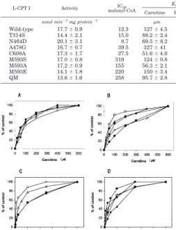
Kinetic Properties of CPT I Wild Type and Mutants—L-CPT I activities of the wild type, quintuple mutant variant T314S/ N464D/A478G/M593S/C608S, and point mutants were similar
(values ranged between 14 and 20 nmol min⫺1mg protein⫺1) when the protein was overexpressed 20 h after galactose induc-tion, showing that the various mutations assayed produce small changes in L-CPT I activity (Table I).
All mutants exhibited standard saturation kinetics when the carnitine concentration was varied relative to a constant con-centration of the second substrate, palmitoyl-CoA, and when palmitoyl-CoA concentration was varied relative to a constant carnitine concentration, a property identical to that of the wild type L-CPT I (Fig. 3). The quintuple mutant produced small changes in the kinetic constants for carnitine and palmitoyl-CoA as substrates (Table I). Catalytic efficiency (Vmax/Km) was increased by a factor of 2.6 (carnitine) and 2.2 (palmitoyl-CoA). The catalytic efficiency for carnitine as substrate of those point
FIG. 1.Sequence space analysis of the carnitine-choline acyltransferase family.A, protein sequences projected onto the plane defined by principle axes 2 and 4. This two-dimensional space allows separation of protein subfamilies according to their malonyl-CoA regulatory characteristics; CPT II, CAT, and ChAT (CACP) enzymes (malonyl-CoA insensitive) are clustered to thelower left cornerof the panel, whereas CPT I (L- and M-isoforms) and COT (malonyl-CoA inhibitable enzymes) are projected on theupperandrightareas of the vertical and horizontal axes, respectively.B, the sequence of each subfamily is represented as a vector point in a multidimensional space (sequence space), with residue positions and types as the basic dimensions. Single residues completely conserved in CPT I or COT subfamilies are projected in the same position as their corresponding protein sequences. Residues conserved in both groups of malonyl-CoA-regulated enzymes occupy theupper right corner, whereas the residues conserved in the nonregulated cluster of acyltransferases (CPT II, CAT, and ChAT) occupy theoppositeone. Residues located in alignment positions present in both opposite corners of the two-dimensional plot are responsible for protein cluster segregation and are predicted to be involved in malonyl-CoA sensitivity.
FIG. 2.Alignment of representative sequences of mammalian carnitine-choline acyltransferases.Amino acid sequence of 18 repre-sentative members of the malonyl-CoA-insensitive enzymes, CPT II (CPT2) from rat, mouse, and human; CAT (CACP) from human and mouse; ChAT (CLAT) from human, pig, rat, and mouse; and malonyl-CoA inhibitable enzymes L-CPT I (CPT1) from rat, mouse, and human; M-CPT I (CPTM) from human, rat, and mouse; and COT (OCTC) from human, rat, and bovine, were obtained from the SwissProt data bank and aligned using ClustalW (22).A, schematic representation of the position of the tree-determinant residues obtained using the SequenceSpace algorithm (23, 24) on the rat CPT II and L-CPTI proteins: Ser223/Thr314, Asp363/Asn464, Gly377/Ala478, Ser490/Met593, Ala505/Cys608. Transmembrane regions of
L-CPT I are also represented (tm1andtm2). Position of the catalytic histidine (His372/His473) as well as the previously three-dimensional modeled
mutants that altered the sensitivity to malonyl-CoA increased (see below). The catalytic efficiency of the methionine mutants increased between 4.2- and 21-fold, C608A increased 8.8-fold, and A478G increased 4.1-fold. T314S, which produced a small change in malonyl-CoA sensitivity (see below), increased the Vmax/Kmvalue by only 2.8, whereas in N464D, in which the sensitivity to malonyl-CoA was unchanged (see below), the catalytic efficiency was modified by a factor of 5.6.
An analogous tendency was also observed inKmfor
palmi-toyl-CoA but the changes were smaller.Kmvalues for palmi-toyl-CoA were 24.3, 15.1, 7.4, 6.1, and 6.3 for mutants C608A, A478G, M593S, M593A, and M593E, respectively (Kmvalue for
the wild type was 4.9) (Table I). Catalytic efficiencies for palmi-toyl-CoA as substrate increased in all mutants, the values ranging between 2.78 and 4.81 (Table I).
Inhibition of CPT I Wild Type and Mutants by Malonyl-CoA—When inhibitory kinetics versus increasing concentra-tions of malonyl-CoA was performed, the quintuple mutant practically abolished the sensitivity toward malonyl-CoA (IC50 of 258versus12.3Mof the wild type) (Fig. 4Band Table I).
Even at concentrations as high as 100 M malonyl-CoA the CPT I quintuple mutant maintained 80% of the activity of the control without malonyl-CoA.
We then addressed the individual responsibility of the sep-arate CPT I mutants for the malonyl-CoA sensitivity. Mutants T314S, N464D, M593S, and C608A expressed inS. cerevisiae were incubated with increasing amounts of malonyl-CoA, and CPT I activity was determined. Mutant A478G had been pre-viously studied in Ref. 20 and showed decreased sensitivity to malonyl-CoA (IC50of 39.5versus12.3Mof the wild type).
The kinetics of inhibition by malonyl-CoA depended on the mutant considered. Whereas mutant M593S (Fig. 4A) showed very low sensitivity at malonyl-CoA inhibition (IC50of 319M), the other mutations produced varied changes in malonyl-CoA sensitivity. L-CPT I C608A slightly modified the sensitivity to malonyl-CoA (IC50is 27.5M), the change in IC50of mutant T314S was small, whereas N464D showed similar sensitivity to malonyl-CoA to the wild type (Fig. 4Band Table I). Because the highest changes in sensitivity to malonyl-CoA andKmvalues
for carnitine were observed in the methionine mutants (point and quintuple mutants), we additionally prepared two new mutants: M593A and M593E to examine whether Met593was essential to the malonyl-CoA interaction in L-CPT I. Results show that the sensitivity to malonyl-CoA was also nearly abol-ished in these mutants (Fig. 4A) (IC50values of 155 and 220M, respectively) as in the M593S mutant, confirming the essential role of Met593in this interaction.
DISCUSSION
We attempted to identify the amino acids in the C-terminal domain of L-CPT I that are responsible for the inhibition of the catalytic activity by malonyl-CoA. Over many years much work has been done to identify the domains in L-CPT I that may bind malonyl-CoA. Different groups have tested different empirical hypotheses and mutated amino acids, mostly in the amino-terminal region of L-CPT I. The results have shown that this domain plays a role in the regulation of CPT I by malonyl-CoA, because in some cases the sensitivity to the inhibitor is impaired.
A different approach was employed by our group very re-cently. This was based on the conservation of two histidine residues, which are present in the inhibitable malonyl-CoA carnitine acyltransferases (CPT I and COT) and absent in noninhibitable enzymes (CPT II and CAT). Mutation of both histidines resulted in the abolition of malonyl-CoA sensitivity in COT (26). Analogous results were observed in CPT I when its concentration at the mitochondrial membranes was not high. Mutation of other amino acids in the domain proximal to the catalytic site (Ala478and Pro479) indicated that a malonyl-CoA-T314S/N464D/A478G/M593S/C608A (QM)
Extracts from yeast expressing wild type and several mutants of L-CPT I were assayed for activity, malonyl-CoA sensitivity, and kinetics as described under “Experimental Procedures.” The results are the mean⫾S.D. of at least three independent experiments with different prepara-tions. In parentheses are shown the increase (in-fold number) of the catalytic efficiency (Vmax/Km) versus to that of the wild type.
L-CPT I Activity IC50
malonyl-CoA
Km Vmax Catalytic efficiency
Carnitine Palmitoyl-CoA Carnitine Palmitoyl-CoA Carnitine Palmitoyl-CoA
nmol min⫺1mg protein⫺1
m nmol min⫺1mg protein⫺1 V
max/Km Wild-type 17.7⫾0.9 12.3 127⫾4.5 4.9⫾0.3 6.6⫾0.8 6.3⫾0.4 0.05 (⫻1) 1.28 (⫻1) T314S 14.4⫾2.1 15.0 88.2⫾2.4 1.7⫾0.5 12.8⫾0.1 6.8⫾0.1 0.15 (⫻2.8) 3.98 (⫻3.1) N464D 20.1⫾3.1 8.7 69.5⫾8.2 4.1⫾0.4 19.4⫾1.4 18.9⫾3.6 0.28 (⫻5.6) 4.63 (⫻3.6) A478G 16.7⫾0.7 39.5 327⫾41 15.1⫾4.0 69.8⫾9.3 50.4⫾17 0.21 (⫻4.1) 3.34 (⫻2.6) C608A 17.3⫾1.7 27.5 51.6⫾4.0 24.3⫾2.0 23.7⫾5.0 67.5⫾9.0 0.46 (⫻8.8) 2.78 (⫻2.2) M593S 17.0⫾0.8 319 124⫾0.8 7.4⫾1.2 133⫾18 20.9⫾1.6 1.07 (⫻21) 2.84 (⫻2.2) M593A 17.2⫾0.9 155 56.3⫾2.1 6.1⫾0.2 32.5⫾4.6 30.3⫾4.7 0.58 (⫻12) 4.81 (⫻3.7) M593E 14.1⫾1.8 220 150⫾3.4 6.3⫾0.5 31.3⫾2.6 27.5⫾1.8 0.21 (⫻4.2) 4.37 (⫻3.4) QM 13.6⫾1.6 258 95.7⫾2.8 4.6⫾1.5 13.1⫾4.7 13.0⫾6.3 0.14 (⫻2.6) 2.84 (⫻2.2)
FIG. 3.Kinetic analysis of wild type and different mutants of L-CPT I.Yeast extracts (10g of protein) of (AandC) wild type (open circles) and mutants M593S (open triangles), M593A (black rhombus), M593E (black squares), and (CandD) T314S (open rhombus), N464D (open squares), A478G (black squares), C608A (black triangles), and quintuple mutant T314S/N464D/A478G/M593S/C608A (black circles) were incubated at increasing concentrations of carnitine (AandB) and palmitoyl-CoA (CandD).
[image:6.621.51.305.122.453.2]inhibitable domain was probably the low-affinity malonyl-CoA binding site. Our previous studies showed that the location of malonyl-CoA in the structural model was compatible with com-petition of the inhibitorversusthe substrate in the malonyl-CoA low affinity binding site (20).
The site-directed mutagenesis study used here to identify amino acids responsible for malonyl-CoA inhibition is based on the comparison of the sequences in a range of carnitine and choline acyltransferases, taking the positive or negative sensi-tivity to malonyl-CoA as a discriminatory criterion. The bio-computing study has shown that five amino acids are present in all CPT I (isoforms L- and M-) and in COT from various organisms and that they are absent not only in other nonma-lonyl-CoA-inhibitable carnitine acyltransferases but also in ChAT. In rat L-CPT I these amino acids are Thr314, Asn464, Ala478, Met593, and Cys608. The corresponding positional amino acids in CPT II, CAT, and ChAT are Ser223, Asp363, Gly377, Ser490, and Ala505, respectively (coordinates of rat CPT II). Therefore, we considered it highly probable that these amino acids were involved in the interaction of malonyl-CoA. Results confirmed in part this supposition. The quintuple mutant re-duced malonyl-CoA sensitivity almost completely (80% activity at 100 M malonyl-CoA (which is outside the physiological range)), supporting the initial hypothesis. The results obtained using separate single mutants indicate that not all of these amino acids have the same role in malonyl-CoA inhibition. Whereas M593S nearly abolished the sensitivity to malonyl-CoA like the quintuple mutant, A478G increased the IC50from 12 to 39.5M(20). The other amino acids are less responsible for the inhibition.
The relevance of Met593as a critical amino acid for malonyl-CoA sensitivity was confirmed by the results of mutation to other two amino acids, Ala and Glu. The mutants equally showed diminished sensitivity to malonyl-CoA like mutant M593S. Met593, when mutated to Ser as it appears in CPT II and CAT, decreased the sensitivity to malonyl-CoA in a stron-ger fashion than when it was mutated to other amino acids like Ala and Glu, which were unrelated to this position in other carnitine acyltransferases. Therefore, we conclude that the oc-currence of Ser in this position has probably been evolutionary conserved in nonmalonyl-CoA-sensitive carnitine acyltrans-ferases because it prevents sensitivity to malonyl-CoA. In any
case, it appears that Met593 is critical in the interaction of malonyl-CoA with L-CPT I.
It was of interest to measure the kinetic constants of all CPT I mutants. Several authors reported the competition between malonyl-CoA and carnitine (27, 28). The tissues in which the sensitivity of CPT I to malonyl-CoA is highest are those that require the highest concentration of carnitine to drive the re-action and the requirement for carnitine and sensitivity to malonyl-CoA appears to be inversely related. The authors con-cluded that the sites to which the two metabolites bind are closely associated (27, 7). Studies by Bird and Saggerson (28) showed on the one hand that malonyl-CoA reduced the effec-tiveness of carnitine as substrate, and on the other hand, that carnitine might diminish the regulatory effect of malonyl-CoA (29). Although a clear mechanism for this competition could not be established, the data strongly supported this idea. In the present study the various CPT I mutants have alteredKmor Vmaxfor carnitine. Whereas theKmfor C608A was half of the
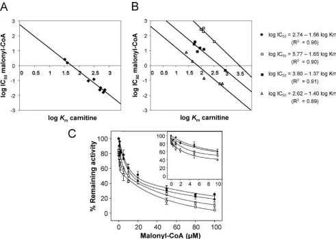
wild type, itsVmaxwas 3.6-fold higher. The mutant M593S had the sameKmvalue for carnitine as the wild type but itsVmax increased 20-fold. The mutant A478G increased both theKm value and theVmaxwith respect to the wild type values. The relationship between these values and catalysis is best re-vealed in the term catalytic efficiency. This term as calculated by theVmax/Kmratio varies considerably among different mu-tants. Carnitine catalytic efficiencies for mutants M593S, M593A, M593E, C608A, and A478G increased 21-, 12-, 4.2-, 8.8-, and 4.1-fold with respect to the wild type. This means that mutations designed to decrease malonyl-CoA sensitivity strongly modified the catalytic efficiency of CPT I mutants measured in the absence of malonyl-CoA. Interestingly, the increase in catalytic efficiency appears to be roughly propor-tional to the extent of the alteration in malonyl-CoA sensitivity. The IC50values for malonyl-CoA run in the same direction to the catalytic efficiency of the mutants. This indicates that those mutants that can locate carnitine better at the catalytic site might displace malonyl-CoA from its site, preventing the bind-ing of the metabolite and thus the inhibition of CPT I.
Because L-CPT I has not been crystallized, we do not know the proximity of Met593 to the site of carnitine binding to perform the catalytic event. However, Met593is very near the tripeptide TET602– 604, which has been reported to play an
FIG. 4.Effect of malonyl-CoA on the activity of yeast overexpressed L-CPT I (wild type) and several mutants.A, L-CPT I wild type
VDN in choline acetyltransferases to TET greatly increased the catalytic efficiency of the reaction (137-fold) using carnitine as substrate. This proximity between Met593 and TET602– 604 would explain the inverse correlation observed between the catalytic efficiency for carnitine and the IC50for malonyl-CoA values of the mutants assayed. A new scenario appears in the mutual interaction between carnitine and malonyl-CoA in CPT I. The domain comprised, at least, between amino acid residues 593 and 604 is probably the site of interaction between carni-tine and malonyl-CoA, which exclude each other. Higher cata-lytic efficiencies for carnitine in the mutants are followed by decreases in the inhibitory sensitivity to malonyl-CoA.
It is equally interesting to note that all mutants tested show higher catalytic efficiency for palmitoyl-CoA as substrate than the wild type. The increase inVmax/Kmranges from 2- to 3-fold.
Previous work with a partially purified preparation of CPT I had indicated that the kinetics of the reaction with respect to carnitine concentration could be highly dependent on the con-centration of the second substrate, palmitoyl-CoA (29). Exper-iments carried out by Bird and Saggerson (28) showed that in fasted animals, in which carnitine concentration was de-creased, the IC50values for malonyl-CoA increased up to 17-fold and the binding of [2-14-C]malonyl-CoA was reduced by 35% at 50Mpalmitoyl-CoA and to even lower values at in-creasing palmitoyl-CoA concentrations.
Only two of these mutated amino acids are located in the three-dimensional CPT I structural model. Ala478is one of the amino acids present in the low affinity site of malonyl-CoA interaction. This amino acid together with Pro479 and His483 conform a domain to which malonyl-CoA appears to bind (20). Mutation of this amino acid would explain a decrease in sen-sitivity to malonyl-CoA, and therefore it would also explain the increase in catalytic efficiency. On the other hand, Asn464 is also present in the catalytic core of the structural model of CPT I (20), but its location does not permit any conclusions about a participation in the malonyl-CoA inhibitory effect. In fact it is located on the opposite site to malonyl-CoA (data not shown). Therefore, it is not surprising that its mutation from Asn464to Asp464does not alter sensitivity to the inhibitor. As a corollary of this study, we conclude that the occurrence of the five other amino acids (Ser223, Asp363, Gly377, Ser490, and Ala505) at the positions, respectively, identical to those amino acids seen in CPT I may be sufficient to prevent the sensitivity to malonyl-CoA not only to carnitine acyltransferases such as CPT II and CAT but also to ChAT.
The use of either the quintuple mutant or the methionine point mutants may allow studies on the influence of these
liver, muscle, and the-cell, in which the metabolism of fatty acids plays important roles in ketone body synthesis, resistance to insulin, and glucose-stimulated insulin secretion, respec-tively. Some of these topics are the subject of current investi-gations in our laboratory.
Acknowledgment—We are grateful to Robin Rycroft of the Language Service for valuable assistance in the preparation of the manuscript.
REFERENCES
1. McGarry, J. D., and Brown, N. F. (1997)Eur. J. Biochem.244,1–14 2. Zammit, V. A. (1999)Biochem. J.343,505–515
3. Anderson, R. C. (1998)Curr. Pharmaceut. Design4,1–16
4. Esser, V., Britton, C. H., Weis, B. C., Foster, D. W., and McGarry, J. D. (1993) J. Biol. Chem.268,5817–5822
5. Yamazaki, N., Shinohara, Y., Shima, A., and Terada, H. (1995)FEBS Lett. 363,41– 45
6. Esser, V., Brown, N. F., Cowan, A. T., Foster, D. W., and McGarry, J. D. (1996) J. Biol. Chem.271,6972– 6977
7. Mills, S. E., Foster, D. W., and McGarry, J. D. (1984)Biochem. J.219,601– 608 8. Bird, M. I., and Saggerson, E. D. (1984)Biochem. J.222,639 – 647 9. Nic a’Bhaird, N., and Ramsay, R. R. (1992)Biochem. J.286,637– 640 10. Saggerson, E. D., and Carpenter, C. A. (1981)FEBS Lett.132,166 –168 11. Cook, G. A., Mynatt, R. L., and Kashfi, K. (1994) J. Biol. Chem.269,
8803– 8807
12. Kashfi, K., Mynatt, R. L., and Cook, G. A. (1994)Biochim. Biophys. Acta1212, 245–252
13. Grantham, B. D., and Zammit, V. A. (1986)Biochem. J.233,589 –593 14. Jackson, V. N., Zammit, V. A., and Price, N. T. (2000)J. Biol. Chem.275,
38410 –38416
15. Swanson, S. T., Foster, D. W., McGarry, J. D., and Brown, N. F. (1998) Biochem. J.335,513–519
16. Shi, J., Zhu, H., Arvidson, D. N., and Woldegiorgis, G. (1999)J. Biol. Chem. 274,9421–9426
17. Shi, J., Zhu, H., Arvidson, D. N., Cregg, J. M., and Woldegiorgis, G. (1998) Biochemistry37,11033–11038
18. Shi, J., Zhu, H., Arvidson, D. N., and Woldegiorgis, G. (2000)Biochemistry39, 712–717
19. Morillas, M., Go´mez-Puertas, P., Roca, R., Serra, D., Asins, G., Valencia, A., and Hegardt, F. G. (2001)J. Biol. Chem.276,45001– 45008
20. Morillas, M., Go´mez-Puertas, P., Rubı´, B., Clotet, J., Arin˜o, J., Valencia, A., Hegardt, F. G., Serra, D., and Asins, G. (2002) J. Biol. Chem.277, 11473–11480
21. Altschul, S. F., Gish, W., Miller, W., Myers, E. W., and Lipman, D. J. (1990)J. Mol. Biol.215,403– 410
22. Thompson, J. D., Higgins, D. G., and Gibson, T. J. (1994)Nucleic Acids Res.22, 4673– 4680
23. Casari, G., Sander, C., and Valencia, A. (1995)Nat. Struct. Biol.2,171–178 24. Pazos, F., Sanchez-Pulido, L., Garcı´a-Ranea, J. A., Andrade, M. A., Atrian, S.,
and Valencia, A. (1997) in Biocomputing and Emergent Computation (Lundh, D., Olsson, B., and Narayanan, A., eds) pp. 132–145, World Scien-tific, Singapore
25. Bauer, B., Mirey, G., Vetter, I. R., Garcia-Ranea, J. A., Valencia, A., Wittinghofer, A., Camonis, J. H., and Cool, R. H. (1999)J. Biol. Chem.274, 17763–17770
26. Morillas, M., Clotet, J., Rubı´, B., Serra, D., Asins, G., Arin˜o, J., and Hegardt F. G. (2000)FEBS Lett.466,183–186
27. McGarry, J. D., Mills, S. E., Long, C. S., and Foster, D. W. (1983)Biochem. J. 214,21–28
Articles
Novel Effect of C75 on Carnitine Palmitoyltransferase I Activity and Palmitate
Oxidation
†Assia Bentebibel,‡ David Sebastia´n,‡Laura Herrero,‡Eduardo Lo´pez-Vin˜as,§Dolors Serra,‡ Guillermina Asins,‡
Paulino Go´mez-Puertas,§and Fausto G. Hegardt*,‡
Department of Biochemistry and Molecular Biology, School of Pharmacy, UniVersity of Barcelona, E-08028 Barcelona, Spain, and Centro de Biologia Molecular “SeVero Ochoa”, UniVersidad Autonoma de Madrid, Consejo Superior de InVestigaciones
Cientı´ficas, Cantoblanco, E-28049 Madrid, Spain
ReceiVed October 26, 2005; ReVised Manuscript ReceiVed January 23, 2006
ABSTRACT: C75 is a potential drug for the treatment of obesity. It was first identified as a competitive, irreversible inhibitor of fatty acid synthase (FAS). It has also been described as a malonyl-CoA analogue that antagonizes the allosteric inhibitory effect of malonyl-CoA on carnitine palmitoyltransferase I (CPT I), the main regulatory enzyme involved in fatty acid oxidation. On the basis of MALDI-TOF analysis, we now provide evidence that C75 can be transformed to its C75-CoA derivative. Unlike the activation produced by C75, the CoA derivative is a potent competitive inhibitor that binds tightly but reversibly to CPT I. IC50values for yeast-overexpressed L- or M-CPT I isoforms, as well as for purified mitochondria
from rat liver and muscle, were within the same range as those observed for etomoxiryl-CoA, a potent inhibitor of CPT I. When a pancreatic INS(823/13), muscle L6E9, or kidney HEK293 cell line was incubated directly with C75, fatty acid oxidation was inhibited. This suggests that C75 could be transformed in the cell to its C75-CoA derivative, inhibiting CPT I activity and consequently fatty acid oxidation. In vivo, a single intraperitoneal injection of C75 in mice produced short-term inhibition of CPT I activity in mitochondria from the liver, soleus, and pancreas, indicating that C75 could be transformed to its C75-CoA derivative in these tissues. Finally, in silico molecular docking studies showed that C75-C75-CoA occupies the same pocket in CPT I as palmitoyl-CoA, suggesting an inhibiting mechanism based on mutual exclusion. Overall, our results describe a novel role for C75 in CPT I activity, highlighting the inhibitory effect of its C75-CoA derivative.
C75 is a chemically stable synthetic inhibitor of fatty acid synthase (FAS).1Structurally, it is a cell-permeableR
-meth-ylene-γ-butyrolactone, designed to be less reactive and potentially safer than cerulenin, a natural product obtained from the fungusCephalosporium caerulens.C75 lacks the reactive epoxide present in cerulenin, which enhances †This study was supported by Grant SAF2004-06843-C03 from the
Ministerio de Educacio´n y Ciencia, Grant C3/08 from the Fondo de Investigacio´n Sanitaria of the Instituto de Salud Carlos III, Red de Centros en Metabolismo y Nutricio´n (RCMN) from the Ministerio de Educacio´n y Ciencia, Research Prize 2004 from Fundacio´n Puleva, and the Ajut de Suport als Grups de Recerca de Catalunya (2001SGR-00129), Spain. A.B. and L.H. are recipients of fellowships from the Ministerio de Educacio´n y Ciencia, and D. Sebastian is a recipient of a fellowship from the University of Barcelona.
* To whom correspondence should be addressed: Department of Biochemistry and Molecular Biology, School of Pharmacy, University of Barcelona, Diagonal 643, E-08028 Barcelona, Spain. Phone: +34 93 4024523. Fax: +34 93 4024520. E-mail: [email protected] and [email protected].
‡University of Barcelona.
§Consejo Superior de Investigaciones Cientı´ficas.
inhibits two additional FAS activities: enoyl reductase and thioesterase. With respect to the overall FAS reaction, C75 is a competitive, irreversible inhibitor against all three substrates: acetyl-CoA, malonyl-CoA, and NADPH (1,2). C75 has been proposed for two therapeutic applications. The first is as an antitumor agent, since it induces cytostatic and cytotoxic effects in cultured tumor cells where the increase in malonyl-CoA levels, due to FAS inhibition, causes cancer cell-specific apoptosis (3). The second is as an anti-obesity agent, since it can alter the metabolism of neurons in the hypothalamus, where an increase in the level of malonyl-CoA serves as a secondary messenger of nutrient status, thereby mediating appetite suppression (4,5). Fur-thermore, it appears that C75 exerts both short- and long-term effects on food intake by preventing the upregulation of orexigenic neuropeptides and the downregulation of anorexigenic neuropeptides (6). In peripheral tissues (7), C75 has been postulated not only to increase the level of malonyl-CoA but also to act as a malonyl-malonyl-CoA analogue that antagonizes the inhibitory effect on carnitine palmitoyltrans-ferase (CPT I), the main regulatory step of mitochondrial â-oxidation (8). Both its central and the peripheral actions could reduce weight in lean and fat mice.
Mammalian tissues express three isoforms of CPT I: liver (L-CPT I), muscle (M-CPT I) (8), and brain (CPT I-C) (9). The liver and muscle isoforms are tightly regulated by their physiological inhibitor malonyl-CoA, which allows CPT I to signal the availability of lipid and carbohydrate fuels to the cell. The malonyl-CoA sensitivity of L-CPT I in the adult rat depends on the physiological state. It is increased by renewed feeding of carbohydrates to fasted rats, by obesity, or following administration of insulin to diabetic rats, whereas it is decreased by starvation and diabetes (10,11). In addition to the physiological inhibition by malonyl-CoA, CPT I activity may also be inhibited by several synthetic epoxy-containing fatty acid compounds such as etomoxir, 2-TDGA (palmoxirate-tetradecyl-2-oxiranecar-boxylate) and POCA{ 2-[5-(4-chlorophenyl)pentyl]oxirane-2-carboxylic acid}. The CoA esters of these compounds formed in the cytosol inhibit long-chain fatty acid oxidation via their potent inhibitory effect on CPT I (12). Taking into account that the CoA derivatives, rather than their free acid forms, are the inhibitory forms of these compounds, we hypothesize that C75 could constitute an acyl-CoA synthetase substrate, with the resulting C75-CoA derivative acting as a potential inhibitor of CPT I activity. We found that C75-CoA is produced in vitro and inhibits CPT I with competitive inhibition kinetics. CPT I activity was also inhibited in mitochondria from pancreas-, muscle-, and kidney-derived cell lines incubated with C75 directly, as observed with etomoxir, revealing that the CoA derivatives of both com-pounds may be produced within the cell. These inhibitory effects were followed by a decrease in the level of fatty acid oxidation. Finally, in mice treated with a single intraperito-neal (ip) injection of C75, CPT I activity decreased but
to CPT I inside the palmitoyl-CoA pocket.
EXPERIMENTAL PROCEDURES
Animals. Six-week-old C57BL/6J male mice were pur-chased from Harlam Co. Animals were maintained under a 12 h dark/light cycle at 23°C with free access to food and water. Experiments were performed following a 1 week acclimatization period. Male Sprague-Dawley rats (180-200 g) bred in our laboratory were used to obtain liver, pancreas, and soleus. All experimental protocols were approved by the Animal Ethics Committee at the University of Barcelona. Materials.L-[methyl-3H]Carnitine hydrochloride and [1-14 C]-palmitic acid were purchased from Amersham Biosciences. C75 was purchased from Alexis Biochemicals, and etomoxir was provided by H. P. O. Wolf (GMBH, Allensbach, Germany). Yeast culture media products were from Difco. The Bradford solution for protein assays was from Bio-Rad. Dulbecco’s modified Eagle’s medium (DMEM), RPMI 1640, and antibiotics were from Gibco-Invitrogen Corp. Defatted bovine serum albumin (BSA), palmitate, malonyl-CoA, and other chemicals were purchased from Sigma-Aldrich. Acyl-CoA synthetase fromPseudomonassp. was obtained from Sigma.
Synthesis of C75-CoA and Etomoxiryl-CoA.Etomoxir and C75 were activated to CoA derivatives by long-chain acyl-CoA synthetase in the presence of acyl-CoA-SH (13). Etomoxir and C75 were dissolved in DMSO to a final concentration of 100 mM. The synthesis was performed with 1 µmol of each drug separately, in a total volume of 1 mL of a buffer containing 0.1% (w/v) Triton X-100, 5 mM CoA-SH, 10
mM ATP, 1 mM DTT, 10 mM MgCl2, 100 mM
MOPS-NaOH (pH 7.5), and 0.25 unit of long-chain acyl-CoA synthetase fromPseudomonassp. The reaction was carried out at 35°C for 2 h. The conversion of etomoxir and C75 to etomoxiryl-CoA and C75-CoA, respectively, was com-plete, as deduced from the spectrophotometric assay of the remaining free CoA, as described elsewhere (14). Stock aliquots, in which the final concentration of each CoA derivative was 1 mM, were stored at-20°C and diluted in 100 mM MOPS-NaOH (pH 7.5) for activity assays.
Mass Spectrometry. The MALDI-TOF mass spectra of C75 and etomoxir, as well as their CoA derivatives C75-CoA and etomoxiryl-C75-CoA, were obtained on a Voyager DE-RP (Applied Biosystems) mass spectrometer equipped with a nitrogen laser (337 nm, 3 ns pulse). The acceleration voltage was set to 20 kV. Data were acquired in the reflector mode with delay times of 320 ns for both positive and negative polarities. Spectra were calibrated externally using a calibration mixture (Calibration Mixture 1, Applied Bio-systems): CHCA, des-Arg1-bradykinin, angiotensin I, Glu1
-fibrinopeptide B, and neurotensinm/z300-1700. Samples were prepared by diluting 1µL of each drug in the activation buffer to 100µL with H2O, and mixing 1µL of this diluted
solution with 1 µL of matrix solution [10 mg/mL 2,5-dihydroxybenzoic acid (2,5-DHB, Aldrich) in a 1:1 methanol/ water mixture]. One microliter of the sample/matrix mixture was spotted onto the stainless steel sample plate, allowed to evaporate to dryness in air, and introduced into the mass spectrometer. Spectra were acquired in the positive and 1Abbreviations: FAS, fatty acid synthase; L-CPT I, carnitine
lona).
Expression of CPT I in Saccharomyces cereVisiae. S. cereVisiaewas chosen as a heterologous expression system because it does not express endogenous CPT I activity. The plasmid pYES2-L-CPT I, which encodes the liver isoform of CPT I, was obtained as previously described (15). The plasmid pYES2-M-CPT I was obtained from the plasmid DS112-36 (16) containing the coding cDNA of the rat muscle CPT I isoform. The fragment that encompassed nucleotides 27-2432, including the coding region of M-CPT I, was subcloned into theS. cereVisiaeexpression plasmid pYES2 (Invitrogen). AHindIII site (underlined) was introduced by PCR immediately 5′of the ATG start codon of M-CPT I to enable cloning into the unique HindIII site of plasmid pYES2. A consensus sequence (in boldface type), optimized for efficient translation in yeast, was also introduced in the same PCR, using the forward primer CPT IHindIII.for (5′
-TCG ATA AGC TTA TAA AAT GGC GGA AGC ACA
CCA GGC AG-3′) and the reverse primer CPT IHindIII.rev (5′-GGA AGC TTG GGC AGT GAT GT-3′). The resulting 550 bp fragment, obtained after the digestion of PCR products with HindIII, was ligated on pYES2 plasmid digested with the same restriction enzyme, thereby yielding the plasmid pYES2-M-CPT I-ATG. This plasmid was digested with SalI (in cDNA of M-CPT I) and SphI (in plasmid pYES2) and ligated with the CPT I SalI-SphI fragment (purified band of 2351 bp), producing pYES2-M-CPT Ipre. The TTTTTTA sequence (nucleotides 387-393) present in the M-CPT I cDNA, which resembles a known yeast polyadenylation signal (17), was subsequently changed by PCR to increase expression levels in yeast without changing the amino acid sequence, producing pYES2-M-CPT I. The appropriate substitutions and the absence of unwanted mutations were confirmed by sequencing the inserts in both directions with an Applied Biosystems 373 automated DNA sequencer. The expression of the plasmids pYES2-L-CPT I and pYES2-M-CPT I in yeast was per-formed as previously described (15).
Cell Cultures.The clonalâcell line INS(832/13), derived and selected from the parental rat insulinoma INS-1 (18), was cultured (passages 48-60) in a humidified atmosphere of 5% CO2in complete medium composed of RPMI 1640,
containing 11 mM glucose and supplemented with 10% heat-inactivated FBS (Wisent Inc.), 10 mM HEPES, 2 mM glutamine, 1 mM sodium pyruvate, 50 mM 2-mercaptoet-hanol, 100 units/mL penicillin, and 100µg/mL streptomycin. The maintenance culture was passaged once a week by gentle trypsinization, and cells were grown to confluence in Falcon dishes.
The L6E9 rat skeletal muscle cells were cultured in a humidified atmosphere containing 5% CO2 in complete
medium composed of DMEM containing 10% FBS (Gibco-Invitrogen Corp.), 100 units/mL penicillin, 100 µg/mL streptomycin, and 25 mM HEPES (pH 7.4) (growth me-dium). Preconfluent myoblasts (80-90%) were induced to differentiate by lowering the level of FBS to a final concentration of 2% (differentiation medium). All experi-ments were performed with completely differentiated myo-tubes (after 4 days in differentiation medium).
tured in a humidified atmosphere containing 5% CO2 in
complete medium composed of DMEM containing 10% FCS (Biological Industries), 100 units/mL penicillin, and 100µg/ mL streptomycin. Cells were grown to 80% confluence.
Preparation of Mitochondrial Fractions. Mitochondria-enriched fractions from yeast overexpressing L- and M-CPT I were obtained as previously described (15). Mitochondria-enriched cell fractions from INS(832/13), L6E9, and HEK293 cells cultured in 15 cm dishes were obtained with a glass homogenizer as previously described (19). The pellet, in which the mitochondria remain largely intact, was used directly for CPT I activity assays. Mitochondria-enriched fractions were obtained from rat and mouse muscle as described elsewhere (20), with minor modifications. Two soleus muscle samples of each animal were homogenized separately in 250 mM sucrose buffer using an omni mixer and then centrifuged at 1000g for 15 min. The pellet was homogenized and centrifuged at 600g for 10 min. The resulting supernatant was centrifuged at 15000gfor 15 min, and the pellet was resuspended in 100 µL of a buffer containing 250 mM sucrose and 150 mM KCl. Mitochondria-enriched fractions from rat and mouse liver were obtained by homogenization in a buffer containing 250 mM sucrose, 1 mM EDTA, and 10 mM Tris-HCl (pH 7.4) (21). The liver suspension was centrifuged at 560g for 15 min, and the supernatant was further centrifuged at 12000g for 20 min. The pellet was resuspended in 2 mL of homogenization buffer, centrifuged for 10 min at 7000g, washed, and resuspended in 1 mL of the homogenization buffer. To obtain mitochondria-enriched fractions from mice pancreas, tissue was homogenized in a buffer containing 250 mM sucrose, 20 mM Tris-HCl (pH 7.4), 0.5 mM EDTA, 0.5 mM EGTA, 1 mM DTT, 10µg/mL leupeptin, 4µg/mL aprotinin, 2µg/ mL pepstatin, and 100 µM PMSF. The homogenate was subjected to differential centrifugation at 900g for 10 min and at 5500g for 10 min. The pellet was resuspended with a Dounce homogenizer and centrifuged at 2000gfor 2 min and at 4000gfor 8 min. Finally, the pellet was resuspended in 250µL of 250 mM sucrose (22). All the processes were performed at 4°C, and fractions were assayed immediately for determination of CPT I activity.
(CPT II activity) in mitochondria obtained from cell cultures was less than 5% and thus was not taken into consideration. Drugs or their CoA derivatives were preincubated with the enzyme between 1 and 5 min depending on the assay. Drug concentrations ranging from 0.01 to 50 µM were used to estimate the IC50 value. IC50 corresponds to the inhibitor
concentration that inhibits 50% of the enzyme activity. Malonyl-CoA (50µM) was used for malonyl-CoA inhibition assays. C75-CoA concentrations were varied from 1 to 5 µM to examine the dependence of CPT I activity on increasing palmitoyl-CoA concentration. In all cases, the molar ratio of palmitoyl-CoA to albumin was kept at 5:1 to avoid the presence of free acyl-CoA and its deleterious detergent effects and to prevent the formation of micelles. Kinetic constants (Km andVmax) were determined by
Lin-eweaver-Burk analysis. Inhibition constants (KI andkinact) were determined at 20 µM palmitoyl-CoA by nonlinear parameter estimation (23,24), using SigmaPlot version 8.0. All protein concentrations were determined using the Bio-Rad protein assay with bovine albumin as a standard.
Washing and Dialysis Assays. The binding of CoA derivatives to CPT I was assessed as described previously (25), with some modifications. Yeast mitochondria-enriched fractions overexpressing L-CPT I were preincubated for 5 min at 30°C with each CoA derivative at 50µM. One aliquot was used directly for the CPT I activity assay (unwashed), and the other aliquot was centrifuged at 13000g for 5 min at 4°C and resuspended (washed) in 5 mM Tris-HCl (pH 7.2), 150 mM KCl, 2µg/mL leupeptine, 0.5 µM benzami-dine, 1µg/mL pepstatin, and 1 mM PMSF before the assay. The CPT I activity assay was conducted for 4 min at 30°C as described above.
To verify the reversibility of the interaction of C75-CoA with CPT I, dialysis assays were performed. Mitochondria-enriched fractions (160 µg) obtained from yeast cells expressing L-CPT I were preincubated at 30°C for 5 min (without the drug) or with a final concentration of 50µM C75-CoA or 50 µM etomoxiryl-CoA and then dialyzed in buffer containing 10 mM Hepes (pH 7.4), 1 mM EDTA, and 10% glycerol at 4°C. Aliquots were taken before dialysis (0 h), and 24 and 36 h thereafter, and assayed for CPT I activity.
C75 Treatment of Cell Cultures and Administration to Mice.Cells were incubated with either C75 at 10, 20, 30, or 40µg/mL or etomoxir at 30 or 40µg/mL in culture medium. Stock solutions of C75 and etomoxir were prepared at 100 mM in DMSO. Control cells were incubated with the same amount of DMSO. L6E9 myotubes were incubated for 2 h at 37°C, and INS(832/13) and HEK293 cells were incubated for 1 h at 37 °C. Subsequently, the cells were washed in PBS, and either the CPT I activity, the level of palmitate oxidation, or cell viability was measured.
Mice were given a single ip injection of either C75 or etomoxir, dissolved in RPMI 1640 medium, at 20 mg/kg of body weight or medium alone for control. Animals were killed at different times postinjection, and mitochondria-enriched fractions from liver, soleus, and pancreas were obtained as described above. Fractions were assayed im-mediately to measure CPT I activity.
On the day of the assay, cells were washed in KRBH with 0.1% defatted BSA, preincubated at 37 °C for 30 min in KRBH with 1% BSA, and washed in KRBH with 0.1% BSA. Cells were then incubated for 2 h at 37°C with fresh KRBH containing 2.5 mM glucose in the presence of 0.8 mM carnitine with 0.25 mM palmitate and 1 µCi/mL [1-14 C]-palmitic acid bound to 1% (w/v) BSA. Oxidation measure-ments were performed by a CO2-capture system assay as
previously described (26).
Viability Cell Culture Assays. To evaluate the cytotoxic effect of the drugs, an MTT [3-(4,5-dimethylthiazol-2-yl)-2,5-diphenyltetrazolium bromide] assay was performed (27). Cells were seeded in 12-well plates and incubated with drugs as described above. Subsequently, 200µL of 0.25% (w/v) MTT was added to each well, and cells were further incubated for 2 h. The resulting formazan crystals were then solubilized by adding 1 mL of MTT lysis solution [10% (w/ v) SDS and 1 mM acetic acid in DMSO], and the absorbance at 570 nm was measured. The results are expressed as the percentage of absorbance related to control cells.
In Silico Molecular Docking. A new three-dimensional model for CPT I was constructed using homology modeling procedures based on structural alignments of CPT I sequence and utilizing the coordinates of the recently described protein structure of the mouse carnitine octanoyltransferase (28). The three-dimensional model of the free C75-CoA molecule was prepared using molecular-orbital calculation methods imple-mented in Mopac (29). The in silico docking programs Autogrid and Autodock (30,31) were used to generate and evaluate low-energy conformational models for the putative ligand position, thus providing a model for the interaction of C75-CoA with the active center of CPT I.
Statistical Analysis.Data are expressed as the mean(the standard error of at least three independent experiments. The significance of differences was assessed using the unpaired Student’st test.
RESULTS
Synthesis and Analysis of C75-CoA.To determine whether C75 is converted to C75-CoA as described for etomoxir, each drug was incubated independently in the presence of CoA and acyl-CoA synthetase, as described in Experimental Procedures. The production of stable CoA derivatives was then analyzed by MALDI-TOF. Figure 1A shows a peak of 1020.4 Da corresponding to the molecular mass of the C75-CoA formed in the reaction. According to this molecular mass, CoA-SH binds to C75 by opening the furan group (Figure 6B), without the loss of a water molecule. A similar analysis was performed for etomoxir (Figure 1B). This figure shows a peak of 1064.0 Da for etomoxiryl-CoA. The chemical structure of the product formed is shown in Figure 6B, in which the CoA-SH binds to etomoxir by opening the epoxid group, without the loss of a water molecule. Other peaks correspond to products derived from CoA, C75, or etomoxir.
CoA derivatives strongly inhibited L- and M-CPT I isoforms with similar kinetics (Figure 2). Almost complete inhibition of CPT I was observed at 50µM C75-CoA. IC50values for
C75-CoA were 0.24 and 0.36 µM for L- and M-CPT I isoforms, respectively (Table 1). IC50values for
etomoxiryl-CoA were 4.06 and 3.10µM for L- and M-CPT I isoforms, respectively (Table 1). Only the CoA derivatives inhibited CPT I activity. When mitochondrial yeast extracts were incubated with 40 or 200µM C75, a 20-30% increase in CPT I activity was observed. However, etomoxir did not produce CPT I activation.
Analysis of Binding of C75-CoA to CPT I. To assess whether binding of C75-CoA to CPT I is stable, yeast
mitochondria-enriched fractions overexpressing L-CPT I were incubated with 50µM C75-CoA, etomoxiryl-CoA, or malonyl-CoA, or with buffer alone as a control. Following preincubation for 5 min, the effects of washing were tested. Extracts were assayed directly (unwashed samples) or centrifuged and resuspended in buffer (washed samples) (see Experimental Procedures) and assayed for CPT I activity. As shown in Figure 3A, the inhibition by malonyl-CoA was lost when extracts were washed, with CPT I activity recovering by 91% with respect to the washed control. However, both C75-CoA and etomoxiryl-CoA produced persistent inhibition, at levels of 75 and 79%, respectively, with respect to washed control fractions. The experiments
FIGURE1: MALDI-TOF spectra of C75-CoA and etomoxiryl-CoA. Spectra were directly obtained from the reaction product of C75 and
[image:13.612.88.533.48.553.2]with C75-CoA and etomoxiryl-CoA demonstrate that they were tightly bound to CPT I.
Fifty and one hundred percent of the CPT I activity was recovered after dialysis for 24 and 36 h, respectively, in C75-CoA-treated fractions, though not in those treated with etomoxiryl-CoA (Figure 3B). The C75-CoA-protein com-plex was undone during dialysis, showing tight but reversible binding. In contrast, there was no CPT I activity recovery when etomoxiryl-CoA was used, which is consistent with data demonstrating that protein and etomoxiryl-CoA formed covalent adducts.
C75-CoA Inhibits CPT I ActiVity.To examine whether the CPT I enzyme source could modify the response to C75-CoA, we carried out additional experiments with freshly isolated mitochondria from rat liver (L-CPT I) and rat muscle (M-CPT I). Both C75-CoA and etomoxiryl-CoA inhibited CPT I activity with similar kinetics (Figure 4A,B). CPT I from fresh mitochondria proved to be more sensitive than that from yeast extract; at 10 µM CoA derivative, CPT I was almost completely inhibited. IC50values for C75-CoA
were 0.25 and 0.015 µM for CPT I from rat liver and rat muscle, respectively (Table 1). IC50values for
etomoxiryl-CoA were 0.70 and 0.04µM for CPT I from rat liver and rat muscle, respectively (Table 1). C75-CoA appears to be a stronger inhibitor for M- than for L-CPT I.
The inhibitory effects of C75-CoA and etomoxiryl-CoA were also tested on purified mitochondria from pancreas [INS(832/13)] and muscle (L6E9) cultured cell lines. In all cases, CPT I activity was strongly inhibited by increasing concentrations of both CoA derivatives (Figure 4C,D). The
IC50values for C75-CoA were 0.25 and 0.46µM for
INS-(812/13) and L6E9 cells, respectively. The IC50values for
etomoxiryl-CoA were 1.21 and 2.87 µM for INS(823/13) and L6E9 cells, respectively (Table 1). C75-CoA was a more potent CPT I inhibitor than etomoxiryl-CoA in all cases.
To define the type of inhibition of C75-CoA on CPT I activity, we performed experiments with varying C75-CoA and palmitoyl-CoA concentrations. Lineweaver-Burk plots for CPT I activity at different palmitoyl-CoA concentrations for the enzyme were linear (Figure 5). The palmitoyl-CoA concentrations ranged from 1 to 100µM. The observedKm
values for palmitoyl-CoA were 4.5, 33.6, 61.6, and 126.0 µM at C75-CoA concentrations of 0, 1, 2, and 5 µM, respectively, but no change was observed in the intrinsic
FIGURE2: Effects of C75-CoA and etomoxiryl-CoA on the activity of liver (L) and muscle (M) isoforms of CPT I overexpressed in yeast
S. cereVisiae. L-CPT I (A) and M-CPT I (B) were overexpressed in yeast and mitochondrial fractions were preincubated for 5 min with
increasing concentrations of etomoxiryl-CoA (Oand4) and C75-CoA (band2). CPT I activity was measured, and data are expressed
relative to control values in the absence of drugs (100%) as the mean of three independent experiments.
Table 1: IC50Values of CPT I for C75-CoA and Etomoxiryl-CoAa
IC50(µM)
C75-CoA etomoxiryl-CoA yeast overexpressing L-CPT I 0.24(0.01 4.06(0.78 yeast overexpressing M-CPT I 0.36(0.18 3.10(0.06
rat liver 0.25(0.13 0.70(0.10
rat muscle 0.015(0.005 0.04(0.01
INS(832/13) cells 0.25(0.16 1.21(0.35 L6E9 myotubes 0.46(0.21 2.87(0.8
aMitochondrial fractions obtained from rat liver, rat muscle, cultured
cells, and yeast overexpressing CPT I were assayed for CPT I activity in the presence of C75-CoA and etomoxiryl-CoA. IC50values were
calculated as described in Experimental Procedures.
FIGURE3: Analysis of binding of C75-CoA to L-CPT I activity
overexpressed in yeast S. cereVisiae. (A) Three micrograms of
[image:14.612.318.558.254.472.2]catalytic activity of the enzyme (3.98 (0.83 nmol min-1
mg-1). Inhibition constants were determined by nonlinear
regression analysis and were 0.23(0.08µM for the apparent inhibition constant (KI) and 0.09 ( 0.004 min-1 for the
inactivation constant (kinact). The results of the inhibition
kinetics revealed that C75-CoA is a competitive inhibitor with respect to palmitoyl-CoA.
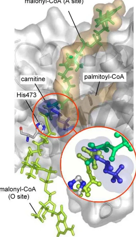
Molecular Model of Docking of C75-CoA into L-CPT I. The recent crystallization of carnitine octanoyltransferase (COT) (28), a member of the carnitine acyltransferase family, allowed us to improve the previous three-dimensional model of CPT I based on the carnitine acetyltransferase crystal (32). Using docking analysis, an in silico model was constructed for the interaction between the active center of CPT I and C75-CoA (Figure 5A). Refined in silico docking techniques were used, allowing free rotation of the ligand acyl chain bonds. This model suggests that the inhibitor C75-CoA fits into the enzyme in a manner similar to that of the physi-ological substrate palmitoyl-CoA. While the CoA segment in both molecules is positioned in almost the same orienta-tion, the aliphatic tail of C75 fits into the hydrophobic cavity of CPT I defined byR-helix 12 andâ-strands 1, 13, and 14 of the protein, just where the acyl group of palmitoyl-CoA is most likely positioned during normal enzymatic processes. The head of C75, bound to the sulfur atom of CoA, is located in the proximity of the catalytic residue His473.
Effects of C75 on CPT I ActiVity and Fatty Acid Oxidation in Cultured Cells. To assess whether CPT I inhibition is followed by a similar decrease in the extent of fatty acid oxidation, the three cultured cell lines [INS(812/13), L6E9, and HEK293] were incubated with C75 or etomoxir. It was not necessary in this case to transform the drugs to their CoA derivatives, since this conversion is assumed to occur inside the cells via endogenous acyl-CoA synthetase. CPT I activity
FIGURE4: Effects of C75-CoA and etomoxiryl-CoA on CPT I activity. Mitochondria isolated from rat liver (A), rat muscle (B), INS(823/
13) cells (C), or L6E9 myotubes (D) were preincubated for 1 min with increasing concentrations of C75-CoA (b) or etomoxiryl-CoA (O),
and CPT I activity was assayed as described in Experimental Procedures. Data represent the mean of at least three independent experiments and are expressed relative to control values in the absence of the inhibitor (100%). The inset shows an expanded dose-response curve for
the two inhibitors.
FIGURE5: Lineweaver-Burk plot for the inhibition of L-CPT I
by C75-CoA. Mitochondria-enriched fractions from yeast-overex-pressed L-CPT I (3-4µg) were preincubated with C75-CoA for 5
min and then incubated for 4 min at 30°C with various palmitoyl-CoA concentrations in the presence of 400 µM carnitine. The concentrations of C75-CoA were (b) 0, (O) 1, (2) 2, and (4) 5
decreased with increasing C75 concentrations (Figure 7). CPT I activity was reduced by 49, 62, and 62% in pancreatic INS(832/13), muscle L6E9, and kidney HEK293 cells, respectively, at maximal C75 concentrations of 30, 40, and 30 µg/mL, respectively. In parallel, the level of [1-14
C]-palmitate oxidation was reduced by 62, 84, and 68% in pancreatic INS(832/13), muscle L6E9, and kidney HEK293 cells, respectively, at maximal C75 concentrations of 30, 40, and 30µg/mL, respectively. When etomoxir was used, CPT I activity was reduced by 80, 52, and 71% in pancreatic INS-(832/13), muscle L6E9, and kidney HEK293 cells, respec-tively, while the level of palmitate oxidation was conse-quently reduced by 71, 78, and 77%, respectively. To rule out the possibility that the inhibition of palmitate oxidation reflected an increase in the level of cell death caused by C75 or etomoxir, we performed viability experiments using the MTT assay. In all cases, cell viability at the drug concentra-tions that were used was higher than 98% of that of control samples.
Effects of C75 Treatment on Whole Animals.We examined the effect of C75 on CPT I activity in vivo. Mice were injected (ip) with a single dose of either C75 or etomoxir (20 mg/kg of body weight) and killed at different time points thereafter. Tissue samples (liver, soleus, and pancreas) were taken to isolate mitochondria, as described in Experimental Procedures. CPT I activity decreased rapidly in all tissues that were assayed (Figure 8) but subsequently recovered, with tissue-dependent kinetics. The level of inhibition of liver CPT I decreased by 56% at 1 h and by 73% after treatment for 3 h, while at 5 h, CPT I values were similar to those of the control. Muscle CPT I was inhibited by 80% after being treated for 30 min, recovering more rapidly than liver CPT I. In the pancreas, CPT I activity decreased by 36% after C75 treatment for 30 min, compared with the control, recovering thereafter. In no case did the level of CPT I activation exceed the control. Etomoxir also provoked an inhibition of CPT I activity in these tissues (Figure 8). After treatment for 3 h, the levels of CPT I inhibition were 97,
FIGURE6: Proposed model for the location of C75-CoA in the CPT I active center. (A) Comparison of the location of a molecule of
palmitoyl-CoA in the active center of CPT I (left) and the proposed model for the location of C75-CoA in the same enzyme locus (right). The positions of carnitine and catalytic His473 (magenta), as well as secondary structure elementsR-helix 12 andâ-strands 1, 13, and 14,
[image:16.612.151.464.45.457.2]71, and 60% in liver, muscle, and pancreas, respectively, and they were always lower than those observed following C75 treatment. These inhibitory effects of etomoxir on CPT I activity, unlike those observed following C75 treatment, were maintained for up to 5 h, producing 96, 82, and 72% CPT I inhibition in liver, muscle, and pancreas, respectively.
DISCUSSION
In this study, we show that C75 and etomoxir were transformed to their CoA derivatives by the action of an acyl-CoA synthetase. Within the cell, acyl-acyl-CoAs are formed as part of the metabolism of a variety of endogenous fatty acids, as well as some xenobiotic carboxylic acids. The synthesis of CoA derivatives is the rate-limiting step for both conjuga-tion and inactivaconjuga-tion of most xenobiotics. CoA conjugates increase the chemical reactivity of these compounds and may function as alternative substrates in intermediate metabolism pathways of short-, medium-, and long-chain fatty acids (33). Distinct mammalian long-chain and xenobiotic/medium-chain fatty acid:CoA ligases [termed acyl-CoA synthetase in revised nomenclature (34)] can activate long-chain,
xenobiotic, and medium-chain fatty acids. The substrate specificity and intracellular location of the acyl-CoA syn-thetases may explain the rate differences in the synthesis of the xenobiotic-CoA derivatives and their possible toxicity. Although C75 is a potential substrate for acyl-CoA syn-thetase, due to its aliphatic C8 chain and its esterifiable carboxylate groups, no studies have addressed this fact. The MALDI-TOF analysis performed in this study showed that C75-CoA was synthesized when C75 was incubated in vitro in the presence of CoA, long-chain acyl-CoA synthetase, and ATP.
Compounds such as etomoxir, TDGA, and POCA must be converted to their CoA derivative before acting as inhibitors of CPT I activity (12). In the same way, when C75 is transformed into its CoA derivative, it also becomes a potent inhibitor of CPT I. Under all the in vitro conditions that were tested, CPT I was clearly inhibited in a dose-dependent manner by C75-CoA. IC50 values observed for
C75-CoA ranged from the micromolar level to the nanomolar level and were lower than those obtained for etomoxiryl-CoA, showing that C75-CoA is a stronger CPT I inhibitor
FIGURE7: CPT I activity and palmitate oxidation in cell cultures. Cells were incubated for 2 h (L6E9) or 1 h [INS(832/13) and HEK293]
with complete medium containing either 0, 10, 20, 30, and 40µg/mL C75 or 30 and 40µg/mL etomoxir. Mitochondria-enriched cell fractions were obtained, and 10µg of protein was used for the CPT I activity assay. In palmitate oxidation assays, cells were preincubated for 30 min at 37°C with KRBH with 1% BSA and then incubated for 2 h at 2.5 mM glucose in the presence of 0.8 mM carnitine, 0.25 mM palmitate, and 1µCi/mL [1-14C]palmitate. Palmitate oxidation to CO
2was assessed as described in Experimental Procedures. Data are
presented as the mean(the standard error of three independent experiments. *P<0.05, **P<0.01, and ***P<0.001 compared with