Expression of MMP-1 and MMP-11 in squamous cell carcinoma of the
nasal cavity and paranasal sinuses
María de Lourdes Suárez Roa,* Juan Asbun,** Angélica Ruiz,* Luz María Ruiz Godoy,*** Abelardo Meneses García****
RESUMEN
Antecedentes: el pronóstico en pacientes con carcinoma epidermoide de la cavidad nasal y de los senos paranasales es malo, sobre todo por invasión, recurrencia y metástasis locorregionales.
Objetivo: evaluar la expresión de MMP-1 y MMP-11 en las células escamosas de los carcinomas epidermoides de la cavidad nasal y los senos paranasales.
Pacientes y método: diecisiete pacientes con historia clínica completa, sin tratamiento previo al diagnóstico inicial con biopsia y material adecuado para el estudio inmunohistoquímico de MMP-1 y MMP-11. Se utilizaron la prueba de la c2 y la exacta de Fisher para evaluar la ex-presión de ambas endopeptidasas de los fibroblastos neoplásicos y peritumorales, y su relación con los parámetros clinicopatológicos. Resultados: encontramos 10 mujeres y siete hombres con edad promedio de 58 años (37 a 85); dos en etapa clínica II, seis en III y nueve en IV. Trece y 12 casos fueron positivos en las células neoplásicas, y 13 y 10 en los fibroblastos peritumorales para MMP-1 y MMP-11, respectivamente.
Conclusiones: estas endopeptidasas son expresadas en carcinomas epidermoides de la cavidad nasal y de los senos paranasales por las células neoplásicas y por los fibroblastos peritumorales, lo que sugiere que podrían tener acciones adyuvantes en los mecanismos de progresión tumoral y que su baja expresión puede indicar un pronóstico más favorable.
Palabras clave: metaloproteasas, carcinoma de células escamosas, cavidad nasal y senos paranasales.
ABSTRACT
Background: Poor prognosis in patients with squamous cell carcinoma of the nasal cavity and paranasal sinuses is associated mainly with invasion, recurrence, and loco-regional metastases.
Objective: To evaluate the expression of MMP-1 and -11 in NCPS squamous cell carcinoma.
Patients and method: Seventeen patients with complete clinical record, no treatment before the initial diagnostic biopsy, and adequate material for the immunohistochemical study of MMP-1 and MMP-11. c2 and Fisher’s exact test were used to assess MMP-1 and MMP-11 expression of neoplastic and peritumoral fibroblasts and its relation to clinicopathological parameters.
Results: We found 10 females and 7 males with 58 years old mean age (37 to 85); two were classified as clinical stage II, six as III, and nine as IV. For MMP-1, 13 cases (76%) were positive in neoplastic cells and 13 in peritumoral fibroblasts. For MMP-11, 12 (71%) were positive in neoplastic cells and 10 (59%) in peritumoral fibroblasts.
Conclusions: MMP-1 and MMP-11 are expressed in squamous cell carcinoma of the nasal cavity and paranasal sinuses by both neoplastic cells and peritumoral fibroblasts, suggesting that they might be exerting adjuvant actions in the mechanisms of tumor progression and that their low expression could be related to a favorable prognosis.
Key words: metalloproteases, squamous cell carcinoma, nasal cavity and paranasal sinuses.
* División de patología, Instituto Nacional de Cancerología, México.
** Escuela Superior de Medicina, Instituto Politécnico Nacional, México.
*** Investigación básica, Instituto Nacional de Cancerología, México.
**** Director médico, Instituto Nacional de Cancerología, México.
Correspondencia: Dr. Abelardo Meneses García. División de pato-logía. Instituto Nacional de Canceropato-logía. San Fernando núm. 22, CP 14080, Delegación Tlalpan, México, DF.
E-mail: [email protected]
Recibido: octubre, 2007. Aceptado: junio, 2008.
La versión completa de este artículo también está disponible en:
www.revistasmedicasmexicanas.com.mx
Revista latinoamericana
Artículo original
Patología 2008;46(3):209-14
M
alignant neoplasms of the nasal cavity and paranasal sinuses (NCPS) are infre-quent tumors representing about 3% of all malignant neoplasms of head and neck.1,2The poor prognosis of patients with these neoplasms is related to the complex anatomy of the region, diagnosis in advanced stages, extension to adjacent structures, and frequent loco-regional recurrences.1-3
Sixty-three percent of the malignant NCPS neoplasms correspond to squamous cell carcinomas (SCC),1-3 which
recurrence. In this tumor, the degree of histological dif-ferentiation and the invasion pattern, either in small groups or individual neoplastic cells, lead to a more aggressive behavior.4,5
Tumor invasion and development of metastasis require degradation of the basal membrane (BM) and extracellu-lar matrix (EM), a process in which several enzymes are involved. Among these enzymes are the matrix metallo-proteases (MMPs) that are able to degrade completely all components of the BM and EM.6 These zinc-dependent
metalloproteases are classified by their specific substrate
in: collagenases, stromelysins, gelatinases, matrilysins, and membranal-type MMPs.6,7 Several MMPs have been
described in different tumors and locations and may be considered as possible tumor markers and/or treatment targets. However, the large amount of available data has generated discrepancies in evaluating their precise involvement.6-8
Of the different MMPs, the interstitial collagenase
(MMP-1) presents specific substrates for collagenases I,
II, III, VII, VIII, X, and for proteoglicans.
These enzymes are produced in small amounts by a variety of normal cells. Nevertheless, it has been reported that some tumors express high levels of this enzyme, mainly the invasive neoplastic cells,9 and this has been
related with poor prognosis in patients with breast, skin,10
gallbladder,11 esophageal,7 stomach, colorectal and oral
cavity7-11 cancers.
Stromelysin-3 (MMP-11) specific substrates are casein, laminin, fibronectin, gelatinin, collagenase IV, and car -boxymethylated transferrin. Its participation in malignant transformation of cells is currently being considered, and it has been correlated with an aggressive biological behavior and poor prognosis in patients with breast, lung, ovarian, laryngeal, and oral cavity cancers.12-15
There are few studies on the participation of these enzymes in NCPS squamous cell carcinoma. Considering that these regions have epidemiological, histological, and clinical characteristics that differ from the rest of head and neck structures, we decided to evaluate the expression of MMP-1 and -11.
MATERIAL AND METHODS
From the pathological records of the National Cancer Institute in Mexico, 55 cases with histological diagnosis
by biopsy of primary SCC of NCPS were identified be
-tween 1998 and 2003. Of these, 17 fulfilled the following
inclusion criteria: complete clinical record, no treatment before the initial diagnostic biopsy, and adequate mate-rial for the immunohistochemical study of MMP-1 and MMP-11. Of the 17 cases, ten were females and seven were males, with an average age of 58 years (range 37 to
85). Two patients were classified as clinical stage II, six
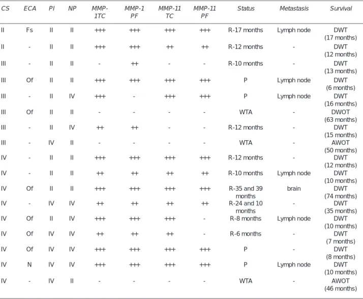
to stage III, and nine to stage IV. Eight patients presented extension to adjacent critical structures. Twelve cases (70%) were moderately differentiated SCC, four (24%) were poorly differentiated, and one (6%) was a well-dif-ferentiated carcinoma. Eleven cases (65%) presented an invasion pattern in layers, cords, and solid bands; six (35%) presented invasion in small groups and individual neoplastic cells. Regarding nuclear polymorphism, 50% to 70% of mature malignant cells (considered as moderate nuclear polymorphism) were found in ten cases (59%). The remnant seven cases (41%) presented less than 25% of mature neoplastic cells (high nuclear polymorphism). Most patients received combined treatment based on radiotherapy and surgery (eight patients), four patients received chemotherapy, and two patients were subjected to chemo- and radiotherapy. Ten patients presented local recurrences, three developed lymphatic node metastasis, and one of the latter metastasized to the brain. Tumor persistence was observed in four patients (23.5%), six developed lymphatic node metastasis. The 14 patients with persistence and/or recurrence with tumor activity died (82%). Of the remainder three patients (18%) two were alive without tumor activity and one died without tumor. The postoperative follow-up of patients was 40 months, with an average 6 to 24 months (table 1).
IMMUNOHISTOCHEMISTRY
Paraffin blocks were cut into 2μ sections containing
rep-resentative areas. They were deparaffinized in xylene and
second-Table 1. Clinical and histological characteristics and MMP-1 and MMP-11 expression in squamous cell carcinoma of the nasal cavity and paranasal sinuses
CS ECA PI NP
MMP-1TC
MMP-1 PF
MMP-11 TC
MMP-11 PF
Status Metastasis Survival
II Fs II II +++ +++ +++ +++ R-17 months Lymph node DWT
(17 months)
II - II II +++ +++ ++ ++ R-12 months - DWT
(12 months)
III - II II - ++ - - R-10 months - DWT
(13 months)
III Of II II +++ +++ +++ +++ P Lymph node DWT
(6 months)
III - II IV +++ - +++ +++ P Lymph node DWT
(16 months)
III Of II II - - - - WTA - DWOT
(63 months)
III - II IV ++ ++ - - R-12 months - DWT
(15 months)
III - IV II - - - - WTA - AWOT
(50 months)
IV - II II +++ +++ +++ +++ R-12 months - DWT
(12 months)
IV - II II ++ ++ ++ ++ R-10 months Lymph node DWT
(10 months)
IV Of II II +++ +++ +++ +++ R-35 and 39
months
brain DWT
(74 months)
IV - IV IV ++ ++ ++ ++ R-24 and 10
months
- DWT
(35 months)
IV Of II IV +++ +++ +++ - R-8 months Lymph node DWT
(10 months)
IV Of IV IV ++ ++ ++ - R-6 months - DWT
(7 months)
IV Of IV IV +++ +++ +++ +++ P - DWT
(8 months)
IV N IV IV +++ +++ +++ +++ P Lymph node DWT
(10 months)
IV - IV II - - - - WTA - AWOT
(46 months)
CS: clinical stage, ECA: extension to adjacent areas, Fs: frontal sinus, OF: orbital floor, N: nasopharynx, PI: pattern of invasion, NP: nuclear polymorphism, TC: tumoral cells, PF: peritumoral fibroblasts, R: recurrence, P: persistence, WTA: without tumor activity, DWT: death with tumor, DWOT: death without activity, AWOT: alive without tumor.
ary antibody labeled with biotin was added for 15 min at room temperature. Afterwards, streptavidin conjugated to peroxidase (LSAB + System HRP Universal, Dako Cyto-mation, USA), was added to react with diaminobenzidine (Liquid DAB, Dako Cytomation, USA) and counterstained with Mayer’s hematoxylin. As positive control, ovary and placental tissue samples, treated under the same condi-tions, were included.
MMP-1 and MMP-11 expression was assessed by two independent observers (AMG and LSR) in the area cor-responding to the invasive front in both neoplastic and
peritumoral fibroblasts, in five high-resolution (40x) fields.
Cases considered positive were those exhibiting more than 20% of moderate to intense expression in the cytoplasm of the studied cells. The surface epithelium and the ducts were used as internal basal control.8
STATISTICAL ANALYSIS
Statistical analysis was based on c2 and Fisher’s exact test
to assess MMP-1 and MMP-11 expression of neoplastic
-pathological parameters, using the Sigma Stat Ver. 3.00 software (SPSS, USA). A p value < 0.05 was considered
statistically significant.
RESULTS
Expression varied from moderate to intense in the positive cases for both MMP-1 and MMP-11. For MMP-1, 13 cases (76%) were positive in neoplastic cells and 13 in
peritu-moral fibroblasts. For MMP-11, 12 (71%) positive cases
were found in neoplastic cells and in ten (59%) positivity
was expressed in peritumoral fibroblasts (figure 1). Table
2 shows the clinical characteristics of each patient and the assessment of MMP-1 and MMP-11 expression.
Figure 1. A) Microphotograph of MMP-1 immunoexpression in SCC of NCPS showing neoplastic and peritumoral stromal cells with an invasion pattern in pushing borders (40x). B) Microphotograph of MMP-11 immunoexpression in SCC of NCPS showing nests of neoplastic and peritumoral stromal cells (40x).
A B
Dependence of MMP-1 expression in tumor and
peritu-moral fibroblasts on clinicopathological parameters
MMP-1 expression in tumor cells was found to be as-sociated with patient status (p = 0.015) but not with metastasis.
Dependence of MMP-11 expression in tumor and
peritu-moral fibroblasts on clinicopathological parameters
MMP-11 expression in tumor cells was found to be as-sociated with all clinicopathological parameters assessed (metastasis p = 0.044 and status p = 0.003)
The literature on different malignant neoplasms of NCPS, revision period among 4 to 20 years with major populations of 80 cases, where the percentages of SCC are between 20 to 30%. In this study of 52 cases, patients had neoplasms in advanced stages when the diagnosis was
made, with non specific, poor prognosis and a maximal survival of five years.11,16,17
In this series of SCC cases of NCPS, clinical (ad-vanced clinical stage and invasion to adjacent structures) and histological (pleomorphism and invasion pattern) characteristics considered as predictors of the status and survival of patients did not correlate with data referred in the literature.1-3 These discrepant findings indicate the
need to understand the diverse mechanisms involved in the biological behavior of neoplasms.
Different enzymes mediate tumoral invasion, with MMPs been associated with tumor progression. However,
Table 2. Positive (+) and negative (-) cases of MMP-1 and MMP-11 expression and their association with the patient status, survival, and metastasis
Clinicopathological parameters MMP-1 MMP-11
Tumor cell Peritumoral fibroblasts Tumor cell Peritumoral
fibroblasts
+ - + - + - +
-Metastasis
With Without
7 6
0 4
6 7
1 3
7 5
0 5
6 4
1 6
p value ns ns 0.044 ns
Status
Recurrence Persistence
Without tumor activity
8 4 1
1 0 3
9 3 1
0 1 3
8 4 0
1 0 4
6 4 0
3 0 4
p value 0.015 ns 0.003 0.005
Survival
Death with tumor Alive without tumor
12 1
1 3
12 1
1 3
12 0
1 4
10 0
3 4
p value 0.022 0.022 0.002 0.015
up to now the role played by the diverse MMPs has not been clearly established.
The main group of MMPs widely studied in SCC of head and neck are the gelatinases (MMP-2 and MMP-9), which have been associated with invasion processes and metastases (the advanced stages in tumoral progression). Those enzymes have specific substrates for collagen types I, II, III, IV, V, VII, X, XI, and XIV; gelatin, elastin,
fibronectin, laminin-1, laminin-5 galectin-3, and proteo -glicans, among others. Nonetheless, contradictory results are reported in the literature regarding their participation, suggesting that they are not the only MMPs involved in these processes.6-11,17
In our series, we detected the presence of MMP-1 and MMP-11 in neoplastic and peritumoral stromal cells by means of immunohistochemistry.
This technique allows the direct assessment of expres-sion intensity in the cells and allows the correlation with invasion at the biological level.17
MMP-1 initiates degradation of collagen I, which is abundant in the extracellular matrix16 and is essential
for keratinocyte migration; several authors consider that these mechanisms facilitate tumoral invasion.6,7,9,11
MMP-1 expression has been described in both neoplastic and peritumoral stromal cells;16,17 however, its presence
is considered more important in the zone of greatest activity corresponding to the tumoral front.9,17 In a study
of SCC of the larynx, in which MMP-1 was evaluated,
no significant tendency was found in its expression in
invasion processes and metastasis.11 On the other hand,
Kurahara et al.,8 in a study of the oral cavity, suggested
that participation of MMP-1 together with other MMPs is essential for the invasion process. In our series, the pres-ence of MMP-1 in both types of cells at the level of the tumoral front suggests its participation in the processes of tumor persistence and recurrence, showing a more ag-gressive behavior and a poor prognosis in patients with SCC of the NCPS.
However, it has been shown that MMP-11 is expressed
in peritumoral fibroblasts of the area corresponding to the
tumoral front, indicating its stromal origin, based on its paracrine stimulation by tumoral cells. It is considered that this stromelysin can also be related with progression of phenotypic alterations acquired during malignant trans-formation.6,7 In some studies on colorectal cancer in mice,
MMP-11 deficiency coincides with a decrease in tumor
genesis. The mechanisms implicating in MMP-11 in tumor progression include inhibition of neoplastic cells death, thereby fostering survival in the stromal environment.6
Studies on MMP-11 in head and neck tumors are scarce; Arora et al. performed a study in patients with SCC of the oral cavity in normal tissue, pre-malignant and invasive lesions also in a cell line.
In our study, we observed a relation between MMP-11 expression and persistence and recurrence of tumors, which could suggest participation of this enzyme in a more aggressive behavior of the disease as seen in other tumors.12,13
For tumoral cells, blood circulation is considered a hostile environment, as they can be affected by mechani-cal deformation, immunologic attack, serum toxicity, or high shear stress. Neoplastic cells acquire a variety of mechanisms in which MMPs are involved. Based on ex-periments, it has been suggested that MMP-11 generates bioactive fragments of the α-1-proteinase inhibitor, which could reduce the action of natural killer cells and, thereby, increase tumor growth and invasion in vivo.6
Immunopositivity for MMP-11 was found in both neoplastic and peritumoral stromal cells in biopsies of pa-tients with metastases. This is relevant because in papa-tients without treatment, expression of this enzyme could be an important predictor of metastasis.
In conclusion, our results show that these enzymes are expressed in SCC of the NCPS in neoplastic and peritumoral stromal cells, suggesting that they might co-participate in tumor progression processes, and that a lower expression of these enzymes might indicate a favorable prognosis. In addition, it could be considered that MMP-11 might play a relevant role in fostering metastasis, but it is necessary to increase the number of cases as well as to perform other types of study to understand the diverse mechanisms in which these enzymes participate in the pathogenesis of tumors and their interaction with the extracellular matrix.
The objective of this study was to explore MMPs -1 and -11 expression in SCC of the NCPS, because these MMPs were found in other similar tumors as well as in oral cavity squamous cell carcinoma. This research could be the start for other studies regarding MMPs expression and activ-ity in these aggressive tumors. It would be important to increase the number of cases as well as patients follow-up
Patología de las miopatías más frecuentes
Autor: Alicia Rodríguez Velasco Tamaño: 14 x 21 cm
Páginas: 157
Editado por: Editorial Prado, S. A. de C. V. País: México
Edición: primera, 2005.
Este libro de la doctora Alicia Rodríguez es un regalo para cualquiera que esté interesado en las enfermeda-des musculares de los niños. En unas cuantas páginas se podrán revisar los hallazgos clínicos, paraclínicos histopatológicos, de histoquímica y ultraestructura característicos de las miopatías más frecuentes. Es muy útil para pediatras, neurólogos, neuropediatras,
ortopedistas, fisioterapeutas, genetistas, patólogos y
estudiantes de medicina. REFERENCES
1. Porceddu S, Martin J, Shanker G, Weih L, et al. Paranasal sinus tumors: Peter MacCallum Cancer Institute experience. Head Neck 2004;26(4):322-30.
2. Dulguerov P, Jacobsen MS, Allal AS, Lehmann W, Calcaterra T. Nasal and paranasal sinus carcinoma: are we making prog-ress? A series of 220 patients and systematic review. Cancer 2001;92:3012-25.
3. Carrillo JF, Guemes A, Ramirez-Ortega MC, Oñate-Ocaña LF. Prognostic factors in maxillary sinus and nasal cavity carci-noma. Eur J Surg Oncol 2005;31:1206-12.
4. Anneroth G, Batsakis J, Luna M. Review of the literature and a recommended system of malignancy grading in oral squamous cell carcinomas. Scand J Dent Res 1987;95:229-49. 5. Bundgaard T, Rossen K, Henriksen SD, Charabi S, et al.
His-topathology parameters in the evaluation of T1 squamous cell carcinomas of the oral cavity. Head Neck 2002;24:656-60. 6. Deryugina EI, Quigley JP. Matrix metalloproteinases and tumor
metastasis. Cancer Metastasis Rev 2006;25:9-34.
7. Curran S, Murray G. Matrix metalloproteinases in tumor inva-sion and metastasis. J Pathol 1999;189:300-8.
8. Kurahara S, Shinohara M, Ikebe T, Nakamura S, et al. Ex-pression of MMPs, MT-MMP, and TIMPs in squamous cell carcinoma of the oral cavity: correlations with tumor invasion and metastasis. Head Neck 1999;21:627-38.
9. Brinckerhoff CE, Rutter JL, Benbow U. Interstitial collagenases as markers of tumor progression. Clin Cancer Res 2000;6:4823-30.
10. Sutinen M, Kainulainen T, Hurskainen T, Vesterlund E, et al.
Expression of matrix metalloproteinases (MMP-1 and -2) and their inhibitors (TIMP-1, -2 and -3) in oral lichen planus, dys-plasia, squamous cell carcinoma and lymph node metastasis. Br J Cancer 1998;77:2239-45.
11. Gorogh T, Beier UH, Baumken J, Meyer JE, et al. Metallopro-teinases and their inhibitors: influence on tumor invasiveness and metastasis formation in head and neck squamous cell carcinomas. Head Neck 2006; 28:31-9.
12. Soni S, Mathur M, Shukla NK, Deo SV, Ralhan R. Stromelysin-3 expression is an early event in human oral tumorigenesis. Int J Cancer 2003;1:107:309-6.
13. Skoglund J, Emterling A, Arbman G, Anglar P, Sun X-F. Clinicopathological significance of stromelysin-3 expression in colorectal cancer. Oncology 2004;67:67-72.
14. Arora S, Kaur J, Sharma C, Mathur M, et al. Stromelysin 3, Ets-1, and vascular endothelial growth factor expression in oral precancerous and cancerous lesions: correlation with microvessel density, progression, and prognosis. Clin Cancer Res 2005;11:2272-84.
15. Muller D, Wolf C, Abecassis J, Millon R, et al. Increased stromelysin 3 gene expression is associated with increased local invasiveness in head and neck squamous cell carcinomas. Cancer Res 1993;53:165-9.
16. Ziober BL, Turner MA, Palefsky JM, Banda MJ, Kramer RH. Type I collagen degradation by invasive oral squamous cell carcinoma. Oral Oncology 2000;36:365-72.