ACOUSTIC PROPERTIES OF CANCELLOUS BONE
AND EVALUATION OF BONE DENSITY
PACS REFERENCE: 43.80.Qf
OTANI Takahiko; SHIMOI Yuichi
Department of Electrical Engineering, Doshisha University 1-3 Miyakodani, Tatara
Kyotanabe-shi 610-0321 Japan
Tel.: (81) 774 65 6267 Fax: (81) 774 65 6801
E-mail: [email protected]
ABSTRACT
The propagation of both fast and slow longitudinal waves in bovine cancellous bone in vitro was experimentally examined by the pulse transmission technique in relation to the bone density. Experimental results show that the propagation speed and the amplitude of the fast wave increase greatly with the bone density and those of the slow wave decrease reversely with the bone density.
I. INTRODUCTION
Osteoporosis is a disease characterized by decrease in bone density and deteriorations in bone microstructure, which cause bone fragility and increase in fracture risk. Bone tissues are elastic and can be classified into two types depending on macroscopic and microscopic features. Bone with a low volume fraction of solid (less than 70%) is called cancellous (spongy) bone and above 70% cortical (compact) bone. Whereas cortical bone is a dense hard solid, cancellous bone is comprised of a porous network of numerous rod-like and plate-like solid trabecular elements with soft tissue (bone marrow) in the pore space. The effect of decreasing bone density (or a symptom of osteoporosis) is stronger for cancellous bone than for dense cortical bone, because cancellous bone is metabolically more active. Accordingly, it is adequate to measure bone density at the position with a high volume fraction of cancellous bone to estimate the onset of osteoporosis. X-ray method or X-ray absorptiometry is now widely used for non-invasive measurement of bone mineral density for the assessment of osteoporosis. Ultrasonic method or Ultrasonic Osteodensitometer has been proposed as an alternative in the diagnosis of osteoporosis. Ultrasonic method takes several advantages over X-ray bone densitometry as it is radiation-free and inexpensive technique which has the potential to evaluate the elastic or mechanical properties of bone. However the reproducibility and accuracy of ultrasonic measurements are a problem peculiar to the ultrasonic wave propagation phenomenon due to the complexity of bone structure. Ultrasonic evaluation of bone density is based on the correlation between the bone mass density measured by X-ray densitometry and the slope of frequency dependent attenuation (or broadband ultrasonic attenuation, BUA) and/or the ultrasonic wave propagation speed (or speed of sound, SOS).
waves is more reasonable than the conventional method by BUA and SOS. In the present study, the effects of the bone density (bone porosity) on the amplitude and the propagation speed of transmitted both fast and slow waves through bovine cancellous bone are experimentally examined.
II. ULTRASONIC WAVE PROPAGATION IN CANCELLOUS BONE II.1. Biot’s Equations
Maurice A. Biot initially proposed a general theory of elastic wave propagation in a system composed of a porous elastic solid saturated with a viscous fluid. Biot’s theory is basically developed by considering a dynamic coupling between the solid and fluid, which is the relative motion of the fluid in the pore spaces to the solid frame. The average motion of the solid and fluid are separately described. The wave equations for compressional (longitudinal) waves are given as[4-6] ), ( ) ( 2 2
2 ζζ ρρ ρρζζ
f e t C He −− ∂∂ ∂∂ == −− ∇ ∇ (1) . ) ( ) ( ) ( 2 2 2 t k F m e t M Ce f ∂∂ ∂∂ −− −− ∂∂ ∂∂ == −− ∇
∇ ζζ ρρ ζζ κκηη ζζ (2)
Here, e is the dilatation of a volume element attached to the solid. ζζ is the fluid volume that flows into or out of that element called “increment of fluid content”. Th e harmonic solutions of eq.(1) and (2) yield the dispersion relation for the longitudinal waves at high frequencies [4-7]
0 / / / / / == −−
−−−−ρρρρf −−αρραρρ f ββ f z M z C z C z H (3)
where z=v2. Equation (3) has two roots corresponding to two speeds of propagation, vfast and
slow
v . Two longitudinal waves have the form as
2 fast v −− −− −− ++ −− −− ++ −− == 2 1 2 2 2 2 ) ( ) ( 4 ) 2 ( ) 2 ( ) ( 2 f f f m C HM C Hm M C Hm M C HM ρ ρ ρ ρ ρ ρ ρ ρ ρ ρ ρ ρ m (4) 2 slow
v .
The terms H, C and M are generalized elastic coefficients deduced by Biot and expressed in terms of the bulk moduli of the solid Ks, the pore fluid Kf, the bulk modulus Kb and the shear modulus µµof the skeltal frame and the porosity ββ[4-7]. According to Gibson, the bulk modulus Kb and shear modulus µµ of the skeltal frame of cancellous bone are expressed as a function of porosity ββ [8].
, ) 2 1 ( 3 ) 1 ( b n s b E K ν ν β β −− −− == (5) , ) 1 ( 2 ) 1 ( b n s E ν ν β β µ µ ++ −− == (6)
where Es is the Young’s modulus of the solid bone, ννb is the Poisson’s ratio of the skeltal frame and n is a va riable depending on the geometrical structure of the cancellous bone. In terms of the porosity ββ and the density of the solid ρρs and fluid ρρf, the total density ρρ of the fluid-saturated medium is given by
. )
1
( ββρρs βρβρf ρ
ρ== −− ++ (7)
The term αα is the structure factor, and is obtained from the relation determined by Berryman[9,10]:
) 1 1 (
1 ββ
α
α== −−r −− (8)
II.2. Experimental Results[1]
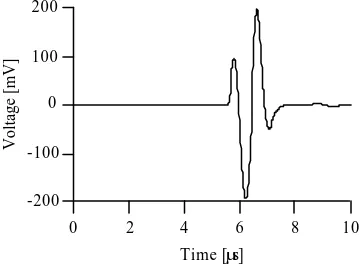
The experimental arrangement for transmission of ultrasonic pulse is shown in Fig.1. Cancellous bone specimens, 20-30 mm in size and 9 or 7 mm thickness, were cut from the distal epiphysis of bovine femora, with soft tissue in situ. A single sinusoidal pulse wave of 1 MHz, transmitted by a wide band PVDF transmitter, was used in order to observe the fast and slow waves separately. Figure 2 shows the pulsed waveform travelling in water, which is applied to the specimens. Figure 3 shows typical waveforms travelling through the cancellous bones in the direction of trabecular alignment. Figure 3(a) is a waveform for the high density (ρρ=1200 kg/m3) specimen and (b) is a low density (ρρ=1120 kg/m3). In both Fig.3(a) and (b), the fast and slow longitudinal waves can be clearly observed in the time domain. As the density increases (i.e., as the volume fraction of the solid bone increase), the amplitude of the fast wave becomes greater. At the same time, the amplitude of the slow wave decreases. Accordingly, it can be deduced that the fast wave is associated with solid core in cancellous bone, and the slow wave with the propagation in soft tissue.
[image:3.596.324.504.254.386.2]The propagation speeds and attenuations of the fast and slow waves were measured using pulse spectrum analysis. Figure 4 shows the propagation speeds of the fast and slow waves in cancellous bone at 1 MHz as a function of porosity ββ. The speed of the fast wave varies from 2700 to 2200 m/s as the porosity increases. The slow wave remains constant at about 1400 m/s, which is close to the propagation speed of 1450 m/s in bone marrow. Thus the ultrasonic properties of the soft tissue in cancellous bone can be expected to be similar to the bone marrow, and it can be therefore assumed that the slow wave is mainly dominated by soft tissue. The propagation speed 2200-2700 m/s of the fast wave is much slower than the propagation speed 3400-4200 m/s of cortical bone (solid bone). This can be explained by the fact that the cancellous bone is not solid but has a spongy or porous structure.
[image:3.596.97.269.256.392.2]Figure 3. Pulsed waveform traveling through cancellous bone: (a) high density; (b) low density (a) (b)
Figure 1. Experimental arrangement for transmission of ultrasonic pulse.
Figure 2. Pulsed waveform traveling in water.
-20 -10 0 10 20
Voltage [mV]
10 8 6 4 2 0
Time [µs]
Fast wave Slow wave
-20 -10 0 10 20
Voltage [mV]
10 8 6 4 2 0
Time [µs] -200
-100 0 100 200
Voltage [mV]
10 8 6 4 2 0
Time [µs] Function
generator Power Amp.
Transmitter
Preamp.
Oscilloscope Specimen
[image:3.596.103.489.442.592.2]Figure 5 shows the frequency dependence of the propagation speeds for a specimen of porosity ββ=0.81 (density ρρ=1140 kg/m3). No data for the fast wave at frequencies over 1.5 MHz was obtained because the weak amplitude of the signal made further measurement difficult. In Fig.5, the propagation speeds of both the fast and slow waves are considered nondispersive in the range 0.5-5 MHz. Figure 6 shows the attenuation for the three specimens of ββ=0.75, 0,81 and 0.83 (density ρρ=1200, 1140 and 1120 kg/m3). From Fig.6(a) it can be seen that the attenuation of the fast wave at porosity ββ=0.75, 0.81 and 0.83 are about the same. The attenuation of the slow wave (Fig.6(b)) increases with frequency and the variation of the slow wave with the porosity ββ is large. The attenuation in cortical bone and bone marrow were also obtained as about 5.0×10⁻² [neper/mmMHz] and 1.3×10⁻² [neper/mmMHz]. Both the fast and slow waves in cancellous bone show much higher attenuation than the bulk wave in cortical bone (ρρ=1960 kg/m3) or bone marrow (ρρ=930 kg/m3). Accordingly, both the fast and slow waves are attenuated not only by the solid core or soft tissue component in the cancellous bone but also by the porous structure of the cancellous bone.
III. BONE DENSITY AND ULTRASONIC WAVE TRANSMISSION THROUGH CENCELLOUS BONE
In Fig.6 of the ultrasonic wave attenuation in cancellous bone, the attenuation of the fast wave is almost independent of the bone density. However, observed pulse waveforms through
4000
3000
2000
1000
Propagation speed [m/s]
1.0 0.8 0.6 0.4 0.2 0.0
Porosity β Fast wave
Slow wave Measured Calculated
4000
3000
2000
1000
Propagation speed [m/s]
0.1 1 10
Frequency [MHz] Fast wave
Slow wave Measured Calculated
[image:4.596.316.484.76.236.2]Figure 4. Propagation speeds of fast and slow waves in cancellous bone at 1 MHz as a function of porosity ββ.
Figure 5. Propagation speeds of fast and slow waves in cancellous bone at a porosity of ββ=0.81 as a function of frequency.
0.01 0.1 1
Attenuation [neper/mm]
0.1 1 10
Frequency [MHz] β=0.75 β=0.81 β=0.83
(a)
0.01 0.1 1
Attenuation [neper/mm]
0.1 1 10
Frequency [MHz]
(b)
[image:4.596.87.494.83.476.2] [image:4.596.100.485.290.473.2]cancellous bone in Fig.3 show that the amplitude of the fast wave becomes greater as the bone density increases. The fact that the fast wave is associated with porous solid core in cancellous bone and the slow wave with soft tissue in the pore space, means the cancellous bone as an acoustic medium has two different characteristic impedances for the fast wave and for the slow wave. The characteristic impedance for the fast wave is essentially dominated by the mass of solid core (bone density) and the characteristic impedance for the slow wave is dominated by the soft tissue. Consequently, the transmission coefficient of the fast wave is strongly dependent on the bone density of the cancellous bone. Thus, it would be deduced that the amplitude of transmitted fast wave depends greatly on the transmission coefficient at the boundary of cancellous bone or on the bone density, and that the amplitude of the slow wave depends mainly on the attenuation coefficient of cancellous bone. As for the propagation speed through cancellous bone, the dependence on bone density is high for the fast wave and very small for the slow wave as shown in Fig.3.
The amplitudes and propagation speeds of the fast and slow waves through cancellous bone in vitro were measured in a water tank. A focused PVDF transmitter was driven by a single sinusoidal impulse voltage of 50 V peak to peak with a frequency of 1 MHz. Pulse waves propagating through the water/specimen/water system was detected by a wide band non focused PVDF hydrophone and the output was amplified by a 40 dB preamplifier. A cancellous
[image:5.596.365.493.323.427.2]bone specimen of about 10 mm thickness was cut from the distal epiphysis of bovine femur with soft tissue in situ as shown in Fig.7. The specimen was mounted between the transmitter and the hydrophone at normal incidence. Ultrasonic wave path was scanned in an area of 15×15 mm and transmitted waves were measured at 1 mm intervals or 225 points. The scanned area was taken in the nearly middle region of the specimen, where the trabecular alignment was approximately parallel to the thickness direction or the direction of ultrasonic wave propagation. The local bone density corresponding to the measured points was obtained by use of a micro focus X-ray CT system. The dependence of measured peak to
Figure 7. Sectional view of a distal epiphysis of bovine femur.
12
8
4
0
Amplitude [mV]
100 80 60 40 20 0
Bone volume fraction [%]
400
300
200
100
0
Amplitude [mV]
100 80 60 40 20 0
Bone volume fraction [%]
3000
2000
Propagation speed [m/s] 0 20 40 60 80 100
Bone volume fraction [%]
2000
1500
1000
Propagation speed [m/s] 0 20 40 60 80 100
[image:5.596.312.502.476.716.2]Bone volume fraction [%]
Figure 8. Amplitude and propagation speed of the fast wave through bovine cancellous bone as a function of bone volum e fraction.
[image:5.596.94.280.479.719.2]peak amplitudes and propagation speeds is shown for the fast wave in Fig.8 and for the slow wave in Fig.9. The bone density has a strong positive correlation with both amplitude and propagation speed for the fast wave and a clear negative correlation for the slow wave.
IV. CONCLUSION
Ultrasonic wave propagation through cancellous bone has been experimentally examined. It was shown that both fast and slow longitudinal waves propagate through cancellous bone in the direction of the trabecular alignment, and that the fast wave is associated with solid core of cancellous bone. The slow wave is associated with soft tissue in the pore spaces. The propagation speed of the fast wave depends strongly on the bone density and that of the slow wave depends slightly and negatively on the bone density. The amplitude of the fast wave is mainly determined by the transmission coefficient and consequently depends strongly on the bone density. The amplitude of the slow wave is mainly given by the attenuation coefficient of the soft tissue and depends on the visco-elastic characteristics of the soft tissue and pore shape. In order to reveal the above facts, the wave propagation phenomenon at the boundary of cancellous bone and the characteristic impedance for both the fast and slow waves should be examined.
REFERENCES
1. A. Hosokawa and T. Otani,: J. Acoust. Soc. Am. 101, 558-562 (1997).
2. A. Hosokawa, T. Otani, T. Suzaki, Y. Kubo and S. Takai: Jpn. J. Appl. Phys. 36, Pt.1, No.5B, 3233-3237 (1997).
3. A. Hosokawa and T. Otani: J. Acoust. Soc. Am. 103 (5), Pt.1, 2718-2722 (1998). 4. M. A. Biot: J. Acoust. Soc. Am. 28, 168-178 (1956).
5. M. A. Biot: J. Acoust. Soc. Am. 28, 179-191 (1956). 6. M. A. Biot: J. Acoust. Soc. Am. 34, 1254-1264 (1962).
7. R. D. Stoll: Physics of Sound in Marine Sediments, ed. L. Hampton, Plenum, New York, 19-39 (1974).
8. L. J. Gibson: J. Biomech. 18, 317-328 (1985).
9. J. G. Berryman: Appl. Phys. Lett. 37, 382-384 (1980). 10. J. G. Berryman: J. Acoust. Soc. Am. 69, 416-424 (1981).
ACKNOWLEDGEMENT