Endomorphin peptides: pharmacological and functional implications in the brain of mammals
Actualización por temas Salud Mental 2010;33:179-196
Endomorphin peptides: pharmacological and functional
implications of these opioid peptides in the brain
of mammals. Part one
Philippe Leff Gelman,
1Norma Estela González Herrera,
2Maura Epifanía Matus Ortega,
1Lenin Pavón Romero,
3Carlos Téllez Santillán,
3Alberto Salazar Juárez,
1Benito Antón Palma
1SUMMARY
The present paper describes several aspects of the biological activities, physiological and behavioral responses displayed by the most recent discovered opioid peptides: endomorphins. Endormorphins comprise two endogenous C-terminal amide tetrapeptides, named as endomorphin-1 (EM1; Tyr-Pro-Trp-Phe-NH2) and endomorphin-2 (EM2; Tyr-Pro-Phe-Phe-NH2), which were discovered a decade ago (1997) by Zadina’s group. Initially, they reported the identification of two endogenous opioid peptides that displayed high binding affinities and selectivities for the µ-opioid receptor among other identified and cloned opioid receptors. These led authors to support the hypothesis that endomorphin peptides represent the endogenous ligand agonists for the µ-opioid receptor. Both peptides were identified and isolated from bovine and human brains. They consist of four amino acids that share a 75% structural homology among amino acids, and which display the structural α-amidated form of C-terminal –Phe- residue, as demonstrated for many other bioactive neuropeptides.
These peptides are structurally distinct from other endogenous opioid substances identified in the brain of mammals, although they share some similarities with other amide terapeptides such as Tyr-W-MIF-1, found also in the mammalian brain. Here, we review the structure-relationship activity of both endomorphin molecules comparing their binding properties to different opioid receptors. Both EM1/EM2 peptides appear to be vulnerable to enzymatic degradation when exposed to the activities of different proteolytic enzymes, as occurs with many other neuroactive peptides found in the SNC of mammals. Immunohistochemical studies showed the wide and asymmetric distribution of both EM1-2 peptides in the brain, leading to the extensive pharmacological, cellular, and physiological studies that demonstrated the wide and varied bioactivities displayed by these peptides at both central and peripheral tissues. These studies led several authors to suggest the potential endogenous role of these peptides in major physiological processes (e.g. analgesia or antinociception). Based on the generation of specific (rabbit) polyclonal antibodies and the use of combined radioimmunoassay
(RIA) techniques and immunohistochemical procedures, it was shown the wide distribution of EM1-2-LI (endomorphin1-2-like immunoreactivities) throughout the brain of different species (e.g. rat, primate, human), particularly co-localized in specific areas where µ-opioid receptor has been shown to be expressed. IHC mapping of endomorphin material in the CNS showed a parallelism with the neuroanatomical distribution of other endogenous opioid peptides (e.g. Met/Leu-enk, Dynorphin A, β-endorphin) previously reported.
These studies showed for instance that, whereas EM1-LI was shown to be widely and densely distributed throughout the brain, particularly in forebrain structures (e.g. nucleus accumbens [NAc]; cortex [Cx]; amygdale [AMG]; thalamus [Th], the hypothalamus [Hyp], the striatum [CPu]), including the upper brainstem (BS); and dorsal root ganglia (DRG); EM2-LI is highly expressed in spinal cord and lower brainstem. Interesting enough is the demonstration of the expression of EM1-2-LI outside the CNS (e.g. spleen, thymus and blood), and detected in immune cells (e.g. macrophages/monocytes, lymphocytes, and polymophonuclear leucocytes) surrounding inflammatory foci. Pharmacological studies showed that these peptides displace with high potency several µ-opioid receptor ligands agonists in a concentration-dependent manner. Moreover, EM1-2 peptides have been shown to modulate the release of several conventional transmitters from neurons (e.g. DA, NA , 5-HT, ACh) besides on active neurohormones. Additionally, in vitro and in vivo studies showed that both EM-1/EM-2 peptides produce their pharmacological and biological effects by stimulating either µ1 or µ2-opioid receptors, which mediate the distinct pharmacological activities detected for each peptide. Cellular studies showed that both EM-1/EM-2 peptides induce a potent granule/vesicle endocytosis and trafficking of µ-opioid receptor in cells transfected with the µ-opioid receptor cDNA; following some endocytosis responses and µ-opioid receptor trafficking mechanisms shown in enteric neurons; cells previously reported to express naturally µ-opioid binding sites on cells.
Endomorphins have been shown to induce potent antinociceptive responses after ICV or IT administration into mice; to modulate nociceptive transmission and pain sensation into the brain after
1 Laboratorio de Neurobiología Molecular y Neuroquímica de Adicciones. Subdirección de Investigaciones Clínicas, Instituto Nacional de Psiquiatría
Ramón de la Fuente Muñiz.
2 Laboratorio de Oncología Molecular. Sección de Posgrado e Investigación. Escuela Superior de Medicina, Instituto Politécnico Nacional. 3 Laboratorio de Psicoinmunología. Dirección de Neurociencias. Instituto Nacional de Psiquiatría Ramón de la Fuente Muñiz.
Corresponding: Philippe Leff. Laboratorio de Neurobiología Molecular y Neuroquímica de Adicciones. Subdirección de Investigaciones Clínicas, Instituto Nacional de Psiquiatría Ramón de la Fuente Muñiz. Calzada México-Xochimilco 101, San Lorenzo Huipulco, Tlalpan, 14370, México DF, email: [email protected]
stimulating peripheral nociceptors on primary neuronal afferents; and to generate cross-tolerance between endomorphin peptides and between EM1 and opiate compounds, such as morphine.
Key words: Endomorphins, peptides, nociception, analgesia, mu-opioid receptor, physiology, immunoreactivity.
RESUMEN
Este artículo resume varios aspectos de las múltiples actividades bio-lógicas, celulares, efectos farmacológicos, respuestas fisiológicas y conductuales de dos nuevas sustancias peptídicas de naturaleza opioide, descubiertas recientemente y denominadas endomorfinas. Las endomorfinas son dos péptidos opioides, clasificados como endomorfina-1 (EM1, Tyr-Pro-Trp-Phe-NH2) y endomorfina-2 (EM2, Tyr-Pro-Phe-Phe-NH2), cuyas secuencias peptídicas fueron identifica-das y aislaidentifica-das del cerebro de bovino y humano por el grupo de Zadina en 1997. Estudios de unión radioligando-receptor demostraron que estos péptidos se unen con alta afinidad de unión al receptor opioide µ en relación con su capacidad de unión a otros subtipos de recepto-res opioides (kappa [κ], delta [δ]), previamente identificados en el SNC de mamíferos. Ambos péptidos están compuestos por cuatro aminoácidos y son estructuralmente distintos de las demás sustan-cias opioides endógenas conocidas.
Esta revisión detalla con precisión diversos aspectos de la farmacología y actividades celulares de estos opioides y sus implicaciones en la modulación de distintas circuitos o vías neurales y funcionamiento del SNC de los mamíferos, respectivamente. Los estudios relacionados con la función estructura-actividad de estos péptidos han mostrado que, al igual que la mayoría de los péptidos bioactivos endógenos de naturaleza opioide y no opioide, son vulnerables a la escisión peptídica por cortes enzimáticos mediante la exposición a distintas enzimas proteolíticas que pudiesen participar en la degradación endógena de las endomorfinas, y la obtención de diversos productos de degradación. Asimismo, este artículo menciona la amplia distribución neuroanatómica que poseen las endomorfinas en distintas regiones del cerebro, particularmente en aquellas que regulan el procesamiento y la transmisión de la información nociceptiva y que, por tanto, reflejan el papel potencial de estos péptidos en procesos fisiológicos de analgesia, entre muchos otros (memoria y otro aprendizaje). En este contexto, diferentes estudios basados en el empleo de ensayos inmunológicos (radioinmunoensayos [RIA] y técnicas de inmunohistoquímica [IHC]) que requieren el uso de anticuerpos específicos generados contra las secuencias consenso de las endomorfinas mostraron una amplia distribución de material inmunoreactivo a endomorfina (vg., EM1-LI, EM2-LI) en tejidos neurales de humano, bovino y roedores. Por ejemplo, la EM1-LI mostró una distribución relativamente abundante en una gran mayoría de las
regiones del SNC de mamíferos estudiados, particularmente en la región rostral y superior del tallo cerebral, así como en el núcleo
accumbens (NAc), la corteza prefrontal y frontal (PFCx), la amígdala (AMG), el tálamo (TH), el hipotálamo (HPT), el estriado (CPu) y fibras nerviosas de la raíz del ganglio dorsal (DRG). En contraste, la expresión de EMZ mostró ser muy abundante en la región de la médula espinal y en la región caudal del tallo cerebral.
La distribución de material inmunoreactivo a EM1-2 en el SNC de mamíferos mostró similitudes en cuanto a la distribución neuroanatómica reportada para otros péptidos opioides endógenos, previamente identificados (vg., encefalinas, dinorfinas, endorfinas). Así mismo, estudios paralelos lograron identificar la presencia de EM1-2-LI en órganos periféricos (vg., bazo, timo, células inflamatorias del tipo de macrófagos-monocitos, linfocitos y leucocitos PMN) y en plasma. Más aún, diversos estudios farmacológicos han mostrado que las actividades biológicas y respuestas fisiológicas de las EM1-2 están mediadas a través de la estimulación de los subtipos de receptores opioides µ1 y µ2. Estudios de inmunohistoquímica (IHC) demostraron la colocalización del receptor opioide µ y las EM1-2 en diversas regiones del SNC de mamiferos. Esto ha permitido proponer que las EM1-2 representan una nueva familia de péptidos opioides con funciones neuromoduladoras relevantes en el SNC, las cuales intervienen en la regulación de los procesos biológicos de percepción del dolor; respuestas de estrés; funciones límbicas de placer y recompensa inducidas por incentivos naturales y/o sustancias psicotrópicas; funciones de estado de alerta y vigilia, funciones cognitivas (de aprendizaje y memoria) y actividades de regulación neuroendócrina. Además, diversos estudios celulares han mostrado que ambos péptidos opioides son capaces de inducir la internalización aguda o endocitosis del receptor opioide µ en células somáticas transfectadas con el ADN (ADNc) que codifica este mismo receptor opioide. Al igual que otros péptidos opioides (v.g., encefalinas), diversos estudios mostraron el catabolismo enzimático de estos péptidos amidados mediante la actividad de enzimas proteolíticas (v.g., carboxipeptidasa Y, aminopeptidasa M), lo que ha permitido sugerir que estos péptidos opioides son degradados por rutas de degradación enzimática similares que rigen para múltiples péptidos bioactivos moduladores en el SNC de los mamíferos. Al igual que otros péptidos endógenos, ambas endomorfinas mostraron la capacidad de modular la liberación neuronal de neurotransmisores (DA, NA, 5-HT, ACh) y hormonas peptídicas en áreas específicas del cerebro de los mamíferos. Asimismo, ambos péptidos mostraron una capacidad de generar efectos antinociceptivos potentes en forma dosis-dependiente posterior a su administración ICV o IT en animales experimentales, además de generar respuestas de tolerancia cruzada entre ambas endomorfinas y/o entre la EM1 y alcaloides opiáceos del tipo de la morfina.
Palabras clave: Endomorfina, péptidos, nocicepción, analgesia, re-ceptor opioide mu, fisiología, inmuno reactividad.
INTRODUCTION
Extensive studies on opiate receptor pharmacology led to the initial identification of the three major families of
endogenous opioid ligands referred to as endorphins,1,2
enkephalins,3 and dynorphins,4,5 and most recently the
nociceptin/OFQ peptide.6,7 Cloning and molecular
characterization of the genes that encode the different
propeptide precursors —namely pro-opiomelonocortin,2
proenkephalin A,8-10 prodynorphin,11 and pronociceptin/
OFQ peptide—12-14 showed that these large opioid
propeptide precursors contain different copies of active peptide sequences flanked by pairs of basic amino acids (e.g. Arg-Arg or Arg-Lys), which upon tissue specific endo and exopeptidases15-17 mature opioid peptides are released
from LDCV vesicles into the extracellular space by
stimuli-coupling dependent mechanisms.18-21 Activation of the
endogenous opioid peptides results in the expression of a vast number of pharmacological, physiological, neuroendocrine, and behavioral responses in the CNS of
mammals and humans.27
I.
STRUCTURE-RELATIONSHIP ACTIVITY
OF ENDOMORPHINS
Naturally occurring opioid peptides identified and cloned from mammalian tissues —such as casomorphin heptapeptide (Tyr-Pro-Phe-Pro-Gly-Pro-Ile), obtained from tryptic digests of β-casein;28 hemorphin-4
(Tyr-Pro-Trp-Thr), obtained from hemoglobin digests;29 the amide
tetrapeptides, Tyr-MIF-11 (Tyr-Pro-Leu-Gly-NH2), and
Tyr-W-MIF-1 (Tyr-Pro-Trp-Gly-NH2), isolated from the bovine
and human brains, respectively— display preferential binding affinities and selectivity for the µ-opioid receptor
when compared to both δ- and κ-opioid receptors,
respectively.30,31 Nonetheless the previous observation,
these peptides displayed low-to-medium binding affinities (Ki= 10-80 nM) for the µ-opioid receptor.32
A large number of synthetic analogs of Tyr-W-MIF-1
peptide (Tyr-Pro-Trp-Gly-NH2), which that contain all
possible natural amino acid substitutions at position 4 with exception of the Gly residue, were used to screen out peptide compounds capable of displaying high and selective binding properties at the µ-opioid receptor
(Kid≤1.0 nM). Zadina’s group reported the identification
of a potent peptide sequence (Tyr-Pro-Trp-Phe-NH2),
named as endomorphin 1 (EM1), which displayed the highest binding affinity (Ki=360 pM, 0.36 nM) and selectivity for the µ-opioid receptor (e.g. 4000-15,000-fold
over δ- and κ-opioid receptors, respectively), when
compared to other endogenous opioid ligands, including
β-endorphin.33,34 This peptide sequence identified and
isolated from cortical tissues of mammalian brains (e.g.
bovine and human).33,35 This newly identified opioid
peptide was extremely potent in the guinea pig ileum assay, a classic test for µ-opioid receptor agonist activity33 shown
to display a potent and specific antinociceptive effect in vivo, as shown in the tail-flick test33,35 and thereby modulate
both supraspinal and spinal antinociception) after ICV or IT injection of the peptide.36-40 A structurally close related
peptide sequence, named as, endomorphin-2 (EM2)
(Tyr-Pro-Phe-Phe-NH2) was subsequently isolated from
mammalian tissues33 and shown to be almost as potent as
endomorphin-1.33,35
Thus, endomorphins molecules represent the first peptides isolated from brain shown to bind the µ-opioid receptor with high affinity and selectivity and categorized as the natural opioid peptide ligands for the µ-opioid
re-ceptor in mammals, including humans.34 However, despite
the whole range of activities reported for these peptides (see below), no existing evidences have been reported on the cloning of specific mRNA(s) encoding a precursor molecule(s) for these opioide peptides, as demonstrated for other major family of opioid peptides found in mammals (e.g. proenkephalin A, prodynorphin, POMC, proNOC/ OFQ peptide).8,12,41
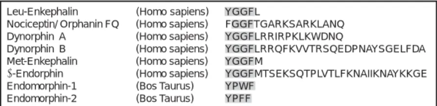
Structurally, the peptide sequences encoded by each endomorphin molecules (EM1, EM2) have been shown to be completely different from the consensus motif encoded by several endogenous opioid peptides (e.g. endorphins, enkephalins, and dynorphins) as depicted in figure 1. As shown, classical opioid peptides share the Tyr-Gly-Gly-Phe motif at the N-terminus domain of each peptide, whereas EM1 and EM2 contain two pharmacophoric amino acid residues (Tyr [1] and Trp[3] in EM1; Phe[3] replacing Trp [3] in EM2) joined together via a Pro(2) amino acid residue, used as molecular spacer in both peptides. Both phenolic groups (Tyr, Phe) and/or aromatic rings (Trp, Phe) encoded by each amino acid residue represent structural and
chemical requirements for opioid receptor recognition.32
Using nuclear magnetic resonance approaches to reveal amino acid conformation of endomorphin molecules and
Figure 1. Representative structural features of active opioid peptides in the CNS of mammals. The figure displays the amino acid sequences of different endogenous opioid peptides previously identified and cloned from the brain of mammals, inclu-ding the most recent EM1/EM2 peptides, identified and isolated from the human and bovine brain. Structural homology shared by each opioid peptide listed is outlined by the shaded amino acids at the N-terminal domain of each peptide. Implications of the structural homology and identity among mammalian and non-mammalian spe-cies (not shown here) demonstrates conservation of shared amino acid sequences along evolution (text and figure were adapted from Darlison and Richter, 1999, and modified for the present review).
Leu-Enkephalin (Homo sapiens) YGGFL
Nociceptin/ Orphanin FQ (Homo sapiens) FGGFTGARKSARKLANQ Dynorphin A (Homo sapiens) YGGFLRRIRPKLKWDNQ
Dynorphin B (Homo sapiens) YGGFLRRQFKVVTRSQEDPNAYSGELFDA Met-Enkephalin (Homo sapiens) YGGFM
β-Endorphin (Homo sapiens) YGGFMTSEKSQTPLVTLFKNAIIKNAYKKGE Endomorphin-1 (Bos Taurus) YPWF
the structural domains required to recognize opioid receptors, these studies revealed that both Tyr(1) and Trp(3) side chains display opposite orientations with respect to Pro(2), which provides the necessary stereochemical properties of endomorphin-1 for binding the µ-opioid
re-ceptor.42 Moreover, using pseudo-proline containing
analogs of EM2, it was shown that the Tyr-Pro amide bond forms a cis-conformation structure along the peptide backbone, allowing its molecular conformation to bind and recognize the µ-opioid receptor.43
II. DISTRIBUTION OF ENDOMORPHINS
IN CENTRAL AND PERIPHERAL TISSUES
OF MAMMALS
Immunological assays (e.g. radioimmunoassays, immuno-cytochemical analyses) using different rabbit antisera raised against synthetic peptides of EM1 and EM2 showed that endomorphin1-2-like immunoreactivities (EM1-2-LIs) are widely distributed throughout the neuroaxis of the rat, pri-mate, and human brains,35,44-50 displaying a preferential
localization in areas where MOR-LI (µ-opioid receptor-immunoreactivity) is highly expressed (see representative
immunoreactive areas in figure 2).51 Although EM1-2-LIs
were localized in cells and fibers in similar regions of the rat CNS (e.g. stria terminalis [ST]; periaqueductal gray [PAG]; locus coeruleus [LC]; parabrachial nucleus [PBN]; hypothalamus [HPTH]; nucleus of the solitary tract [NTS]), major differences about the neural distribution of these
peptides were detected.44,47,49 For instance, EM1-LI was
shown to be widely and densely distributed in several forebrain structures (e.g. nucleus accumbens [NAC]; the cortex [Cx]; amygdale [AM]; thalamus [TH]; the hypothalamus [HTH]; striatum [ST]), together with the upper brainstem (e.g., nucleus of the solitary tract [NTS])
and the dorsal root ganglia,47,50 whereas EM2-LI was
localized mostly in the spinal cord and lower brainstem,47,49
showing densely stained cell bodies and axon fibers within the nucleus of the solitary tract (NTS); substantia gelatinosa of the medulla and superficial layers of the spinal cord/dor-sal horn, including the lateral hypothalamus.44,47,50 Although
these studies showed that the anatomical distribution of EM1-2-like immunoreactive material along the rat CNS parallels the regional localization of several endogenous opioid peptides and their cognate opioid receptors;52,53 differences
observed between the endomorphin material, enkephalin,54,55
and dynorphin56 peptide systems showed that EM2-LI in the
mammalian brain47,50 is highly analogous to the distribution
of the µ-opioid receptor peptide ligand, β-endorphin (BE).57
Moreover, it has been shown that endomorphins do not co-localize with brain areas expressing µ-opioid receptors (e.g. striatum) containing low-to negligible amounts of
EM1-2-LI material.49 This asymmetric localization between
endomorphins peptides and their corresponding binding sites in areas of the rat brain does not preclude the synaptic activation of membrane bound opioid receptors. However, µ-opioid receptors and EM1-2-LI fibers have been shown to co-localize in limbic structures of the rat brain (e.g. septal nuclei, the bed nucleus of the stria terminalis, NAC, amygdaloid complex, hypothalamic nuclei), whereas in other rat brain regions (e.g. amygdala, thalamus, hypothalamus, PAG), showing for instance the co-localization of low levels of EM1-2-LI in areas displaying high density of opioid-binding sites (figure 2).47,49,58
Very few reports have documented the extraneuronal distribution of endomorphin material in peripheral tissues,32 in contrast with the vast reports describing the
distribution and expression of µ-opioid receptors in extraneuronal tissues.27 For instance, using RIA assays in
combination with reversed phase high-performance liquid
chromatography techniques (RP-HPLC),59 it was found the
expression of detectable amounts of EM1-LI and EM2-LI in human spleen, as compared to high levels of EM1-2-like immunoreactive material detected in non-neuronal tissues of the rat, such as spleen and thymus, including plasma. These results contrast the low amounts of EM1-2-LI detected in anterior and posterior rat pituitaries, showing that secretion of opioid material from the pituitary gland does not contribute at all to the significant high levels of EM1-2
detected in plasma.59 Based on these results, authors
proposed that EM1-2-like immunoreactive material secreted from the nerve fibers and/or presynaptic terminals localized in the spinal cord may explain the high content of endomorphin material observed in human and/or rat
plas-ma samples.59 Moreover, endomorphins were shown to be
expressed by immune cells (e.g., macrophages/monocytes,
lymphocytes, and polymorphonuclear leukocytes),60,61
including inflamed subcutaneous tissue.62 Endomorphins
have been found to alter a variety of immune parameters and functions,63-66 as well as to inhibit spleen cell antibody
formation when added to in vitro culture systems at femtomolar concentrations (10-13 M to 10-15 M) and reverted
after application of specific EM1/EM2 polyclonal rabbit antisera.67
III. BINDING ASSAYS AND CELLULAR ACTIONS
OF ENDOMORPHINS
A large number of pharmacological and molecular studies have clearly demonstrated that the µ-opioid receptor mediates the spectrum of opiate-related activities of
endomorphins in the CNS of mammals.32 The proposed
hypothesis suggesting that endomorphin molecules are the endogenous ligands for the µ-opioid receptor, aroused from classic binding assays and functional studies, which showed that both endomorphin peptides displaced DAMGO (Tyr-DAla-Gly-MePhe-Gly-ol); naloxone, and other µ-opioid re-ceptor-selective ligands, in a concentration-dependent manner68 and stimulate the binding of [35S]guanosine
5'-O-(3-thio)triphosphate ([35S]γGTP) in riched mu-opioid
receptors-membrane preparations, isolated from thalamus
69-71 periacuectal gray area (PAG)72 and pons/medulla.73
Moreover, autoradiographic studies using radiolabeled tracers of endomorphin molecules showed that these ligands matched the neuroanatomical distribution of DAMGO-binding sites (a conventional µ-opioid receptor selective ligand used as standard in binding studies) in the rodent’s brain, postulating thus the role of endomorphins «as the preferential endogenous ligands of the µ-opioid receptor in the brain».32
However, based on the lower efficacy of endomorphins in stimulating µ-opioid receptors in several bioassays (e.g.
([35S]γGTP binding) compared to DAMGO and morphine,
both peptides have been regarded as partial agonists of µ-opioid receptors.71,74 The extent to which these relative
efficacies of endomorphins apply in the expression of functional bioactivities is still unclear and further analysis is needed to solve this issue.
Moreover, pharmacological studies showed that both EM1 and EM2 produce their biological effects by functionally stimulating different µ-opioid receptors subtypes, µ1 (MOR1)/µ2 (MOR2)-opioid receptors, which appear to be responsible for the distinct pharmacological activities
mediated by opiate alkaloids, including both EM1-2 peptides.38,40,75 For instance, the µ1 receptor antagonist,
naloxonazine, displayed a potent efficacy in blocking the antinociception response induced by EM2 compared to that exhibited by EM1. Converserly, the µ-opioid receptor antagonist, β-funaltrexamine, was capable of inhibiting the antinociceptive responses induced by both EM1-2 molecules.38,40,75
Pharmacological and molecular studies, showed that spinal administration of specific antisense oligodeoxy-nucleotides (used to knockdown the expression of the µ-opioid receptor genes) differentially attenuated the antinociception induced by either EM1 and/or EM2
peptides.76,77 These studies proved that endomorphin
molecules activate and modulate µ2-receptor induced-responses (e.g. spinal analgesia, respiratory depression, and
inhibition of gastrointestinal motility),78 whereas EM2
appears to modulate µ1-dependent responses, which include supraspinal analgesia, acetylcholine (ACh), and
prolactin release from hypothalamus.78 Thus, these data
provided significant insights on the differential regulation of µ-opioid receptor subtypes and their interaction with endomorphin peptides.
Quite interesting to note is that in vivo studies using radioligand binding techniques, combined with autoradio-graphic procedures, demonstrated that endomorphin peptides labeled some binding sites shown for DAMGO in specific reports of the rat brain,36,70,71 support the endogenous
role of EMI-2 as µ-opioid receptor peptide ligands (partial agonists of µ-opioid receptor) involved in the regulation of several µ1/µ2 opiate-receptor related bioactivities.32
Furthermore, a vast number of cellular and pharmaco-logical studies showed that chronic exposure of µ-opioid receptor-agonists mediate long-lasting changes and cellular adaptations that underlie the development of opiate tolerance and physical dependence in animals and
humans.79-81 These observations led pharmacologists to
limit a wide range of µ-opioid receptor-binding agents, clinically used to treat pain–related disorders.82,83 In vitro
studies have shown that chronic administrations of EM1-2 peptides stimulated the development of tolerance in specific neuronal-tumor cell lines (e.g. SH-SY5Y neuroblastoma cells).84,85 For instance, chronic exposure
of EM1/EM2 peptides on Chinese hamster ovary cells (functionally transfected with a µ-opioid receptor cDNA) induced a naloxone-dependent increase of forskolin-stimulated adenylyl cyclase activity above baseline responses, whereas in monkey kidney cells a specific EM1-2-dependent increase of type I and V-adenylyl cyclase
isozymes was observed.86
peptide ligand agonists.87,88 For instance, endomorphins have
been shown to induce potent granule/vesicle endocytosis and trafficking of µ-opioid receptor in cells (transfected with the µ-opioid receptor cDNA), inducing similar sort of endocytosis and trafficking mechanisms for µ-opioid receptors in enteric neurons (shown to synthesize and
express naturally opioid-receptor binding sites).85 This
peptide-inducing regulated endocytosis and trafficking of µ-opioid receptor in cells has been shown to mediate desensitization, resensitization, and down-regulation of distinct molecular mechanisms that mediate the cell responsiveness to ligand stimulation, involved in the development of opioid tolerance and drug addiction.89
In vivo experiments showed that endomorphin peptides induced a naloxone-precipitated withdrawal response in rats, effects similar to those induced using same dose-unit of morphine.90 Using a preestablished scoring system used to
detect specific withdrawal signs in rats (e.g. chewing, sniffing, grooming, wet-dog shakes, stretching, yawning, rearing, jumping, teeth grinding, ptosis, diarrhea, penile erection), Chen et al. demonstrated that endomorphin pre-treatment for 5 days (20 µg, ICV administration), followed by naloxone (4mg/kg, IP) administration, rats displayed physical signs of opioid dependence, exhibiting different potency for different withdrawal signs.90
The documented µ-opioid receptor ligand agonists (involved in the development of tolerance and physical dependence in animals and humans) have been suggested to operate through the interaction of conventional transmitter systems (e.g. dopamine[DA];91-93 norepinephrine [NE];94-97
cholinergic [ACh];98,99 benzodiazepine;100 GABA101 and
N-methyl-D-aspartate [NMDA],102 imidazoline103 and
glutama-tergic neural pathways).104 However, no data on the
interactions between endomorphin peptides and
aforemen-tioned transmission systems have been reported.32
Nitric oxide (NO) has been suggested to participate in the cellular mechanisms involved in peptide(s)-inducing physical dependence, based on observations that NO synthase inhibitors attenuated some signs of naloxone-precipitated withdrawal and cell-hyperactivity at the locus coeruleus (LC).105 In addition, the intrathecal (IT) infusion of morphine
was shown to increase the N-methyl-D-aspartate (NMDA) binding activity, besides of inducing an up-regulation of NO synthase expression in neurons as well.106
IV. DEGRADATION OF ENDOMORPHINS:
PEPTIDE CATABOLITES AND BIOACTIVITIES
Endomorphins have been shown to be vulnerable to enzymatic cleavage and several enzymes have been
proposed as participants in endomorphin degradation.107
Studies performed to analyze the enzymatic degradation of endomorphins upon exposure to the activity of
proteolytic enzymes (e.g. carboxipeptidase Y and A, proteinase A, and aminopeptidase M) showed a chromato-graphic profile of recovered endomorphin metabolites
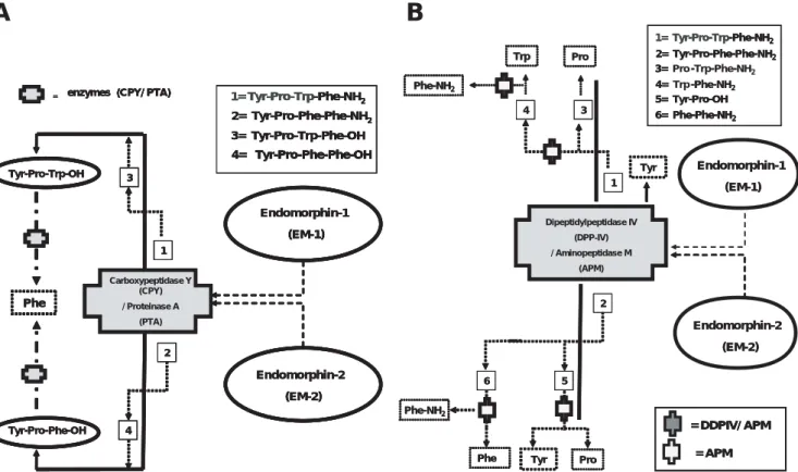
eluted from the HPLC column (figures 3A-B).108 These
studies led to the proposal that enzymatic cleavage mediated by aminopeptidase M (EC 3.4.11.2) and/or aminopeptidase P (EC 3.4.11.9) degrade N-terminal Tyr-Pro peptide bonds, thus releasing the N-terminal amino acid (Tyr) and generating a tripeptide fragment from endomorphin molecules.109,110 Moreover, it has been shown
that peptide sequences containing N-terminal hydrophobic residues followed by a Proline (Pro) residue; the preceding two amino acids (e.g., H2N-Tyr-Pro) in the peptide moiety
may be released by aminopeptidase M, as an intact dipeptide (e.g. Tyr-Pro).111 In addition, enzymes —such as
carboxypeptidase Y (a serine peptidase, EC 3.4.16.5) and/ or proteinase A (a non-pepsin-like acid endopeptidase [EC 3.4.23.6]), shown to display deamidase activities— have been postulated to catalyze the C-terminal amide group and catalyze the hydrolysis of the Xaa(3)–Phe(4) peptide bond (Xaa= natural amino acid), possibly by hydrolyzing EM1-2 peptide sequences into peptide acids, after releasing ammonia and cleaving off the C-terminal Phe(4) amino acid residue (figures 3A-B).108,112
Other enzymes, shown to display peptidase activities on distinct polypeptides, have been included as potential degrading enzymes of endomorphins. For instance, the membrane-bound serine proteinase, dipeptidyl-peptidase IV (DPPIV) (EC.3.4.14.5), has been shown to remove N-termi-nal dipeptide sequences from different polypeptides that contain the Pro residue at the penultimate position (H2
N-Tyr-Pro…..Xn-COO-).113 In vivo studies have shown that two
groups of enzymes (e.g. proteinase A, aminopeptidase M/ P), working together in related catalytic pathways, may catalyze the degradation of endomorphins from formed dipeptides into single amino acids. In this context, the serine proteinase/DPP-IV system could trigger the catalytic activity of EM1/EM2 by cleaving the Pro(2)–Trp(3) and Pro(2)–Phe(3) peptide bonds, respectively, followed by the aminopeptidase M/P activities, recruited to catalyze secondary peptide cleavages. That is, hydrolyzing released dipeptides (Tyr-Pro/ Trp-Phe in EM1; Tyr-Pro/Phe-Phe in EM2) into single amino acids.114 Although the Tyr(1)–Pro(2) peptide bond might also
be cleaved during the first step of enzyme processing pathway (dipeptide formation), some authors argued that EM1 appears to be more resistant to enzymatic degradation
in vivo than EM2.115 Such proposals appear to be in good
agreement with the observations showing that the spinal antinociceptive effects induced by EM1 are significantly longer than those induced by EM2.116 In a similar context,
the analgesia induced by EM1 required a longer pre-treatment time than that required for EM2, before tolerance is detected in mice.117 In addition, the antidepressant-like
duration than that detected for EM1 in mice, after ICV administration of each peptide (figures 3A-B).32
One crucial aspect to elucidate the byproducts formed during the degradation of endomorphins is that the catabolites produced during peptidase activity could dis-play related bioactivities similar to the parent compound. Thus, competition between endomorphin byproducts and full peptide sequences for the m-opioid receptor binding sites would lead to the expression of cellular and related
opioid-bioactivities.32,118 In such context, radioligand
binding studies demonstrated that the primary degradation products of EM1 (Tyr-Phe-OH and/or Pro-Trp-Phe-OH) displayed a very low m-opioid receptor binding affinity, showing no substantial activation/stimulation on Go/Gi proteins and non-antinociceptive activity after ICV administration of cleaved catabolites of EM1 in mice
(figu-res 3A-B).118 Parallel studies used to evaluate the
relationship between the binding activity of EM1/EM2 peptides and byproducts of peptide catabolism showed that peptidase inhibitors (e.g. diprotin A [Ile-Pro-Ile] a specific DPP-IV inhibitor) have no effect on the binding properties displayed by [3H]-EM2 on rat brain membrane preparations.
However, when no inhibitor was added to the incubation mixture, 40% of the radioligand was degraded by
endogenous membrane–bound proteinases.119 Moreover,
ICV administration of a different DPP-IV inhibitor (e.g. Ala-pyrrolidonyl-2-nitrile) produced a significant increase in the antinociception activity induced by EM2, as shown for the altered analgesic related-parameters (e.g., magnitude, duration, and potency) detected in the rat tail-flick test.120
Similar EM2- induced analgesic effects were observed with diprotin A (a DPP-IV inhibitor), which produced a five-fold increase in potency with respect to EM2 itself in the paw withdrawal test114 or with actinonin (a selective
serine-Figure 3. Schematic representation of the proposed enzymatic pathways involved in the catalysis of endomorphin peptides in the brain. Both panels illustrate enzymes and pathways implicated in the degradation of endomorphin peptides in both neural and non-neuronal tissues. Panel A depicts the enzymatic degradation of endomorphins at the C-terminal domain of both molecules. Endomorphins exposed to
Carboxypeptidase Y (CPY, a serine peptidase, EC 3.4.16.5) and/or Proteinase A (PTA, a non-pepsin type acid endopeptidase, EC 3.4.23.6) have been proposed as the enzymatic route implicated in the initial catalysis of the C-terminal domain of both amide tetrapeptides. Thus, both CPY and PTA may hydrolyze the C-terminal amide group (Phe-CO-NH2) into a carboxyl group (-Phe-OH) followed by enzymatic hydrolysis of the Xaa(3)-Phe(4) peptide bond in EM1 and EM2 molecules, releasing the Phe residue, as shown (see text for more details).
Panel B depicts the proposed degradation of endomorphin peptides at the N-terminal domain of both molecules, where the aminopeptida-se M (APM, EC 3.4.11.2) and/or aminopeptidaaminopeptida-se P (APM, EC 3.4.11.9)(not shown) enzymes could hydrolyze the N-terminal domain of both opioid amide tetrapeptides; releasing from either EM1 and/or EM2 peptides, the N-terminal amino acid (Tyr) together with the correspon-ding tripeptide sequence (EM1; Pro-Trp-Phe-NH2; EM2; Pro-Phe-Phe-NH2). Both APM and the membrane bound serine proteinase, dipeptidyl-peptidase IV (DPP-IV, EC.3.4.14.5), have been proposed to participate in the initial release of the dipeptide (Tyr-Pro) from both EM1-2-molecules, where APM may hydrolyze the dipeptide bond enhancing the releasing of both N-terminal amino acids from the peptide structure (see text for more details) (text and figure were adapted from Fichna et al., 2007 and modified for the present review).
Tyr-Pro-Phe-OH Endomorphin-1 (EM-1) Endomorphin-2 (EM-2) Carboxypeptidase Y (CPY) / Proteinase A
(PTA) Tyr-Pro-Trp-OH Phe 1 3 2 4
3= Tyr-Pro-Trp-Phe-OH 1= Tyr-Pro-Trp-Phe-NH2 2= Tyr-Pro-Phe-Phe-NH2
4= Tyr-Pro-Phe-Phe-OH = enzymes (CPY/ PTA)
Tyr-Pro-Phe-OH Endomorphin-1 (EM-1) Endomorphin-2 (EM-2) Carboxypeptidase Y (CPY) / Proteinase A
(PTA) Tyr-Pro-Trp-OH Phe 1 3 2 4
3= Tyr-Pro-Trp-Phe-OH 1= Tyr-Pro-Trp-Phe-NH2 2= Tyr-Pro-Phe-Phe-NH2
4= Tyr-Pro-Phe-Phe-OH = enzymes (CPY/ PTA)
Tyr-Pro-Phe-OH Endomorphin-1 (EM-1) Endomorphin-2 (EM-2) Carboxypeptidase Y (CPY) / Proteinase A
(PTA) Tyr-Pro-Trp-OH Phe 1 3 2 4
3= Tyr-Pro-Trp-Phe-OH 1= Tyr-Pro-Trp-Phe-NH2 2= Tyr-Pro-Phe-Phe-NH2
4= Tyr-Pro-Phe-Phe-OH 3= Tyr-Pro-Trp-Phe-OH 1= Tyr-Pro-Trp-Phe-NH2 2= Tyr-Pro-Phe-Phe-NH2
4= Tyr-Pro-Phe-Phe-OH = enzymes (CPY/ PTA)
= enzymes (CPY/ PTA)
Endomorphin-1 (EM-1) Endomorphin-2 (EM-2) 1 3 2 5 Dipeptidylpeptidase IV (DPP-IV) / Aminopeptidase M
(APM) Tyr Pro 4 Trp Phe-NH2
= DDPIV/ APM
= APM Tyr Pro
6
Phe Phe-NH2
3= Pro -Trp-Phe-NH2
1= Tyr-Pro-Trp-Phe-NH2
2= Tyr-Pro-Phe-Phe-NH2
4= Trp -Phe-NH2
5= Tyr-Pro-OH 6= Phe-Phe-NH2
Endomorphin-1 (EM-1) Endomorphin-2 (EM-2) 1 3 2 5 Dipeptidylpeptidase IV (DPP-IV) / Aminopeptidase M
(APM) Tyr Pro 4 Trp Phe-NH2
= DDPIV/ APM
= APM = DDPIV/ APM
= APM Tyr Pro
6
Phe Phe-NH2
3= - -
-1= Tyr-Pro-Trp-Phe-NH2
2= Tyr-Pro-Phe-Phe-NH2
4= -Phe-NH2
5= Tyr-Pro-OH 6= Phe-Phe-NH2
3=
1= Tyr-Pro-Trp-Phe-NH2
2= Tyr-Pro-Phe-Phe-NH2
4= 5= -Pro-OH 6= Phe-Phe-NH2
proteinase/peptidase inhibitor), which blocked the peptide catabolism of EM1, when incubated in a 6-day-old rat spinal cord homogenate, producing a potent antinociception effect compared to the peptide alone.121
V. ENDOMORPHINS REGULATING
CONVENTIONAL NEUROTRANSMITTERS
SYSTEMS AND RELEASE
OF PEPTIDE HORMONES
Both exogenous and endogenous opioid receptor ligands display their biological activites by modifying the function and release of conventional transmitters in the CNS of mammals, including the evoked-release of various
neurohormones from neuroendocrine tissues.32 Opioid
ligands regulating neurotransmitter release have been extensively reviewed and reported elsewhere.32,122,123
In this context, µ-opioid receptor agonists have been shown to regulate the neuronal release of dopamine (DA);124-127
norepinerphrine (NA);125,128,129 serotonin (5-HT),122,123,125 and
acetylcholine (ACh),130,131 in areas where µ-opioid receptors
have been found to be co-localized with each neurotransmitter in specific areas of the mammalian brain.32
a) Dopamine (DA) transmission system
Immunohistochemical studies (IHC) revealed that µ-opioid receptors are densely expressed on dopamine neurons in the NAC and striatum in the CNS of rodents, including
primates and humans,132-134 and they play an important
regulatory role on the DA transmission system in these brain structures.32 In the striatum, opioid ligands have been
shown to act on presynaptic opioid-receptors, producing indirect effects on DA turnover. This effect on DA activity varies according to the sort of opioid receptor bound by ligands.135-137 For instance, striatal administration of ligand
agonists acting on postsynaptic µ-opioid receptors induced
an increased DA efflux from neuronal cells138 without
affecting the DA uptake or the levels of DA metabolites
(DOPAC, HVA).139 Interesting enough is that the
modulatory activity of DA release from striatal neurons by µ-opioid receptor agonists, requires the integrity of both
cholinergic and GABAergic neurons,140 including the
nigrostriatal neural pathway, where µ-opioid receptor agonists (e.g. morphiceptin) acting at a presynaptic level prevented the neuronal release of DA.139,141
In the accumbal region (NAC), microdialysis studies showed that µ-opioid receptor ligands agonists infused into the NAC induced an increased accumbal DA release or DA
efflux in rats142 as shown for EM1-2 peptides, which
produced a dose-dependent increase in DA efflux from
NAC in freely moving rats.143 Whereas an accumbal
infusion of EM1 showed a dose-dependent increase of DA efflux, which was abolished by intraccumbal perfusion of
CTOP or by systemic administration of naloxone and/or
naloxonazine,143 DA efflux induced by local infusion of
EM2 was not abolished by selective µ-opioid receptor antagonists. This suggess that the EM2-induced effects may be mediated through different receptors other than the
µ-opioid receptor.143 These studies support previous data
showing that EM2 induces a dose-dependent stimulation of DA release and levels of DA metabolites (e.g. DOPAC, HVA) from the accumbal shell region126 as a result of the
disinhibition of local GABA neurons.126
b) Noradrenergic (NA) transmission system Noradrenergic system arises from a clustered group of NA-containing cell bodies, that arises from the locus coeruleous (LC) localized at the rostral area of the pons,144 and which
projects axon fibers to forebrain areas and limbic structures. NA projecting fibers from LC represent the primary source of NA in cortical and hippocampal areas of the
brain.145-147 The LC-NA neurons innervate almost all
regions of the neuraxis, influencing several bioactivities and
behaviors.148-150 LC-NA neurons also provide descending
fibers to both medulla and spinal cord involved antinociceptive bioactivies.151-153 Neuroanatomical studies
using autoradiographic and electrophysiological procedures using intracellular recording techniques revealed the expression of a high-density of µ-opioid re-ceptor at the LC,154-156 where endogenous opioid peptides
(e.g. morphiceptin, hemorphin-4, Tyr-MIF-1, Tyr-W-MIF-1), including endomorphins and non-natural synthetic peptides
(e.g., Tyr-D-Arg-Phe-Sar; Tyr-D-Arg-Phe-Lys-NH2), shown
to bind and activate µ-opioid receptors, were shown to modify the physiological activity of LC neurons.32,155
All peptides tested displayed opiate-alkaloid activities
on LC-NA cells,155 producing a dose- and time- dependent
electrophysiological responses (e.g. decreased neuronal excitability; inhibition of spontaneous cell-firing, opioid-induced hyperpolarization effect mediated through the opening of inward-rectification potassium channels, and other membrane bioactivies).155 Among the tested peptides,
EM1 and EM2 represented the most effective peptides displaying equipotent electrophysiological responses (e.g. hyperpolarizations) on LC-NA neurons at very low
concentrations, when compared to β-endorphin, DAMGO,
or morphine.32,155,157 These results led authors to assume
that the presence of the aromatic group on the Phe residue at position 4, in addition of the structural C-terminal amidation found in both EM1-2 peptides, represent crucial structural features by which these peptides exert their electrophysiological responses on LC-NA neurons after binding their cognate µ-opioid receptors on these cells.32,155
the brainstem dorsal pontine tegmentum) in the rat brain.157
Both areas were shown to express high-densities of µ-opioid receptors158,159 and were suggested to be targeted by
EM1-2 peptides, producing a significant reduction of LC-NA
neuronal excitability.157 Thus, EM1 could modulate LC
neurons directly by tonically inhibiting neurons within the Barrington’s nucleus.157 Futhermore, physiological studies
showed the interaction between EM1 and NA at the level of the spinal cord, showing that the IT administration of EM1 in rats induced an evoked-release of NA from presynaptic axon terminals that arise from LC-NA descending neural pathways impinging into the dorsal horn
of the rat’s spinal cord.160 Thus, EM1 produced a potent
analgesia activity (measured in different behavioral models of analgesia) by activating µ2-opioid receptors.32,160
c) Serotonin (5-HT) transmission system
The serotonin (5-HT) transmission system has been shown to mediate a vast number of physiological and behavioral responses in mammals (e.g. nociceptive, appetitive, emotional, stress and anxiety, motor, cognitive, and autonomic functions).161-165 Several bioactivities mediated
by serotoninergic tone in the brain have been found to be
influenced by the endogenous opioid system (EOS).32 The
serotonin (5-HT) transmission system is functionally dependent on the serotoninergic tone of the CNS, which is regulated by the firing activity of cell bodies (somata) which are clustered in different nuclei along the midline of the brainstem; namely, the dorsal raphe nucleus (DRN) and the
median raphe nucleus (MRN).162 Both nuclei have been
shown to project axon fibers to almost all forebrain structures along the neuroaxis (e.g. mesolimbic areas, hippocampus, and cortex), where 5-HT axons projecting to terminal fields regulate the expression of different bioactivities and behavioral repertoires required for survival and adaptive functions in species.162 Thus, the 5-H/DRN located at the
ventral aspect of the PAG has been shown to be crucial for the integration stress-responses,166 and pain and stressful
stimuli activating peptidergic neurons at the PAG have been shown to modulate the firing activity of 5-HT neurons (at the somatodendritic level) via activation of µ-opioid receptors expressed at synaptic endings in short axons, innervating
the 5-HT/DRN.167,168 Finally, 5-HT/DRN neurons project
axon fibers to several limbic structures (e.g. amygdale, PFCx, hippocampus, NAC) involved in handling emotional and stress-related responses.167,169,170 However, the cellular
mechanisms by which opiod agonists affect the activity 5-HT neurons have not been completely elucidated.
For instance, previous electrophysiological studies showed, on the one hand, that µ-opioid receptor agonists induced significant inhibitory effects on cell-firing of DRN/ 5-HT neurons.171 On the other hand, in vivo microdialysis
showed that opioid agonists induced an increased activity
on 5-HT neurons.167,172 Several mechanisms have been
offered to explain such inconsistencies based on the structural organization of the DRN. This brainstem nucleus receives neural inputs from local GABAergic neurons and from descending glutamatergic axon fibers from the cortex, which control the cellular excitability of 5-HT neurons and the net equilibrium between both neural systems, will provide the result on the neuronal release or efflux of 5-HT into the external milieu.162 In this context, it has been shown that
µ-receptor agonists move the net equilibrium of both neural systems into an increased release and extracellular levels of 5-HT at the DRN, possibly mediated through an opioid-enhanced activity of the glutamatergic (excitatory) input on 5-HT/DRN neurons and/or opioid-induced disinhibitory effect of GABAergic neurons.122
Microdialysis studies revealed that endomorphins peptides influenced the activity of DRN/5-HT neurons by increasing the net efflux of 5-HT (increased extracellular levels of the aminergic transmitter) within the DRN. This effect was blocked by selective µ-opioid receptor antagonists
such as β-funaltrexamine.122 In a similar context,
endomorphins have shown to modulate 5-HT activity in other brain areas. For instance, EM1-2 peptides infused into the VTA induced a significant decreased of 5-HT activity in the rat PFCx (cortex) and NAC (ventral striatum), thus suggesting that µ-opioid receptors could potentially mediate
a neuronal depletion of 5-HT in these brain areas.125
However, no effect on the HT metabolite, 5-hydroxyindoleacetic acid (5-HIAA), was found in the
NAC.126 In forebrain areas, DAMGO and EM1 produced a
strong, CTOP-reversible suppression of the 5-HT2A-induced
excitatory post-synaptic currents on layer V-pyramidal cells, localized at the medial prefrontal cortex (PFCx).173 These
studies showed that µ-opioid receptor agonists act presynaptically in cortical layer V pyramidal cells,174 and
the interaction of 5-HT2A and µ-opioid receptor ligands
appear to ocurr on projecting glutamatergic afferents sent from thalamic or amygdale nuclei (e.g. basolateral nucleus)175-177 that locally synapse layer V-piramidal cells.174
d) Acetylcholine (ACh) transmission system
With regard to the ACh-transmision system, several studies showed that EM1-2 peptides modulate cholinergic neurotransmission in peripheral tissues, such as the respiratory system and the gastrointestinal (GI) tract (see corresponding sections xiv and xv in the following chapter, Part II, in this journal ) acting on prejunctional µ-opioid receptors that inhibit the release of ACh that mediate the contractile muscle-activity and peristalsis in the GI tract.32
In a similar context, recent experiments showed that EM1 regulates ACh-transmission system in the rat inner hair
cells.178 Using patch-clamp recording techniques, these
e) Release of neuropeptides hormones
Electrophysiological studies in vivo showed that EOS regulates the electrical activity of the neuron cells that synthesize and secrete oxytocin (OT) (from supraoptic nuclei [SON] localized within the hypothalamus) and arginine vasopressin (AVP) (from the anterior paraventricular nucleus of the hypothalamus [PVN]).179,180 Previous studies showed
that µ-opioid receptor agonists produced a potent inhibition on OT-secreting cells (SON), whereas neuronal-activity of AVP-secreting cells (PVN) is decreased by the same recep-tor agonists.181 In a similar context, endormorphin peptides
showed to display related activities on OT/SON and AVP/ PVN cells, respectively, showing that ICV administration of EM1 into rats induced a more potent inhibition effect of OT cells than on AVP cells.182 These effects detected on SON
versus PVN-cells were attributed to the number of cell-surface opioid receptors expressed on each neurosecretory cell.182 Recent studies showed that the oxytocin (OT)
indu-ces important antinociceptive effects in rodents and humans,183-188 as shown after ICV,189 IP,183 or IT/IC184
administration of the peptide hormone, in addition of the injection of the peptide directly into the PAG and NRM (nucleus raphe magnus)190,191 (sections x-xiii in the following
chapter, Part II, in this journal).
Recent studies shed important data concerning the interaction between µ-opioid receptors and OT-inducing
antinociceptive responses in the rat CNS.192,193 These
studies showed that µ-opioid receptors antagonists (e.g.
β-FNA) inhibited OT-induced antinociceptive effects in
response to the application of either thermal or mechanical stimuli in rats. So far, no experimental data have elucidated yet any role for the endogenous opioid system
(e.g. β-endorphin), including both EM1-2 peptides,
mediating the OT-induced antinociception responses in animals.32
Most of the experimental data reporting on the interaction between the AVP/PVN system and µ-opioid re-ceptor agonists have been shown in stress-related behaviors32
(section xiii, in the following paper Part II, in this journal). However, no experimental works have clearly elucidated the mechanisms by which EOS and endormorphin peptide interact with the AVP/PVN peptide system. Nonetheless, recent documents showed that acute stress leads to an increase synaptic concentration of AVP in the rat amygdala
or hypothalamus;194,195 where AVP released from PVN–
neuronal cells was shown to activate its cognate membrane receptor (e.g. V1 receptor) and mediate crucial anxiogenic and depressive behavioral responses in rodents.195
With regard to somatostatin, µ-opioid receptors ligand
agonists,196,197 including endomorphins,198 have been
reported to regulate gastric-somatostatin and neuroendocrine activity at the GI tract (section xv, in the following chapter Part II, in this journal). For instance, EM1 was shown to exert
an important inhibition on somatostatin-induced stimulation in the perfused rat stomach, which was partially blocked with CTOP.198 These results posit that EM1 acting on µ-opioid
receptors mediate in large part the opioid-inhibitory effect on somatostatin activity.198 Mediated by the EM1 peptide,
this inhibitory effect might be similar for other regulatory
peptide-hormones in several systems.32 However, the
interaction between peptide-hormone activity and EM1-2 peptides needs further investigation.
VI. ENDOMORPHINS MODULATING
NOCICEPTIVE TRANSMISSION
AND TOLERANCE
With regard to µ-opioid receptor-mediating pain modulation, in vivo studies showed that the intracerebro-ventricular (ICV) administration of endomorphins produced a potent antinociceptive effect in wild type mice,33,34,68 showing no significant effect in µ-opioid
recep-tor mutant knockout mice.34,199-201 In a similar context,
intrathecal (IT) administration produced significant antinociception effects in adult rodents exposed to the tail-flick, paw-withdrawal, tail pressure, and/or flexor-reflex tests, respectively (figures 4A-B).31,33,36,116,117,202,203
Based on the aforementioned description of the neuroanatomical distribution of endomorphins and µ-opioid receptors in the rodent, primate and human CNS (see section ii); several authors have categorized the functional role of EM1-2 peptides as neurotransmitters or neuromodulators, regulating different biological and physiological processes, as previously described.32 Such modulatory-related activities
mediated by endomorphins include pain perception, stress-related responses, complex neural functions —such as reward, arousal, and vigilance—, as well as autonomic, cognitive, neuroendocrine, and limbic homeostasis.32
Endomorphin peptides have been implicated in the modulation of neural pathways of pain and/or nociceptive
transmission in several areas of mammals brain,32
particularly, the spino(trigemino)-ponto-amygdaloid pathway, where nuclei and neuronal elements (e.g. caudal nucleus of spinal trigeminal tract, parabrachial nucleus, the NTS, PAG, nucleus ambiguous, LC, and midline thalamic nuclei) have been shown to express EM1-2-LI material (see
reviews in204,205) (figures 2 and 4A-B). Based on the
anatomical distribution of EM2-LI34,62 and its co-localization
with µ-opioid receptors —densely expressed in spinal cord neurons and along primary afferent fibers penetrating the superficial layers of the dorsal horn—,46 EM2 was found to
modulate nociceptive transmission pathway, modulating the release of excitatory transmitters (e.g. glutamate [Glu],
substance P,γ-aminobutyric acid [GABA], glycine [Gly],
expressed in axon terminals along the Lissahuer tract that penetrates the superficial dorsal horn and established synaptic contacts with intrinsic and relaying neurons of the spinal cord (figure 4A).206,207
Supporting evidences for the modulatory role of EM2 on primary nociceptive transmission pathways come from electrophysiological studies that showed the evoked-release
of EM2 from dense-cored vesicles from spinal neurons after electrical stimulation from the dorsal root ganglion fibers;208,209 in addition to the EM-2-induced hyperpolarizing
responses on cell-membranes of intrinsic spinal neurons, and the decreased excitability of postsynaptic µ-opioid receptors in intrinsic relay neurons as well.210
Furthermore, supraspinal (ICV) and spinal (IT) administration of EM1/EM2 showed that both amide tetrapeptides influence several neurotransmitter systems, in a similar fashion as demonstrated for morphine and DAMGO (preferential agonists of the µ-opioid receptor
subtypes MOR1/MOR2) in the rat’s CNS.33,34 In such a
context, anatomical and pharmacological studies have
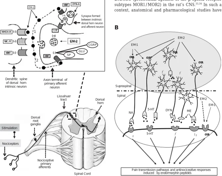
Figure 4. Schematic representation of the putative role of endomorphin-2 modulating nociceptive transmission and pain perception in the brain of mammals. Panel A illustrates a hypothetical situation whereby endogenous EM-2 peptide might be involved in the regula-tion of nociceptive transmission through the activity of intrinsic spinal neurons. Stimularegula-tion of peripheral nociceptors will relay nociceptive transmission enhancing the release of EM2 from vesicles localized in axon terminals of penetrating primary neuronal afferents to superficial dorsal horn (Lissahuer tract). (1) EM-2, exerting a regulatory function on postsynaptic cells, may decrease the activity of postsynaptic µ 2-opioid receptors. (2) EM-2 may exert an autoregulatory function via the activation of its presynaptic cognate µ1-opioid receptor, expressed either on axon terminals of incoming neural afferents or on intrinsic dorsal horn neurons. Thus, presynaptic activation of µ1-opioid recep-tors will lead to significant inhibition on the local release of excitatory peptide (Substance P [SP]; calcitonin gene-related peptide [CGRP]) and
non-peptide transmitters Glu from primary neuronal afferents; and conversely, EM-2 may enhance the release of dynorphin A (DYN-A) from local spinal neurons (see inset, upper left) (see text for more details).
Panel B depicts different neural pathways involved in nociceptive transmission and modulation of pain during supraspinal and supraspinal administration of EM-1 and EM-2. Abbreviations: DYN, dynorphin A; ME, met-enkephalin; 5-HT, serotonin; NA, noradrenaline; α2-R, alpha-2 noradrenergic receptor, δ2-R, delta-2 enkephalin-opioid receptor subtype; µ-R, mu-opioid receptor subtype (see text for more details) (text and figures were adapted from Fichna et al., 2007, and modified for the present review).
Spinal Cord Stimulation
Nociceptors
DORSAL ROOT GANGLIA
NOCICEPTIVE PRIMARY
Lissahuer
tract Dorsal horn
Nociceptors
Dorsal root ganglia
Nociceptive primary afferents Stimulation
Dendritic spine of dorsal horn intrinsic neuron
Axon terminal of primary afferent
neuron
(-) -R EM-2
(+)
(+)
µ2-R NMDA-R
(-) (-) (-)
µ1-R
(-) -R
(+)
-NMDA-R
(-) (-) (-)
-(-) k
(+)
SP GLU
CGRP
-NK1-R NK -R NK
R
(-) (-) (-)
(-) (-) (-)
2 1
-µ1-R
-DYN-A DYN-A DYN-A
Synapse formed between instrinsic dorsal horn neuron and afferent neuron
induced by endormorphin peptides NA
2 5-HT 2
5-HT DYN EM-1
EM-2
EM-2
EM-1 Spinal
Supraspinal
ME
Pain transmission pathways and antinociceptive responses NA
5-HT
5-HT DYN
µ µ
µ
κ κ
EM-1
EM-2
EM-2 EM-1
µ
ME
δ2
α2 κ µ
A
extensively shown that neurotransmitters systems and chemical substrates that mediate mesencephalic analgesia induced by morphine (at the rostral ventromedial medulla) include the serotonin (5-HT),211,212 the inhibitory GABAergic
system;213,214 the Glutamatergic and N-methyl-D-aspartate
excitatory system,215,216 and the neurotensin peptide system
as well (figures 4A-B).217
Pharmacological studies showed that both NA and 5-HT (acting on spinal cord, α2- and 5-HT receptors subtypes) mediate antinoceptive/analgesia responses induced by ICV
administration of DAMGO,218,219 and conversely, the
induced-depletion of both neurotransmitters (NA, 5-HT) after IT administration of 6-hydroxydopamine (6-OHDA) and/or 5,7-dihydroxytryptamine (5,7-DHT), respectively, or IT administration of yohimbine and methysergide (used to block spinal α2-adrenoreceptors and serotonin receptors) respectively; attenuated the antinociception response induced after ICV administration of morphine.220-223 In a
similar context, the antinoceptive responses induced after ICV administration of EM1/EM2 peptides were completely abolished after depleting (> 90%) the spinal NA system with IT administered 6-OH-DA (3-day pretreatment period with
neurotoxic compound).129 However, administration of
5,7-DHT (which depleted both 5-HT [³ 90%] and NA [≅ 25%])
attenuated, but did not block the endomorphin-induced antinociception. These results clearly showed the relevance of the NA system over the 5-HT system in mediating the induced-antinociception by the supraspinal (ICV) administration of endomorphin molecules (figure 4B).32,129
Complementary results showed that the antinoception-induced effect detected after the spinal administration of EM1/EM2 peptides into mice was abolished with µ-recep-tor antagonists (e.g. naloxone, β-funaltrexamine), but not by the neurotoxic agents, 6-OHDA or 5,7-DHT,
respectively,129,202 suggesting that the spinal
induced-antinociception by EM1/EM2 peptides is caused by the direct stimulation of µ-opioid receptors, without affecting the NA or 5-HT transmission.32,129,202 Moreover, parallel studies
showed that the supraspinal antinociception induced by ICV administration of EM2, but not EM1, were shown to be
mediated by the activity of dynorphin A1-17 and
Met-enkephalin peptides, acting on their cognate κ- and δ2-opioid
receptor subtypes, respectively.40 Thus, these data
demonstrated functional differences in the spinal and supraspinal antinociceptive effects induced by EM1 and EM2 substances.40
Several studies provided strong evidence that the peripheral administration of endomorphins produce simi-lar analgesic effect shown after IT or ICV administration of same opioid tetrapeptides. Albeit that the EM1/EM2 peptide-induced analgesia was demonstrated to be
mediated through the activation of µ- and δ-opioid
receptors expressed on primary neuronal afferents penetrating the dorsal horn (see above and figures 4A-B),
224-226 the precise mechanisms and neural pathways mediating
antinociception from peripherally-administered opioids are still unclear. In this context, several studies showed that EM1 produced a dose-dependent and naloxone-dependent reversible analgesia after i.p. administration in rats, exhibiting a delayed and less pronounced analgesic response when compared to the faster and increased antinociceptive effect induced from ICV or IT administered opioid peptides.227 The reduced analgesic effect observed
after peripheral administration of opioid peptides could be explained by the rapid enzymatic degradation of EM1/ EM2 peptides in peripheral tissues (by unspecific membrane proteasas or esterases), besides the low permeation capability of these peptides to cross the brain-blood barrier (BBB), producing a small amount of active substances to elicit a centrally-mediated analgesia
response.228,229 Whether peripheral administered-opioid
peptides produced their antinociceptive/analgesia effect via this route or after permeating the BBB, is still a controversial issue, which requires further investigation (figures 4A-B).32
With regard to opioid-peptides inducing tolerance, in vivo studies showed that pre-treatment with different doses of EM1 or EM2 peptides in rodents (mice, rats)37,84,117 or the
initial administration of single high doses of EM1 or EM2 peptides230-233 produced acute tolerance to the antinociceptive
responses, induced by EM1/EM2 peptides after IT or ICV administration. Although tolerance development by EM1/ EM2 was much faster than that induced by morphine, tolerance induced by EM1 required a longer pretreatment time-period when compared to pretreatment with EM2, before the acute antinociceptive tolerance was observed. Such variations in the EM1/EM2 producing acute antinociceptive tolerance responses were proposed to be due to several pharmacological and cell-related mechanisms (e.g. different peptide half-lives, differences in µ-opioid receptor selectivity, divergent neuronal mechanisms), but not to the degree of how EM1-2 peptides stimulate membrane-bound µ-opioid receptors or induced µ-opioid receptor desensitization.232
Moreover, cross-tolerance studies (evaluating the induced-antinociceptive effects of EM1/EM2 peptides and morphine in both tail-flick and paw pressure tests in rats) showed that animals displayed a tolerance effect to EM2, exhibiting a partial antinociceptive cross-tolerance to EM1. However, rats displaying tolerance to EM1 showed no cross-tolerance to EM2.231
physical state of the functionally expressed µ-opioid recep-tor subtype.231
Moreover, cross-tolerance studies (evaluating the induced-antinociceptive effects of EM1-2 peptides and morphine in both tail-flick and paw pressure tests in rats) showed that animals made tolerant to EM2 exhibited a partial antinociceptive cross-tolerance to EM1, whereas rats tolerant to EM1 showed no cross-tolerance to EM2.231 Similarly, these
studies showed a cross-tolerance between morphine and EM1, but not for EM2, thus suggesting that morphine/EM1 mediate their analgesic/tolerance effects via a common target opioid receptor (e.g. the µ2-opioid receptor subtype), whereas EM2 could mediate their opioid-related responses via a different membrane-bound opioid receptor (e.g., the µ1-opioid receptor subtype) or via a spliced variant of the µ2-opioid receptor and/or the same receptor displaying a different physical state of the functionally expressed µ-opioid
receptor subtype.231 Albeit the several studies used to
characterize the development of tolerance to the antinociceptive effects mediated by EM1-2 peptides in ani-mal models of pain, tolerance responses to other endomorphin-inducing activities have not been elucidated and/or compared to morphine, its parent compound. For instance, tolerance does not develop for all of the alkaloid-mediating or inducing behavioral or functional responses (e.g. tolerance does not develop during morphine-inducing locomotor activity responses) (see corresponding sections vii-ix in the following chapter, part II, in this journal).234
ACKNOWLEDGEMENTS
This review is dedicated to the memory of profesor Ramon de la Fuente Muñiz, whose vision and knowledge in distinct areas on the neuroscience research led to the support and development of several projects in our lab. Moreover, we gratefully thank the following institutions for the funding of the present manuscript: National Institute of Psychiatry (Ins-tituto Nacional de Psiquiatria) Mexico-Project INPRF-NC092318.1; INP-2040, ICYTDF-2007; CONACYT-SALUD-2003-C01-14; CONACyT/SEP-COI-47804; CONACyT-FOSSIS/SALUD-2007-COI-69373; Megaproyecto UNAM MP6-16; Fundación Gonzalo Rio Arronte A.C. CONADIC and BIRMEX are also acknowledged for supporting in the preparation of this manuscript.
REFERENCES
1. Birdsall NJ, Bradbury AF, Burgen AS et al. Interactions of peptides deri-ved from the C-fragment of beta-lipotropin with brain opiate receptors [proceedings]. Br J Pharmacol 1976;58:460P-461P.
2. Li CH, Chung D. Isolation and structure of an untriakontapeptide with opiate activity from camel pituitary glands. Proc Natl Acad Sci USA 1976;73:1145-1148.
3. Hughes J, Smith TW, Kosterlitz HW et al. Identification of two related pentapeptides from the brain with potent opiate agonist activity. Natu-re 1975;258:577-580.
4. Chavkin C, James IF, Goldstein A. Dynorphin is a specific endogenous ligand of the kappa opioid receptor. Science 1982;215:413-415.
5. Goldstein A, Tachibana S, Lowney LI et al. Dynorphin-(1-13), an ex-traordinarily potent opioid peptide. Proc Natl Acad Sci USA 1979;76:6666-6670.
6. Meunier JC, Mollereau C, Toll L et al. Isolation and structure of the en-dogenous agonist of opioid receptor-like ORL1 receptor. Nature 1995;377:532-535.
7. Reinscheid RK, Nothacker HP, Bourson A et al. Orphanin FQ: a neuro-peptide that activates an opioidlike G protein-coupled receptor. Science 1995;270:792-794.
8. Comb M, Seeburg PH, Adelman J et al. Primary structure of the human Met- and Leu-enkephalin precursor and its mRNA. Nature 1982;295:663-666.
9. Gubler U, Seeburg P, Hoffman BJ et al. Molecular cloning establishes proenkephalin as precursor of enkephalin-containing peptides. Nature 1982;295:206-208.
10. Noda M, Furutani Y, Takahashi H et al. Cloning and sequence analysis of cDNA for bovine adrenal preproenkephalin. Nature 1982;295:202-206. 11. Horikawa S, Takai T, Toyosato M et al. Isolation and structural
organi-zation of the human preproenkephalin B gene. Nature 1983;306:611-614.
12. Nothacker HP, Reinscheid RK, Mansour A et al. Primary structure and tissue distribution of the orphanin FQ precursor. Proc Natl Acad Sci USA 1996;93:8677-8682.
13. Mollereau C, Simons MJ, Soularue P et al. Structure, tissue distribution, and chromosomal localization of the prepronociceptin gene. Proc Natl Acad Sci USA 1996;93:8666-8670.
14. Okuda-Ashitaka E, Tachibana S, Houtani T et al. Identification and cha-racterization of an endogenous ligand for opioid receptor homologue ROR-C: its involvement in allodynic response to innocuous stimulus. Brain Res Mol Brain Res 1996;43:96-104.
15. Fugere M, Day R. Inhibitors of the subtilase-like pro-protein converta-ses (SPCs). Curr Pharm Des 2002;8:549-562.
16. Muller L, Lindberg I. The cell biology of the prohormone convertases PC1 and PC2. Prog Nucleic Acid Res Mol Biol 1999;63:69-108. 17. Perone MJ, Castro MG. Prohormone and proneuropeptide synthesis and
secretion. Histol Histopathol 1997;12:1179-1188.
18. Branchaw JL, Hsu SF, Jackson MB. Membrane excitability and secretion from peptidergic nerve terminals. Cell Mol Neurobiol 1998;18:45-63. 19. Burgoyne RD, Morgan A. Secretory granule exocytosis. Physiol Rev
2003;83:581-632.
20. Glombik MM, Gerdes HH. Signal-mediated sorting of neuropeptides and prohormones: secretory granule biogenesis revisited. Biochimie 2000;82:315-326.
21. Tse FW, Tse A. Regulation of exocytosis via release of Ca(2+) from in-tracellular stores. Bioessays 1999;21:861-865.
22. Bunzow JR, Saez C, Mortrud M et al. Molecular cloning and tissue dis-tribution of a putative member of the rat opioid receptor gene family that is not a mu, delta or kappa opioid receptor type. FEBS Lett 1994;347:284-288.
23. Kosterlitz HW. Endogenous opioids and their receptors. Pol J Pharma-col Pharm 1987;39:571-576.
24. Minami M, Satoh M. Molecular biology of the opioid receptors: structu-res, functions and distributions. Neurosci Res 1995;23:121-145. 25. Mollereau C, Parmentier M, Mailleux P et al. ORL1, a novel member of
the opioid receptor family. Cloning, functional expression and localiza-tion. FEBS Lett 1994;341:33-38.
26. Reisine T, Law SF, Blake A et al. Molecular mechanisms of opiate recep-tor coupling to G proteins and effecrecep-tor systems. Ann NY Acad Sci 1996;780:168-175.
27. Olson GA, Olson RD, Vaccarino AL et al. Endogenous opiates: 1997. Peptides 1998;19:1791-1843.