Endomorphin peptides: pharmacological and functional implications in the brain of mammals
Actualizaci
ón por temas
Salud Mental 2010;33:257-272
Endomorphin peptides: pharmacological and functional
implications of these opioid peptides in the brain
of mammals. Part two
Philippe Leff Gelman,
1Norma Estela González Herrera,
2Maura Epifanía Matus Ortega,
1Enrique Beceril Villanueva,
3Carlos Téllez Santillán,
3Alberto Salazar Juárez,
1Benito Antón Palma
11 Laboratorio de Neurobiología Molecular y Neuroquímica de Adicciones. Subdirección de Investigaciones Clínicas, Instituto Nacional de Psiquiatría,
Ramón de la Fuente Muñiz.
2 Laboratorio de Oncología Molecular. Sección de Posgrado e Investigación. Escuela Superior de Medicina, Instituto Politécnico Nacional. 3 Laboratorio de Psicoinmunología. Dirección de Neurociencias Instituto Nacional de Psiquiatría Ramón de la Fuente Muñiz.
Corresponding author. Philippe Leff Gelman. Laboratorio de Neurobiología Molecular y Neuroquímica de Adicciones. Subdirección de Investigaciones Clínicas, Instituto Nacional de Psiquiatría, Ramón de la Fuente Muñiz. Calzada México-Xochimilco 101, San Lorenzo Huipulco, Tlalpan, 14370, México DF, email: [email protected]
SUMMARY
Endomorphin-1 (EM1) and Endomorphin-2 (EM2) represent the two endogenous C-terminal amide tetrapeptides shown to display a high binding affinity and selectivity for the µ-opioid receptor as reported previously (see previous paper, Part I). Endomorphins injected into the VTA were shown to enhance the development of behavioral sensitization responses to amphetamine (AMPH), besides of inducing an increase of locomotion (horizontal) activity in animals. These studies showed that EM2 was significantly more potent than EM1 in modulating the increased opioid-mediated ambulatory responses by altering the dopamine (DA) projecting system in the globus pallidus in tested animals. Several transmission systems (e.g., GABA) have been shown to participate in the endormorphin-induced locomotor responses. EM1 injected into the VTA produced potent rewarding effects in rodents, similar to the rewarding responses produced by distinct opiate compounds. The opioid rewarding responses induced by EM1-2 were shown to be mediated via the activation of both GABAergic and the dopamine (VTA-NAc-PFCx) transmission systems in the brain. Moreover, EM1-2 peptides injected into the VTA, but not in the NAc, produced similar related-rewarding responses induced by low doses of morphine. However, ICV administration of EM1 was shown to enhance a significant conditioned-place preference (CPP); whereas EM2 displayed a place aversion in tested animals.
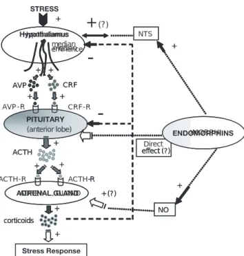
With regard to stress-related behaviors and physiological responses in mammals, endomorphin peptides have been proposed to modulate the HPA axis function via activation of the NTS-projecting neural system impinging on hypothalamic neurons, and/or via activation of the PAG (ventrolateral area) mediating analgesic responses-induced by stress. EM1-2 peptides have been shown to induce mood-related behaviors. For instance, administration of EM1 induced an increased anxiolytic response in mice when tested in elevated plus maze paradigms, results that showed that the µ-opioid receptor modulates mood-related responses in animals and humans, as well. Interesting enough is the recent observation that EM1-2 peptides may induce antidepressant-like behaviors in animals models
of stress and depression, whereby EM1-2 peptides have been shown to up-regulate in a dose-dependent manner the neuronal expression of the BDNF mRNA in rat limbic areas involved in stress and depressive-like behaviors. Thus, these studies led to the proposition that endomorphin peptides may play crucial roles in psychiatric disorders (e.g., depression, schizophrenia). Furthermore, over the past years, it has been shown that µ-opioid receptor agonists (e.g., morphine, DAMGO; morphine-6β-glucuronide) displayed potent orexigenic activities in the CNS of mammals, similar to that displayed by EM1-2 peptides, whose dose-dependent orexigenic activity appears to be mediated by the endogenous opioid peptide, Dynorphin A, acting on its cognate κ-opioid receptor at the hypothalamus.
activation of µ-opioid receptors. Studies on respiratory function showed that EM1-2 peptides attenuate and produce significant respiratory depression in tested animals. Finally, EM1-2 peptides have been shown to induce important inhibitory gastrointestinal effects via the activation of µ-opioid receptors localized in myenteric-plexus neurons that innervate smooth-muscle cells producing a dose-dependent- and CTOP-reversible inhibition of electrically-induced twitch ileum contractions, probably mediated through a reduced release response of several peptide and non-peptide transmitters.
Key words: Endomorphins, physiology, locomotor sensitization responses, opioid reward, stress, HPA axis, sex, feeding, cardio-vascular, respiratory, anxiolytic, social defeat.
RESUMEN
La endomorfina-1 (EM1) y la endomorfina-2 (EM2) son dos péptidos bioactivos que poseen la más alta afinidad de unión selectiva por el receptor opioide µ en comparación con la unión de distintos ligandos agonistas a este subtipo de receptor opioide (véase resumen y texto del capítulo anterior, parte I). Estudios farmacológicos y conductuales han demostrado que la inyección de las EM1-2 en el área ventrotegmental (AVT) genera respuestas conductuales de sensibili-zación locomotora a la anfetamina (AMPH), además de incremen-tar la actividad locomotora de tipo horizontal en los roedores trata-dos. Estos estudios mostraron que la EM2 fue significativamente más potente que la EM1 en inducir las respuestas locomotoras detecta-das, mediadas a través de la alteración de la actividad sináptica de dopamina (DA) y en el globus pallidus de los animales tratados. Asimismo, estudios fármaco-conductuales similares demostraron que otros sistemas de transmisión participan conjuntamente con el siste-ma dopaminérgico en la generación de los efectos locomotores in-ducidos por las EM1-2, como es el caso del sistema gabaérgico (GABA). Más aún, la inyección de EM1 en la región AVT del cerebro de roedores mostró generar respuestas potentes de recompensa pla-centera, similares a las reportadas por distintos alcaloides opiáceos de alto potencial adictivo, posterior a su administración sistémica. Más aún, la inyección de endomorfinas en la región AVT del cerebro del roedor, mas no en el núcleo accumbens (NAc), mostró generar respuestas de recompensa paralela a la generada posteriormente a la administración de dosis bajas de morfina.
En línea con los efectos farmacológicos inducidos por las EM1-2, estudios fármaco-conductuales demostraron que la administración ICV de la EM1 fue capaz de generar respuestas de preferencia de lugar en roedores tratados CPP, por sus siglas en inglés, conditioned place preference, en tanto que la administración de EM2 generó respuestas opuestas, esto es, respuestas de aversión al lugar. Estudios conductuales relacionados con el fenómeno de estrés mostraron que las EM1-2 son capaces de modular la actividad funcional del eje HHA (eje hipotálamo/hipófisis/glándula adrenal) a través de la activación del sistema de proyección neuronal del tracto solitario (NTS, por sus siglas en inglés), al hipotálamo y/o a través de la activación del área ventrolateral de la sustancia gris periacueductal (PAG, por sus siglas en inglés); componente importante del sistema opioide endógeno, que median respuestas analgésicas (antinociceptivas) inducidas por estímulos estresantes. Asimismo, la administración de endomorfinas (v.g., EM1) mostró generar incrementos de conductas de naturaleza ansiolítica en ratones expuestos a paradigmas experimentales de generación de conductas estresantes (v.g., laberinto elevado). Estos estudios sugieren que la generación de conductas de estrés-emocional inducidas por las endomorfinas es mediada a través de la activación del receptor
opioide µ en neuronas del hipotálamo responsables de regular la secreción de factores liberadores de distintas hormonas hipofisiarias (v.g., CRH, LHRH). Más aún, resulta interesante que las endomorfinas sean capaces de inducir conductas antidepresivas o de tipo antidepresivos como se ha reportado recientemente en modelos animales de estrés y depresión. Estos estudios mostraron que las respuestas conductuales de reacción al estrés y las conductas antidepresivas mediadas por las EM1-2 están ligadas con la expresión neuronal del mensajero de RNA que codifica para el factor trófico (BDNF, por sus siglas en inglés, brain derived neurotrophic factor), en áreas del sistema limbico, y que es inducida en forma dosis-dependiente por las endomorfinas, posterior a su administración ICV. Por lo tanto, estos estudios han permitido proponer que las endomorfinas cumplen un papel relevante durante el curso o desarrollo de las enfermedades mentales (v.g., esquizofrenia y depresión). En extensión a estos estudios conductuales, estudios recientes han demostrado la actividad orexigénica de las endomorfinas en forma similar a lo previamente detectado con distintos ligandos agonistas del receptor opioide µ (v.g., morfina, DAMGO; morfina-6β-glucurónido). Si bien estos estudios mostraron que tanto las EM1-2 como diversos agonistas del receptor opioide µ exhiben potentes actividades orexigénicas en el SNC de roedores, la actividad de las EM1-2 parece depender de la actividad de la dinorfina A y su unión sobre su receptor opioide κ en neuronas hipotalámicas. Más aún, diversos estudios han mostrado que el sistema opioide endógeno (a través de la β-endorfina) regula conductas de naturaleza sexual y apareamiento (v.g., lordosis), además de modular la secreción y/o actividad de hormonas de origen gonadal (estrógenos, progesterona).
VII. OPIOID RECEPTOR AGONISTS AND
BEHAVIORAL SENSITIZATION
Repeated injections of psychoactive drugs into animals or
humans usually lead either to a decrement of behavioral
responses (tolerance) or an increase (sensitization) of
psychomotor effects.
1Behavioral sensitization is a term often
used to describe the neurochemical responsiveness and
behavioral effects detected from repeated and intermittent
administration of lower doses of a psychoactive drug.
2This
phenomenon creates a drug «preference» state, which allows
a drug to be used frequently once the substance of abuse has
«sensitized» the active sites in the brain. Thus,
drug-sensitization plays a crucial role in the development and
maintenance of drug addiction,
3which may persist for
long-term periods after drug withdrawal,
4enhancing an
overwhelming urge (craving) for increased drug-seeking and
drug-taking behaviors associated with a loss of behavioral
control during long-term periods of abstinence.
3,5Interesting
enough is that all drugs abused by humans have been shown
to generate drug-rewarding effects and behavioral
sensitization responses in animal models of drug addiction.
6The neuroanatomical and neurochemical bases of drug
sensitization led researchers to focus on the mesocorticolimbic
projecting dopamine (DA) pathway (neuron and axon fibers that
emerge from the VTA and project to both NAC and mPFCx).
7Several interacting transmission systems besides of the
dopamine (DA) system
8,9impinging directly or indirectly on
VTA neurons; have been shown to mediate several of the
evoked-behavioral sensitization responses to drug of abuse
in animals, which includes the inhibitory GABAergic
system,
10the glutamate (GLU)/aspartate (ASP) excitatory
neurotransmission system,
11and the endogenous
µ
and
δ
-dependent opioid receptor systems
12(figure 1). Microdialysis
experiments demonstrated that rats exposed to either
systemic administration or direct injection of
µ
-opioid
receptor agonists (e.g., DAMGO and morphine) into the VTA
(but not in the NAc)
12,13enhanced a significant increase of
DA release in the NAc,
14-17and conversely, administration
of
µ
-opioid receptor antagonists (e.g., CTOP) attenuated the
release of DA in the NAc.
12Likewise, chronic administration
of endomorphins in the VTA produced a significant effect
on the development of locomotor sensitization responses to
amphetamine (AMPH).
18Endomorphin treatment
significantly increased the tissue concentration of GLU and
its metabolites in several limbic structures (e.g., NAc, mPFCx,
CPu) in either EM1-2 plus treated rats or
AMPH-treated animals, used as controls. These results demonstrated
that
µ
-opioid receptor agonists, including both EM1-2
peptides, induce behavioral sensitization responses in animals
mediated via the activation of both GABA and GLU
transmission systems in the VTA.
10,19,20VIII. ENDOMORPHINS MODULATING
LOCOMOTOR BEHAVIORAL RESPONSES
Specific subcortical structures of the brain have been shown
to play an important role in the control of movement.
21,22a través de una reducción sostenida en la liberación de neuro-transmisores de terminales sinápticas del plexo mientérico, mismas que inervan el tejido muscular liso del tracto gastrointestinal.
Palabras clave: Endomorfinas, fisiología, sensibilización locomo-tora, estrés, recompensa a opiacios, eje HPA, sexo, alimentación, cardiovascular, respiratorio, ansiolítico, conducta social.
DA NAc
D2R D1R
Reward
VTA (A10) DYN-A
+
Mesolimbic pathway
GABA-R
+
-GABA intern euro n
EM-2
EM-1
GABA
µ
+
DYN neur on
EM-2 µ
κ
VTA (A10)
NAc
Within the basal ganglia, the globus pallidus contains a
subpopulation of neurons that expresses high levels of
µ
-opioid receptor mRNA.
21,22µ
-opioid receptor agonists have
been shown to increase locomotor sensitization responses
that are influenced by a number of variables (e.g., ligand
concentration, experimental paradigm, and timing of
experiment performed). Most studies using psychomotor
paradigms have shown that
µ
- and
δ
-opioid receptor
agonists enhanced an increment of ambulatory responses,
whereas
κ
-opioid receptor agonists produced opposite
responses.
23,24In this context, locomotor responses (e.g.,
horizontal and vertical locomotor) mediated by activation
of the
µ
-opioid receptor by morphine-binding its cognate
µ
-opioid receptor
25-27and grooming behavior mediated by
activation of
δ
-opioid binding sites (localized in the VTA,
NAc, and PAG)
26,28,29have been shown to depend on the
synthesis and release of DA from both nigrostriatal and
mesolimbic dopaminergic neurons.
30Interactions between
DA and opioid systems in the brain have been extensively
reported. These studies showed that opiates (e.g., morphine,
heroin) increase behavioral sensitivity responses to DA
agonists, enhancing an increase supersensitivity of DA
receptors and the expression of stereotypic behaviors
mediated by the activation of D1R/D2R-receptor by ligand
agonists.
31For instance, morphine potentiated an
apomorphine-dependent climbing behavior in wild-type
mice, as opposed to the mutant-
µ
-opioid receptor knockout
mice.
32Thus, these set of results demonstrated that
µ
-receptor ligands alter the DA projecting system by
potentiat-ing the climbpotentiat-ing behavior responses in mice induced after
administration of D1R/D2R-ligand agonists.
33Endomorphin peptides, like morphine, were found to
increase locomotion (horizontal) activity
34,35without
affecting the vertical locomotor activity
19in mice. These
studies showed that low concentrations of EM2 (0.3 and
1.0
µ
g/animal, ICV administration) induce similar
behavioral responses displayed by higher concentrations
of EM1 peptide (10-30 µg/mouse, ICV administration).
These results led researchers to posit that EM1-2 peptides
not only activate different
µ
-opioid receptor subtypes in the
basal ganglia,
36,37but could modulate different opioid
systems (i.e. enkephalinergic, dynorphinergic) that appear
to be implicated in the expression of the sensitization
responses mediated by endomorphins.
38,39Similar studies
demonstrated that morphine injected into the globus pallidus
produced a robust increase in locomotor activity in mice,
40whereas EM1 induced localized stereotyped behavioral
responses (e.g., orofacial dyskinesia).
41These
opioid-mediated behavioral responses led authors to propose that
the locomotor activity induced by morphine could be
mediated via the activation of
δ
- and
κ
-opioid receptors,
whereas EM1-inducing inhibitory activities would depend
mostly on the activation of
µ
-opioid binding sites.
41As shown
for the interaction of GABA and several endogenous opioid
peptides (e.g., enkephalins),
42EM1 and GABA could mediate
opposite behavioral responses in the control of movement
at the globus pallidus. The resulting chemical unbalance
induced in both neurotransmission systems could lead to
the development of motor dysfunctions and the
manifestation of localized dyskinesias.
19IX. ENDOMORPHINS REGULATING OPIATE
REWARDING RESPONSES
Extensive studies have shown that the chronic administration
of
µ
-opioid-receptor agonists (e.g., DAMGO, morphine,
codeine, and sufentanyl)
43-45produce potent drug- and/or
stress-rewarding effects, associated to the development of
drug withdrawal symptoms and physical dependence in
animals.
46,47Quite interesting to note is that opposite
(non-rewarding) aversion responses in rodents appeared after
administration of selective
κ
-opioid receptor agonists.
48Drug-rewarding effects have been shown to be mediated
via the interaction between GABAergic neurons and the
mesolimbic/VTA-dopaminergic transmission system (see
extensive reviews in
49-51) (figure 1). As described above,
opioid receptor agonists inhibit GABAergic inputs to VTA/
dopaminergic principal cells that project the NAc, inducing
a disinhibitory effect, which in turn enhances a potent release
of DA into this limbic structure.
10,52Endomorphin peptides
injected into the posterior area of the VTA induced a
conditioned-place preference (CPP), displaying similar
behavioral responses to those exhibited by morphine or
DAMGO.
53Moreover, EM1 injected into the VTA produced
a potent rewarding effect in rodents exposed to the drug
self-administration paradigm. However, injection of same
peptide or DAMGO into the NAc produced poor and
delayed rewarding effects compared to the VTA-detected
responses.
53These data suggested the absence or poor
expression of µ-opioid-receptor sites in this mesolimbic
area.
54ICV administration of endomorphin peptides shed
inconsistent results on the induced rewarding responses.
For instance, some authors reported that EM1 mediated a
significant CPP, whereas EM2 displayed significant place
aversion effects in mice.
55Conversely, other authors
reported that ICV administration of EM1-2 peptides (at low
doses, 15
µ
g) induced significant antinociceptive responses
in mice
56producing no-effects on the CPP paradigm.
57However, higher doses of EM1 (30
µ
g) produced barrel
rotation of the body trunk, whereas EM2 evoked a
significant place preference condition in tested mice.
57Such
asymmetric expression of opioid receptor sites in targeted
brain areas and cells; and/or to the activation of different
molecular mechanism that drive the rewarding effects and
behavioral responses to opioid substances.
19Moreover, differences in the behavioral effects induced
by endomorphin peptides could be due to the activation of
µ
-opioid receptors expressed in neurons localized at the
brainstem PAG region, involved in the generation of
aversive behaviors.
58These effects have been associated to
the disruption of the HPA axis, in addition to the
deregulation of different mesolimbic transmission systems
involved in rewarding functions, as shown in addicted
humans exhibiting a history of long-term opiate abuse
59(figures 1 and 2).
X. ENDOMORPHINS IMPLICATED IN
STRESS-INDUCING ALTERED BEHAVIORS
Stressors have been implicated in the development of
several psychiatric illnesses, where the HPA axis and
endogenous opioid system have been shown to play a
crucial role in stress responses. Although the precise role
of endogenous opioid peptides and receptors to stress
stimuli has not been fully elucidated, over the past years
several works showed the existing interactions between
stressors, the HPA axis and the endogenous opioid system,
(see reference,
19and references therein). These works
showed a close relationship between the levels of the opioid
ligands, corticosteroids, pituitary hormone levels, and
immune-borne hormones (e.g., cytokines).
19Activation of
the HPA axis by external/internal stressful stimuli (e.g.,
stress, immune challenge) leads to the increase secretion of
corticotrophin-releasing factor (CRF) and Arg-vasopressin
(AVP) from the median eminence of the hypothalamus,
enhancing the cell-release of ACTH from the anterior lobe
of the pituitary.
60Increased serum levels of ACTH enhance
the release of glucocorticoids from the adrenal gland, which
exert a negative feedback on pituitary adrenocorticotrophs
and limbic regions of the mammalian CNS (e.g., amygdale),
enhancing the homeostatic and neuroendocrine balance
along the HPA axis.
61Some authors have postulated that
the endogenous opioid system, driven through
β
-endorphin
(binding
µ
-opioid receptors) at the hypothalamus exerts a
potent inhibitory activity on the HPA axis.
62,63In this context,
pharmacological studies showed that acute morphine
administration, acting on
µ
-opioid receptors expressed along
the HPA axis,
64produced an important increase of ACTH
and adrenal secretion of corticosterone
65(figure 2).
Based on the aforementioned results, ICV
administration of EM1 or EM2 peptides (10
µ
g) produced
no stimulatory activity on the HPA axis and displayed no
neuroendocrine effect on the ACTH and corticosterone
secretion.
60Moreover, EM1 failed to block the stimulatory
effect of morphine on the ACTH-induced increased levels
of corticosterone in plasma. Furthermore, chronic activation
of the HPA axis by exposure of animals to chronic stress
paradigms (e.g., chronic inflammatory stress of
adjuvant-induced arthritis, the restraint stress model, and the
immune-based lipopolysaccharide stress model) showed
that plasma corticosterone, ACTH and
β
-endorphin were
dramatically increased, whereas levels of endomorphin
peptides showed no detectable changes compared to
controls. These data suggested that EM1-2 peptides appear
to display no significant roles on the neuroendocrine
modulation of the HPA axis, mediating stress responses to
challenging stimuli.
60Based on that
µ
-opioid receptors are expressed on
pituitary cells and neuronal cells within the hypothalamus,
and receptor ligand agonists (e.g., morphine) properly induce
a potent activation of the HPA axis, it has been proposed
Direct effect (?) +
Hypothalamus
median
AVP CRF
+ +
PITUITARY
+ +
(anterior lobe)
AVP-R CRF-R
ACTH +
+
ADRENAL GLAND +
+
corticoids
ACTH-R ACTH-R
Stress Response +
ENDOMORPHINS NTS
+
NO + + (?)
effect (?) +
median eminence
AVP CRF
+ +
+ +
(anterior lobe)
ACTH +
+ +
+
corticoids
--R
+
+
+
(?)
NO + + (?)
that the lack of endomorphin-induced neuroendocrine effects
on the HPA axis could be due merely to the central
metabolism or degradation of these peptides (see degradation
of EM1-2 peptides in previous paper, part I) (figure 2).
Other plausible explanation offered relies on that
µ-opioid receptor agonists display different stimulating
properties of Gi/Go protein, as demonstrated in animal
models of pain.
38,66Thus, intracellular molecules may
provide important insights into the differential cell-responses
of endomorphin peptides modulating several neural systems
involved in distinct physiological effects and responses.
For instance, different works showed recently that nitric
oxide (a chemical messenger molecule involved in different
physiological and pathological processes in mammals)
released from cells mediates several physiological responses
induced by opioid peptides (e.g.,
β
-endorphin) or
morphine
67-69besides of the aforementioned endomorphin
bioactivities (e.g., vasodilatory responses, modulation of
HPA axis activity).
35,67This situation led researchers to
propose the term «endomorphin-NO-HPA axis»,
35,70where
endomorphin peptides acting either on hypothalamic
neurons, may stimulate the HPA axis; or through the
proposed NTS-endomorphin projecting neural system
impinging on hypothalamic neuron cells (this neural
component has been shown previously to activate directly
the HPA axis in animals) (see previous studies in
references
71,72) (figure 2).
Other neural pathways emerging from the PAG have
been shown to mediate stress-induced immobility and
analgesia in adult rats and rat pups.
73Pharmacological
experiments showed that
µ
-opioid receptor antagonists (e.g.,
naltrexone, CTOP) acting on the ventrolateral PAG blocked
several analgesic responses mediated by endogenous
opioids, providing evidences that endomorphins could
mediate PAG-inducing stress-related analgesic effects in rats,
as shown for different
µ
-opioid receptor ligand agonists.
73Thus, these neural and neuroendocrine driving mechanisms
provide important insights that endomorphin peptides might
be directly or indirectly involved in the activation of the HPA
axis, enhancing the release of CRH from PVN/hypothalamic
neurons.
74,75XI. ENDOMORPHINS INVOLVED IN
MOOD-RELATED AND PSYCHIATRIC DISORDERS
Over the past decades extensive studies demonstrated that
ICV (central)
76,77or IP (peripheral)
78,79administration of
µ
-opioid receptor agonists (e.g., morphine) produce anxiolytic
responses, whereas
µ
-opioid receptor antagonists promote
anxiogenic effects.
80These responses were shown to be
mediated through the interaction of the EOS and the
GABAergic system (e.g., GABA and BZD);
81,82the
monoaminergic (e.g., 5-HT, BZD) and peptidergic systems.
19Moreover, these pharmacological and behavioral studies
demonstrated that the anxiolytic and anxiogenic activities
of opiates substances are dose- and site-dependent after their
local administration into the rat neural tissue. For instance,
low doses of morphine injected into rat midbrain tectum
induced anxiolytic–like responses, whereas the injection of
higher doses displayed anxiogenic-like effects.
83Converserly,
morphine injected into the dorsal PAG
84or lateral septum
of the rat brain
85has been shown to produce aversive
responses. Previous reports describing the neuroanatomical
co-localization between endomorphins and
µ
-opioid
receptors in both limbic and brainstem regions and nuclei
in the CNS of rodents,
86,87led to postulate the hypothesis
that EM1-2 could modulate mood-related behaviors (e.g.,
anxiety and stress-related behaviors) in animals and
humans.
19For instance, ICV administration of EM1 into mice
induced an increased anxiolytic behavior responses in the
elevated plus maze
88supporting previous observations that
administrations of
µ
-opioid receptor agonists in humans
produce anxiolytic symptomology (e.g., drowsiness, warmth
feelings, and sensation of well-being).
80Earlier findings showed the expression of high density
µ
-opioid receptors and high concentration of endogenous
opioid ligands (e.g.,
β
-endorphin) in limbic areas of animals
exposed to stressful challenges.
89-92These data led authors
to postulate that the EOS and endomorphins play a crucial
role in modulation in psychiatric disorders,
93such as
depression and schizophrenia,
94-98in spite of the absence
of a clear therapeutic benefit of opioid ligands to treat
mental illnesses.
19Molecular and behavioral studies
showed that knockout mice lacking the
µ
-opioid receptor
display altered emotional states consistent of
depressive-like behaviors, similar to those studies that have extensively
demonstrated the use of a wide variety of
µ
-opioid receptor
agonists, as antidepressant agents (e.g., oxycodone and
oxymorphone)
99for treating depressive symptoms among
many other mental illnesess.
100-102For instance, morphine
has been used as an antidepressant-like agent to relieve
stress behaviors in experimental animals.
103These data
demonstrate the importance of the EOS in the etiology of
mental disorders, besides of the controversial issue on the
clinical use of
µ
-opioid receptor agonists as therapeutical
agents to relieve psychiatric disorders.
19Based on the immunoreactive co-localization of
EM1-2 peptides and
µ
-opioid receptors in both forebrain (e.g.,
septum, NAc, amygdala, thalamic nuclei) and brainstem
regions (e.g., LC) in the CNS of mamamals, and shown
previously with regard to their functional implication in
the pathophysiology of depression;
86,104,105several authors
have postulated the importance of these amide
tetra-peptides in the etiology of depressive disorders, based on
their potential antidepressant-like effects observed in
animal models of depression and stress.
19The
(0.3–30
µ
g/animal, ICV) in mice were dose-dependent and
short-lasting (enduring only 10-15 min after their brain
administration).
19The magnitude of the antidepressant
responses displayed by both peptides was comparable to the
several compounds shown to display potent antidepressant
activities.
106,107These studies provided strong evidences that
these opioid peptides, like conventional antidepressants, may
generate antidepressant-like responses (reducing
behavioral-immobility in paradigms of stress and depression [e.g.,
forced swimming test, FST])
108and which may be blocked
by µ-opioid receptor antagonists (e.g., naloxone,
β
-funal-trexamine) but not with selective
δ
- or
κ
-opioid receptor
antagonists (e.g., naltrindole, nor-binaltorphimine,
respective-ly).
19Additional studies regarding the implication of EM1-2
in the pathophysiology of depression showed that ICV
administration of endomorphins (e.g.,20-50
µ
g/animal), into
rats induces a dose-dependent up-regulation of BDNF
mRNA expression in limbic areas of the rat brain (mPFCx,
hippocampus, amygdala) that was blocked by specific
µ
-opioid receptor antagonists (e.g., naltrexone), but not with
specific
δ
-opioid receptor antagonists (e.g., naltrindole).
109This neurotrophic factor (BDNF) has been shown to
modulate primordial functions, such as neuronal survival,
differentiation, and plasticity,
110and shown recently to play
an important role in the therapeutic actions of several
antidepressants acting on different neurotransmission
systems.
111-115XII. ENDOMORPHINS MEDIATING
FOOD-INTAKE BEHAVIOR
The neural pathways and transmission systems that regulate
food-intake behavior in mammals are complex.
19Several
regulatory peptides (i.e., NPY, GHRH, 26RFa peptide) have
been shown to display orexigenic activities in the brain.
116-119In a similar context,
µ
-opioid receptor ligand agonists
(e.g., morphine, DAMGO), including the active morphine
metabolite (e.g., morphine-6
β
-glucuronide), have been
shown to display an orexigenic activity
120-123regulating
gustatory neural pathways arising from the NTS
neurons
124,125(a neural pathway projecting to hypothalamic
areas and other limbic structures).
126In a similar context,
ICV administration of EM1 or EM2 (0.03–30 nmol)
produced a dose-dependent food-intake behavior in non
food-deprived mice for up to 4 h after peptide injection.
88These EM1-2 mediated effects were attenuated by the
specific
µ
-opioid receptor antagonist,
β
-funaltrexamine.
127,128However, the endogenous
κ
-opioid receptor ligand peptide,
Dynorphin A (DYN A), was shown to display a potent
stimulatory food-intake bevavior compared to EM1-2
peptides. These results posit that the orexigenic activity
induced by EM1-2 peptides appears to be mediated, via
activation of
κ
-opioid receptors, where
µ
-opioid receptors
appear to play a minor role in this opioid and non-opioid
dependent physiological activity.
19XIII. ENDOMORPHINS MODULATING
SEXUAL BEHAVIOR RESPONSES
Extensive studies demonstrated the direct and indirect
effects of endogenous opioid peptides (e.g.,
β
-endorphin)
on gonadal hormones, regulating both sexually-induced
behaviors (e.g., lordosis) and reproductive functions in
female rats.
129-131These effects have been shown to be
mediated through the activation of
µ
-opioid receptors
expressed along the limbo-hypothalamic neural circuits that
mediate the release of gonadal hormones (LH, FSH) from
the pituitary and the release of its releasing peptide
hormone (LHRH) from the hypothalamus.
132LH/FSH have
been extensively shown to influence female rat sexual
behavior,
133besides of modulating the release and
expression of sexual steroids during mating behavior.
134Based on the neuroanatomical distribution and cell
expression of
µ
-opioid receptors within specific
hypothalamic and mesencephalic regions that coordinate
and regulate female reproductive behavior (e.g., VMH,
mPOA, MCG),
104,135several authors showed that
µ
-opioid
receptor ligand agonists produce dual effects on lordosis
in hormonally-primed female rodents during mating.
135,136In a similar context, ICV or local administration of low
doses of
β
-endorphin into the MCG or mPOA produces a
potent inhibition of lordosis in gonadectomized,
steroid-primed female rats.
137-140Converserly, similar route of
administration of high doses of
β
-endorphin or
morphiceptin facilitated lordosis in estrogen- or estrogen/
progesterone primed rats.
137,138,141-143Moreover, ICV
administration of EM1-2 peptides into the third ventricle
or bilateral infusion into the diagonal band (DB)
(septum-horizontal limb of the diagonal band, MS-HDB, an area
shown to project axon fibers to the mPOA of the
hypothalamus)
144produced dose- and time-dependent
lordosis responses in female rats
135which was attenuated
with naloxone.
134However, similar responses were not
detected when peptides were injected into VMH, mPOA
or MCG.
134These results led authors to propose that
EM1-2 peptides modulate
104,145the release of active
neuropeptides (e.g., LHRH) and non-peptide (Ach, GABA)
transmitters within the MS-HDB, inducing their
opioid-dependent behavioral effects.
19XIV. ENDOMORPHINS INVOLVED IN
LEARNING AND MEMORY PROCESSING
studied and reported elsewhere.
19For instance, the EOS
has been shown to play important roles in operant and
classic conditioning and different cognitive tasks, including
memory processing.
19These studies showed that either
µ
-opioid receptor (e.g., DAMGO and Tyr-D-Arg-Phe-
β
-Ala)
and
δ
-selective opioid receptor (e.g.,D-Pen
2,L-Pen
5-enkephalin and D-Ala
2-deltorphin II) agonists, respectively,
induced an impairment of both short-term and long-term
memory processing in mice exposed either to passive
avoidance paradigms
146-148or spatial memory tasks.
149Conversely,
µ
-opioid receptor antagonists enhanced
memory retention in animals exposed to different learning
tasks.
150Similarly,
κ
-opioid receptor agonists (e.g.,
dynorphin A
1-13) have been shown to attenuate aberrant
learning and memory processing in rodents exposed to
aversive and non-aversive memory tasks.
151With regard to endomorphin molecules, a single report
from Ukai et al.
152showed that endomorphins impaired
short-term memory processing in mice exposed to
spontaneous alternation performance task. Whereas both
tetrapeptides induced an important inhibitory activity on
long-term memory processing when tested in passive
avoidance learning task in mice,
153,154EM2 peptide was
shown to mediate its memory attenuating effect
155by
inducing an opioid receptor dependent cytosolic and
mitochondrial protein synthesis mechanism in the lobus
paraolfactorius in chicks (a brain area structurally related to
the caudate putamen in mammals).
156,157These studies
showed that EM2 reverted the amnesic effects induced by
anisomycin administration into chicks, and blocked the
inhibitory effect on protein synthesis induced by this drug
in this related striatal-brain structure.
155However, other
authors proposed that this EM2-dependent inhibition of
passive avoidance learning task resulted from a functional
disconnection of the hippocampus, a brain area known to
be crucially important for processing and conversion of
short-term memories into long-term memories (see
reference
158). Besides of the aforementioned studies, several
authors proposed that both cholinergic and dopaminergic
transmitter systems could mediate or participate in the opioid
peptide-induced long-term memory impairment, although
their exact roles have not been clearly elucidated.
19In this
context, behavioral and pharmacological studies showed that
spatial working memory requires at least the interaction
between
µ
-opioid receptor ligand agonists (including both
EM1/EM2 peptides) and the ACh transmission system.
159Cellular studies demonstrated that endomorphin peptides
decreased significantly the release of ACh from neuronal
cells in brain areas associated with memory processing and
storage
153,154,160and that physostigmine (a cholinesterase
inhibitor) reverted the endomorphin induced passive
avoidance learning impaired response.
146,154Besides of the interaction between ACh and opioid
system, several authors showed that D2-receptor antagonists
were capable of attenuating the EM2-induced passive
avoidance learning impairment in rodents.
153These results
suggested that the inhibitory effect of EM2 would be mediated
from stimulation of heterosynaptic D2 receptors expressed
in dopaminergic neurons innervating both the striatum and
the NAc, during acquisition and consolidation of memory.
153In addition, ICV administration of EM1-2 peptides was shown
to increase BDNF mRNA expressions in the hippocampus
and amygdala.
109This trophic factor, acting via its neuronal
NT-3 receptor subtype, has been shown to mediate several
plastic events in the brain, such as development and
establishment of long-term potentiation (LTP) in
hippocampal neurons; morphologic changes in active
synapses and neurons in brain regions involved in learning
and memory processing (e.g., hippocampus and cortex).
161,162Thus, based on the information described above,
endomorphins acting via
µ
-opioid receptors could be
implicated in learning and memory processing in several
areas of the brain regulating BDNF activity on neuronal cells.
19XV. ENDOMORPHINS REGULATING
CARDIOVASCULAR AND RESPIRATORY
BIOACTIVITIES
Over the past years it has been shown that several regions
along the rostracaudal axis (e.g., VLM, NTS, LH, PVN) of
the rat brain, including the dorsal hippocampus and limbic
system, regulate cardiovascular and respiratory bioactivities,
areas shown also to express a high density of
µ
-opioid
receptors in neurons within each functional brain region.
191. Cardiovascular effects
Although several works have acknowledged the important
role of the EOS mediating cardiovascular responses, the
effects of distinct opioid receptor ligand agonists on blood
pressure and heart rate have been unclear and confusing.
For instance, local injections of
µ
,
δ
and
κ
-opioid receptor
agonists into the specific areas of the rat brain (e.g., PVN,
DH, RVLM) in normotensive and/or hypertensive animals,
were shown to reduce heart rate and blood pressure,
163whereas ICV administration of
µ
-opioid receptor agonists
(e.g., morphine,
β
-endorphin, DAMGO) produced
hypotension in different species.
93,164-166In a similar context, ICV or IV administration of
EM1-2 produced a significant reduction of heart rate and
blood pressure in normotensive and hypertensive
rats
167-170showing a reversible and dose-dependent
biphasic change in systemic arterial pressure
165upon
differences in the administration route of EM2 into rats (IV
versus ICV) produced different graded responses,
171which
led authors to suggest that although peripheral
µ
-opioid
receptors might play an important role in the opioid
peptide-inducing hypotensive effects in animals,
172the
mechanisms by which endomorphin peptides mediate both
peripheral and central cardiovascular activities are not
completely elucidated.
This assumption is based on previous results that
demonstrated, on the one hand, that bradycardia results
from activation of the vagus nerve, and bilateral vagotomy
(or atropine) abolished EM1 effect on heart rate in rats,
suggesting thus, that EM1-2 mediate their vascular effects
via activation of vagal afferents.
171On the other hand,
EM1-2 peptides which exert potent inhibitory activities on
neurons,
173-175their cellular activities are expected to
increase blood pressure and heart rate due an increase
response on neuronal firing on cardiovascular regulatory
areas at both medulla and cervical spinal cord.
176-178Recent
studies showed that infusion of EM2 into the rat NTS
(mNTS) attenuated reflex responses upon stimulation of
the carotid sinus and aortic baroreceptors,
176-178showing
that depressor and bradycardic responses resulted from a
peripheral inhibitory effect on the baroreflex response and
from the excitatory activity of mNTS neurons. These
authors proposed that EM2 mediated its cardiovascular
responses after being injected into the mNTS (a brainstem
area control under local inhibitory-GABAergic
neurons
179,180and glutamate [GLU] projecting neurons
descending from insular cortex)
181,182through direct
activation of
µ
-opioid receptors expressed on mNTS-GABA
neurons, producing an hyperpolarization response,
decreased of GABA release and increase of GLU release
from presynaptic terminals, enhancing an overall reduction
of GABAergic activity on mNTS postsynaptic neurons
(disinhibitory effect) leading thus, to a final increase of
neuronal excitability of mNTS neurons.
19,176-178Moreover, EM2 acting on µ-opioid receptors expressed
on glutamate baroreceptor afferent terminals in response to
baroreceptor stimulation were shown to decrease GLU
release, resulting in attenuation of the baroreflex activity.
19Despite that the depressor and bradycardic responses induced
by EM2 could be explained through the aforementioned
cellular mechanisms,
19,183the endogenous mechanisms by
which EM1-2 induced vasoactive responses are still unclear
and further research may clarify this issue. However, several
authors proposed different mechanisms by which
endomorphins may mediate their hypotensive responses.
One proposed mechanism is through the intracellular
stimulation of NO synthesis
164,184and NO release from the
vessels
185after binding their cognate opioid receptor on
endothelial cells. Other mechanisms proposed is through the
hyperpolarization of vascular smooth muscle cells and/or
the cell-release of vasodilator prostanglandins or via an
endomorphin inhibition of presynaptic release of NA from
nerve endings distributed along vessel walls.
172Whichever
the mechanisms involved, none of these have been completely
confirmed, which requires further research to clarify the exact
endogenous opioid-dependent mechanism.
2. Respiratory effects
Several studies demonstrated that
µ
-opioid receptor agonists
induce a potent respiratory depression.
186Opioid-receptor
agonists, such as morphine (see reference
19and references
therein), heroin,
187-189fentanyl,
190,191buprenorphine,
192-194and
DAMGO
171,195have been shown to induce a respiratory
depression by decreasing the sensitivity of brainstem nuclei
regulating-respiratory activity to carbon dioxide, followed
by a decrement of respiratory rate (see reviews in
46,196,197).
Although immunochemical studies showed that both
EM1-2-LI
198and
µ
-opioid receptors
199,200appear to be
co-localized at specific neuroanatomical areas and nuclei of the
brainstem of mammals (e.g., NTS and PBN)
104shown to play
a crucial role in the respiratory control,
19very few studies
have focused on the effects of endomorphin peptides on
respiratory activity. For instance, IV administration of
supra-analgesic doses of EM1 and EM2 peptides in rats produced
biphasic responses, characterized by rapid initial ventilatory
depression (inhibitory effect that lasts for 4-6 s) followed
by an increase ventilation activity (excitatory effect that lasts
for 10-12 min).
201In contrast to the monotonically-induced
decreased ventilation activity by morphine, EM1-2 appear
to mediate their depressant respiratory activity via
activation of central
µ
-opioid receptors, based on that
methylnaloxone (a peripheral restricted
µ
-opioid receptor
antagonist) was unable to block peripherically the opioid
effects, whereas naloxone prevented the
respiratory-depressed activity induced by endomorphin peptides.
201Interestingly, EM1-2 peptides administered (I.V.) at doses
higher above their corresponding analgesic threshold or
higher above their respiratory depression-threshold dose
(as shown for DAMGO or morphine) showed to attenuate
hypercapnic ventilatory responses in rats, producing a
respiratory depression in tested animals.
201Although
EM1-2 peptides induced a weaker depression activity on
ventilatory responses compared to the hypercapnia effects
induced by either DAMGO or morphine,
201the reduced
effects mediated by EM1-2 peptides have been extensively
discussed based on several pharmacokinetic and
pharmacodynamic parameters and factors discussed
elsewhere.
19,201In this context, the increased ventilation
activity induced by EM1-2 peptides appeared to be
mediated through a central non-opioid mechanism, based
on that conventional
µ
-opioid receptor antagonists were
unable to block the increased respiratory response.
201showed that both EM1 and EM2 produced dose-dependent
inhibition of tachykinin-mediated contractions of the guinea
pig bronchus, with the exception of EM1 activity which was
blocked by naloxone, whereas that of EM2 was not
antagonized by any µ-opioid receptor antagonist.
202In the same context, Patel et al.
203showed that EM1-2
peptides produced a dose-dependent inhibition and
naloxone-reversible antagonism activity on cholinergic
induced-contractile responses of the guinea pig trachea.
These studies showed that EM1-2 peptides, acting on
µ
-opioid receptors distributed in rodent airways,
204interact
with both cholinergic and tachykinergic fiber types.
202,203Thus, EM1-2 peptides were shown to induce a potent
inhibition on the electrically-evoked release of ACh from
cholinergic nerves innervating guinea pig trachea,
203including the release of NA from nonadrenergic
postganglionic nerve fibers innervating the airway-smooth
muscle cells.
203Recent studies showed that ICV
administration of EM1 in mice induced an increased oxygen
consumption that was blocked by naloxone, suggesting a
µ
-opioid receptor peptide dependent effect.
205XVI. ENDOMORPHINS INVOLVED IN
GASTROINTESTINAL ACTIVITY
Opioid agonists regulating gastrointestinal activity have
been extensively reported. IHC techniques and binding
studies, using neuromuscular preparations, showed that
µ
-opioid receptors may be localized in smooth muscle cells
and neurons, as well
206where
µ
-opioid receptor agonists
(e.g.,morphine) have been shown to exert, in a
naloxone-reversible fashion, their inhibitory effects on GI activity
and/or motility,
207-209besides of modulating the evoked
release of neurotransmitter release from nerve terminals
(e.g., ACh, NA)
209-211and influencing peristaltic reflex.
207In a similar context, EM1-2 peptides were shown to
modulate different GI activities. For instance, application
of a concentration range of EM1-2 peptides (10
-12M to 10
-6M) on a specific guinea pig ileum preparation (longitudinal
muscle-myenteric plexus preparations from ileum)
produced a dose-dependent- and CTOP-reversible
inhibition of the amplitude of electrically-induced twitch
ileum contractions.
212However, EM1-2 peptides failed to
inhibit muscle contractions induced by ACh stimulation.
Moreover, both peptides displayed a potent inhibitory effect
on the ascending excitatory reflex and increased stimulation
of the descending inhibitory reflex, inducing an increase
latency on the onset of ileum muscle-contractile
responses.
213Similar inhibitory responses were detected at
the smooth and striated muscles of the rat esophagus.
214Overall, these results led authors to suggest that
EM1-2 induce their inhibitory GI effects, via activation of
µ
-opioid
receptors localized either in presynaptic terminals of
non-adrenergic/non-cholinergic inhibitory neurons and/or in
myenteric-plexus neurons that innervate smooth-muscle
cells.
212,214Activation of
µ
-opioid receptors may lead to a
reduced release response of different neurotransmitters
(e.g., NO, VIP), including ACh and NA from local and
myenteric neurons, respectively.
210-213XVII. ENDOGENOUS OPIOIDS
AND SOCIAL BEHAVIORS
A clear example of stress-inducing increased HPA activity
occurs in foraging and defensive behaviors in animals.
215For example, after social defeat, subordinate animals display
physiological, neuroendocrine, neurochemical and
behavioral changes induced by the endogenous
stress-driving mechanisms in socially-interacting species.
216These
behavioral and functional changes have been suggested to
be highly connected to the development of fear, anxiety,
depression, and panic disorders,
217,218including the
development of drug-seeking and drug-taking behaviors in
both animals and humans.
219Moreover, few studies have
shown that
µ
-opioid receptor results to be up-regulated in
restricted regions of the rat brain (e.g., VTA) after social
defeat.
220ICV administration of EM1 into Syrian hamsters
failed to inhibit the consolidation of conditioned defeat
(without stimulating locomotor activity or inducing
sedation),
221whereas morphine impaired the consolidation
of newly acquired memories in rats and mice.
222-225These
authors suggested that such reduced or failed behavioral
responses mediated by EM1 could be due to cellular
responses and pharmacological activities mediated through
the binding of the peptide to its cognate receptor (see detailed
explanations in
19and references therein). For instance,
morphine, DAMGO, besides of other potent endomorphin
peptide analogs (e.g., Tyr-D-Arg-Phe-
β
-Ala),
88,226,227have
been shown to inhibit memory retrieval, increase anxiolytic
responses and produce fear-conditioning responses in
animals.
77,78,228,229Such responses appear to depend on the
activation of NA neurons and NA neural system.
230ACKNOWLEDGEMENTS
REFERENCES
1. Segal DS, Kuczenski R. Repeated binge exposures to amphetamine and methamphetamine: behavioral and neurochemical characterization. J Pharmacol Exp Ther 1997; 282:561-573.
2. Spyraki C, Fibiger HC, Phillips AG. Attenuation of heroin reward in rats by disruption of the mesolimbic dopamine system. Psychopharma-cology (Berl) 1983; 79:278-283.
3. Robinson TE, Berridge KC. The neural basis of drug craving: an incenti-ve-sensitization theory of addiction. Brain Res Brain Res Rev 1993; 18:247-291.
4. Robinson TE, Becker JB. Enduring changes in brain and behavior pro-duced by chronic amphetamine administration: a review and evalua-tion of animal models of amphetamine psychosis. Brain Res 1986; 396:157-198.
5. Wise RA, Bozarth MA. A psychomotor stimulant theory of addiction. Psychol Rev 1987; 94:469-492.
6. Stewart J, Badiani A. Tolerance and sensitization to the behavioral effects of drugs. Behav Pharmacol 1993; 4:289-312.
7. Spanagel R. Modulation of drug-induced sensitization processes by en-dogenous opioid systems. Behav Brain Res 1995; 70:37-49.
8. Hooks MS, Kalivas PW. Involvement of dopamine and excitatory ami-no acid transmission in ami-novelty-induced motor activity. J Pharmacol Exp Ther 1994; 269:976-988.
9. Vezina P. D1 dopamine receptor activation is necessary for the induc-tion of sensitizainduc-tion by amphetamine in the ventral tegmental area. J Neurosci 1996; 16:2411-2420.
10. Johnson SW, North RA. Opioids excite dopamine neurons by hyperpo-larization of local interneurons. J Neurosci 1992; 12:483-488. 11. Karler R, Chaudhry IA, Calder LD et al. Amphetamine behavioral
sen-sitization and the excitatory amino acids. Brain Res 1990; 537:76-82. 12. Spanagel R, Herz A, Shippenberg TS. Opposing tonically active
endo-genous opioid systems modulate the mesolimbic dopaminergic pathway. Proc Natl Acad Sci U S A 1992; 89:2046-2050.
13. Devine DP, Leone P, Wise RA. Mesolimbic dopamine neurotransmis-sion is increased by administration of mu-opioid receptor antagonists. Eur J Pharmacol 1993; 243:55-64.
14. Di CG, Imperato A. Opposite effects of mu and kappa opiate agonists on dopamine release in the nucleus accumbens and in the dorsal cauda-te of freely moving rats. J Pharmacol Exp Ther 1988; 244:1067-1080. 15. Di CG, Imperato A. Drugs abused by humans preferentially increase
synaptic dopamine concentrations in the mesolimbic system of freely moving rats. Proc Natl Acad Sci U S A 1988; 85:5274-5278.
16. Pentney RJ, Gratton A. Effects of local delta and mu opioid receptor activation on basal and stimulated dopamine release in striatum and nucleus accumbens of rat: an in vivo electrochemical study. Neuros-cience 1991; 45:95-102.
17. Spanagel R, Herz A, Shippenberg TS. The effects of opioid peptides on dopamine release in the nucleus accumbens: an in vivo microdialysis study. J Neurochem 1990; 55:1734-1740.
18. Chen JC, Liang KW, Huang EY. Differential effects of endomorphin-1 and -2 on amphetamine sensitization: neurochemical and behavioral aspects. Synapse 2001; 39:239-248.
19. Fichna J, Janecka A, Costentin J et al. The endomorphin system and its evolving neurophysiological role. Pharmacol Rev 2007; 59:88-123. 20. Wolf ME, Xue CJ. Amphetamine and D1 dopamine receptor agonists
produce biphasic effects on glutamate efflux in rat ventral tegmental area: modification by repeated amphetamine administration. J Neuro-chem 1998; 70:198-209.
21. Delfs JM, Kong H, Mestek A et al. Expression of mu opioid receptor mRNA in rat brain: an in situ hybridization study at the single cell level. J Comp Neurol 1994; 345:46-68.
22. Peckys D, Landwehrmeyer GB. Expression of mu, kappa, and delta opioid receptor messenger RNA in the human CNS: a 33P in situ hybri-dization study. Neuroscience 1999; 88:1093-1135.
23. Obeso JA, Rodriguez-Oroz MC, Rodriguez M et al. Pathophysiology of levodopa-induced dyskinesias in Parkinson’s disease: problems with the current model. Ann Neurol 2000; 47:S22-S32.
24. Kuzmin A, Sandin J, Terenius L et al. Dose- and time-dependent bimo-dal effects of kappa-opioid agonists on locomotor activity in mice. J Pharmacol Exp Ther 2000; 295:1031-1042.
25. Austin MC, Kalivas PW. Enkephalinergic and GABAergic modulation of motor activity in the ventral pallidum. J Pharmacol Exp Ther 1990; 252:1370-1377.
26. Schildein S, Agmo A, Huston JP et al. Intraaccumbens injections of subs-tance P, morphine and amphetamine: effects on conditioned place pre-ference and behavioral activity. Brain Res 1998; 790:185-194. 27. Stinus L, Robert C, Karasinski P et al. Continuous quantitative
monito-ring of spontaneous opiate withdrawal: locomotor activity and sleep disorders. Pharmacol Biochem Behav 1998; 59:83-89.
28. Joyce EM, Iversen SD. The effect of morphine applied locally to mesen-cephalic dopamine cell bodies on spontaneous motor activity in the rat. Neurosci Lett 1979; 14:207-212.
29. Morgan MM, Whitney PK, Gold MS. Immobility and flight associated with antinociception produced by activation of the ventral and lateral/dorsal regions of the rat periaqueductal gray. Brain Res 1998; 804:159-166. 30. Cunningham ST, Kelley AE. Opiate infusion into nucleus accumbens:
contrasting effects on motor activity and responding for conditioned reward. Brain Res 1992; 588:104-114.
31. de la BS, Patey G, Marcais H et al. Changes in dopamine receptors in mouse striatum following morphine treatments. Life Sci 1979; 24:2333-2342.
32. Ritzmann RF, Walter R, Bhargava HN et al. Blockage of narcotic-indu-ced dopamine receptor supersensitivity by cyclo(Leu-Gly). Proc Natl Acad Sci U S A 1979; 76:5997-5998.
33. Jang C, Park Y, Tanaka S et al. Involvement of mu-opioid receptors in potentiation of apomorphine-induced climbing behavior by morphine: studies using mu-opioid receptor gene knockout mice. Brain Res Mol Brain Res 2000; 78:204-206.
34. Bujdoso E, Jaszberenyi M, Tomboly C et al. Behavioral and neuroendo-crine actions of endomorphin-2. Peptides 2001; 22:1459-1463. 35. Bujdoso E, Jaszberenyi M, Gardi J et al. The involvement of dopamine
and nitric oxide in the endocrine and behavioural action of endomor-phin-1. Neuroscience 2003; 120:261-268.
36. Sakurada S, Zadina JE, Kastin AJ et al. Differential involvement of mu-opioid receptor subtypes in endomorphin-1- and -2-induced antinoci-ception. Eur J Pharmacol 1999; 372:25-30.
37. Sakurada S, Hayashi T, Yuhki M et al. Differential antagonism of endo-morphin-1 and endomorphin-2 spinal antinociception by naloxonazine and 3-methoxynaltrexone. Brain Res 2000; 881:1-8.
38. Sanchez-Blazquez P, Rodriguez-Diaz M, DeAntonio I et al. Endomor-phin-1 and endomorphin-2 show differences in their activation of mu opioid receptor-regulated G proteins in supraspinal antinociception in mice. J Pharmacol Exp Ther 1999; 291:12-18.
39. Tseng LF, Narita M, Suganuma C et al. Differential antinociceptive effects of endomorphin-1 and endomorphin-2 in the mouse. J Pharmacol Exp Ther 2000; 292:576-583.
40. Anagnostakis Y, Krikos Y, Spyraki C. Pallidal substrate of morphine-induced locomotion. Eur Neuropsychopharmacol 1992; 2:65-72. 41. Mehta A, Bot G, Reisine T et al. Endomorphin-1: induction of motor
be-havior and lack of receptor desensitization. J Neurosci 2001; 21:4436-4442. 42. Bayon A, Anton B, Leff P et al. Release of proteins, enzymes, and the neuroactive peptides, enkephalins, from the striatum of the freely mo-ving rat. Ann N Y Acad Sci 1986; 473:401-417.
43. Devine DP, Wise RA. Self-administration of morphine, DAMGO, and DPDPE into the ventral tegmental area of rats. J Neurosci 1994; 14:1978-1984.