Annals of Hepatology 6(2) 2007: 92-96
92
edigraphic.com
Annals of Hepatology 2007; 6(2): April-June: 92-96Annals of Hepatology
Original Article
What is the reason of elevated alanine
aminotransferase level in HBeAg negative patients
with low viremia: NAFLD or chronic hepatitis?
Kadir Demir;1 Filiz Akyuz;1 Sadakat Ozdil;1 Nevzat Aksoy;1 Sabahattin Kaymakoglu;1 Sule Poturoglu;1 Ümit Akyüz;2 Fatih Besisik;1 Gungor Boztas;1 Zeynel Mungan;1
Ugur Cevikbas;3 Yýlmaz Cakaloglu;1 Atilla Okten1
Abstract
Background and study aims: Increased alanine ami-notransferase (ALT) levels with negative hepatitis B vi-rus (HBV) DNA by hybridization is a common prob-lem in Turkey where is a mild endemic region. We aimed to evaluate the causes of elevated ALT levels in patients who are negative for hepatitis B e antigen (HBeAg) and HBV DNA (by hybridization) for at least 6 months. Patients-methods: Forty-nine patients were enrolled in this study. Histological changes [histologi-cal activity index (HAI), and the extent of fibrosis] were assessed according to the Knodell scoring system and steatosis were graded by Brunt’s classification for NAFLD in all patients. Results: A mean age of the pa-tients was 34.9 ± 12.1 years (16-70). 43 (87.8%) of them were male. Mean ALT level was 95 ± 39.7 IU/L (50-258). Hyperglycemia (>100 mg/dL) and hyperlipi-demia were found in 12 and 24 patients, respectively. Hepatic steatosis (7 patients grade 1; 5 patients grade 2; and 7 patients grade 3), ground-glass hepatocyte,
1Istanbul University, Istanbul Medical Faculty, Department of
Gastroenterohepatology.
2International Hospital, Department of Internal Medicine.2 3Istanbul University, Istanbul Medical Faculty, Department of
Pathology.
Abbreviations
Chronic hepatitis B (CHB), hepatitis B e antigen (HBeAg), antibody to hepatitis B e antigen (antiHBe), nonalcoholic fatty liver disease (NAFLD), hepatitis C virus (HCV), alanine aminotransferase (ALT), aspartate aminotransferase (AST), histological activity index (HAI), body mass index (BMI)
Address for correspondence: Kadir Demir MD.
Ístanbul Üniversitesi, Ístanbul Týp Fakültesi, Íç Hastalýklarý, Endoskopi Bölümü Çapa 34590, Ístanbul/TURKEY
Tel: +90 4142000/ 32117 Fax: +90 212 6319743 E-mail: [email protected]
Manuscript received and accepted: 25 December 2006 and 12 March 2007
chronic hepatitis, and Wilson disease were found in liv-er biopsy in 38.8%, 32.6%, 26.6%, 2%, respectively. Mean HAI was 6.5 ± 3.6 (4-12) in chronic hepatitis. Seven patients (53.9%) were in stage 1 and 2 while 6 patients (46.1%) were in stage 3 and 4. Conclusions: Nonalcoholic fatty liver disease is the most common cause of elevated ALT levels in HBeAg negative/HBV DNA negative patients. Chronic hepatitis B was found in 26.6% of these patients.
Key words: HBeAg negative chronic hepatitis B, ste-atosis.
Introduction
Chronic hepatitis B (CHB) remains an important health problem. There are two separate CHB patient pop-ulations: Hepatitis B e antigen (HBeAg) positive and HBeAg negative. The clinical characteristics of these two groups are different. One HBeAg negative CHB appears to be the most common form of CHB in Southern Europe and Asia. The diagnosis of HBeAg negative CHB is based on the detectable serum hepatitis B virus (HBV) DNA by molecular hybridization, increased alanine ami-notransferase (ALT) levels, and histological liver necro-inflammation. Recently, 104-105 copies/mL HBV DNA
levels were accepted as cut-off level or 103-104 copies/
mL as gray zone for HBeAg negative CHB. Viremia and transaminases fluctuate in these patients. The other HBeAg positive CHB should be monitored. Spontaneous suppression of viral replication may be observed. Treat-ment is usually started in patients with elevated ALT lev-els and high HBV DNA levlev-els (>104-105 copies/mL).1
Meanwhile, elevated ALT levels with negative HBV DNA by hybridization is a common problem in Turkey where is a mild endemic region.
On the other hand, the clinical importance of nonalco-holic fatty liver disease (NAFLD) increased in last ten years. Recently, investigators have became curious about the relationship between viral hepatitis and nonalcohol-ic fatty liver disease (NAFLD). Although there are many reports concerning the relationship hepatitis C virus
Artemisa
edigraphic.com
SUSTRAÍDODE-M.E.D.I.G.R.A.P.H.I.C :ROP ODAROBALE FDP
VC ED AS, CIDEMIHPARG ARAP
ACIDÉMOIB ARUTARETIL :CIHPARGIDEM
(HCV) and steatosis, there is a limited data about HBV and steatosis.
We aimed to evaluate the causes of elevated ALT lev-els in patients negative for HBeAg and HBV DNA (by hy-bridization).
Patients and methods
Forty-nine HBeAg negative, antibody to hepatitis B e antigen (antiHBe) positive and HBV DNA negative (hy-bridization) patients were enrolled to this study. The study protocol was approved by the local ethic committee, and the informed consent was taken from all patients. Ami-notransferases levels were high in all patients (>1.3 x up-per limit of normal) for at least 6 months. HBeAg negativi-ty and antiHBe positivinegativi-ty were known for at least one year in all patients. Other causes of chronic liver disease, hetotoxic drugs, and alcohol usage were excluded in all pa-tients. Physical examination [including body mass index (BMI)] and biochemical analysis [glucose, ALT, aspartate aminotransferase (AST), alkaline phosphatase (ALP), gam-ma glutamyl transpeptidase (GGT), total bilirubin, triglyc-erides, cholesterol, and complete blood count] were done and liver biopsy was performed in all patients. Liver biop-sies were obtained by Menghini technique using a 16 G Braun needle. Tissue sections were fixed in buffered for-malin and cut-from paraffin-embedded blocks. They were stained with Heamatoxylin-Eosine; Masson’s Trichrome, Orcein and Perl’s stains. Histological changes [histologi-cal activity index (HAI), and the extent of fibrosis] were assessed according to the Knodell scoring system2 and
steatosis were graded by Brunt’s3 classification for
NAFLD. All biopsy specimens were evaluated blindly by a one and same pathologist. Patients were divided in three groups according to biopsy findings [1-NAFLD (he-patic steatosis or steatohepatitis) 2-chronic hepatitis 3-Others (only ground glass hepatocyte without compati-ble findings with chronic hepatitis and NAFLD]. Anti-body to hepatitis C virus (antiHCV) and total antiAnti-body to hepatitis delta virus (antiHDV) were also negative in all patients. HBsAg, antiHBs, antiHBe and HBeAg were tested by using immunoenzymatic assays [Organon Teknika (Holland) for HBsAg and antiHBs, Pasteur (France) for antiHBe and HBeAg]. AntiHCV and antiH-DV total were determined by UBI EIA 4.0 [Organon Teknika (Holland) and Pasteur (France)]. HBV-DNA was investigated in all serum samples using molecular hy-bridization (Digene, USA). The lowest detection limit of this assay was 4 pg/mL. HBV DNA negativity (by hy-bridization) was confirmed at least two times in all pa-tients during six months period before enrollment.
Data are presented as mean ± SEM. Data analysis was made by the Chi-Square, Fisher’s exact, unpaired t-tests, and Kruskal-Wallis test by using SPSS for Windows (vers. 10.0; SPSS, Chicago, IL, USA). A value of p < 0.05 was considered statistically significant.
Results
Biochemical and demographic features of all patients and three divided groups (chronic hepatitis, NAFLD and the others) are presented on table 1. Mean age of the all patients were 34.9 ± 12.1 years (16-70) and 43 (87.8%) of them were male. Mean ALT level was 95 ± 39.7 IU/L (50-258). Hyperglycaemia (> 100 mg/dL) rate was 24.5%. Four (8.2%) of these patients were diabetic (> 125 mg/ dL). Hyperlipidemia (hypertriglyceridemia or hypercho-lesterolemia) was found in 24 patients. Hypercholester-olemia (> 200 mg/dL) and hypertriglyceridemia (> 150 mg/dL) rates were 38.8% and 30.6%, respectively. Pa-tients with NAFLD were younger than chronic hepatitis group (p = 0.057) but the youngest group was group 3
(Table I). Glucose levels were higher in chronic hepatitis patients than in NAFLD group (< 0.05), but there was no significant difference when all three groups were com-pared. 44.4% of patients were normal weight (< 25 kg/ m2). Overweight (25-30 kg/m2) patients’ rate was 40%
and 15.6% of these patients were obese (BMI > 30 kg/ m2). Table II shows obesity, hyperglycaemia and
dyslipi-demia rates of patients with and without NAFLD. There was no significant difference in any.
Hepatic steatosis (7, 5, 7 patients, grade 1, 2, 3, respec-tively), ground-glass hepatocyte, chronic hepatitis and histological findings compatible with Wilson disease (Mallory bodies, periportal glycogenated nuclei and moderate copper storage) were found in liver biopsies in; 38.8%, 32.6%, 26.6%, and 2% respectively. Steatohepa-titis was detected in only 2 patients (Grade 2 and stage 3 in one patient, grade 3 and stage 4 in the other) with he-patic steatosis (Figures 1 and 2). Biopsy specimens were also evaluated for iron accumulation, no accumulation was detected. Mean HAI was 6.5 ± 3.6 (4-12) in chronic hepatitis. Seven patients (53.9%) were in stage 1 and 2; while 6 were (46.1%) in stage 3, 4.
Discussion
In Turkey, HBV is the most common cause of chronic hepatitis and HBeAg negative CHB is more frequent than HBeAg positive CHB, like in other Mediterranean and Asian countries.4,5
During the clinical course of the disease in HBeAg negative patients, ALT flares. Normalization can be seen in 44.5% of these patients. HBV DNA levels fell below the sensitivity limits of hybridization assay more than once annually in about 90%, and six or more times, in 60% of patients.6 It is not easy to say that HBV infection
is the cause of elevated ALT levels in patients with low level replication.
edigraphic.com
NAFLD.7 Type 2 DM, obesity and hyperlipidemia rates
in NAFLD were 28-55%, 60-95% and 20-92%, respec-tively.8
Natural course and progression to cirrhosis are differ-ent in HBeAg negative and HBeAg positive patidiffer-ents. Spontaneous remission is uncommon in patients with HBeAg negative CHB (6%-15%) and the long term prog-nosis is poor compared with HBeAg positive patients.4
Therefore, therapy chance should be tried. There is no ac-cepted suggestion about the management of patients with elevated ALT levels and low viremia in the last consen-sus.9 This subject is contradictory. We aimed to
investi-gate the causes of elevated ALT levels in these patients which may be important for the management of therapy. We have to answer the question of which patients to treat. The clinical significance of low HBV DNA level is un-certain. Also, threshold HBV DNA level that is associat-ed with progressive liver disease is unknown.
Our results showed that NAFLD (38.8%) and chronic hepatitis (26.6%) seem to be the main causes of elevated aminotransferase in patients with low viremia. The rela-tionship between chronic viral hepatitis and hepatic ste-atosis has been investigated. While there are many re-ports about chronic hepatitis C and steatosis, few studies about HBV and steatosis have been reported.10-13 Hepatic
steatosis rate is approximately 31%–72% in patients with chronic hepatitis C.14 Czaja et al.15 reported that steatosis
rate in these patients (52%) was significantly higher in those with chronic hepatitis B (22%). In recent studies, it is thought that anti-inflammatory response in chronic HCV infection acts as a second hit in the pathogenesis of NAFLD, and steatosis is a cytopathic lesion induced by HCV of some genotypes.11 Another study has shown that
hepatic steatosis increases the presence and progression rate of fibrosis in patients with HCV infection but not in patients with chronic hepatitis B.16 Our steatosis rate is
higher than in the other studies. In other studies,10-13 they
evaluated all HBV infected patients without any consid-eration of subgroup and viremia. The difference, we un-covered may be related to characteristic features of our patients (HBeAg negative, HBV DNA negative by hy-bridization). HBV may affect steatosis not as much as HCV, but in a similar or different way. This subject is worth to investigate in HBeAg negative patients.
Table II. Obesity, hyperglycaemia and dyslipidemia rates of patients with and without NAFLD.
Patients without Patients with
NAFLD NAFLD
n 3 0 1 9
BMI, kg/m2 (n/%)*
< 25 16/53.3 6/31.5
25-30 9/30 11/57.9
> 30 5/16.7 2/10.5
Glucose, mg/dL*
< 100 22/66.7 15/78.9
100-125 4/13.4 4/21
> 125 4/13.4 0/0
Cholesterol, mg/dL*
< 200 19/63.4 11/57.9
> 200 11/ 36.7 8/42.1
Triglycerides, mg/dL*
< 150 21/70 13/68.4
> 150 9/30 6/31.5
* p > 0.05
Table I. Demographic and biochemical features of patients.
All patients Chronic hepatitis Patients with NAFLD Others p *
n 4 9 1 3 1 9 1 7
Age, years 34.9 ± 12.1 44.6 ± 11.3 36.2 ± 10.7 26.2 ± 7.2 < 0.05
(mean, range) (16-70) (26-70) (20-51) (16-42)
Female/male (n) 6/43 3/10 1/18 2/15 NS
BMI, kg/m2 25.5 ± 4.1 27.7 ± 3.9 26.1 ± 3.2 22.9 ± 4.1 < 0.05
(mean, range) (17.4-35.9) (22-35.9) (20-33) (17-31)
Glucose, mg/dL 97.5 ± 28.5 120 ± 45.2 90.7 ± 14.4 87.7 ± 10.4 NS
(mean, range) (63-206) (77-206) (63-119) (75-109)
ALT, IU/L 95 ± 39.7 105.3 ± 119.7 80.6 ± 27.9 74 ± 10.7 NS
(mean, range) (60-258) (65-258) (60-164) (56-95)
AST, IU/L 55.4 ± 40.9 76.6 ± 72.6 48.2 ± 20.2 47 ± 10 NS
(mean, range) (22-305) (27-305) (22-108) (34-70)
ALP, IU/L 160.2 ± 119 160 ± 71 133 ± 130.5 164 ± 115 NS
(mean, range) (42-596) (69-272) (42-593) (75-596)
GGT, IU/L 46.1 ± 31.5 58 ± 31 48.4 ± 39 34 ± 17 < 0.05
(mean, range) (15-189) (15-121) (20-189) (16-80)
T. bilirubin, mg/dL 0.8 ± 0.4 0.8 ± 0.3 0.9 ± 0.4 0.7 ± 0.4 NS
(mean, range) (0.2-1.9) (0.4-1.6) (0.2-1.6) (0.2-1.9)
Cholesterol, mg/dL 178 ± 41.1 187 ± 36.7 190 ± 34.1 160 ± 46 NS
(mean, range) (49-257) (127-257) (125-248) (49-230)
Triglycerides, mg/dL 141 ± 130.7 116 ± 39.5 145 ± 91 88 ± 10 NS
(mean, range) (54-919) (61-190) (59-472) (75-109)
edigraphic.com
SUSTRAÍDODE-M.E.D.I.G.R.A.P.H.I.C :ROP ODAROBALE FDP
VC ED AS, CIDEMIHPARG ARAP
ACIDÉMOIB ARUTARETIL :CIHPARGIDEM
We also evaluated risk factors of hepatic steatosis. In-terestingly, risk factors are not statistically different be-tween patients with and without steatosis. Glucose levels were higher in chronic hepatitis patients than NAFLD. On the other hand, patients with chronic hepatitis are old-er than the othold-ers. This may explain why this group had a higher blood glucose level because the prevalence of di-abetes increases with age. Hyperlipidemia and hypergly-caemia rates in NAFLD were varied. But insulin resis-tance is the main factor in the pathogenesis of NAFLD. Unfortunately, we did not evaluate insulin resistance in our patients. Also, BMI was not statistically different be-tween chronic hepatitis and steatosis groups. Hepatic ste-atosis characterized by insulin resistance in normal weight subjects is independent of BMI and overall obesi-ty.17 Also, Stranges et al18 showed increased levels of
he-patic enzymes are independent from BMI in predicting unrecognized fatty liver.
Another lack of our study is not detecting HBV DNA by polymerase chain reaction. If we evaluated HBV DNA by both hybridization and PCR, and all had negative HBV DNA. It would be easy to say NAFLD is the most common cause of this group of patients. However, this study shows the importance of liver biopsy in determin-ing the causes of high ALT levels and may affect the choice of therapy in these patients. Liver biopsy must be performed in all patients with elevated ALT levels and low viremia. Another important data obtained from our study is the presence of chronic hepatitis in 26.6% of
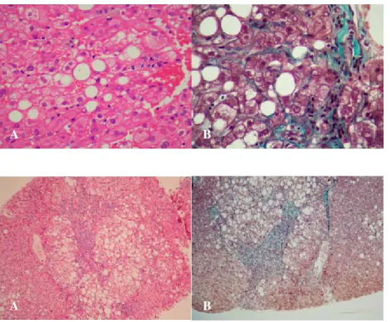
pa-Figure 2A. Grade 2 steatohepati-tis (Hematoxylin-Eosine) 2B: sta-ge 3 fibrosis (Masson’s Trichro-me).
A
B
A
B
Figure 1A. Macrovesicular stea-tosis and ground-glass hepatocyte (HBsAg positive) by Hema-toxylin-Eosine; 1B: Pericellular and perisinusoidal fibrosis in por-tal region and macrovesicular steatosis by Masson’s Trichrome.
tients and six (46.1%) of these patients were in stage.3,4
That means nearly half of the patients with chronic hepa-titis were in advanced stage of chronic hepahepa-titis B in this group.
As a conclusion, NAFLD seems to be the most com-mon cause of elevated ALT levels in patients with low viremia (HBV DNA negative by hybridization). Chronic hepatitis B is the second common cause of elevated ALT levels and half of these patients are in advanced stage. Liver biopsy which should be performed in these pa-tients is the main method to determine the cause of the disease and management of therapy. Interestingly, risk factors of NAFLD are not different between patients with NAFLD and the others in HBeAg negative group, on the contrary to the general population.
The causative factors of high ALT levels in hepatitis with low viremia are various. While liver biopsy indica-tions are clear in replicative HBV infection, in daily prac-tice nonreplicative group may be followed without biop-sy and treatment. This study shows that liver biopbiop-sy should be performed in low replicative HBV infection if hepatotoxic drugs are absent.
References
1. Bonino F, Brunetto MR. Chronic hepatitis B e antigen (HBeAg) negative, anti-HBe positive hepatitis B: overview. J Hepatol 2003; 39: S160-163.
edigraphic.com
scoring system for assessing histological activity inasymptom-atic chronic active hepatitis. Hepatology 1981; 1: 431-35. 3. Brunt EM, Janney CG, Di Bisceglie AM, Neuschwander-Tetri
BA, Bacon BR. Nonalcoholic steatohepatitis: a proposal for grad-ing and staggrad-ing the histological lesions. Am J Gastroenterol 1999; 94: 2467-74.
4. Okten A, Demir K, Kaymakoglu S, Cakaloglu Y, Dincer D, Besisik F. Etiologies of chronic hepatitis. Turk J Gastroenterol 1998; 2: 113-15.
5. Bozdayi AM, Bozkaya H, Turkyilmaz AR, Saryodlu M, Cetinkaya H, Karayalcin S, Yurdaydin C, et al. Nucleotide divergences in the core promoter and precore region of genotype D hepatitis B virus in patients with persistently elevated or normal ALT levels.
J Clin Virol 2001; 21: 91-101.
6. Brunetto MR, Oliveri F, Coco B, Leandro G, Colombatto P, Gorin JM, Bonino F. The outcome of chronic antiHBe-positive chronic hepatitis B in alpha interferon treated and untreated patients; a long term cohort study. J Hepatol 2002; 36: 263-70.
7. Day CP, James OFW. Hepatic steatosis: innocent bystander or guilty party? Hepatology 1998; 27: 1463-1466.
8. Bellentani S, Saccoccio G, Masutti F, Croce LS, Brandi G, Sasso F, Cristanini G, et al. Prevalance of and risk factors for hepatic steatosis in northern Italy. Ann Intern Med 2000; 132; 112-7. 9. Keeffe EB, Dieterich DT, Han SH, Jacobson IM, Martin P, Schiff
ER, Tobias H, et al. A treatment algorithm for the management of chronic hepatitits B virus infection in the United States. Clin Gastroenterol Hepatol 2004; 2: 87-106.
10. Sharma P, Balan V, Hernandez J, Rosati M, Williams J, Rodriguez-Luna H, Schwartz J, et al. Hepatic steatosis in hepatitis C virus genotype 3 infection: does it correlate with body mass index, fibrosis, and HCV risk factors? Dig Dis Sci 2004; 49: 25-29.
11. Lonardo A, Adinolfi LE, Loria P, Carulli N, Ruggiero G, Day CP. Steatosis and hepatitis C virus: Mechanisms and significance for hepatic and extrahepatic disease. Gastroenterology 2004; 126: 586-597.
12. Rubbia-Brandt L, Fabris P, Paganin S, Leandro G, Male PJ, Giostra E, Carlotto A, et al. Steatosis affects chronic hepatitis C progres-sion in a genotype specific way. Gut 2004; 53: 406-412. 13. Adinolfi LE, Gambardella M, Andreana A, Tripodi MF, Utili R,
Ruggiero G. Steatosis accelerates the progression of liver dam-age of chronic hepatitis C patients and correlates with specific HCV genotype and visceral obesity. Hepatology 2001; 33: 1358-1364.
14. Bach N, Thung SN, Schaffner R. The histological features of chronic hepatitis C and autoimmune chronic hepatitis: a com-parative analysis. Hepatology 1992: 15: 572-577.
15. Czaja AJ, Carpenter HA, Santrach PJ, Moore B. Host and disease specific factors affecting steatosis in chronic hepatitis C. J Hepatol
1998; 29: 198-206.
16. Wieslaw K, Antoinette U, Magdalena C, Dorota M, Katarzyna P. The significance of hepatic steatosis in chronic hepatitis B and C.
J Hepatol 2003; 38: 119.
17. Seppala-Lindroos A, Vehkavaara S, Hakkinen AM, Goto T, Westerbacka J, Sovijarvi A, Halavaara J, et al. Fat accumulation in the liver associated with defects in insulin suppression of glu-cose production and serum free fatty acids independent of obe-sity in normal men. J Clin Endocrinol Metab 2002; 87: 3023-3028.
18. Stranges S, Dorn JM, Muti P, Freudenheim JL, Farinaro E, Russell M, Nochajski TH, et al. Body fat distribution, relative weight, and liver enzyme levels: a population-based study. Hepatology