Bone metastases as the initial presentation of hepatocellular
carcinoma. Two case reports and a literature review
Jose M. Ruiz-Morales,* Rita Dorantes-Heredia,** Fredy Chable-Montero,** Sara Vazquez-Manjarrez,*** Nahum Méndez-Sánchez,**** Daniel Motola-Kuba*
* Oncology center. Medica Sur Hospital. Mexico City, Mexico. ** Anatomical Pathology Department. Medica Sur Hospital. Mexico City, Mexico. *** Department of Radiology. Instituto Nacional de Ciencias Médicas y Nutrición Salvador Zubirán. Mexico City. Mexico.
**** Liver Unit. Medica Sur Clinic & Foundation. Mexico City. Mexico.
ABSTRACT
Hepatocellular carcinoma (HCC) is the most common primary tumor of the liver and is the fifth most com-mon cancer in the world; its incidence has been increasing in recent years. Extrahepatic spread is present at the time of diagnosis in only about 5 to 15% of patients. Skeletal metastasis of HCC occurs less frequent-ly compared with other cancers and is considered a rare primary form of presentation. We report two ca-ses of unsuspected HCC presenting with multiple bone lesions as the initial presentation. The first patient was a 76-year-old man with symptoms of fatigue and back pain. The PET-CT revealed the hypercaptant bone lesions and a liver lesion. The pathology report showed that the metastases were positive for the he-patic marker HEPAR-1, indicating that they had originated from the HCC. The second patient was a 56-year-old man. He presented to the emergency department for right shoulder pain and weakness of the entire right arm with no history of trauma. During hospitalization, the patient became quadriplegic. MRI revealed osseous blastic lesions in the cervical vertebrae and right shoulder. A CT-guided biopsy was per-formed in the cervical lesion and showed poorly differentiated carcinoma. Immunohistochemistry staining was positive for HEPAR-1. In conclusion, this cases show an unusual presentation of HCC with skeletal me-tastasis.
Key words. Liver. Cancer. Skeletal. HEPAR-1. Ostelytic.
Correspondence and reprint request: Daniel Motola-Kuba, M.D.
Oncology center at Medica Sur Hospital Puente de Piedra, No. 150. Col Toriello Guerra. Deleg Tlalpan. Mexico City. Mexico.
E-mail: [email protected]
Manuscript received: January 21, 2014. Manuscript accepted: March 28, 2014. INTRODUCTION
Hepatocellular carcinoma (HCC) is the most com-mon primary tumor of the liver and is the fifth most common cancer in the world; its incidence has been increasing in recent years. HCC usually develops in the setting of chronic liver disease, particularly in patients with chronic hepatitis B or hepatitis C vi-rus infection.1 The diagnosis of HCC can be difficult
and often requires the use of one or more imaging modalities.2 Ideally, tumors should be detected when
about 2 cm in size so that all treatment options can be offered. However, HCC is frequently diagnosed
late in its course because of the absence of symp-toms. As a result, many patients have untreatable disease when first diagnosed. The median survival following diagnosis is 6 to 20 months. Large tumor size, vascular invasion, poor functional status, and nodal metastases are all associated with a poor out-come.3
HCC should be suspected in patients with previ-ously compensated cirrhosis evolving with complica-tions such as ascites, encephalopathy, jaundice, or variceal bleeding.4 These complications are often
as-sociated with extension of the tumor into the hepat-ic or portal veins, or are secondary to arteriovenous shunting induced by the tumor.5
Extrahepatic spread is present at the time of di-agnosis in only about 5 to 15% of patients. Extrahe-patic metastases are more common in patients with advanced-stage primary tumors (> 5 cm and large vessel vascular invasion). Extrahepatic recurrence is uncommon after locoregional therapy (5 to 24%).6
tion.8 Bone metastases from HCC commonly appear
as expansive soft tissue masses with bone destruc-tion. Fifty percent of patients with bone metastasis have poorly controlled pain. Radiation therapy is considered the standard care for management of painful bony metastases.9 We report two cases of
unsuspected HCC presenting with multiple bone le-sions as the initial presentation.
CASE REPORT
Case 1
The first patient was a 76-year-old man without underlying hepatitis B or C, who was a nonsmoker and not a heavy drinker. He had a medical history of hypertension and major depressive disorder, both of which had received adequate medical treat-ment. He presented to our hospital with symptoms of fatigue and back pain. Clinical examination re-vealed no further information. Patients body mass index was 21 kg/m2. The patient’s laboratory
stud-ies showed: hemoglobin (Hb) concentration, 10.5 g/ dL; creatinine serum concentration, 1.66 mg/dL;
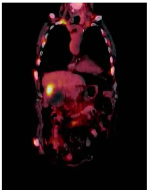
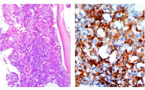
(LDH), 923 IU/L. Because of low hemoglobin levels gastric endoscopy was performed and no signs of bleeding or portal hypertension (variceal disease) were seen. Because of the alterations in the hepatic panel, noncontrast CT was ordered. The CT demon-strated bilateral pleural effusion, a small liver, and osseous blastic and lytic lesions in every vertebral body, the seventh left rib, fifth right rib, sacrum, and ilium. A normal prostate-specific antigen (PSA) concentration of 0.8 ng/mL and no altera-tion in protein electrophoresis were found. Because of these findings, an 18FDG PET-CT was ordered. The PET-CT revealed the hypercaptant bone le-sions described earlier and a liver lesion measuring 5 x 4 cm that was not seen in the noncontrast CT (Figures 1 and 2). Because we could not identify whether the lesion had originated in the liver or gall bladder, a CT-guided biopsy of the pubic bone was performed. The pathology report showed that the metastases were positive for the hepatic marker HEPAR-1, indicating that they had originated from the HCC (Figure 3). The lesions were negative for TTF-1, PSA, synaptophysin and chromogranin. Se-rum AFP concentration was 7,724 μg/L.
Figure 1. PET-CT with hypermetabolic lesion in hepatic area. Blastic bone lesiones are also observed.
Case 2
The second patient was a 56-year-old man. He was a nonsmoker and had consumed beer since the age of 17 years, and binge drinking at least once a week. He presented to the emergency department for right shoulder pain and weakness of the entire right arm with no history of trauma. Physical exploration identified the “stigmata” of chronic liver disease: sclera icterus, gynecomastia, and palmar erythema. Initially, Parsonage-Turner syndrome was diag-nosed. Laboratory values were Hb concentration, 11 g/dL; BUN, 28.2 mg/dL; creatinine, 1.47 mg/dL; ALT, 89 U/L; AST, 181 U/L; ALP, 1,300 IU/L; GGT, 200 IU/L; and LDH, 700 IU/L. During hospi-talization, the patient became quadriplegic. MRI re-vealed osseous blastic lesions in the cervical vertebrae and right shoulder (Figures 4 and 5). A CT-guided biopsy was performed in the cervical le-sion and showed poorly differentiated carcinoma. Immunohistochemistry staining was positive for HEPAR-1 and negative for TTF-1, PSA, synapto-physin and chromogranin. These findings were com-patible with metastatic HCC (Figure 6). Serum AFP concentration was 5,234 μg/L. Abdominal US showed two liver lesions, a 2 x 2 cm lesion in the left lobe and a 4.5 x 4.5 cm lesion in the 6th seg-ment, both lesions were heterogeneous primary hy-Figure 3. Malignant epithelial neoplasm with growth pat-tern in nests and cords. The variable atypia cells showed granular eosinophilic cytoplasm.
Figure 4. Magnetic resonance imaging: sagittal STIR; At metastasis area, there are central hyperintensity (edema) and altered morphology of the bone cylinder, which means edema and inflammatory compression secondary to metastasis.
Figure 5. Magnetic resonance imaging: T1 gadolinium FATSAT where expansive spinal injury shows invasion of soft tissue with contrast medium enhancement.
DISCUSSION
Although much less common in Western Europe and North America, HCC still accounts for 1 to 2% of all malignant tumors. HCC occurs frequently in sub-Saharan Africa and in Asia. Hepatitis B and C viruses, dietary aflatoxin B1, and cirrhosis from any cause are the common risk factors in that order of importance. Several lines of evidence, including mo-lecular biology and animal studies, support these etiological linkages.10 HCC survival depends on
stage at diagnosis. AJCC/UICC staging system have shown to povide the best stratification of prognosis, in patients with HCC undergoing liver transplanta-tion, with 5 year survival rate of:11
• 55% for stage I. • 37% for stage II. • 16% for stage III. • < 7% for stage IV.
Improved HCC survival rates because of recent therapeutic advancements may have increased the frequency of remote metastasis. Metastatic spread to the bones occurs in 13 to 16% of HCC patients and has been described thoroughly. The most common sites of skeletal involvement are, in descending or-der, the vertebrae, pelvis, ribs, skull, humerus, and sternum. Much less is known about the initial clini-cal presentation of unsuspected HCC associated with bone metastasis. The literature documents a modest number of isolated reports, most of which in-volve the vertebrae. Most reported cases of sacral/ lumbosacral metastasis are accompanied by either multiple metastatic spread elsewhere in the body or previously known HCC.12
The presence of bone metastasis have not been associated with absence of cirrhosis, in a consecu-tive series of 395 patients with pathologically veri-fied hepatocellular carcinoma, 20 patients (5%) had bone metastasis at initial presentation. The age, sex, hepatitis B surface antigen seropositivity, al-pha-fetoprotein level, and frequency of associated cirrhosis were not statistically different from those in patients without initial bone metastasis.13
Skeletal metastasis appears to be unique among various hematogenous metastasis of HCC because it can occur before clinical manifestations of liver dis-ease and is usually symptomatic, as in the present cases. The pulmonary and systemic circulation is thought to be the main route of metastasis to the skeletal system.14 Because the clinical importance of
diagnosing bone metastasis from HCC has increased with improved HCC patient prognosis, and because radiation therapy can palliate symptoms, radiolo-gists should take care not to miss base metastasis at an early stage when the initial symptoms appear. Al-though HCC should be included in the differential diagnosis of osteolytic, hypervascular lesions, bone metastasis from renal cell carcinoma, thyroid gland cancer, parotid gland cancer, and pheochromocyto-ma pheochromocyto-may show similar ipheochromocyto-maging findings.15
sis of hepatocellular carcinoma is made. Hepar-1 (human hepatocyte paraffin-1) is an antigen that re-flects hepatocyte differentiation in approximately 90% of hepatocellular carcinomas and to be consid-ered positive, the staining must be granular and cy-toplasmic. It must be noted that in the study of any tumor, immunohistochemistry is nonspecific. Immu-nohistochemistry is used to supplement the morpho-logical study of neoplasms.16
In conclusion, this cases show an unusual pres-entation of HCC with skeletal metastases. A plausi-ble explanation for this rarity is that both the lymphangitic and hematogenous spread of HCC is closely associated with tumor staging. Thus, a more advanced hepatic tumor should unveil itself with lo-cal symptoms and lead to the diagnosis of HCC be-fore the symptoms of metastatic spread are present.
REFERENCES
1. Bruix J, Sherman M. Practice Guidelines Committee, Ameri-can Association for the Study of Liver Diseases. Manage-ment of hepatocellular carcinoma. Hepatology 2011; 53: 1020-2.
2. Forner A, Llovet JM, Bruix J. Hepatocellular carcinoma.
Lancet 2012; 379: 1245.
3. Manghisi G, Elba S, Mossa A, Giorgio A, Aloisio V, Perrotta A, Tardio B, et al. A new prognostic system for hepatocel-lular carcinoma: a retrospective study of 435 patients: the Cancer of the Liver Italian Program (CLIP) investiga-tors. Hepatology 1998; 28: 751.
4. Motola-Kuba D, Zamora-Valdés D, Uribe M, Méndez-Sán-chez N. Hepatocellular carcinoma. An overview. Ann
He-patol 2006; 5: 16-24.
5. Sugano S, Miyoshi K, Suzuki T, Kawafune T, Kubota M. In-trahepatic arteriovenous shunting due to hepatocellular carcinoma and cirrhosis, and its change by transcatheter arterial embolization. Am J Gastroenterol 1994; 89: 184.
6. Yoo DJ, Kim KM, Jin YJ, Shim JH, Ko GY, Yoon HK, Sung KB, et al. Clinical outcome of 251 patients with extrahepatic metastasis at initial diagnosis of hepatocellular carcino-ma: does transarterial chemoembolization improve survi-val in these patients? J Gastroenterol Hepatol 2011; 26: 145-54.
7. Qureshi SS, Shirkhande SV, Borges AM, Shukla Parul J. Chest wall metastases from unknown primary hepatocellular carcinoma. J Postgrad Med 2005; 51: 41-2.
8. Fukutomi M, Yokota M, Chuman H, Harada H, Zaitsu Y, Funakoshi A, Wakasugi H, et al. Increased incidence of bone metastases in hepatocellular carcinoma. Eur J
Gastroenterol Hepatol 2001; 13: 1083-8.
9. Nakamura N, Igaki H, Yamashita H, Shiraishi K, Tago M, Sasano N, Shiina S, et al. A retrospective study of radio-therapy for spinal bone metastases from hepatocellular carcinoma (HCC). Jpn J Clin Oncol 2007; 37: 38-43. 10. Nayak NC. Hepatocellular carcinoma-a model of human
cancer: clinico-pathological features, etiology and patho-genesis. Indian J Pathol Microbiol 2003; 46: 1-16.
11. Vauthey JN, Ribero D, Abdalla EK, Jonas S, Bharat A, Schu-macher G, Lerut J, et al. Outcomes of liver transplanta-tion in 490 patients with hepatocellular carcinoma: validation of a uniform staging after surgical treatment. J
Am Coll Surg 2007; (5): 1016.
12. Piccirillo M, Granata V, Albino V, Palaia R, Setola SV, Petri-llo A, Tatangelo F, et al. Can hepatocellular carcinoma (HCC) produce unconventional metastases? Four cases of extrahepatic HCC. Tumori 2013; 99(1): e19-e23.
13. Liaw CC, Ng KT, Chen TJ, Liaw YF. Hepatocellular carcino-ma presenting as bone metastasis. Cancer 1989; 64: 1753-7.
14. Fukusato T, Aoyama H, Mori W. Age and sex differen-ces in bone metastasis of hepatocellular carcinoma in Japanese autopsy cases. Gastroenterol Jpn 1989; 24: 127-34.
15. Miura T, Hirabuki N, Kozuka T. Cranial metastasis from he-patocellular carcinoma. Clin Radiol 1990; 42: 445-6. 16. Radwan NA, Ahmed NS. The diagnostic value of arginase-1