Estudio de la ecología de Bacillus thuringiensis en la hoja
130
0
0
Texto completo
(2) 2.
(3) Estudio de la ecología de Bacillus thuringiensis en la hoja. Memoria presentada por Pau Maduell Soler para conseguir el título de doctor en Microbiología por la Facultad de Biociencias de la Universidad Autónoma de Barcelona. Bellaterra, septiembre de 2007. 3.
(4) UNIVERSIDAD AUTÓNOMA DE BARCELONA DEPARTAMENTO DE GENÉTICA Y MICROBIOLOGÍA. ESTUDIO DE LA ECOLOGÍA DE Bacillus thuringiensis EN LA HOJA. Memoria. presentada. por. el. Licenciado en Biología Pau Maduell Soler para optar al grado de Doctor en Ciencias Biológicas por la Universidad Autónoma de Barcelona.. Con el Vo Bo de la Directora. Dra. Montserrat Llagostera Casas BARCELONA 2007. 4.
(5) AGRADECIMIENTOS. 5.
(6) 6.
(7) INDICE Página Resumen. 9. Summary. 11. 1. Introducción. 13. 1.1. Bacillus thuringiensis. 14. 1.2. Marco histórico. 14. 1.3. Mecanismo de acción. 16. 1.4. Identificación y caracterización de B. thuringiensis. 18. 1.5. Ecología de B. thuringiensis. 20. 1.6. Modelos vegetales. 24. 2. Objetivos. 27. 3. Artículos. 29. 3.1. Artículo I. 30. 3.2. Artículo II. 31. 3.3. Artículo III. 32. 3.4. Artículo IV. 33. 4. Materiales y métodos. 34. 4.1. Distribución y caracterización de B. thuringiensis en el filoplano de especies de Piper (Piperaceae) en tres estratos altitudinales (artículo I). 35. 4.2. Diversidad de cepas de B. thuringiensis en el filoplano de maíz y fríjol y en sus respectivos suelos en Colombia (artículo II). 41. 4.3. Migración de B. thuringiensis a hojas de fríjol desde el suelo o desde hojas distantes (artículo III). 45. 4.4. Determinación de la capacidad de B. thuringiensis de colonizar la superficie de las hojas (artículo IV). 49. 4.5. Capacidad de B. thuringiensis de crecer in vitro en medios que simulan el medio ambiente de la hoja (artículos III y IV) 5. Resultados y discusión. 52 54. 5.1. Distribución y caracterización de B. thuringiensis en el filoplano de especies de Piper (Piperaceae) en tres estratos altitudinales (artículo I). 55. 7.
(8) 5.2. Diversidad de cepas de B. thuringiensis en el filoplano de maíz y fríjol y en sus respectivos suelos en Colombia (artículo II). 60. 5.3. Migración de B. thuringiensis a hojas de fríjol desde el suelo o desde hojas distantes (artículo III). 67. 5.4. Determinación de la capacidad de B. thuringiensis de colonizar la superficie de las hojas (artículo IV). 70. 5.5. Capacidad de B. thuringiensis de crecer in vitro en medios que simulan el medio ambiente de la hoja (artículos III y IV). 76. 5.6. Consideraciones generales acerca de la ecología de B. thuringiensis en el filoplano. 80. 6. Conclusiones. 84. 7. Bibliografía. 87. 8.
(9) RESUMEN La ecología de Bacillus thuringiensis, un bioinsecticida muy común, es poco conocida. Nuestro principal objetivo era investigar acerca de la ecología de esta bacteria en la filosfera. En un primer estudio se recogieron 35 muestras de hojas del género Piper de bosques andinos colombianos. Se obtuvieron 256 aislamientos de B. thuringiensis del 74% de las muestras estudiadas. Los aislamientos fueron caracterizados según la morfología del cristal, la presencia de genes cry por PCR y la toxicidad contra insectos. Además, se estimaron las poblaciones de células vegetativas viables y endosporas por unidad de área obteniéndose 2-5x10³ cfu/cm2 de hoja. En general, no se encontraron diferencias estadísticamente significativas en el número de aislamientos de B. thuringiensis por cm2 de hoja ni en las características de los aislamientos de B. thuringiensis entre las diferentes especies de Piper. Después de comprobar que B. thuringiensis estaba presente en el filoplano, se quiso comparar las poblaciones de esta bacteria en el suelo y en las hojas. Se aisló B. thuringiensis del filoplano y del suelo de cultivos de maíz y fríjol. Se recuperaron 214 aislamientos de 96 muestras de filoplano (0-34 cfu/cm2) y 59 aislamientos de 24 muestras de suelo. Todos los aislamientos fueron caracterizados como se ha explicado anteriormente. Las poblaciones predominantes de B. thuringiensis en el filoplano contenían genes cry1 y eran activas contra Spodoptera frugiperda, mientras que los aislamientos del suelo tenían genes cry11 y eran activos contra Culex quinquefasciatus. El hecho de que predominaran poblaciones específicas de B. thuringiensis en las hojas diferentes a las del suelo sugiere que existe una selección diferencial en las poblaciones de B. thuringiensis en el filoplano y en el suelo. Entonces, se investigó la capacidad de migración de B. thuringiensis desde el suelo a las hojas. Se inocularon dos cepas diferentes de B. thuringiensis en suelos, semillas y hojas jóvenes de plantas de fríjol, para determinar si. 9.
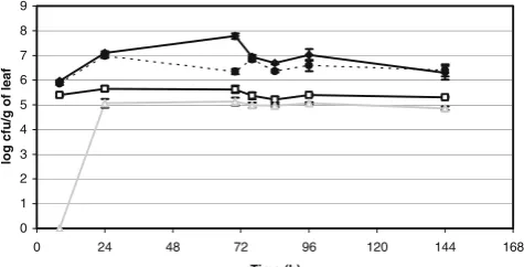
(10) podían migrar a las hojas superiores en condiciones controladas. Aunque se recuperaron aislamientos de B. thuringiensis en las hojas, las poblaciones fueron muy bajas (menos de 10 cfu/cm2 de hoja). Además el número de células recuperado disminuía a medida que las hojas estaban más distantes del suelo o de las hojas inoculadas. Todo esto indicaba que B. thuringiensis migra pobremente desde el suelo o la semilla a las hojas o entre hojas de la misma planta. También se evaluó la capacidad de varias cepas de B. thuringiensis de colonizar la superficie de las plantas y se comparó con otras bacterias epífitas. Mientras que todas las cepas de B. thuringiensis se multiplicaron en cierta medida después de la inoculación sobre hojas de fríjol, las poblaciones máximas alcanzadas fueron de 106 cfu/g de hoja, muy inferiores a las conseguidas por otras bacteria epífitas, como Pseudomonas fluorescens. Muy poco tiempo después de la inoculación, una porción importante de las células de B. thuringiensis estaban en forma de endospora. Además el crecimiento de B. thuringiensis no se vio afectado por la presencia de Pseudomonas spp. cuando fueron co-inoculados y viceversa. Por otro lado, cuando se observaron al microscopio las cepas de B. thuringiensis (marcadas con el gen de la proteína verde fluorescente) sobre hojas de fríjol, se observó que no formaban agregados celulares y no estaban asociadas con otras bacterias epífitas ni con estructuras de la hoja. Finalmente se investigó la capacidad de B. thuringiensis de crecer en un medio diseñado para simular la composición de nutrientes del filoplano. Sin embargo el crecimiento fue inferior al de otras bacterias. Aparentemente, B. thuringiensis tiene unos requisitos nutricionales mayores que otras especies bacterianas habitantes naturales del filoplano.. 10.
(11) SUMMARY The ecology of Bacillus thuringiensis, a common biopesticide, is poorly understood. Our main objective was to investigate the ecology of this bacteria on the phylloplane. In a first study 35 leaf samples of the genus Piper were collected from the Colombian Andean forest. Two hundred and fifty-six isolates of B. thuringiensis were obtained from 74% of the samples studied. The isolates were characterized by crystal morphology, the presence of cry genes by PCR and toxicity against insects. The populations of viable vegetative cells and spores per unit area were estimated (2-5x10³ cfu/cm2 of leaf). Overall, no significant differences in the number of B. thuringiensis isolates per cm2 of leaf nor in the B. thuringiensis characteristics were found among the different Piper species evaluated. After observing B. thuringiensis on the phylloplane, a comparison was performed between soil and leaf populations. B. thuringiensis was isolated from the phylloplane and soil in plantings of maize and bean from Colombia; and 214 isolates were recovered from 96 phylloplane samples (0-34 cfu/cm2) while 59 isolates from 24 soil samples. All the isolates were characterized as above-mentioned. The predominant population of B. thuringiensis on the phylloplane harbored cry1 gene and was active against Spodoptera frugiperda, whereas in soil the isolates harboring cry11 gene and active against Culex quinquefasciatus predominated. The predominance of specific B. thuringiensis populations both on the leaves and in the soil, suggests the presence of differential selection in B. thuringiensis populations on the phylloplane and in soil. Then, we addressed the process of immigration of B. thuringiensis from soil to leaves. Two different B. thuringiensis strains were inoculated into soils, onto seeds or onto lower leaves of bean plants to determine if they were able to disperse to upper leaves under controlled conditions. While B. thuringiensis isolates were commonly recovered from leaves exposed to such inocula, populations were very low (less than 10. 11.
(12) cfu/cm2 of leaf). In addition, the number of cells of B. thuringiensis recovered decreased with increasing distance from the soil or from the inoculated leaves. This indicates that B. thuringiensis disperses poorly from the soil or the seed to the leaves or between leaves of the same plant under controlled conditions. Moreover, the ability of several B. thuringiensis strains to colonize plant surfaces was assessed and compared with that of more common epiphytic bacteria. While all B. thuringiensis strains multiplied to some extent after inoculation on bean plants, their maximum epiphytic population sizes of 106 cfu/g of leaf were always much less than that achieved by other resident epiphytic bacteria or an epiphytically fit Pseudomonas fluorescens strain. Many cells were in a spore form soon after inoculation onto plants. The growth of B. thuringiensis was not affected by the presence of Pseudomonas syringae spp. when co-inoculated, and vice versa. B. thuringiensis strains harboring a green fluorescent protein marker gene did not form large cell aggregates, were not associated with other epiphytic bacteria, and were not found associated with leaf structures when directly observed on bean leaves by epifluorescent microscopy. Finally, we analyzed the capacity of B. thuringiensis to grow on a medium designed to simulate the nutrient composition of the phylloplane but the growth observed was very poor compared to other bacteria. This bacterium apparently has greater nutrient requirements than other bacterial species that are prominent inhabitants of the phylloplane.. 12.
(13) 1. INTRODUCCIÓN. 13.
(14) 1.1. Bacillus thuringiensis Bacillus thuringiensis es una bacteria Gram positiva con la capacidad de esporular. Es muy semejante a otras especies del género Bacillus, como B. cereus y B. anthracis, pero se diferencia de ellas por la formación de un cristal proteico en el momento de la esporulación (Schnepf et al., 1998). El cristal está compuesto por proteínas, algunas de las cuales son extremadamente tóxicas contra insectos, principalmente de los órdenes Lepidoptera, Diptera y Coleoptera; mientras que los mamíferos, incluido el hombre, no se ven afectados (Siegel, 2001). Cabe destacar que gran parte de las plagas agrícolas y forestales son provocadas por insectos de los órdenes Lepidoptera y Coleoptera, y que la mayoría de los vectores de enfermedades humanas son del orden Diptera (Blas y Vives, 1987). Además, estas proteínas son biodegradables, por lo que no contaminan suelos ni aguas. Por estos motivos, esta bacteria está siendo utilizada como una alternativa ecológicamente sostenible a los insecticidas químicos para controlar plagas agrícolas, plagas forestales y vectores de enfermedades (Whalon y Wingerd, 2003).. 1.2. Marco histórico B. thuringiensis fue aislado por primera vez en Japón por Ishiwata en 1901 como patógeno del gusano de seda, Bombyx mori (Lepidoptera, Bombycidae), causándole la enfermedad de sotto (Ishiwata, 1901). En 1911 Berliner, en Alemania, aisló la misma bacteria a partir de larvas del lepidóptero Anagasta kuehniella (Lepidoptera, Pyrallidae), la palomilla de la harina del Mediterráneo, e hizo una descripción formal de esta bacteria: bacilo Gram positivo, que presenta un cristal paraesporal de naturaleza proteica, endospora y flagelos peritricos, en distintas fases de su ciclo de crecimiento. La denominó Bacillus thuringiensis, en honor a la región alemana de donde la aisló: Thuringia (Berliner, 1915).. 14.
(15) El uso de B. thuringiensis para el control de insectos se inició en los años 30 contra el barrenador europeo del maíz, Ostrinia nubilalis (Lepidoptera, Pyrallidae). El primer producto comercial salió en Francia en 1938 bajo el nombre de Sporeine®; en EEUU se comercializó por primera vez en 1957, bajo el nombre de Thuricide®. Desde entonces se ha desarrollado la producción de forma masiva en varios países de todo el mundo (Whalon y Wingerd, 2003).. En 1970 Dulmage descubrió la cepa HD-1 de B. thuringiensis serovar. kurstaki en EEUU a partir de larvas enfermas de lepidópteros. Esta serovariedad resultó ser entre 2 y 200 veces más tóxica que las variedades anteriormente utilizadas en la producción comercial de bioinsecticidas. La Agencia de Protección del Medio Ambiente de EEUU (EPA) la designó como estándar de potencia o unidad internacional de toxicidad (van Frankenhuyzen, 1993).. En 1977, dos entomólogos israelíes aislaron un bacilo de larvas del mosquito Culex sp., el cual se designó con el nombre de B. thuringiensis serovar. israeliensis, y fue considerado útil en salud pública para el control de vectores de enfermedades tropicales por ser patógeno de larvas del mosquito Aedes aegypti (Diptera, Culicidae), vector del dengue (Golberg y Margalit, 1977).. En los años 80 fueron descritas otras cepas de B. thuringiensis serovar. morrisoni patovariedades tenebrionis y san diego, activos contra coleópteros (Krieg, 1983). Las serovariedades de mayor impacto a nivel industrial han sido kurstaki, israeliensis y morrisoni, si bien no son las únicas. Desde el descubrimiento e identificación de B.. 15.
(16) thuringiensis, el interés en esta bacteria ha sido cada vez mayor, tanto que a la fecha se han descrito 83 serovariedades distintas según su antígeno flagelar, en el intento de encontrar nuevas toxinas con nuevas actividades biológicas (Lecadet et al., 1999; Khyami-Horani et al., 2003).. La mayoría de variedades estudiadas actualmente son activas contra larvas de diferentes grupos de lepidópteros, y algunas de ellas también lo son contra dípteros y coleópteros (de Maagd et al., 2001; Whalon y Wingerd, 2003). Algunos estudios han mostrado que B. thuringiensis también podría ser utilizado contra insectos de otros órdenes (Hymenoptera, Homoptera y Mallophaga), nemátodos, ácaros y protozoos (Drummond et al., 1995; Zhioua et al., 1999; Raps et al., 2001; Pinto et al., 2003; Wei et al., 2003).. En los últimos años se han desarrollado ampliamente técnicas moleculares que han permitido manipular genes de B. thuringiensis para desarrollar bacterias acuáticas recombinantes que forman parte de la dieta de larvas de insectos (Armengol et al., 2005), así como para generar plantas transgénicas para conferirles resistencia a los insectos plaga (Romeis et al., 2006). En el año 2002 ya había 14 millones de ha con plantas trasgénicas con genes de B. thuringensis (Shelton et al., 2002).. 1.3. Mecanismo de acción B. thuringiensis tiene un ciclo de vida que comprende la formación de endosporas cuando las condiciones del medio en el que se encuentra son adversas. La endospora es una forma de resistencia frente a situaciones de estrés ambiental como la desecación o la falta de nutrientes, entre otros. Junto con la endospora también forma un cristal paraesporal constituido por δ-endotoxinas. Cuando estas protoxinas contenidas en el. 16.
(17) cristal son ingeridas por las larvas de los insectos susceptibles les causan intoxicación. Las condiciones alcalinas del intestino medio de las larvas y sus enzimas digestivas (principalmente tripsina y quimotripsina) disuelven los cristales y activan las protoxinas por cortes proteolíticos, conviertiéndolas en toxinas. La toxina es reconocida por receptores de membrana de las células epiteliales del intestino medio del insecto, se ancla a ella y forma canales iónicos, causando un desequilibrio osmótico y por tanto la lisis celular. Esto provoca en último término la muerte del insecto. Las endosporas de B. thuringiensis se mantienen en el canal alimentario, donde, después de una disminución del pH provocada por el desequilibrio osmótico, pueden germinar (Schnepf et al., 1998; de Maagd et al., 2001; Whalon y Wingerd, 2003).. Una característica importante de estas toxinas es su alto grado de especificidad. En la relación toxina-insecto susceptible se han descrito cuatro niveles de especificidad. El primer nivel corresponde a la conducta alimentaria del insecto; éste debe ingerir junto con su alimento las endosporas y los cristales de B. thuringiensis. El segundo nivel viene definido por el pH del intestino medio del insecto, que debe ser alcalino, pues la mayoría de los cristales paraesporales de B. thuringiensis se solubilizan a pH alto, excepto en un caso, en el que también solubilizan a pH ácido (Koller et al., 1992). El tercer nivel es debido a las proteasas alcalinas del intestino del insecto; éstas han de ser las adecuadas para poder digerir parcialmente las proteínas, es decir, realizar el paso de protoxina a toxina. Por último, el cuarto nivel de especificidad corresponde a los receptores de membrana de las células epiteliales del intestino medio del insecto; las toxinas deben ser reconocidas por los receptores para poder anclarse a la membrana. Además de la unión al receptor, es necesaria la inserción de parte del dominio I de la toxina, la agregación y la formación del canal (Whalon y Wingerd, 2003).. 17.
(18) 1.4. Identificación y caracterización de B. thuringiensis Los métodos que se utilizan habitualmente para la caracterización fenotípica y genotípica de los aislamientos de B. thuringiensis son: el serotipo flagelar, la descripción morfológica del cristal paraesporal, la caracterización por métodos moleculares y los bioensayos (Hastowo et al., 1992; Bernhard et al., 1997; Helgason et al., 1998; Iriarte et al., 1998; Kim et al., 1998; Hongyu et al., 2000).. El antígeno flagelar ha sido usado para caracterizar B. thuringiensis desde 1962 (de Barjac y Bonnefoi, 1962). Los aislamientos de B. thuringiensis son incubados con anticuerpos policlonales de cepas conocidas, los cuales reconocen los antígenos flagelares del bacilo, dándose una reacción de aglutinación específica. Actualmente se conocen 83 serovariedades (Lecadet et al., 1999; Khyami-Horani et al., 2003). Sin embargo, existen algunos trabajos en los que se concluye que no existe una relación clara entre la clasificación por la técnica del antígeno flagelar y las clasificaciones filogenéticas elaboradas mediante marcadores moleculares (Hastowo et al., 1992; Nakamura, 1994; Bourque et al., 1995; Helgason et al., 2000). De todas formas esta técnica sigue considerándose como la única herramienta disponible para identificar las distintas serovariedades de B. thuringiensis (Lecadet et al., 1999).. El cristal paraesporal puede presentar varias morfologías: bipiramidal, ovoidal, redondo, cuadrado, triangular, "plate-like" (forma de plato) y amorfo, entre otras (Hastowo et al., 1992; Ohba, 1996; Bernhard et al., 1997). Existe una correlación bastante alta entre la forma del cristal y la actividad tóxica, por ejemplo, los cristales bipiramidales están. 18.
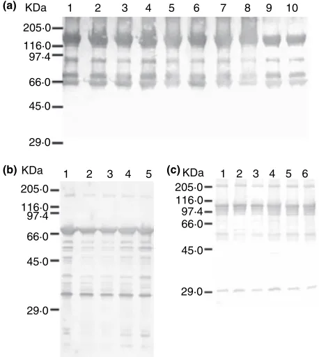
(19) generalmente asociados a actividad contra lepidópteros y los cristales redondos están asociados a actividad contra dípteros (Lerecluse et al., 1993; Damgaard, 1996).. Seguramente la técnica más utilizada para caracterizar cepas de B. thuringiensis es la PCR (Bourque et al., 1993; Brousseau et al., 1993; Gleave et al., 1993; Ceron et al., 1994; Ceron et al., 1995; Bravo et al., 1998). Usando cebadores específicos de DNA, se pueden identificar los genes que codifican las distintas proteínas que forman los cristales, y así hacer una aproximación de la toxicidad de cada aislamiento. En la actualidad están catalogadas genéticamente más de 350 proteínas cristalinas (Crickmore, 2006).. La composición proteica del cristal puede ser analizada mediante electroforesis en geles desnaturalizantes de poliacrilamida con dodecil sulfato de sodio (SDS-PAGE). De esta forma se separan las protoxinas según su peso molecular y, comparando con patrones de bandas de los cristales de cepas conocidas, se puede obtener información sobre la posible toxicidad de los cristales de las cepas evaluadas (Kim et al., 1998; Hongyu et al., 2000; Marquez et al., 2000). Esta técnica puede ser complementada combinándola con Western blot, en la que los componentes separados electroforéticamente son transferidos a una membrana que se incuba con anticuerpos específicos contra secuencias de aminoácidos conocidas. Así se pueden identificar las protoxinas (Kaelin et al., 1994; Marquez et al., 2000).. Otra técnica importante para estos estudios es la evaluación de la toxicidad de los aislamientos sobre distintas especies de insectos de interés agronómico y para la salud. Estos estudios se denominan bioensayos, en los cuales se alimentan a las especies de. 19.
(20) insectos a evaluar con toxinas y endosporas. Existen dos tipos de bioensayos: los cualitativos, que evalúan la patogenicidad con altas concentraciones de la muestra, contando el número de insectos muertos, ya que sólo se pretende ver la efectividad de la muestra como insecticida; y los cuantitativos, en los que se utilizan diversas dosis de toxina para evaluar la cantidad necesaria de toxina con la que se obtiene el 50% de mortalidad del insecto estudiado (Dosis Letal 50 (DL50) y Concentración Letal 50 (CL50)) (Ohba y Aizawa, 1986; Bernhard et al., 1997; Iriarte et al., 1998). 1.5. Ecología de B. thuringiensis Desde el punto de vista ecológico, no se conoce con exactitud cual es el hábitat natural de B. thuringiensis. Históricamente se ha buscado en el suelo, donde se ha encontrado abundantemente y por todo el mundo (Martin y Travers, 1989; Bernhard et al., 1997; Bravo et al., 1998; Iriarte et al., 1998; Uribe et al., 2003; Armengol et al., 2006); en el suelo persiste principalmente en forma de endospora (Yara et al., 1997). De todas formas también se ha encontrado B. thuringiensis en partículas de polvo en suspensión (Martin y Travers, 1989), cuerpos de insectos enfermos o muertos (Porcar y Caballero, 2000), productos almacenados (Meadows et al., 1992; Kaelin y Gadani, 2000), alimentos (Damgaard et al., 1996), piel de animales (Akhurst et al., 1997), sedimentos marinos (Maeda et al., 2000), fangos de depuradora (Mizuki et al., 2001), e incluso como patógeno humano oportunista (Damgaard et al., 1997a). Además, se ha encontrado abundantemente en hojas de plantas (Smith y Couche, 1991; Ohba, 1996; Damgaard et al., 1997b; Damgaard et al., 1998; Hansen et al., 1998; Rizali et al., 1998; Mizuki et al., 1999; Kaur y Singh, 2000; Vaid y Bishop, 2000; Nair et al., 2002; Collier et al., 2005).. 20.
(21) Los primeros trabajos enfocados al estudio de la ecología de B. thuringiensis tenían como objetivo el de analizar la distribución y abundancia de esta especie en el ambiente, principalmente en el suelo. En la década de los 80 se hicieron varios muestreos de suelos de distintas partes del mundo (DeLucca et al., 1981; Padua et al., 1982; Ohba y Aizawa, 1986). En 1989 Martin y Travers (Martin y Travers, 1989) publicaron un trabajo donde analizaron muestras de suelos de todos los continentes. A pesar de encontrar B. thuringiensis en todo el mundo, concluyeron que el papel de B. thuringiensis en el medio ambiente era un enigma. Después de este trabajo se han realizado varios estudios de búsqueda de B. thuringiensis en suelos de diferentes países: Indonesia (Hastowo et al., 1992), Korea (Kim et al., 1998), Noruega (Helgason et al., 1998), México (Bravo et al., 1998), Colombia (Uribe et al., 2003; Armengol et al., 2006) y España (Iriarte et al., 1998), entre otros. De todos estos trabajos se concluye que ésta es una especie cosmopolita.. A principios de la década de los 90, Smith y Couche (1991) publicaron el primer trabajo de búsqueda de B. thuringiensis en hojas de árboles. Aislaron B. thuringiensis del 5070% de las hojas muestreadas. Estos autores lanzaron una hipótesis en la que proponían B. thuringiensis como comensal o simbionte de las plantas, ofreciendo protección a éstas de los insectos fitófagos. A su vez, las plantas proveerían nutrientes mediante los exudados foliares y además proveerían un nicho libre de competencia con otras bacterias esporogénicas del suelo. Smith y Couche también proponían que se debería determinar si B. thuringiensis es metabólicamente activa en el filoplano y examinar las yemas de las plantas para evaluar si son fuentes adicionales de nuevos aislamientos de B. thuringiensis. Los resultados de este estudio sugerían que el papel de B. thuringiensis en el medio ambiente debía ser reconsiderado.. 21.
(22) Posteriormente, Ohba (1996) buscó B. thuringiensis en el filoplano de hojas de morera (Morus alba), de la cual se alimenta la larva del gusano de la seda, ampliamente cultivado en Japón. Se encontró B. thuringiensis en 24 de los 25 árboles estudiados. No se observaron diferencias entre la cara inferior y la superior de las hojas en cuanto a presencia de B. thuringiensis, lo cual era muy inesperado, ya que B. thuringiensis, al igual que muchos microorganismos, es susceptible a la radiación ultravioleta del sol (Ignoffo et al., 1977; Burges y Jones, 1998). También se describió que las serovariedades de B. thuringiensis encontradas en las hojas de morera eran diferentes de las serovariedades que se habían reportado previamente en los suelos donde se cultivan dichos árboles.. Damgaard et al. (1997b) publicaron un trabajo sobre búsqueda de B. thuringiensis en hojas de repollo (Brassica oleracea). El 64% de los aislamientos de B. thuringiensis correspondían a la serovariedad kurstaki (tóxica contra lepidópteros). En ninguno de los estudios anteriores de colecciones de B. thuringiensis del suelo, la serovariedad kurstaki había alcanzado proporciones tan altas. Con esto parecía confirmarse lo que habían sugerido Smith y Couche (1991) y Ohba (1996): las poblaciones de B. thuringiensis que se encuentran en las hojas no sólo son producto de la transferencia de bacterias del suelo a las hojas, sino que debe haber una selección de dichas poblaciones en las hojas. En el trabajo de Damgaard et al. (1997b) también se aisló B. thuringiensis de insectos vivos. Se postuló que algunos insectos pueden soportar una cierta cantidad de individuos de B. thuringiensis sin sufrir un perjuicio grave.. 22.
(23) Posteriormente se continuaron los trabajos de búsqueda de B. thuringiensis en hojas de plantas, en los que se dedicaron a describir las poblaciones de B. thuringiensis aisladas de diferentes especies vegetales (Hansen et al., 1998; Kaur y Singh, 2000; Nair et al., 2002). Algunos estudios en hojas de césped y de morera (Damgaard et al., 1998; Rizali et al., 1998) aislaron mayor cantidad de aislamientos de B. thuringiensis activos contra dípteros que contra lepidópteros, lo que contradecía la hipótesis que se venía postulando.. En otro trabajo se compararon los aislamientos de B. thuringiensis de 35 especies de plantas agrupadas en cuatro categorías: árboles caducifolios, árboles perennes de hoja entera, coníferas y plantas herbáceas (Mizuki et al., 1999). No se observaron diferencias en cuanto al número de aislamientos de B. thuringiensis entre los cuatro grupos de plantas. De todas formas concluyeron que se debían seguir haciendo trabajos en este campo.. En el año 2000 se introdujo una nueva técnica para estudiar las poblaciones de B. thuringiensis: PCR en células (in situ) (Vaid y Bishop, 2000). Sin embargo, concluyeron que la muestra de hoja debía contener al menos 5x10⁵ células con el gen a amplificar para que esta técnica funcionara de forma confiable.. Eliot et al. (2000) retomaron la hipótesis de Smith y Couche (1991) y la denominaron “la hipótesis guardaespaldas”, que postula que las plantas favorecen la presencia de B. thuringiensis en sus hojas para beneficiarse de la capacidad que tiene la bacteria de matar a los insectos defoliadores antes de que consigan poblaciones elevadas.. 23.
(24) Entre los años 2002 y 2003 Lee y Ohba publicaron tres artículos (Lee et al., 2002a; Lee et al., 2003; Ohba y Lee, 2003) en los que aislaron B. thuringiensis de excrementos de diferentes especies de vertebrados. Observaron que la frecuencia de aislamientos de esta especie bacteriana en excrementos de animales herbívoros era mucho mayor que los encontrados en los excrementos de animales carnívoros, lo que era otra evidencia indirecta de que B. thuringiensis está presente en las hojas de las plantas. De todos los aislamientos de B. thuringiensis caracterizados algunos resultaron ser tóxicos contra lepidópteros y otros contra dípteros.. Recientemente, Collier et al. (2005) estudiaron B. thuringiensis en el filoplano de Rumex obtusifolius y mostraron que la población de B. thuringiensis sigue una distribución normal en las hojas. No obstante, según estos autores debe determinarse si estas distribuciones son el resultado de procesos dinámicos, tales como emigración, inmigración, nacimiento y muerte. Además concluyeron que la unidad mínima de muestreo en estudios de B. thuringiensis como especie epífita debe ser la hoja individual.. 1.6. Modelos vegetales La familia Piperaceae conforma uno de los clados más basales de las Angiospermas y ciertamente uno de los más especiosos. Este grupo de plantas contiene cerca de 2000 especies y se caracteriza fácilmente por sus inflorescencias en espiga, sus hojas alternas y nudos prominentes. Contiene taxa de interés económico y medicinal, de las cuales las más conocidas son la pimienta negra (Piper nigrum L.) y la kava (Piper methysticum Fost.) (Cronquist y Takhtajan, 1981; Judd, 1999).. 24.
(25) Las piperáceas exhiben una distribución pantropical, donde la mayor concentración de especies se encuentra restringida a los países de la zona andina. En Colombia son muy comunes en las tierras bajas y el piedemonte de las cordilleras (200-3000 m sobre el nivel del mar, msnm), ocupando prácticamente todo tipo de zonas de vida y todos los estados sucesionales (Callejas, 1997; Callejas y Betancur, 1997). La amplia tolerancia a diversos hábitats, la ocurrencia en todo un rango altitudinal y la presencia simultánea de numerosas especies en un solo sitio, ha convertido a esta familia en objetivo favorito de estudios sobre fragmentación, fisiología vegetal, dispersión de semillas, herbivoría, arquitectura arbórea y recambio foliar, en los trópicos (Chiariello et al., 1987; Williams et al., 1989; Chazdon y Kaufmann, 1993; Tinoco-Ojanguren y Pearcy, 1993). Además, esta familia no posee a nivel regional la incertidumbre nomenclatural y de taxonomía que subyace en la clasificación de tantos grupos de plantas en el neotrópico. Por todo lo anterior, las piperáceas son un grupo ideal para el estudio de B. thuringiensis en el filoplano en bosques tropicales.. Por otro lado, en el campo agrícola unos de los cultivos más comunes en Latinoamérica son el maíz (Zea mays L.) y el fríjol (Phaseolus vulgaris L.), por eso se seleccionaron para el estudio de B. thuringiensis en ambientes agrícolas. Además, para hacer los estudios de colonización y migración de B. thuringiensis se escogió como modelo vegetal el fríjol por las siguientes razones: porque en estudios previos ya se había detectado B. thuringiensis en hojas de fríjol, porque crece rápida y fácilmente, y porque ha sido utilizado como modelo vegetal en otros estudios de bacterias epífitas (Wilson y Lindow, 1992; Wilson et al., 1999; Leveau y Lindow, 2001; Sabaratnam y Beattie, 2003).. 25.
(26) 26.
(27) 2. OBJETIVOS. 27.
(28) El objetivo de este trabajo ha sido estudiar la ecología de B. thuringiensis en la hoja. Para ello se han abordado los siguientes objetivos específicos:. •. Estudio de la distribución y caracterización de B. thuringiensis en el filoplano de especies de Piper (Piperaceae) en tres estratos altitudinales.. •. Estudio de la diversidad de cepas de B. thuringiensis en el filoplano de maíz y fríjol y en sus respectivos suelos en Colombia.. •. Determinación de la capacidad de migración de B. thuringiensis a hojas de fríjol desde el suelo o desde hojas distantes.. •. Determinación de la capacidad de B. thuringiensis de colonizar la superficie de las hojas.. •. Determinación de la capacidad de B. thuringiensis de crecer in vitro en medios que simulan el medio ambiente de la hoja.. 28.
(29) 3. ARTÍCULOS. 29.
(30) 3.1. Artículo I P. Maduell, R. Callejas, K. R. Cabrera, G. Armengol, S. Orduz (2002). Distribution and characterization of Bacillus thuringiensis on the phylloplane of species of Piper (Piperaceae) in three altitudinal levels. Microbial Ecol. 44: 144-153.. 30.
(31) Microb Ecol (2002) 44:144±153 DOI: 10.1007/s00248-002-1018-z Ó 2002 Springer-Verlag New York Inc.. Distribution and Characterization of Bacillus thuringiensis on the Phylloplane of Species of Piper (Piperaceae) in Three Altitudinal Levels P. Maduell,1 R. Callejas,2 K.R. Cabrera,3 G. Armengol,1 S. Orduz1 1. Unidad de BiotecnologõÂa y Control BioloÂgico, CorporacioÂn para Investigaciones BioloÂgicas, MedellõÂn, Colombia 2 Instituto de BiologõÂa, Universidad de Antioquia, MedellõÂn, Colombia 3 Facultad de Ciencias, Universidad Nacional de Colombia, Sede MedellõÂn, MedellõÂn, Colombia Received: 21 January 2002; Accepted: 29 April 2002; Online Publication: 28 June 2002. A. B S T R A C T. Bacillus thuringiensis is found naturally on the phylloplane. In this study 35 samples from 13 species of the genus Piper (Piperaceae) were collected from three altitudinal levels located between 1800 and 2900 m above sea level in the Colombian Andean forest of Central Cordillera. Two hundred and ®fty-six isolates of B. thuringiensis were obtained from 74% of the samples studied. B. thuringiensis index (number of isolates of B. thuringiensis/number of isolates of sporulated bacilli) was 0.2. The isolates were characterized by crystal morphology, the presence of cry genes by PCR, and toxicity against insects. Fifty-®ve percent of the isolates found presented bipyramidal-crystal morphology, and 42% had round-crystal morphology. Seventy percent of the isolates ampli®ed cry1 [cry one] genes (generally toxic to lepidopterans); 41.4% ampli®ed cry4 and/or cry11 [cry eleven] genes (generally toxic to dipterans), and none of the isolates ampli®ed cry3 genes (generally toxic to coleopterans). The most abundant genotype of cry genes (54.7% of the total) was cry1Aa, cry1Ab, cry1Ac, cry1Ad, and cry1B. From the total isolates found, 7.8% presented both cry1 and cry11 genes, and ®ve isolates (2.0%) harbored cry1, cry4, and cry11 genes; all these isolates were toxic to Culex quinquefasciatus (Diptera) but not to Spodoptera frugiperda (Lepidoptera). To our knowledge, these genotypes have not been previously reported. Overall, almost 60% of the isolates were toxic to S. frugiperda, and a little more than 40% of the isolates were toxic to C. quinquefasciatus. The populations of viable vegetative cells and spores per unit area were estimated and studied statistically. No signi®cant differences in the number of B. thuringiensis isolates per cm2 of leaf among the three altitudinal levels were found, nor were they found among the different Piper species evaluated. This study increases the knowledge of the ecology of B. thuringiensis. Correspondence to: S. Orduz; Carrera 72A No. 78B-141, MedellõÂn, Colombia; Phone: 57 4 4410855; Fax: 57 4 4415514; E-mail: [email protected].
(32) B. thuringiensis on Piperaceae in Three Altitudinal Levels. Introduction Bacillus thuringiensis is a spore-forming Gram-positive bacterium. It has a particular characteristic that differentiates this species from others of the Bacillus group II (characterized by ellipsoidal spores that do not swell the mother cell): the formation of a proteic crystal at sporulation [36]; some of the proteins are toxic for a large number of insects. This characteristic has made B. thuringiensis the most studied and utilized bacterium in the biological control of pests in agriculture, forestry, and disease vectors [35, 42]. From an ecological point of view, the natural habitat of B. thuringiensis is not known exactly. Historically, it has been found in soil, sick or dead insects, and stored products, such as tobacco, ¯our, and grain [2, 3, 12, 17±20, 22, 27, 28], although it has also been detected in dust particles in suspension, on plant leaves, in food, animal skin, and even in marine sediments [1, 2, 14, 26, 29, 39, 40]. There are very few articles in the scienti®c literature describing the ecological niche of B. thuringiensis and the role that this bacterium plays in the natural environment. Martin and Travers [28], who collected B. thuringiensis from soils around the world, concluded that the role of B. thuringiensis in the environment was an enigma. In 1991, Smith and Couche [40] published the ®rst study on the search for B. thuringiensis on leaves of vascular plants, and they isolated B. thuringiensis from 50 to 70% of the leaves sampled. In their conclusions, they suggested that the role of B. thuringiensis in the environment should be reconsidered. Afterward, Ohba [32] and Damgaard et al. [15] concluded that the origin of B. thuringiensis populations on leaves was not due solely to B. thuringiensis populations in the soil where these plants grew. Since then, studies on the search for B. thuringiensis on plant leaves have continued. The researchers have tried to understand in depth the role of this organism in this habitat [16, 23, 30, 37]. The Piperaceae family (black pepper family) forms one of the most basal clades of the angiosperms and certainly it is the one that contains the most species. This group of plants contains approximately 2000 species and is easily characterized by its spicate in¯orescence, its alternate leaves, and its prominent knots. Ninety percent of the species known are grouped in two genera: Piper and Peperomia [6, 13, 21]. The Piperaceae exhibit a pan-tropical distribution in which the largest concentration of species is restricted to. 145. the Andean regions of South America. In Colombia, they are very common in lowlands and at the base of mountain ranges, normally occurring between 100 and 2500 meters above sea level (masl), occupying practically all kinds of life zones and all the succession stages [4, 5]. The wide tolerance of diverse habitats, the occurrence in a large altitudinal range, and the simultaneous presence of numerous species in a particular site have made this family a favorite subject of study on fragmentation, plant physiology, seed dispersion, arboreal architecture, and foliar change in the tropics [10, 11, 45, 47]. Also, this family does not suffer the uncertainty of nomenclature and taxonomy at a regional level that underlies the classi®cation of so many groups of plants in the neo-tropics. For all of the above reasons, the Piperaceae is an ideal group for the study of B. thuringiensis on the phylloplane. In this study, the distribution of varieties of B. thuringiensis on the phylloplane of different species of the Piperaceae family, at an altitudinal range from 1800 to 2900 masl, is described in order to answer some of the questions that exist around the ecology of this bacterium.. Methods Sample Collection, Transportation, and Processing During the morning hours of three days [10/09/1999, 04/02/2000, and 26/05/2000 (dd/mm/yyyy)], leaves from 35 plants belonging to 13 species of the Piperaceae family were collected (Piper aduncum L., P. artanthe C. DC., P. calcerolarium C. DC., P. crassinervium H. B. K., P. daniel-gonzalezii Trel., P. glanduligerun C. DC., P. holtonii C. DC., P. imperiale C. DC., P. marequitense H. B. K., P. montanum C. DC., P. otophorum C. DC., P. scobinifolium Yun., and P. sphaeroides C. DC.). Collections at different times of the year are supposed not to be critical at equatorial latitudes. All leaves were taken from at least 30 cm from the ground level. Vouchers for each species were collected and deposited in the University of Antioquia herbarium (HUA), under R. Callejas numbering. The sampling was carried out in the Santa Rosa de Osos high plateau, in the jurisdiction of the municipalities of Bello, San Pedro, and MedellõÂn, located on the Colombian Central Cordillera between 1800 and 2900 masl and approximately at the coordinates 6° 21¢ 30" N, 75° 38¢ 30" W. The sampled zone includes areas of pre-mountainous rain forest and pre-mountainous very wet rain forest, with average annual precipitations between 1000 (the lowest parts) and 4000 (the highest parts) mm, and with average annual temperatures between 16 and 24°C. This zone was divided into three altitudinal levels: low (1800±1950 masl), medium (1960±2300 masl), and high (2310±2900 masl). In Table 1 the distribution of the samples collected in the three levels is shown. Samples were taken from all three altitudes at each sampling time..
(33) 146. P. Maduell et al.. Table 1. Distribution and number of leaf samples in relation to the Piper species and the altitude where they were collected in the Andean Colombian Central Cordillera (masl, meters above sea level) Altitudinal levels Species P. aduncum P. artanthe P. calcerolarium P. crassinervium P. daniel-gonzalezii P. glanduligerun P. holtonii P. imperiale P. marequitense P. montanum P. otophorum P. scobinifolium P. sphaeroides Total. Low (1800±1950 masl). Medium (1960±2300 masl). High (2310±2900 masl). 1 2 2 1 3 1 1 2 12. From each plant a minimum of four leaves were collected (depending on the leaf size of each species). We selected healthy leaves (no sign of damage), at their maximum physiological development, not contaminated with soil, and not affected by direct sunlight. Each leaf sample was placed in a hermetically sealed, sterile plastic bag. Leaves were transported at 4°C to the laboratory, where they were cut under aseptic conditions into square sections of approximately 0.5 to 1 cm2 until a total area of 120 cm2 per sample was obtained. The samples were then stored in 1 15 mL of liquid B (LB) medium [33] with 20% glycerol at )20°C until they were analysed.. B. thuringiensis Isolation The technique described by Smith and Couche [40] was used with the following modi®cations. The tubes with the samples were defrosted, then shaken in a vortex for 1 min, and the liquid was recovered. It was centrifuged for 15 min at 5000 rpm, and the pellet was resuspended in 1 mL of sterile distilled water. It was centrifuged again for 5 min at 12,500 rpm and the pellet was resuspended in 200 lL of sterile water. Serial dilutions of this suspension were made and spread plated on petri dishes of LB culture medium. It is believed that the sample treatment and storage did not kill any vegetative cells or cause any spores to germinate before they reached the agar surface. After 48 h the petri dishes were observed and the colonies that had a B. thuringiensis-like morphology were selected. B. thuringiensis colonies have a dull or frosted glass appearance and often an undulate margin from which extensive outgrowths do not develop [41]. Colonies were incubated in M1 liquid-sporulation medium [34] and observed 24 h later under phase-contrast microscopy. That suspension of crystals and spores was designated ®nal whole culture (FWC). Those isolates that had parasporal crystals were identi®ed as B. thuringiensis and stored at )20°C for further. 3 2 1 1 1. 3 1 1 1. 1 1. 2. 1 1 13. 1 1 10. analysis, while the others (most of them sporulated bacilli) were rejected. The B. thuringiensis index was calculated for each sample as the number of isolates of B. thuringiensis/number of isolates of sporulated bacilli. The original population of spores and/or viable cells per unit of leaf area was also estimated from the colony-forming units (CFU) of the two highest dilutions in which B. thuringiensis had been isolated. The following serovars of B. thuringiensis were grown: kurstaki, aizawai, israelensis, medellin, morrisoni pathovar. tenebrionis, darmstadiensis, galleriae, and tolworthi HD125. All were obtained from the collection of microorganisms of the Biotechnology and Biological Control Unit of the CorporacioÂn para Investigaciones BioloÂgicas, MedellõÂn, Colombia, to be used as controls during isolate characterization.. DNA Extraction and PCR Analysis All isolates were plated on petri dishes of LB medium. After 18 h, a loop full of cells was collected and resuspended in 200 lL of sterile water. The cellular suspensions were kept at )20°C for 2 h and then immediately placed in a 100°C bath for 10 min, centrifuged for 45 s at 12,500 rpm, and the supernatants containing total DNA were recovered. The DNA was then stored at )20°C for further analysis. General primers gral-cry1, Col, Dip, and gral-cry11 [3, 7] were used to detect the presence of cry1, cry3, cry4, and cry11 genes, respectively. The primer groups CJ1-7, CJ8-13, and CJ14-19 [8, 9] were also used to detect the presence of the speci®c cry1 genes (cry1Aa, b, c, d; cry1B, C, D; and cry1E, F, cry9A, respectively). The PCR mixtures contained PCR buffer (50 mM KCl, 10 mM Tris-HCl, pH 8.8), 2.5 mM MgCl2, 200 lM of each dNTP, 0.2±0.4 lM of each primer, 0.5±2 lL Taq polymerase, and 1±2.5 lL of DNA; the volume was completed to 25 lL with sterile H2O. The ampli®cation conditions were the following: a denaturation step.
(34) B. thuringiensis on Piperaceae in Three Altitudinal Levels for 5 min at 94°C, 35 cycles (for the general primers) or 40 cycles (for the speci®c primers): denaturation 45 s at 94°C, annealing 45 s at 49°C (for gral-cry1, and CJ14-19), 46°C (Col), 52°C (Dip), 45°C (gral-cry11), or 48°C (CJ1-7 and CJ8-13), and a 1 min extension at 72°C; ®nally, an extra extension step of 6±10 min was carried out at 72°C. After the ampli®cation, DNA fragments were separated by electrophoresis in a 1% agarose gel (for the general primers) or in a 2% agarose gel (for the speci®c primers) at 100 V for 45 min (general and CJ14-19 primers) or 70 min (CJ1-7 and CJ8-13 primers). B. thuringiensis serovar. kurstaki and B. thuringiensis serovar. aizawai (for gral-cry1, CJ1-7 and CJ8-13), B. thuringiensis serovar. morrisoni pathovar. tenebrionis (for Col), B. thuringiensis serovar. israelensis 1884 (for Dip), B. thuringiensis serovar. israelensis 1884 and B. thuringiensis serovar. medellin (for gral-cry11), and B. thuringiensis serovar. darmstadiensis, B. thuringiensis serovar. galleriae, and B. thuringiensis serovar. tolworthi HD125 (for CJ14-19) were used as positive controls.. Bioassays The toxicity of all B. thuringiensis isolates was bioassayed with ®rst-instar Spodoptera frugiperda larvae (Lepidoptera, Noctuidae). Four hundred lL of the FWC was spread on the surface of arti®cial bean-based diet [38] in plastic cups (3 cm diameter). These were allowed to air dry, and ®ve S. frugiperda neonatal larvae were added. Bioassay cups were placed at 30°C, and the mortality was observed 72 h later. The range of toxicity of B. thuringiensis isolates that were toxic was calculated by using FWC serial dilutions (100±10)5) by duplicate. B. thuringiensis serovar. kurstaki and B. thuringiensis serovar. aizawai were used as positive controls. All the isolates were also bioassayed with third-instar Culex quinquefasciatus larvae (Diptera, Culicidae). One hundred lL of FWC was added to 100 mL of water in plastic cups, with 10 larvae of C. quinquefasciatus, and they were placed at 30°C. The mortality of the larvae was assessed 24 h later. The range of toxicity of the isolates was also calculated using FWC serial dilutions (100±10)5) by duplicate, B. thuringiensis serovar. israelensis 1884 was used as a positive control. In both bioassays, an isolate was considered to be toxic if all the larvae were dead.. Statistical Analysis An initial analysis was carried out to evaluate the differences between the numbers of isolates of B. thuringiensis among the three altitudinal levels and the different species of Piper. In this study, each one of the leaf samples was de®ned as the experimental unit and the number of viable B. thuringiensis cells and/or spores per cm2 of leaf was taken as the dependent variable. In a second analysis, the aim was to observe if there were any differences regarding the characteristics of the B. thuringiensis isolates found in the three altitudinal levels and in the different vegetal species. This analysis was carried out in two phases. In the ®rst, each one of the vegetal samples was considered as the experimental unit; in second, each one of the B. thuringiensis. 147 isolates was considered as the experimental unit. In both phases, the numbers of viable B. thuringiensis cells and/or spores that had round crystal, bipyramidal crystal, toxicity for S. frugiperda, toxicity for C. quinquefasciatus, presence of cry1 genes, presence of cry4 genes, and presence of cry11 genes were taken as the dependent variables. In the ®rst phase of this analysis, the value of the variables was the percentage of viable cells that presented a given characteristic in each vegetal sample; in the second phase, absolute values of the dependent variables were used. The independent variables utilized in the two phases were the same as in the ®rst analysis. The results obtained were analyzed by oneway analysis of variance (ANOVA) for each variable, with the Statistica 5.0 program (StatSoft, Inc.).. Results Distribution of B. thuringiensis B. thuringiensis isolates were found from 26 out of the 35 plants studied (74.3%). Altogether, 1269 isolates were studied, of which 256 were characterized as B. thuringiensis (B. thuringiensis index of 0.2). The highest number of B. thuringiensis isolates was found from leaves from P. calcerolarium located at a medium altitude (2110 masl) with a total of 44 isolates, and with a B. thuringiensis index of 0.8. Five samples were found with a B. thuringiensis index equal to 1 (i.e., all the isolates were B. thuringiensis) in P. artanthe, P. crassinervium (2 samples), P. glanduligerun, and P. imperiale. The distribution of B. thuringiensis isolates at different altitudes and in Piper species are shown in Tables 2 and 3. B. thuringiensis was found at each altitudinal level and on all the vegetal species (except one). B. thuringiensis Characterization The characteristics of B. thuringiensis isolates obtained from the samples of the Piper species at the different altitudes can be observed in Tables 4 and 5. Of the 256 B. thuringiensis isolates, 140 (54.7%) had bipyramidal crystals, 108 (42.2%) round crystals, 3 (1.2%) amorphous crystals, and 5 (2.0%) polymorphic crystals, composed of a mixture of round and bipyramidal crystals. Only 7 isolates (2.7%) ampli®ed cry4 genes in the PCR tests. No isolates ampli®ed cry3 genes, whereas 99 isolates (38.7%) ampli®ed cry11 genes. With regard to the ampli®cation with the gral-cry1 primers, 149 isolates exhibited an ampli®cation product equivalent to the positive controls and 31 isolates exhibited a low-intensity ampli®cation product, which were also considered as positive;.
(35) 148. P. Maduell et al.. Table 2. Distribution of Bacillus thuringiensis isolates from several Piper species in three altitudinal levels of the Andean Colombian Central Cordillera Plants with B. thuringiensis/total (%). B. thuringiensis isolates/sporulated bacilli (Bt index). B. thuringiensis isolates per plant (SD). Estimated population of B. thuringiensis per cm2 (SD)a. Low Medium High. 7/12 (58.3) 11/13 (84.6) 8/10 (80). 90/197 (0.45) 136/941 (0.14) 30/131 (0.23). 7.5 (10.5) 10.5 (13.1) 3 (4.1). 1940 (3232) 5422 (16522) 1204 (3176). Total. 26/35 (74.3). 256/1269 (0.2). 7.3 (10.5). 3023 (10295). Altitudinal levels. a. Estimation of the population of spores and/or viable cells per area unit, calculated from the CFU of the two highest dilutions positive for B. thuringiensis.. therefore, 180 isolates (70.3%) ampli®ed cry1 genes. From the 180 isolates that ampli®ed with the gral-cry1 primers, 155 isolates (86.1%) ampli®ed cry1A genes, 148 isolates (82.2%) ampli®ed cry1B genes, 5 isolates (2.8%) ampli®ed cry1D genes, whereas cry1C, cry1E, cry1F, or cry9A genes were not found in the population studied. Also, 20 isolates (7.8%) did not amplify any of the above cry1 genes. Finally, two isolates were found that did not amplify any of the evaluated genes (cry1, cry3, cry4, cry9, and cry11), though they killed S. frugiperda at low dilutions (10)2). The set of cry genes within an individual B. thuringiensis isolate is referred to as its cry genotype. The cry genotypic frequencies of the B. thuringiensis isolates found on the phylloplane of individuals belonging to the genus Piper are described in Table 6. The number of cry genes present in the B. thuringiensis isolates found in this study varied between one and ®ve. Overall, 15 combinations of cry genes (genotypes) were found, and more than one cry genotype was found in 10 out of the 26 B. thuringiensisTable 3.. Distribution of Bacillus thuringiensis isolates on different Piper species sampled in the Andean Colombian Central Cordillera. Species P. aduncum P. artanthe P. calcerolarium P. crassinervium P. daniel-gonzalezii P. glanduligerun P. holtonii P. imperiale P. marequitense P. montanum P. otophorum P. scobinifolium P. sphaeroides Total a. positive leaf samples (38%). Some phylloplane samples contained B. thuringiensis isolates displaying as many as six different cry genotypes, although other leaf samples contained isolates with one to four cry genotypes. One hundred and ®fty B. thuringiensis isolates were toxic to S. frugiperda, which represent 58.6% of the population found. Five of these presented toxicity similar to that of B. thuringiensis serovar. kurstaki, widely used in commercial products. On the other hand, it was observed that 104 isolates (41.0%) were toxic to third-instar C. quinquefasciatus larvae, 11 of which exhibited toxicity equal to or higher than that of B. thuringiensis serovar. israelensis. One isolate was toxic against the two species evaluated and two isolates were not toxic in either S. frugiperda or C. quinquefasciatus. Very high percentage relations were observed among toxicity, crystal morphology, and presence of cry genes. One hundred percent of the isolates that were toxic to the S. frugiperda larvae had a cry1 gene, and 94.2% of the. Plants with B. thuringiensis/ total (%). B. thuringiensis isolates/sporulated bacilli (Bt index). B. thuringiensis isolates per plant (SD). Estimated population of B. thuringiensis per cm2 (SD)a. 1/1 (100) 2/3 (66.6) 4/6 (66.6) 4/5 (80) 3/3 (100) 2/4 (50) 1/1 (100) 1/1 (100) 1/1 (100) 2/3 (66.6) 0/1 (0) 2/2 (100) 3/4 (75) 26/35 (74.3). 2/6 (0.33) 2/70 (0.03) 60/125 (0.48) 51/61 (0.83) 36/270 (0.13) 29/320 (0.09) 1/7 (0.14) 7/7 (1.0) 18/154 (0.11) 3/6 (0.5) 0/3 (0.0) 12/128 (0.09) 39/112 (0.35) 256/1269 (0.2). 2 ())b 0.66 (0.6) 10 (17.2) 10.2 (14.3) 10.6 (11.9) 7.25 (9.5) 1 ()) 7 ()) 18 ()) 1 (1) 0 ()) 6 (7) 9.75 (8.4) 7.3 (10.5). 0.06 ()) 0.02 (0.02) 624 (1490) 1073 (2289) 3476 (5665) 2294 (3788) 0.03 ()) 216 ()) 60000 ()) 668 (940) 0 ()) 1.8 (2.5) 4458 (4351) 3023 (10294). Estimation of the population of spores and/or viable cells per area unit, calculated from the CFU of the two highest dilutions positive for B. thuringiensis. b The species with only one sample do not have SD..
(36) B. thuringiensis on Piperaceae in Three Altitudinal Levels. 149. Table 4. Characterization (in percentage) of Bacillus thuringiensis isolates according to the altitudinal levels, by means of crystal morphology, presence of cry genes, and toxicity to S. frugiperda and C. quinquefasciatus larvae Genotypea (%). Crystal morphology (%) Altitudinal levels. Toxicity (%). Bipyramidal. Round. Other. cry1. cry4. cry11. S. frugiperda. C. quinquefasciatus. Low Medium High. 77 38 67. 17 63 33. 7 0 0. 97 48 87. 0 0 26. 3 63 33. 93 34 67. 6 66 33. Total. 55. 42. 3. 70. 3. 38. 58. 41. a. No isolates ampli®ed neither gene cry3 nor cry9.. isolates that were toxic to C. quinquefasciatus ampli®ed cry4 and/or cry11 genes. On the other hand, 99.3% of the isolates with bipyramidal crystals ampli®ed cry1 genes and 91.6% of the isolates that had round crystal ampli®ed cry4 and/or cry11 genes; 93.3% of the isolates that were toxic to the S. frugiperda larvae had bipyramidal crystals, and 100% of the isolates that killed C. quinquefasciatus had round crystals.. of isolates with those features per vegetal sample, no signi®cant differences were found between the Piper species, yet signi®cant differences were found between the altitudinal levels. It was found that the number of isolates with a round crystal morphology and the number of isolates toxic to C. quinquefasciatus were signi®cantly higher in the medium level (p = 0.02 in both cases) (data not shown). In the second analysis of this study, differences among the characteristics of the isolates, measured in absolute value, were sought. No signi®cant differences were found among the altitudinal levels, or among the vegetal species for the variables evaluated (data not shown).. Statistical Analysis No statistically signi®cant differences were found in the number of viable B. thuringiensis cells and/or spores per cm2 among the three altitudinal levels, or among the different vegetal species (data not shown). With regard to the analysis of the B. thuringiensis characteristics (round crystal, bipyramidal crystal, toxicity for S. frugiperda, toxicity for C. quinquefasciatus, presence of cry1, cry4, and cry11 genes), measured as the percentage. Discussion As in studies in the United States, Japan, Denmark, Indonesia, and India [15, 16, 23, 30, 32, 37, 40], B. thuringiensis was isolated from plant leaves in Colombia. In this. Table 5. Characterization (in percentage) of Bacillus thuringiensis isolates according to the Piper species, by means of crystal morphology, presence of cry genes, and toxicity to S. frugiperda and C. quinquefasciatus larvae Genotypea (%). Crystal morphology (%) Species P. P. P. P. P. P. P. P. P. P. P. P. P.. aduncum artanthe calcerolarium crassinervium daniel-gonzalezii glanduligerun holtonii imperiale marequitense montanum otophorum scobinifolium sphaeroides. Total a. Bipyramidal Round. Toxicity (%). Other. cry1. cry4. cry11. S. frugiperda C. quinquefasciatus. 0 0 75 92 12 31 0 100 0 100 0 8 41. 0 100 25 8 88 69 100 0 100 0 0 92 44. 100 0 0 0 0 0 0 0 0 0 0 0 15. 100 0 93 98 22 45 100 100 17 100 0 8 92. 0 50 0 0 0 0 0 0 0 0 0 0 18. 0 100 23 0 84 69 0 0 100 0 0 92 18. 100 0 75 94 12 31 0 100 0 100 0 8 80. 0 100 25 8 88 69 0 0 100 0 0 92 18. 55. 42. 3. 70. 3. 38. 58. 41. No isolates ampli®ed neither gene cry3 nor cry9..
(37) 150. P. Maduell et al.. Table 6. Genotypic frequencies of Bacillus thuringiensis isolates from several Piper species Number of isolates 140 73 13 5 5 3 2 2 2 2 2 2 1 1 1 2. Genotype cry1Aa, cry1Ab, cry1Ac, cry1Ad, cry1B cry11 cry1 (speci®c not recognized),a cry11 cry1Aa, cry1Ab, cry1Ac, cry1Ad, cry1B, cry1D cry1 (speci®c not recognized)a cry1Aa, cry11 cry1Ad, cry4, cry11 cry1 (speci®c not recognized),a cry4, cry11 cry4, cry11 cry1Aa, cry1Ad, cry11 cry1Ad, cry11 cry1Aa, cry1B cry1Aa, cry1Ad cry1Aa, cry1Ab, cry1Ac, cry1Ad cry1Aa, cry1Ad, cry4, cry11 No ampli®cation of any gene. Percentage 54.7 28.5 5.1 2.0 2.0 1.2 0.8 0.8 0.8 0.8 0.8 0.8 0.4 0.4 0.4 0.8. a. Samples that were positive for gral-cry1 primers and negative for CJ1-7, CJ8-13, and CJ14-19 primers.. study, B. thuringiensis was isolated in 74% of the samples evaluated, which con®rms the conclusion from previous studies in which it was observed that B. thuringiensis is a bacterium naturally occurring in the phylloplane [40, 46, Damgaard PH PhD thesis]. The presence of B. thuringiensis in such a high percentage of samples, compared with other reports [15, 30, 32], could be due to the absence of seasons in the tropics, which avoids frequent prolonged changes of environmental conditions and, therefore, favors the stability of the bacterial micro biota. The B. thuringiensis index found in this study (0.2) was also greater than the indexes obtained in other studies in which B. thuringiensis was isolated from phylloplane [15, 30, 32], but was very similar to what Jara et al. [MSc thesis] found in a study searching for B. thuringiensis in leaves of crops grown in Colombia; in that study a B. thuringiensis index of 0.25 was found. The most abundant genotype of cry genes (54.7% of the total) was cry1Aa, cry1Ab, cry1Ac, cry1Ad, and cry1B. Twenty isolates were positive in the PCR with gral-cry1 primers, but negative with all speci®c primers for cry1 genes. It is probable that these isolates have another cry1 gene (cry1G, cry1H, cry1I, cry1J, or cry1K) undetected with the speci®c CJ primers, or some could have new cry1 genes. Moreover, 20 isolates (7.8%) had both cry1 and cry11 genes, and ®ve isolates (2.0%) had cry1, cry4, and cry11 genes; all these isolates were toxic to C. quinque-. fasciatus but not to S. frugiperda. To our knowledge, these genotypes have not been previously reported. Therefore, these strains might be good candidates in the search for biocontrol agents with a wider spectrum of action, since they could present dual activity against dipteran and lepidopteran (other than the species tested here) insects. When the cry genotype composition of the B. thuringiensis isolates was analyzed, it was found that some phylloplane samples contained B. thuringiensis isolates with less than three cry genes and some other bacterial isolates with more complex cry genotypes containing four or ®ve cry genes; in most of these cases, the complex cry genotypes were a combination of the simpler cry genotypes. It has been shown that plasmid transfer can occur between different strains of B. thuringiensis in natural environments, such as soil, insects and water [43, 44]; moreover, gene transfer has been demonstrated between different strains of Pseudomonas putida on the phylloplane [31]. It would not be surprising if the more complex B. thuringiensis cry genotypes found in the present study were the result of a conjugation process of bacterial cells containing simpler cry genotypes. If this were correct, it would con®rm that B. thuringiensis could be found as a vegetative cell on the leaf, capable of exchanging genetic material. The presence of cry1 genes was associated with bipyramidal crystal morphology and toxicity in the lepidopteran S. frugiperda, whereas cry4 and/or cry11 genes were associated with round morphology and toxicity in the mosquito C. quinquefasciatus. This relationship agrees with the conclusion of Carozzi et al. [7] and Lereclus et al. [25] that the type of cry genes might determine the form of the crystal and its toxicity, but contrasts with the later reports of Ohba [32] and Kim [24]. The large proportion of B. thuringiensis isolates ob2 tained in this study that were toxic (99%), contrasts with the results reported by others. Ohba [32] showed that 90% of the 186 B. thuringiensis isolates found were not toxic against Bombyx mori or against Aedes aegypti; Mizuki et al. [30] found that 76% of 120 isolates were not toxic against C. pipiens, Anopheles stephensi, or Plutella xylostella; furthermore, Bernhard et al. [2] showed that 48.8% of the 5000 B. thuringiensis isolates obtained from samples collected worldwide were not toxic to a wide range of insects. It is probable that these discrepancies are due to the different insect species used in the bioassays, or to a difference in the distribution of B. thuringiensis genotypes, and, in this later case, that would be explained by the higher biological diversity of the tropics..
(38) B. thuringiensis on Piperaceae in Three Altitudinal Levels. In the present study, an approximation to the numerical population of B. thuringiensis (vegetative cells and/or viable spores) per cm2 of leaf was attempted. This population per unit area has been estimated from the dilution that has been necessary to obtain those isolates, and gives a much better population estimate than the mere number of bacteria found in samples. Values between 2 and 5 ´ 103 viable B. thuringiensis cells per cm2 of leaves were obtained. These values contrast with values between 3 and 1 ´ 102 per cm2 found by Smith and Couche [40], and with those of Jara [MSc thesis], who found between 1 and 30 spores per cm2. It is very probable that these differences of one order of magnitude could be due to the use of heat shock in their isolation procedures to eliminate non-spore-forming microorganisms. This comparison could lead to the conclusion that the major part of the viable cells of B. thuringiensis on leaves are in vegetative form. However, it is necessary to say that the conclusion has been obtained by comparing two different studies with different vegetal species; therefore, further studies are needed to shed light on this subject. B. thuringiensis was found in all the altitudinal levels, and in all but one of the species evaluated. In the statistical study, it was observed that the number of B. thuringiensis did not differ in an altitudinal range between 1800 and 2900 masl, and none of the plant species sampled bore a greater number of isolates than the others. Regarding the characteristics of the B. thuringiensis isolates found, no statistically signi®cant differences were observed among the different vegetal species samples, but differences among the altitudinal levels were found. The isolates with round crystals and the isolates toxic to C. quinquefasciatus were more abundant in the medium level than in the other two. It would be necessary to carry out a detailed study of the population of dipterans in that altitudinal range to see if there is some relationship between the insects of this order and the plant species at this altitude or an unknown factor that could select such a type of strains. In a published analysis [19], a greater number of isolates of B. thuringiensis were found in the indoor environment of warehouses in the mountainous areas than in lower zones. From the absolute data shown in that study, it is dif®cult to conclude whether altitude had some effect on B. thuringiensis distribution because the actual numbers, of bacteria in the samples studied could not be estimated. B. thuringiensis has a distribution that includes various habitats. It is possible that the B. thuringiensis spores pass to the plant leaves by means of the wind or by direct contact with the soil during plant germination. On the. 151. leaves, B. thuringiensis can live as a spore and/or vegetative cell, where the populations of different strains grow and accommodate themselves in a different way than in the soil [Damgaard PH PhD thesis]. It is at this time that they can be ingested by some phytophagous insect. Later, the leaf and/or the insect falls to the soil or water, where a reservoir of strains can be maintained, and the cycle can be reinitiated in the soil. There should be some factors that favor the development of speci®c B. thuringiensis serovars on the leaves, although up to this time, no differences have been found in the number of B. thuringiensis isolates among the different vegetal species [30, 40]. According to Damgaard [PhD thesis], leaves could act as a reservoir of B. thuringiensis strains toxic against species of defoliating insects (e.g., B. thuringiensis serovar. kurstaki). The facts that the kurstaki serovar is one of the most frequently found on leaves [15, 30, 32], that the pattern of the crystal protein found the most among B. thuringiensis leaf isolates is similar to that of B. thuringiensis serovar. kurstaki [40], and that in the present study the most frequent cry genes are cry1 (present in serovars toxic to lepidopterans, although not exclusively) add weight to that theory. One of the main orders of defoliators is, without a doubt, the Lepidoptera, and it is precisely this order that is most susceptible to the Cry1 toxins. What is still far from being proved is whether plants maintain a mutual relationship with B. thuringiensis to ®ght against the insect species that feed on them, as Smith and Couche [40] proposed. For this reason, it is of enormous interest to continue studying the ecology of this microorganism in order to elucidate its behavior, habitats, and interactions with other organisms. In summary, isolation, enumeration, and genotypic and toxicological characterization of B. thuringiensis from 13 species within the genus Piper, collected at three different altitudes in the Colombian Andean forest, were performed. B. thuringiensis was con®rmed to naturally occur in plant phyllospheres. No altitudinal or species-speci®c distribution was found. Moreover, characterization of the isolates revealed many different genotypes, including some rare and some not previously reported.. Acknowledgments The authors thank Dr. Dennis Burges for critical review of the manuscript. This study has been carried out with the ®nancial support of COLCIENCIAS contract 4501-07-53297, the CorporacioÂn para Investigaciones BioloÂgicas (CIB),.
Figure


+7


Outline
bean phylloplane and their respective soils in Colombia
Diversidad de cepas de B thuringiensis en el filoplano de maíz y fríjol y en sus respectivos suelos en Colombia (Artículo II)
Migración de B thuringiensis a hojas de fríjol desde el suelo o desde hojas distantes (Artículo III)
Consideraciones generales acerca de la ecología de B thuringiensis en el filoplano
Documento similar
The high crystallinity of studied graphite makes central Spain graphite suitable for 360. applications in advanced technologies, as graphene
It has been demonstrated that Bt is capable of degrading other pesticides [2], although no studies are available about neem oil compounds degradation due to Bt
alteration of marine circulation patterns, the abundance, distribution, and structure of marine species, and the frequency and intensification of atmospheric processes such
Identification of expressed proteins from the identified putative insecticidal protein genes in the concentrated supernatant and in the solubilized proteins from the
This section presents a brief introduction to the physics of uorescence on the nanoscale, and considers the most relevant topics for understanding the results obtained from the
Of special concern for this work are outbreaks formed by the benthic dinoflagellate Ostreopsis (Schmidt), including several species producers of palytoxin (PLTX)-like compounds,
This Thesis is focused on the enzymatic production and characterization of bioactive glycoconjugates: fructosylated derivatives of hydroquinone and hydroxytyrosol; and three
Strains belonging to Bacillus cereus group include six different species among which are Bacillus thuringiensis, Bacillus weihenstephanensis and Bacillus cereus sensu stricto, a
