Otras secciones de este sitio:
☞ ☞ ☞ ☞
☞ Índice de este número ☞
☞ ☞ ☞
☞ Más revistas ☞
☞ ☞ ☞
☞ Búsqueda
Others sections in this web site:
☞ ☞ ☞ ☞
☞ Contents of this number ☞
☞ ☞ ☞
☞ More journals ☞
☞ ☞ ☞ ☞ Search
Artículo:
Sensibilidad antimicrobiana y estudio de adherencia en cepas de Staphylococcus spp coagulasa-negativas
Derechos reservados, Copyright © 2001: Asociación Latinoamericana de Microbiología
Revista Latinoamericana de Microbiología
Julio-Septiembre July-September 2001 Volumen
Volume4 3
edigraphic.com
Antimicrobial sensitivity and adherence study in
stra-ins of coagulase-negative
Staphylococcus
spp.
Isabel L. Barberis,* María C. Pájaro,*
Sergio D. Godino,* Liliana Pascual,*,**
María D. Daniele*
* Departamento de Microbiología, Universidad Nacional de Río Cuarto, Córdoba Argentina.
** Laboratorio de Bacteriología, Hospital de Río Cuarto, Córdoba Argentina.
Rev
ista
Lat
inoamer
Vol. 43, No. 3 July - September. 2001 pp. 109 - 113
INTRODUCTION
Coagulase-Negative staphylococci emerged as impor-tant pathogens by the late 70’s, when knowledge of their Biology and sensitivity to antimicrobial agents increased, thus assuming their value as real pathogens and not pollu-ting agents. They are frequently opportunistic pathogens, but none of them should be underestimated, until their
cli-nical relevance is resolved.8
Of all CoNS species, S. epidermidis seems to have the greatest pathogenic potential, and is present in a larger diversi-ty of clinical processes. It is the most common cause for pros-thesis-valve endocarditis, but it is also responsible in a small percentage of native-valve endocarditis. S. epidermidis is the primary pathogen in infections associated to cerebro-spinal fluid in shunt receiving patients, as much as it is in peritonitis
ABSTRACT. Antimicrobial sensitivity was evaluated from 120
strains of coagulase-negative Staphylococcus (CoNS), isolated from urinary infections. The isolated species were identified by conven-tional methods and sensitivity to vancomycin, tetracyclin, norfloxa-cin, cephalothin erythromynorfloxa-cin, clindamynorfloxa-cin, oxacillin, penicillin, cyprofloxacin, ampicillin and ampicillin/sulbactam was tested by the agar dilution technique and the diffusion in disc technique. The distribution of the species was: Staphylococcus epidermidis 26 (21.6%), S. haemolyticus 48 (40.0%), S. saprophyticus 40 (33.4%) and S. simulans 6 (5%). CoNS strains extracted from urinary infec-tions showed a high percentage of vancomycin and tetracyclin
sen-sitivity, and a low sensitivity to β-lactamic antibiotics, except
ampi-cillin/sulbactam and cephalotin. Isolation percentage resistance to oxacillin was less than 48%. Strains resistant to oxacillin were con-sidered as resistant to both penicillin and ampicillin. From 42% to 80% of S. haemolyticus, S. saprophyticus, Staphylococcus
epidermi-dis and S. simulans were positive to the adherence test. Results from
this work showed that slime producing CoNS strains, isolated in Río Cuarto Hospital, had different percentages of resistance to the stud-ied antimicrobial agents.
Key words: Coagulase-Negative Staphylococcus, adherence,
glyco-calyx, antibiotics.
in ambulatory patients subjected to dialysis, and it is also
com-monly isolated from infections of the urinary tract.9,11,18
Among CoNS, S. haemolyticus is the second most fre-quently isolated species from clinical infections. S.
sapro-phyticus has been clearly identified as a species which
commonly produces urinary tract infections (UTI),
espe-cially in sexually active young women.9 Other CoNS
spe-cies such as S. hominis, S. warneri, S. simulans, S. cohnii,
S. saccharolyticus, S. capitis and S. xylosus present a low
incidence in a variety of human infections.15
Some strains excrete a glycocalyx cover that helps them resist humoral and cell-mediated immunological mechanis-ms, as well as antibiotics. This adherent cover, or “slime”, allows bacteria to adhere to surfaces, particularly those of patients with catheters and prosthesis or
immuno-compro-mised patients.20 Adherence is evidenced by slime
produc-tion, and this could be quite important for colonizaproduc-tion,
be-ing this, a virulence factor.20 Christensen et al3 described a
test for adherence, or slime production, that can measure virulence, and therefore, clinical importance.
The ability of an organism to produce slime would be sig-nificantly associated with its capability to produce diverse
ill-RESUMEN. La susceptibilidad antimicrobiana de 120 aislados de
estafilococos coagulasa negativos (CoNS) fue evaluada. Los aisla-dos fueron identificaaisla-dos a nivel de especie por métoaisla-dos convencio-nales y la susceptibilidad a vancomicina, tetraciclina, norfloxacina, cefalotina, eritromicina, clindamicina, oxacilina, penicilina, cipro-floxacin, ampicilina y ampicilina/sulbactam fue probada por las téc-nicas de dilución en agar y de difusión en disco. La distribución de especies fue la siguiente: Staphylococcus epidermidis 26 (21.6%), S.
haemolyticus 48 (40%), S. saprophyticus 40 (33.4%), y S. simulans
6 (5%). Las cepas CoNS provenientes de infecciones urinarias pre-sentaron un alto porcentaje de sensibilidad a vancomicina y
tetraci-clina y una baja sensibilidad a antibióticos β-lactámicos, excepto
ampicilina/sulbactam y cefalotina. El porcentaje de aislados resis-tentes a oxacilina fue menor al 48%. Esos aislados con reducida sus-ceptibilidad a oxacilina fueron resistentes a penicilina y ampicilina, pero se mantuvieron sensibles a vancomicina. Del 42 al 81% de las cepas de aislados clínicos de S. epidermidis, S. haemolyticus, S.
saprophyticus, y S. simulans fueron positivos en la prueba de
adhe-rencia. Nuestros resultados demostraron que la resistencia a un con-junto de once agentes antimicrobianos ha emergido entre algunos aislados en nuestra Institución.
Palabras clave: Staphylococcus coagulasa negativo, adherencia,
Barberis et al. Antimicrobial sensitivity and adherence study in strains of coagulase-negative Staphylococcus spp.
Rev Latinoam Microbiol 2001; 43 (3): 109-113
110
edigraphic.com
nesses.19 Slime production by CoNS could play an important
role in the adherence of these microorganisms to the mucous epithelia. During the last years, an increase in the number of clinical samples with CoNS isolating has been observed.
The frequency of ampicillin resistance of S. epidermidis
has also increased.16,19 A high frequency of colonization by
multiresitant CoNS strains in hospital workers has been evidenced in a recent study. Vancomycin resistant strains of methicillin resistant staphylococcus represent a potential
therapeutic problem.16,19
The object of this work is to study the sensitivity patter-ns of CoNS species to 11 antimicrobial agents of clinical use, as well as the probable relationship between antibiotic resistance and slime production of CoNS species most fre-quently isolated in Río Cuarto, Argentina.
MATERIAL AND METHODS
Strains: This study included 120 CoNS species,
isola-ted from urine at the Bacteriology Laboratory of the Hospi-tal Central de Río Cuarto, Argentina.
All strains were Gram-positive coccus, catalase positive. The guideline that was followed, in order to consider the studied bacterial strains as causes of urinary tract
infectio-ns, was the presence of more than 105 UFC/ml of the
bacte-ria, and a pathologic urinary sediment. Strains were isola-ted from samples of non-hospitalized female patients, within 19 and 40 years old.
Identification: Coagulase reaction was performed for
all strains, in slide and tube, using rabbit blood. Coagulase negative strains were identified upon the basis of a variety of conventional phenotypic characteristics, following the
method of Kloos.10
Sensitivity study by the disc diffusion method:
Diffu-sion in Muller Hinton agar plate (AMH) technique was
per-formed, using antibiotics according to the NCCLS rules.14
The sensitivity spectrum of CoNS was tested, to 11 antibio-tics commonly used in urinary infections. A suspension was prepared, with colonies from an 18 h, 37°C culture, re-sus-pended in sterile tubes containing Soya Tripticase Medium (STM) until a turbidity equivalence to that of the 0.5 Mc Farland scale tube (nephelometer) was achieved. This me-thodology was implemented for each bacterial species in stu-dy. The bacterial suspension was absorbed with a sterile hys-sop, eliminating the liquid excess by pressing the hyssop against the walls of the tube. Afterwards, the suspension was plated in several directions until the plate’s surface was ho-mogeneously covered, and the testing antibiotic disks were applied. The plates were incubated at 35°C during 24 h aero-bically, determining sensitivity or resistance for each anti-biotic by measuring the diameter of the bacterial growth in-hibition haloes. Selected antibiotics for this study, were:
van-comycin, tetracyclin, norfloxacin, cephalotin, erythromycin, clindamycin, oxacillin, penicillin, cyprofloxacin, ampicillin, and ampicillin/sulbactam.
The criteria for the interpretation of inhibition haloes,
was the recommended by the NCCLS.14 Values were
mea-sured by triplicate, and S. epidermidis ATCC 12228 was implemented as sensitivity control.
Determination of the minimal inhibitory concentra-tion (MIC), plate diluconcentra-tion method: Colonies isolated in
STA plate from 18-24 h incubations were re-suspended in sterile medium, and turbidity was adjusted to that of the 0.5 standard of the McFarland scale. AMH plates with 2% NaCl were prepared, using serial dilutions at double anti-biotic, and punctual inoculation in the plate was realized with a 1 mm ase (.001 ml), for a later 24 h incubation at
35°C.4 Also, a control plate without antibiotic was
inocula-ted to testify the viability of the cultures.
MIC was defined according to the international rules su-ggested by NCCLS, as the lowest antibiotic concentration that inhibited visible growth (except two colonies) after 24 h
incubation at 35°C.14
The cut point for sensitivity to each of the tested antimi-crobial agents corresponded to the MIC. Values were mea-sured by triplicate and S. epidermidis ATCC 12228 was implemented as a sensitivity control.
Adherence or slime: Tests of adherence to glass were
performed for all strains, as described by Christensen et al.3
Slime production was observed as a film adhered to the bo-ttom of the tube. A sterile polyester or cotton hyssop was used to transfer bacteria from a Soya Tripticase Agar (STA) plate incubated 24 h in a 5 ml STM tube. The hys-sop was agitated in the medium until an inoculum with a turbidity approximated to that of the tube NR 1 of the McFarland scale was achieved. The tubes were incubated aerobically at 35°C for 18-20 h. Afterwards, content was discarded from the tubes, these were washed with distilled water and a safranin solution was added for 30 min at room temperature. Safranin was discarded and the tubes were gently washed with phosphate saline buffer, then inverted to eliminate remaining buffer and then dried. Dried tubes were observed with a fluorescent lamp to make evident the presence of a film adhered to the inner walls of the tubes. Results were registered as follows: negative (absence of film), weakly positive, moderately positive and strongly positive. As a positive control, S. epidermidis ATCC 35984 was used.
RESULTS
Identification. Of the 120 CoNS isolated species, 48
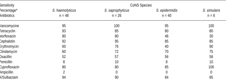
Sensitivity patterns. Sensitivity patterns of CoNS
spe-cies to 11 antibiotics most commonly used clinically are shown in Table 1.
This work has demonstrated a clear difference in the sensitivities of the diverse CoNS species. Frequencies of sensitivity to erythromycin was of a 90% for S. simulans and 40% for S. epidermidis; to norfloxacin, sensitivity from S. saprophyticus and S. haemolyticus was of 80%, compared with a 30% from S. simulans.
CoNS strains isolated from urinary tract showed a high sensitivity percentage to vancomycin and tetracyclin, and a
low sensitivity to β-lactamic antibiotics, except ampicillin/
sulbactam and cephalotin.
All S. saprophyticus strains were sensitive to vancomy-cin, and presented a high sensitivity to cephalotin, ampici-llin/sulbactam, tetracyclin, norfloxacin and cyprofloxacin. This species showed a moderated sensitivity to erythromy-cin, clindamycin and oxacillin. Only 10% of the strains showed sensitivity to penicillin, while all strains were resis-tant to ampicillin.
S. haemolyticus presented a high sensitivity to most of
the studied antibiotics. Only 8% of the strains were sensiti-ve to penicillin, and 2% to ampicillin.
S. epidermidis sensitivities to vancomycin, tetracyclin,
cephalotin, cyprofloxacin and ampicillin/sulbactam ranged from 80% to 95%. All strains were resistant to ampicillin, and only 8% were sensitive to penicillin.
All S. simulans strains were sensitive to vancomycin and cyprofloxacin, besides showing a high sensitivity to most antibiotics (80%-90%). 70% of the strains were re-sistant to norfloxacin, 90% to penicillin and all to ampi-cillin.
S. saprophyticus, S. simulans, S. epidermidis and S. haemolyticus showed a moderate sensitivity to oxacillin
(< 60%). From 80% to 95% of CoNS strains were sensi-tive to tetracyclin, cephalotin and ampicillin/sulbactam (Table 1).
Strains that were resistant to oxacillin showed a dia-meter of the halo of bacterial growth inhibition < 17 mm
and a MIC of 16 µg/ml, but remained sensitive to
van-comycin (MIC < 4 µg/ml). Resistance to vancomycin
(MIC > 32 µg/ml) was detected in 2.5% of S.
epidermi-dis and in 5% of S. haemolyticus, while an intermediate
resistance to vancomycin (MIC 8-16 µg/ml) was found
in 2.5% of S. epidermidis. Strains that were resistant to oxacillin were considered as resistant to penicillin and ampicillin. The cut points to the different antimicrobial agents tested, with respect to the diffusion and dilution tests of sensitivity in agar, are shown in Table 2. Also, a correlation between both antibiogramic techniques em-ployed was observed.
Adherence or slime. In this study, frequency of slime
pro-duction was determined for 120 CoNS strains, classifying them according to the intensity of production as: negative, weakly positive, moderately positive and strongly positive.
From the total number of strains tested for adherence the number of positive ones were: 20 S. haemolyticus, 19 S. saprophyticus, 32 S. epidermidis and 4 S.
simu-lans. Percentages of positives ranged from 42% to 80%,
moderately positive being the most common classifica-tion (Table 3).
All moderately and strongly positive slime producing CoNS strains presented the highest resistance percentage to
Table 1. Percentage of sensitivity of CoNS species to 11 antimicrobial agents.
Sensitivity CoNS Species
Percentage* S. haemolyticus S. saprophyticus S. epidermidis S. simulans
Antibiotics n = 48 n = 26 n = 40 n = 6
Vancomycine 95 100 95 100
Tetracyclin 93 85 80 80
Norfloxacin 80 80 46 30
Cephalotin 92 95 85 85
Erythromycin 60 76 40 90
Clindamycin 60 72 70 75
Oxacillin 52 57 56 58
Penicillin 8 10 8 10
Cyprofloxacin 80 80 85 100
Ampicillin 2 0 0 0
A/Sulbactam 94 90 84 85
Barberis et al. Antimicrobial sensitivity and adherence study in strains of coagulase-negative Staphylococcus spp.
Rev Latinoam Microbiol 2001; 43 (3): 109-113
112
edigraphic.com
cyprofloxacin and cephalotin of 15%, 14% and 10% were found, respectively. Results for norfloxacin from S.
epi-dermidis and S. simulans are not comparable to those of
Laverdiere et al.12
The high frequency of sensitivity to cyprofloxacin (80% to 100%) found in this study might be related to the limited use of fluorochinolones in our environment.
Isolated CoNS presented a high sensitivity (> 80%) for tetracyclin, cephalotin, cyprofloxacin, ampicillin/sulbac-tam, and for vancomycin > 95%. These results are
compa-rable to those carried out in Paris by Vu-Thien.21
In this study, we observed that methicillin resistant stra-ins were frequently resistant to several antimicrobial agents, while CoNS oxacillin resistant strains were genera-lly resistant to clindamycin and erythromycin. In a work realized in Brazil, the authors report oxacillin resistance
percentages comparable to those obtained in this study.4
Sensitivity to cyprofloxacin was more frequent among oxacillin sensitive strains than among oxacillin resistant CoNs. In this present work, it was observed that norfloxa-cin and/or cyprofloxanorfloxa-cin resistant S. epidermidis strains
were simultaneously oxacillin resistant.
In 4 CoNS strains we observed that 2 S. haemolyticus and 1 S. epidermidis presented an intermediate resistance to vancomycin (MIC 8-16 mg/ml). They were classified as sensitive (> 17mm) by the disk diffusion method, while 1 S. epidermidis strain was considered resistant to an MIC of 32 mg/ml. Our results showed that the disk diffusion me-thod cannot detect isolates with a diminished sensitivity to antimicrobial agents. Strausbaugh obtained similar results
in S. epidermidis for vancomycin in 1999.17
Slime production has been reported in strains of all
Sta-phylococcus species associated with human
infectio-ns.3,15,20 In this study we observed that 42% to 80% were
positive to adherence. 80% of S. epidermidis, 73% of S.
saprophyticus, 67% of S. simulans and 42% of S. haemoly-ticus isolated were positive for slime production. A couple
of S. simulans strains were found that were moderately po-sitive, and another couple weakly positive for the adheren-ce test. These results are lightly inferior to those found by
Drozenova et al.6
Table 2. Antimicrobial sensitivity patterns by the methods of disk diffu-sion and agar dilution of CoNS species.
CoNS
Antibiotics Inhibition haloes Cut point
(mm) µg/ml
Vancomycine > 17 < 4
Tetracyclin > 19 < 4
Norfloxacin > 17 < 4
Cephalotin > 18 < 8
Erythromycin > 23 < 0.5
Clindamycin > 21 < 1
Oxacillin > 18 < 8
Penicillin > 29 < 0.12
Cyprofloxacin > 21 < 1
Ampicillin > 29 < 0.25
A/Sulbactam > 15 < 8/4
Table 3. Classification of isolated CoNS species by slime production.
N° (%) of Slime production (%)
positive Strongly Moderately Weakly
CoNS strains positive positive positive
S. haemolyticus 42% (20/48) 3% 27% 12%
S. saprophyticus 73% (19/26) 7% 59% 7%
S. epidermidis 80% (32/40) 30% 40% 10%
S. simulans 67% (4/6) 0% 50% 50%
the tested antibiotics, which was not observed from weakly positive strains.
The S. epidermidis strain that was resistant to
vancomy-cin, with an MIC of 32 µg/ml, was a weak slime producer.
Our results show that approximately 50% of slime pro-ducers S. epidermidis, S. saprophyticus and S.
haemolyti-cus strains presented resistance to 2 or more antimicrobial
agents, and that strongly positive slime producers were more resistant to some antimicrobial agents.
DISCUSSION
Throughout the last years, the frequency of S.
epider-midis isolation has increased, as much as methicillin
re-sistance has increased in such microorganism.5 In this
study, the frequencies of sensitivity to oxacillin observed from S. epidermidis and S. haemolyticus were 56% and 52%, respectively. These results are not comparable to those obtained by Del’ Alamo in the study realized in São
Paulo, Brazil (19.2% and 4.2%).4 Results obtained in this
study are comparable to those found by Laverdiere et al in
In this study, S. epidermidis presented a significant as-sociation between slime production and resistance to vario-us antimicrobial agents. These results are similar to those
obtained by Nayak et al,13 who observed that slime
produ-cing S. epidermidis isolated from ocular infections was multiresistant to antimicrobial agents.
Results in this work suggest that slime producing CoNS was responsible for a significant decrease in sensitivity to some antimicrobial agents.
Our observations confirm that CoNS isolated in our zone presented resistance to several antibiotics of frequent use in the therapy of urinary infections.
ACKNOWLEDGMENT
This work was realized with economic support from the Secretaría de Ciencia y Técnica of the Universidad Nacio-nal de Río Cuarto.
REFERENCES
1. Akiyama, H., O. Yamasaki, H. Kanazaki, J. Tada, and J. Arata.
1998. Adherence characteristics of Staphylococcus aureus and coa-gulase-negative staphylococci isolated from various skin lesions. J. Dermatol. Sci. 18:132-136.
2. Baldassarri, L., G. Donelli, A. Gelosia, M. C. Voglino, S. W.
Simp-son, and G. D. Christensen. Purification and characterization of the Staphylococcal slime-associated antigen and its occurrence among
Staphylococcus epidermidis clinical isolates. Infect. Immunol.
64:3410-3415.
3. Christensen, G. D., A. Simpson, A. Bisno, and E. Beachey. 1982.
Adherence of slime-producing strains of Staphylococcus
epidermi-dis to smooth surfaces. Infect. Immun. 37:318-326.
4. Del’Alamo, L., R. F. Cereda, I. Tosin, E. A. Miranda, H. S. Sader.
1999. Antimicrobial susceptibility of coagulase-negative sta-phyloccoci and characterization of isolates with reduced susceptibi-lity to glycopeptides. Diagn. Microbiol. Infect. Dis. 34:185-191.
5. Domaracki B. E., K. E. Preston, and R. A. Venezia. 1998. Increased
oxacillin activity associated with glycopeptides in coagulase-nega-tive staphylococci. Eur. J. Clin. Microbiol. Infect. Dis. 17:143-150.
6. Drozenova, J., P. Oetras. 2000. Characteristics of
coagulase-negati-ve staphylococci isolated from hemocultures. Epidemiol. Mikro-biol. Inmunol. 49:51-58.
7. Garrett, D. O., E. Jochimsen, B. Zimmer, and W. R. Jarvis. 1999.
The emergence of decreased susceptibility to vancomycin in
Sta-phylococcus epidermidis. Infect. Control. Hosp. Epidemiol. 20:
167-170.
8. Hubner, J., A. Kropec. 1995. Cross infections of
coagulase-negati-ve staphylococci in high-risk patients. Zentralbl. Bakteriol. 283:169-174.
9. Kleeman, K., T. L. Bannerman, W. E. Kloos. 1993. Species
distri-bution of coagulase-negative staphylococcal isolates at a communi-ty hospital and implications for staphylococcal identification pro-cedures. J.Clin. Microbiol. 31:13-18.
10. Kloos W. E., and D. W. Lambe Jr. Staphylococcus. 1991. In: Balo-ws, Hamler, Hcrrmann, Isenberg, Shadomy (Ed.). Manual of Clini-cal Microbiology, 5th edition. American Society for Microbiology, Washington. Pp. 222-237.
11. Krcmery, V. Jr., J. Trupl, L. Drgona, J. Lacka, E. Kukuckova, and E. Oravcova. 1996. Nosocomial bacteremia due to vancomycin-re-sistant Staphylococcus epidermidis in four patients with cancer, neutropenia, and previous treatment with vancomycin. Eur. J. Clin. Microbiol. lnfect. Dis. 15:259-261.
12. Laverdiere, M. K. Weiss, R. Rivest, and J. Delorme. 1998. Trends in antibiotic resistance of staphylococci over an eight-year period: diffe-rences in the emergence of resistance between coagulase positive and coagulase- negative staphylococci. Microb. Drug. Resist. 4:119-122. 13. Nayak, N., and G. Satpathy. 2000. Slime production as a virulence
factor in Staphylococcus epidermidis isolated from bacterial kerati-tis. Indian J. Med. Res. 111:6-10.
14. NCCLS. Performance standards for antimicrobial susceptibility tes-ting. Documents: M2-T4 & M7-A2, 1998; M100-S3, 1991. NC-CLS, Vilanova, Pa, USA.
15. Pinna, A, S. Zanetti, L. Sotgiu, L. A. Sechi, G. Fadda, and F. Carta. 1999. Identification and antibiotic susceptibility of coagulase nega-tive staphylococci isolated in corneal/external infections. Br. J. Ophthalmol. 83:771-773.
16. Sieradzki, K., P. Villari, and A. Tomasz. 1998. Decreased suscepti-bilities to teicoplanin and vancomycin among coagulase-negative methicillin-resistant clinical isolates of staphylococci. Antimicrob. Agents Chemother. 42:100-107.
17. Strausbaught, L. J. 1999. Vancomycin-intermediate Staphylococcus
epidermidis: curio or omen? Infect. Control. Epidemiol. 20:163-5
18. Sung L, et al. 1999. Bacteriemia due to persistent strains of coagu-lase-negative staphylococci in a neonatal intensive-care unit. In-fect. Control Hosp. Epidemiol. 20:349-351.
19. Tenover, F.C., M. V. Lancaster, and B. C. Hill. 1998. Characteriza-tion of staphylococci with reduced susceptibilities to vancomycin and other glycopeptides. J. Clin. Microbiol. 36:1020-1027. 20. Veenstra, G.; Cremers, F.; Dijk, H. and Fleer, A. 1996.
Ultrastruc-tural organization and regulation of a biomaterial adhesin of
Sta-phylococcus epidermidis. J. Bacteriol. 178:537-541.
21. Vu-Thien, H. 1998. Antibiotic sensitivity to isolated bacteria in pe-diatric urinary tract infections. Arch. Pediatr. 3:266S-268S.
Correspondence to:
Isabel L. Barberis
[email protected] Address: Ruta 36 Km 601
Dpto. de Microbiología e Inmunología, UNRC. 5800, Río Cuarto,