M. Zamir
The Physics of
Coronary Blood Flow
With269Illustrations
Series Preface
The fields of biological and medical physics and biomedical engineering are broad, multidisciplinary and dyanmic. They lie at the crossroads of frontier re-search in physics, biology, chemistry, and medicine. The Biological & Medi-cal Physics/BiomediMedi-cal Engineering Series is intended to be comprehensive, covering a broad range of topics important to the study of the physical, chemi-cal and biologichemi-cal sciences. Its goal is to provide scientists and engineers with textbooks, monographs, and reference works to address the growing need for information.
Books in the series emphasize established and emergent areas of science in-cluding molecular, membrane, and mathematical biophysics; photosynthetic en-ergy harvesting and conversion; information processing; physical principles of genetics; sensory communications; automata networks, neural networks, and cellular automata. Equally important will be coverage of applied aspects of bio-logical and medical physics and biomedical engineering such as molecular elec-tronic components and devices, biosensors, medicine, imaging, physical princi-ples of renewable energy production, advanced prostheses, and environmental control and engineering.
M. Zamir
Department of Applied Mathematics University of Western Ontario London, Ontario, N6A 5B7 CANADA
Library of Congress Cataloging-in-Publication Data Zamir, M. (Mair)
The physics of coronary blood flow / M. Zamir.
p. cm. — (Biological and medical physics, biomedical engineering) Includes bibliographical references and index.
1. Coronary circulation. 2. Hemodynamics. 3. Blood flow. I. Title. II. Series. QP108.Z36 2005
612.1′7—dc22 2005042502
ISBN-10: 0-387-25297-5 e-ISBN: 0-387-26019-6 Printed on acid-free paper. ISBN-13: 978-0387-25297-1
©2005 Springer Science+Business Media, Inc.
AIP Press is an imprint of Springer Science+Business Media, Inc.
All rights reserved. This work may not be translated or copied in whole or in part without the written permission of the publisher (Springer Science+Business Media, Inc., 233 Spring Street, New York, NY 10013, USA), except for brief excerpts in connection with reviews or scholarly analysis. Use in connection with any form of information storage and retrieval, electronic adap-tation, computer software, or by similar or dissimilar methodology now known or hereafter de-veloped is forbidden.
The use in this publication of trade names, trademarks, service marks, and similar terms, even if they are not identified as such, is not to be taken as an expression of opinion as to whether or not they are subject to proprietary rights.
Printed in the United States of America. (MP)
9 8 7 6 5 4 3 2 1
Foreword
Everybody is interested in his/her coronary blood flow. I am delighted to read Dr Zamir’s clear exposition of the dynamics of the coronary blood flow in this book. I have read many of his scientific papers published in
profes-sional journals, as well as his bookThe Physics of Pulsatile Flow, published
by Springer-Verlag New York in 2000. His writing is a model of clarity. In an unhurried manner, he dwells on points of conceptual difficulty, and takes his reader to look at the difficulties from as many angles as possible. He offers solutions of difficulties. He enhances true understanding, but never
dogmati-cally. As a reader, I am grateful for that. He loves the wordconundrum. For
example, the heading of Section 10.3 of this book is “Coronary Heart Disease and the Conundrum of Coronary Flow Reserve.” That section is, of course, the heart of this book: every reader would be interested in this topic. What does conundrum mean? According to Webster’s Dictionary, conundrum means “(1) a riddle whose answer involves a pun, (2) anything that puzzles [1590-1600; pseudo-L word of obscure orig.]” That word prepares the reader. Be patient. Listen! Then I became patient, and I got a great deal of enlightenment out of the book.
in the cells, heart muscle tissue remodeling, gene expression activities, protein configuration changes, cell membrane behavior, integrins, enzymes, kinases, and their activities. Before long, besides learning about the heart, we have to learn how to engineer the coronary blood vessels for health and longevity. Physics and mathematics will have a lot more to do with biology!
Preface
Coronary blood flow is blood flow to the heart for its own metabolic needs. In the most common form of heart disease there is a disruption in this flow because of obstructive disease in the vessels that carry the flow. The subject of coronary blood flow is therefore associated mostly with the pathophysiology of this disease, rarely with dynamics or physics. Yet, the system responsible for coronary blood flow, namely the “coronary circulation,” is a highly
sophis-ticateddynamical system in which the dynamics and physics of the flow are
as important as the integrity of the conducting vessels. While an obstruction in the conducting vessels is a fairly obvious and clearly visible cause of dis-ruption in coronary blood flow, any discord in the complex dynamics of the system can cause an equally grave, though less conspicuous, disruption in the flow.
This book is devoted specifically to the dynamics and physics of
coro-nary blood flow. While it upholds the clinical and pathophysiological issues involved, the book focuses on dynamics and physics, approaching the sub-ject from a strictly biomedical engineering viewpoint. The rationale for this approach is simply that the coronary circulation involves many issues in dy-namics and physics, as the book will demonstrate. Also, with this particular focus, the book will complement other books on the subject, that have so far focused largely on clinical and pathophysiological issues.
systematic or factual way, but only tangentially in how they may affect the dynamics of the system. These omissions reflect the degree of complexity of the coronary circulation and serve as a sober reminder that it may never be possible or practical to deal with this complexity in a single book.
What seems possible at this time is to use known elements or properties of the system in such a way as to construct a meaningful, though incomplete, model of the system. This is the spirit in which the content of this book is presented. The book deals essentially with the dynamics of that part of the coronary circulation extending from the coronary ostia at the base of the aorta to the capillary level of coronary vasculature. It is meaningful to consider this part of the system in isolation because this is where the largest part of the pressure drop driving the flow occurs. While the dynamics of this part of the system may not represent the dynamics of the system as a whole, they demonstrate clearly the role of dynamics in the coronary circulation and illustrate how a disruption in these dynamics can affect coronary blood flow as significantly as can the obstruction of a blood vessel. This is indeed what the present book is about. Other books have in the past focused in a similar way on clinical or pathophysiological aspects of the system, or on the microcirculation. Each of these must clearly be seen as representing an important, though equally incomplete, view of the system.
My foray into the subject of coronary blood flow began in earnest in 1984 when I spent a sabbatical leave in the Department of Pathology at University Hospital in London, ON, and it is fair to say that this book would not have come into being without my ensuing collaboration with Professor Malcolm D. Silver, then department chair and chief of pathology. His passion for the heart and coronary arteries, and the depth of his expertise in cardiovascular pathol-ogy in general, was a haven for an engineer/applied mathematician seeking entry into the subject. With his help I came to know the coronary arteries lit-erally “in the flesh” as I attended weekly autopsy review sessions and learned to dissect, cast and measure coronary vasculature. The collaboration was not a hit from the start - he was as puzzled by my preoccupation with branching angles and branch diameters as I was by his preoccupation with shades of pink in myocardial tissue. But a meeting of the minds soon prevailed, and together we embarked on several studies that have since formed the basis of all my subsequent work on the subject. I am deeply indebted to Dr. Silver not only for his continued guidance over the years but also for reading a draft of this book and offering many valuable comments and suggestions.
Preface XIII
as for reading a draft of the book and offering many valuable comments. I received valuable comments also from my friend and colleague Dr. Erik L Ritman, Professor of Physiology and Medicine at the Mayo Clinic College of Medicine, who kindly took time from his ever busy schedule to read the manuscript.
But the mission of this book was to be in Science and Engineering rather than in Pathology or Medicine. For many years I have been inspired by and learned enormously from the books of Professor Y.C. Fung who, in my eyes, is the father of biomechanics and of the notion that engineers should not only “dabble” in the mechanics of the cardiovascular system but should do so deliberately and with no apologies. His books on the subject exemplify this notion and have provided me with the inspiration to write a book in the same spirit. I am grateful to Professor Fung not only for this but for agreeing to read this book and to write the Foreword to it.
It was important for me to have the book scrutinized by experts on both sides of the divide, and I am grateful to three colleagues who were kind enough to plow through the analytical aspects of the book and the tedium of equa-tions and algebra. Professor Guy Kember from the Department of Engineering Mathematics at Dalhousie University, Professor Matt Davison from the De-partment of Applied Mathematics at the University of Western Ontario, and Dr. Hope Alderson, formerly of the Department of Mathematics at the Uni-versity of New Brunswick. I am indebted to all three for laboring tirelessly through the manuscript, particularly to Dr. Alderson who did so heroically with a baby in one hand and the manuscript in the other.
I am grateful to my friend and long-time aide, Mira Rasche, for technical help with the manuscript, and to the secretarial brigade in Applied Mathemat-ics - Gayle McKenzie, Pat Malone, and Audrey Kager– for always being there. My deepest thanks go to my wife, Lilian, who may not share my enthusiasm for the subject, yet so willingly plowed through the manuscript and shared the burden in so many different ways. I am grateful to Dr. Elias Greenbaum, Editor-in-Chief of the Biological and Medical Physics, Biomedical Engineering Series, for his encouragement and continued support, and, at Springer NY, to David Packer and Lee Lubarsky for walking the manuscript to production, to MaryAnn Brickner for finally taking the manuscript away from my hands and setting the production wheels in motion, and to Frank McGuckin and Frank Ganz for helping with the nightmare of electronic typsetting.
Contents
Foreword . . . VII
Series Preface . . . IX
Preface. . . XI
1 Static Design Issues . . . 1
1.1 The Lone Pump . . . 1
1.2 Heart “Disease”? . . . 3
1.3 Origin of Coronary Blood Supply . . . 5
1.4 Coronary Arteries . . . 7
1.5 Left/Right Dominance . . . 12
1.6 Branching Structure . . . 14
1.7 Underlying Design? . . . 21
1.8 Coronary Flow Reserve . . . 27
1.9 Design Conflict? . . . 31
1.10 Summary . . . 32
2 Modelling Preliminaries . . . 35
2.1 Why Modelling? . . . 35
2.2 The “Lumped Model” Concept . . . 37
2.3 Flow in a Tube . . . 38
2.4 Fluid Viscosity: Resistance to Flow . . . 41
2.5 Fluid Inertia: Inductance . . . 45
2.6 Elasticity of the Tube Wall: Capacitance . . . 56
2.7 Elasticity of the Tube Wall: Wave Propagation . . . 62
2.8 Mechanical Analogy . . . 66
2.9 Electrical Analogy . . . 71
XVI Contents
3 Basic Lumped Elements . . . 79
3.1 Introduction . . . 79
3.2 RLC System in Series . . . 81
3.3 Free Dynamics of the RLC System in Series . . . 84
3.4 R1,R2in Parallel . . . 88
3.5 R,L in Parallel . . . 92
3.6 R,C in Parallel . . . 97
3.7 RLC System in Parallel Under Constant Pressure . . . 101
3.8 RLC System in Parallel Under Constant Flow . . . 103
3.9 Summary . . . 112
4 Forced Dynamics of the RLC System . . . 115
4.1 Introduction . . . 115
4.2 The Particular Solution . . . 116
4.3 Using the Complex Exponential Function . . . 117
4.4 Overdamped Forced Dynamics . . . 119
4.5 Underdamped Forced Dynamics . . . 122
4.6 Critically Damped Forced Dynamics . . . 124
4.7 Transient and Steady States . . . 126
4.8 The Concept of Reactance . . . 131
4.9 The Concepts of Impedance, Complex Impedance . . . 137
4.10 Summary . . . 142
5 The Analysis of Composite Waveforms . . . 145
5.1 Introduction . . . 145
5.2 Basic Theory . . . 148
5.3 Example: Single-Step Waveform . . . 151
5.4 Example: Piecewise Waveform . . . 157
5.5 Numerical Formulation . . . 164
5.6 Example: Cardiac Waveform . . . 169
5.7 Summary . . . 174
6 Composite Pressure-Flow Relations. . . 177
6.1 Introduction . . . 177
6.2 Composite Pressure-Flow Relations Under Pure Resistance . . . 179
6.3 Example: Cardiac Pressure Wave . . . 181
6.4 Composite Pressure-Flow Relations Under General Impedance 186 6.5 Composite Pressure-Flow Relations Under Inertial Effects . . . . 190
6.6 Composite Pressure-Flow Relations Under Capacitance Effects 198 6.7 Composite Pressure-Flow Relations Under RLC in Series . . . 207
6.8 Composite Pressure-Flow Relations Under RLC in Parallel . . . 213
7 Lumped Models . . . 221
7.1 Introduction . . . 221
7.2 LM0:{R,C} . . . 222
7.3 LM1:{R1,{R2+C}} . . . 229
7.4 LM2:{{R1+L},{R2+C}}. . . 235
7.5 LM3:{{R1+(pb)},{R2+C}} . . . 241
7.6 Inflow-Outflow . . . 249
7.7 Summary . . . 252
8 Elements of Unlumped-Model Analysis . . . 255
8.1 Introduction . . . 255
8.2 The Streamwise Space Dimension . . . 256
8.3 Steady Flow along Tube Segments . . . 258
8.4 Steady Flow Through a Bifurcation . . . 265
8.5 Pulsatile Flow in a Rigid Tube . . . 272
8.6 Pulsatile Flow in an Elastic Tube . . . 279
8.7 Wave Reflections . . . 287
8.8 Summary . . . 297
9 Basic Unlumped Models. . . 299
9.1 Introduction . . . 299
9.2 Steady Flow in Branching Tubes . . . 300
9.3 Pulsatile Flow in Rigid Branching Tubes . . . 307
9.4 Elastic Branching Tubes . . . 313
9.5 Effective Impedance, Admittance . . . 317
9.6 Pulsatile Flow in Elastic Branching Tubes . . . 329
9.7 Cardiac Pressure Wave in Elastic Branching Tubes . . . 343
9.8 Summary . . . 358
10 Dynamic Pathologies . . . 361
10.1 Introduction . . . 361
10.2 Magic Norms? . . . 362
10.3 Coronary Heart Disease, Physical Exercise, and the Conundrum of Coronary Flow Reserve . . . 370
10.4 Wave Propagation Through a Coronary Bypass . . . 378
10.5 Wave Propagation Through a Coronary Stent . . . 381
10.6 Sudden Cardiac Death . . . 384
10.7 Broken Heart Syndrome . . . 387
10.8 Summary . . . 388
References. . . 391
1
Static Design Issues
1.1 The Lone Pump
There are approximately 1014living cells within the human body, each
need-ing to receive food (nutrients, metabolic products) and to dispose of waste
productson an individual basis. The situation is not unlike that of the
(some-what lower, though equally overwhelming) number of humans on this planet,
approximately 6×10⁹in this case, each needing to receive food and to dispose
of waste products, again, on an individual basis. The cardiovascular system is responsible for bringing a line of supply and a line of return within reach of every living cell within the human body. These service lines are fully cen-tralized, in the sense that they all originate from one location and all return to it. In the world analogy this would amount to having only one location for the dispatch of food for the entire world population, only one location for the return of all waste products, and a line of supply and one of return within reach of every individual on the planet.
The cardiovascular system achieves this mammoth task within the human
body not bystoringfood and waste products in one location, but by having
them carried to and from every cell by a circulating fluid. The circulating
fluid is blood, and the service lines along which it circulates are blood vessels. The central location which all lines of supply originate from and return to is
not a massive warehouse but a small, very small,pump. The pump keeps the
fluid in circulation (Fig.1.1.1). As it circulates, food is added to the fluid and waste products are extracted from it, on a continuing basis, somewhat in the manner of a conveyor belt.
The most important element of the cardiovascular system is therefore not so much the place where nutrients and metabolic products come from but the pump that circulates the fluid which carries these products. That pump is, of course, the heart. Circulation of blood that carries the food is as critical as the food itself because circulation keeps the food coming to the cells. Any disruption in that circulation is a disruption in food supply. Yes, life is not
HEART
LUNGS
BODY
pulmonary circulationsystemic circulation
Fig. 1.1.1.Circulation of a fluid medium makes it possible to meet the metabolic needs of several billion cells within the human body by means of only a small pump (HEART). Metabolic substances are carried by the fluid to and from cells in every part of the body (systemic circulation), and the fluid itself is reconditioned by a second circulation to and from the lungs (pulmonary circulation). Circulation of the fluid is therefore as critical as the metabolic goods which it carries. The pump must maintain both circulations at all times and without fail. There is no backup, no contingencies.
of blood. The heart is the pump that drives that circulation. It must pump at all times, which it does by contracting and relaxing in a rhythmic pattern, approximately once every second, more than 86 thousand times every day, and about 2 billion times in a lifetime of 75 years, nonstop. If it fails, the entire body is deprived of its lifeline and fails with it.
1.2 Heart “Disease”? 3
1.2 Heart “Disease”?
While the heart, like other parts of the body, is subject to genetic disorders and infectious diseases [179], only a very small proportion of heart failures in the developed world are caused by such conditions. In all other cases a lack of
blood supply to the heart muscle, or a lack offuelwhich the muscle needs for
doing its pumping work, is the principal cause of heart failure [83, 206, 14]. Yet, “heart disease” is the term most widely used in association with heart failure. The term is somewhat misleading because it suggests that the
failure is due to a diseaseof the heart itself which, as stated above, occurs
in only a very small proportion of cases. The situation is analogous to using the term “malfunction” to describe a car engine failure caused by lack of fuel. Terms that are less commonly used but that are somewhat more accurate are “coronary heart disease” and “coronary artery disease”, both at least hinting
at a problem in thelines of blood supplyto the heart.
In this book the term “heart disease” shall be reserved for only the small proportion of cases where the heart is diseased in the true sense of a genetic or infectious disorder. In all other cases the term “heart failure” shall be used
instead, to meanfailure of the heart as a pump caused by lack of blood supply.
The lack of blood supply may be theimmediate cause of heart failure as in
the case of a thrombus or an embolus, or it may be theprecipitatingfactor as
in the case of myocardial infarction.
It is appreciated that this usage of the terms “heart disease” and “heart failure” differs from common usage of these terms in the clinical setting. The intention here is to emphasize the strictly biomedical engineering view of the coronary circulation to be adopted in this book. According to this view, blood supply to the heart is provided by a highly dynamic system which can be disrupted by not only a problem in the lines of supply but also by
a disturbance in the dynamics of the system. Indeed, thedynamics of blood
supply to the heart are the principal subject of this book.
The complication, of course, is that the organ responsible for blood supply to the heart is the heart itself. When the heart fails, it fails to provide blood supply to not only every other part of the body but to itself, too. The situation
has the elements of a control system withpositivefeedback. In such a system,
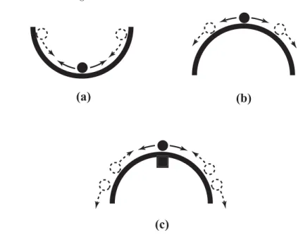
a small failure produces a signal which leads to more failure, while in a system withnegativefeedback, a small failure produces a corrective signal which leads to less failure. The second system is regarded as stable and the first unstable, in the sense that after a small departure from equilibrium the second system can recover and return to equilibrium while the first cannot. A small steel ball at the bottom of a big bowl constitute a stable system, but if the bowl is turned upside down and the steel ball is positioned precariously at its peak, the system becomes unstable (Fig.1.2.1).
(a)
(b)
(c)
Fig. 1.2.1.Stability of coronary blood supply. Blood supply to the heart is provided by the heart itself: Does heart failure, therefore, lead to more failure? Shown here is the analogy of a small steel ball in equilibrium at the bottom of a large bowl (a). A small departure from equilibrium in this case is corrected by a negative signal which acts to move the ball back towards its neutral position. If the bowl is turned upside down as in (b), however, and the ball is positioned precariously at the peak, a small departure from equilibrium produces a positive signal which moves the ball yet further away from the peak. Regulatory mechanisms in coronary blood flow ensure that a small reduction in blood supply to the heart muscle does not lead to further reduction. In the bowl analogy it is as if a small magnet is placed under the bowl’s peak as in (c), so that a small departure of the ball from the peak is corrected by the pull of the magnet. But if blood supply is prevented from reaching the heart muscle because of coronary artery disease, this mechanism becomes less effective or inoperative. In the bowl analogy it is as if the magnet is contaminated and can no longer pull back the ball effectively.
part of the body, including the heart muscle itself. But the situation is not in fact as precarious as the steel ball at the top of the upside-down bowl. Regulatory mechanisms in coronary blood flow (autoregulation) ensure that a reduction in the ability of the heart to pump does not translate immediately into a reduction of blood supply to the heart itself [83, 128, 136, 100, 183]. This mechanism provides a local reprieve and allows recovery to occur. In the bowl analogy it is as if a magnet is placed under the peak of the upside-down bowl, so that a small excursion of the steel ball away from the peak is corrected by the magnet pull before the situation becomes beyond recovery (Fig.1.2.1).
1.3 Origin of Coronary Blood Supply 5
the heart muscle isnota reduction in the innate ability of the heart muscle to
pump but a gradual occlusion or sudden blockage of some of the vessels that
carry this supply to the heart, caused in turn by a disease processwithin the
vessels. Thus, heart failure (as defined in this book) is not usually mediated by
positive feedback but by adirectreduction of blood supply to the heart muscle,
which is not triggered by failure of the muscle to pump in the first place. The recovery mechanism that compensates for the reduction in blood supply to the
heart muscle is thus disrupted because of an inability of blood supply toreach
the heart. In the bowl analogy it is as if the magnet is contaminated and its ability to bring the steel ball back to the neutral position is diminished. “Heart disease” does not fairly describe the situation in hand, “heart starvation” would be a more accurate term. As stated earlier, the term “coronary heart disease”, used less often, is somewhat more accurate in that it hints at the involvement of the coronary arteries, while “coronary artery disease” is yet more accurate because it places the problem precisely where it belongs.
1.3 Origin of Coronary Blood Supply
As a pump, the heart consists of four chambers, two ventricles and two atria, which contract and relax rhythmically. The two ventricles eject blood and the two atria act as receiving chambers for returning blood [133]. Output from the left ventricle is carried by the aorta to every part of the body, then re-turns to the right atrium, thus producing the so-called “systemic circulation” (Fig.1.1.1). Output from the right ventricle is carried by the pulmonary artery to the lungs where blood is oxygenated and then returned to the left atrium, constituting the “pulmonary circulation” [48].
The two systems must clearly operate in tandem to avoid accumulation (congestion) of fluid proximal to or in any of the four chambers. That is, on average, the systemic and pulmonary circulations must move the same volume of blood per cardiac contraction (Fig.1.1.1). However, the systemic circulation operates at a much higher pressure than the pulmonary, hence the work per-formed by the two ventricles is not the same. The pumping power produced by the left ventricle and hence its metabolic requirements are considerably higher than those of the right ventricle [135, 141]. Accordingly, the muscular walls of the left ventricle are the main focus of blood supply to the heart. While blood supply must reach every part of the heart for the organ to re-main viable, blood supply to the left ventricle dominates coronary blood flow because of its intense requirements.
RCA LCA
RV LV
RA LA
AO
Fig. 1.3.1. Origin of blood supply to the heart. As the aorta (AO) leaves the left ventricle (LV) with oxygenated blood for every part of the body, its first two branches, the so-called left main and right coronary arteries (RCA, LCA), are des-tined to serve the heart itself. The figure is highly schematic, to show functional topology only.
body the aorta gives rise to many branches, destined to different parts of the body or to specific organs [48]. Each organ or territory within the body has its own supply line from the aorta, followed by a vascular tree structure to reach different parts of the organ or territory, and a system of capillaries to reach every cell. The heart is served in the same manner [81].
It would seem appropriate, therefore, that as the aorta leaves the left ventricle, laden with oxygenated blood for every part of the body, its first two branches are destined to serve the heart itself (Fig.1.3.1). They are known as the left main and right coronary arteries [133, 228, 216]. While this may seem as if the heart is being given “first priority”, it is more likely the result of physical proximity of the heart to the root of the aorta (Fig.1.3.1).
1.4 Coronary Arteries 7
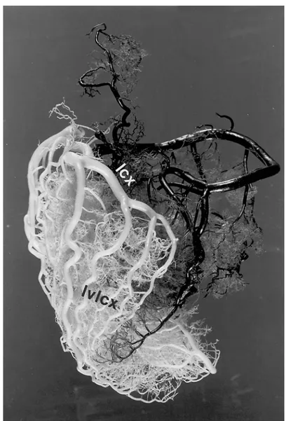
Fig. 1.3.2. Origin of the left main and right coronary arteries (LCA, RCA) and two additional branches (black arrows) as they arise from the aorta. From a cast of a human coronary network [216].
1.4 Coronary Arteries
As the left main and right coronary arteries leave the aorta they circle the heart in the manner of a crown, hence the name “coronary” arteries. The attributes “left” and “right” relate to the fact that the left coronary artery circles the left side of the heart while the right coronary artery circles the right side, though the situation is not accurately so. Furthermore, while the term “coronary” was first applied to the two main supplying arteries, it is now used to include all branches and sub-branches of these vessels as well as the capillary and venous vasculature, which in total comprise the “coronary circulation”. Thus, in general terms, the word “coronary” has come to mean any element of blood supply to the heart for its own metabolic needs.
Fig. 1.4.1.(left) Approximate anatomical orientation of the heart (in a front view) in relation to the median and transverse planes of the body (mp, tp). (right) A “theoretical” upright position in which the heart is turned so that its atrioventricular plane (avp) is approximately parallel to the transverse plane of the body, and the interatrial and interventricular planes (iap, ivp) are approximately parallel to the median plane of the body. This position is convenient for discussion since here the left and right sides of the heart coincide approximately with the left and the right sides of the body. The main coronary arteries circle the heart in the atrioventricular plane, which is hence also known as the coronary plane (cp). From [228].
the coronary arteries in a given heart, and approximatefunctional layout of
these vessels in every heart. The first may be important for the purpose of clinical intervention and treatment in a particular heart. The second relates to fluid dynamic design and function of the coronary network in general, and it is the main focus in this book.
func-1.4 Coronary Arteries 9
APEX
Fig. 1.4.2.The way in which the main coronary arteries circle the heart (in a front view), in relation to the coronary plane (dashed circle) and the interventricular plane (hatched plane). The right coronary artery (RCA) circles the right side of the heart while the left circumflex artery (LCX), which is a main branch of the left main coronary artery (LCA), circles the left side of the heart. The left anterior descending artery (LAD), which is the other main branch of the left main coronary artery, descends along the edge of the interventricular wall toward the apex of the heart. From [216].
tions as part of the left ventricle, hence blood supply to it is particularly
important. The other branch, known as the left circumflex artery, turns to circle the left side of the heart along the atrioventricular groove. As it does so it gives rise to a number of small branches which head up to serve the right atrial region, and larger branches which head down to serve the lateral and posterior walls of the left ventricle (Fig.1.4.3).
As the right coronary artery reaches the atrioventricular groove, it turns to circle the right side of the heart, coursing along the groove and giving rise to branches heading up to serve the right atrial region and down to serve the anterior and posterior walls of the right ventricle (Fig.1.4.3). In approximately 90% of human hearts, as the right coronary artery reaches the atrioventricular
groove at theback of the heart, it gives rise to an important branch known
1.4 Coronary Arteries 11
Fig. 1.4.4. The left anterior descending artery, which descends along the front edge of the interventricular wall (Fig.1.4.2), is seen here wrapping itself around the apex of the heart (see also Fig.1.4.5). It then begins toascendalong the back edge of the interventricular wall, as if to meet the posterior descending artery which is descending toward it along that edge. From [228].
both arteries head towards each other as they move towards the “apex”, the pointed bottom of the heart. In most cases the anterior descending artery then wraps around the apex (Fig.1.4.4) and the two vessels stop short of meeting thereafter (Fig. 1.4.5). In 70% of human hearts the right coronary artery continues to circle the heart, after giving rise to the posterior descending artery, to serve some of the posterior wall of the left ventricle [133].
1.5 Left/Right Dominance
With the heart in an upright position, the right coronary and left circum-flex arteries circle the heart in a horizontal (coronary) plane, and in opposite directions (Fig. 1.4.2). As they reach the back of the heart, the two arteries move toward each other and terminate short of actually meeting (Fig. 1.5.1). The point at which this occurs is a measure of the extent to which the right coronary artery supplies the left side of the heart, which is an important functional aspect of the coronary network usually referred to as left/right dominance [133]. An important anatomical landmark in this subject is the “crux”, the point at which the horizontal atrioventricular groove crosses the vertical interventricular groove at the back of the heart (Fig. 1.5.1). At ap-proximately this point the posterior descending artery arises and begins its descent along the interventricular groove to serve the interventricular wall. Whether this artery arises from the right main coronary artery or from the left circumflex artery depends on which of the two arteries reaches the crux.
In only 10% or so of human hearts, the left circumflex artery reaches the crux and gives rise to the posterior descending artery [133]. Such cases are known as “left dominant” since, functionally, blood supply to the left ventricle then depends entirely on the left coronary artery. In approximately 20% of human hearts, known as “balanced” cases [133], the right coronary artery reaches the crux, gives rise to the posterior descending artery and terminates at that point. In the remaining 70% of cases, known as “right dominant”, the right coronary artery continues beyond the crux to serve part of the posterior wall of the left ventricle.
1.6 Branching Structure 13
Fig. 1.5.1. As the right coronary artery (RCA) and the left circumflex artery (LCX) reach the back of the heart they move toward each other. Only one of them reaches the “crux” and gives rise to the posterior descending artery (PD). The numbers identify individual walls of the two ventricles: (1) interventricular septum, (2,3) lateral and posterior walls of the left ventricle, respectively, (4,5) posterior and anterior walls of the right ventricle, respectively. It is seen that the interventricular septum, which separates the two ventricles, is in fact an important part of theleft
ventricle. From [216].
may be regarded as an “anatomical risk factor”. While an exact numerical measure of that risk factor based on a measure of left/right dominance is not easy to calculate, the connection between the two measures is clear.
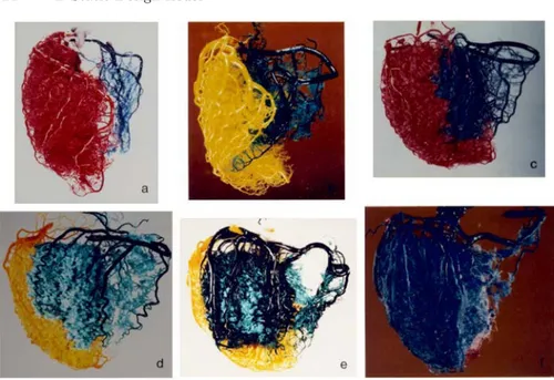
Fig. 1.5.2.Casts of the arterial networks of human hearts in which the distribution of the left main coronary artery is seen in red (a,c,f) or yellow (b,d,e) and the distribution of the right coronary arteries is seen in blue (a,b,c,d,f) or green (e). The hearts, in all cases, are in an upright posterior view. The figure illustrates the range of variability in the way in which the two arteries share the responsibility of cardiac blood supply. At one extreme (a), known as “left-dominant”, the left coronary artery gives rise to the posterior descending artery and therefore blood supply to the left ventricle depends on vasculature arising entirely from the left coronary artery. In a “balanced” case (b), the right coronary artery gives rise to the posterior descending artery and thereby contribute to blood supply to the interventricular wall, which is an important part of the left ventricle. In “right-dominant” cases, the right coronary artery continues beyond the crux to supply part or all the posterior wall of the left ventricle (c,d,e,f), thereby taking a greater share of the supply to that ventricle. From [216]. (See color insert.)
1.6 Branching Structure
1.6 Branching Structure 15
Fig. 1.6.1.In an open tree structure, shown schematically here, flow from the main supplying artery (top) must proceed along the strict branching hierarchy of the tree, that is from parent to branches at each junction. Each destination (bottom) can be reached via only one distinct path. Symmetry and uniformity are not necessary but are used here to emphasize these hierarchical features. Arterial trees are generally nonsymmetrical and highly nonuniform, but their underlying architecture is most often that of an open tree structure.
mesh structure, or in an open tree structure with an overlay of collateral connections, by contrast, there may be several paths to any given destination (Figs. 1.6.2, 3).
This issue is of particular functional and clinical importance. In the pres-ence of multiple paths blood supply can bypass an occluded vessel segment and reach its destination via a different path, an important consideration since occluded or obstructed blood vessels are the cause of most heart failures [3, 17, 209, 83, 81, 14]. But the subject is highly controversial, for several rea-sons. First, because the presence of collateral vessels in the vascular system of the heart is highly variable in different species. Second, because the presence of collateral vessels in the human heart is found to be highly variable not only from heart to heart but also in the same heart at different times. Thus, the way in which collateral vasculature fits in the hemodynamic design of the coronary circulation in general, and in that of the human heart in particular, is not fully established.
Fig. 1.6.2.In an interconnected mesh structure flow from the main supplying artery (top) can reach its destinations (bottom) via different routes. Early studies of dog coronary vasculature indicated that its underlying architecture has this intercon-nected structure. Subsequent work on the human heart, on the other hand, showed repeatedly that the underlying architecture of its vasculature is that of an open tree structure. Interconnected mesh structures in the vascular system occur mostly at the capillary level. At higher levels of the system they are rare because an open tree structure is, fluid dynamically, more efficient than an interconnected mesh.
demonstrated clearly the existence of such collateral vasculature, abundant in the dog heart, rarely or scarcely found in the pig, and variable in the human heart [185, 173, 27, 15, 69, 16, 74, 95]. While differences between species suggest that collateral vasculature may be part of the hemodynamic design of the coronary circulation in some species, the matter is far from settled. Much of the focus has been on the situation in the human heart.
An extensive study by Baroldi and Scomazzoni [16] sums up much of our current understanding of collateral vasculature in the human heart and resolves much of the controversy relating to variability. By a systematic survey of human hearts the authors found that both the number and size of collateral vessels were strongly linked to the presence of an ischemic history or conditions within the heart. Very fine collateral vasculature may be found sporadically here and there when ischemic conditions are absent, but in the presence of such conditions collateral vasculature becomes more significant in size and clearer in its mission. The results clearly established the phenomenon of collateral
vasculature as a compensatorymechanismin coronary heart disease [15, 14],
and this is how the matter rests.
But the question of the effectiveness of this mechanism is far from
1.6 Branching Structure 17
Fig. 1.6.3.When interconnections occur sporadically (dashed lines) in an otherwise open tree structure, they are referred to as “collateral” vasculature. The presence of collateral vasculature has been demonstrated in the coronary circulation of the human heart (see Figs. 1.6.4–6) but controversy continues over its origin, rate of growth, and hemodynamic significance. Collateral vasculature is to be distinguished frompermanentcollateral bridges such as the communicating arteries in the circle of Willis of the human brain. The latter are part of the fluid dynamic design of the cerebral circulation, that is, part of the normal anatomy of the cerebral vasculature. Collateral vasculature in the human heart is not part of the normal anatomy of the coronary vasculature.
The overwhelming majority of heart failures caused by insufficient myocar-dial blood supply [185, 173, 27, 15, 69, 16, 74, 95] point clearly to a failure of this mechanism in these cases. The difference between the time scale of development and growth of collateral vessels and the time scale of vascular obstruction, whether by a slow disease process or sudden occlusion, is clearly an important factor. The indications are that the mechanism of collateral vasculature cannot deal with sudden occlusion, first because the vasculature would likely not be present before the occlusion occurs, and second because new vasculature cannot develop within the fast time scale of a sudden occlu-sion. In the case of the much slower pace of stenosis by atherosclerotic disease,
the indications are that collateral vasculaturecandevelop in time to make a
significant contribution, but whether it does or not in every case, and the magnitude of the contribution it makes in each case, is not clear. The subject is decidedly far from settled [172, 44, 180, 77].
The ultimate question, of course, is the evolutionary origin of the
1.6 Branching Structure 19
Fig. 1.6.6.The absence of collateral connections is further confirmed by the ability to physically separate the distributions of the left and right coronary arteries. In fact, in (b) and (d) it is seen that the beds of individual vessels on the same side could also be separated from each other. See also caption for Fig. 1.6.4. From [216]. (See color insert.)
cap-1.7 Underlying Design? 21
illary level, of course, but at that level it can no longer serve the function of collateral vasculature since the meshing is highly localized and cannot reach (hemodynamically) from the distribution territory of one major artery to that of the next.
1.7 Underlying Design?
Wide variability in the anatomical layout of the coronary arteries and their major branches calls into question the “naming” of these vessels, because the names imply that they are clearly defined anatomical entities. Yet we have seen that the left or the right coronary artery in one heart is not the same functional entity as the left or the right coronary artery in another. This variability also calls into question the notion of “one-artery” or “two-artery” disease as measures of the severity of coronary heart disease, since, again, these terms suggest that one artery in one heart is the same functional entity as an artery with the same name in another heart, or indeed that two arteries have twice the functional value of one artery. More accurately, wide variability in their size and distribution suggests that the coronary arteries, as represented by their anatomical names, do not represent elements of the
underlying functional design of the coronary network as a fluid conveying
system. Elements of that functional design would be represented by features of the coronary network which do not vary from heart to heart.
The situation is not unlike the functional design of the heart itself as a double pump for the maintenance of two circulations. The characteristic four chambers of the heart are essential elements of this design that do not change from heart to heart. On the other hand, the exact size and shape of these chambers, or the size and shape of the heart as a whole, are secondary features that vary considerably from heart to heart. They are not essential
elements of the underlying design of the heart as a double pump. Thus, by
analogy, we ask: are there features of the coronary network that do not change from heart to heart?
A study of human hearts with the purpose of addressing this issue found, in summary, that the coronary network serves the heart by dividing the my-ocardium into six distinct zones [228]. Each zone is served by two types of
vessels: “distributing vessels” that run along thebordersof these zones, and
1.7 Underlying Design? 23
Borders defining the six zones of the myocardium in this scheme coincide with certain anatomical landmarks of the heart, namely the “sulci” of the two main dividing walls of the heart: the atrioventricular septum and the inter-ventricular septum. Topographically, the sulci represent the intersections of the two dividing planes with the outer surface of the heart. They appear as faint grooves on the surface of the heart. The distributing vessels run along these grooves, usually covered by a thin layer of fat. The atrioventricular sep-tum, for example, which lies in the coronary plane (Figs. 1.4.1, 2), produces the atrioventricular sulcus on the surface of the heart as a mark of its inter-section with it. This sulcus runs as a “belt” around the “waist” of the heart and it is the groove along which the right coronary artery and the left cir-cumflex artery run (Figs. 1.4.1, 2). The analogy used more commonly is that of a “crown” (presumably around the “head” of the heart), hence the term “coronary” as mentioned earlier.
1.7 Underlying Design? 25
Another important inner wall of the heart is the interventricular septum, which acts as a divider between the two ventricles but is functionally part of the left ventricle. The intersection of the interventricular plane with the outer surface of the heart produces the interventricular sulcus along which the anterior and posterior descending arteries run (Figs. 1.4.1, 5). By what appears to be a clear design, and in a pattern which does not vary from heart to heart, the two arteries circle the wall as distributing vessels, while branches from them run into the plane of the wall as delivering vessels (Fig. 1.7.1).
Two other borders coincide with the “acute margin” on the right side of the heart, and the “obtuse margin” on the left (Fig. 1.4.5). The names of the six zones and of the borders defining them are shown in Fig. 1.7.4.
The zones and borders are anatomical landmarks which are permanent features that do not change from heart to heart (Fig. 1.7.3). The underlying fluid dynamic design of the coronary network appears to be based on these
landmarks. By this functional design,distributing arteries run along the
di-viding borders, giving rise to delivering branches that enter the zones and
implement blood delivery.This scheme by which the coronary network serves
the heart does not change from heart to heart [228].
Variability in the size and shape of the zones, and wide variability in the length and size of coronary arteries referred to earlier, do not alter this scheme.
A border may be occupied bydifferentdistributing vessels in different hearts,
but the scheme remains constant. The posterior interventricular sulcus, for example, may be occupied by a posterior descending artery arising from the right coronary artery or one arising from the left circumflex. The left posterior atrioventricular sulcus may be occupied fully by the left circumflex artery, fully by the right coronary artery, or partly by both. In these different scenarios there is wide variability in the length and size of the coronary arteries involved, but the underlying design of the coronary network remains the same. In all cases distributing vessels bring blood supply to the borders of zones while
delivering vessels enter the zones and implement delivery. Only theidentities
of the vessels vary from heart to heart, hence these identities, as represented
by the anatomical names of the vessels, do not represent accuratefunctional
elements of the coronary network.
This subject is clearly important in coronary heart disease where one or more arteries may be affected by disease that limits its fluid dynamic func-tion. The foregoing discussion suggests that an accurate clinical assessment of coronary heart disease in a particular heart should be based not on the
anatomical namesof the affected vessels but on thefluid dynamic roleof each affected vessel in the scheme of blood supply to that heart. What is clearly important is whether the affected vessel is a distributing or a delivering vessel, and what particular position it occupies in that particular heart. Only then
is it possible to deduce the effect of the diseased vessels on particular zones
1.8 Coronary Flow Reserve 27
1.8 Coronary Flow Reserve
The intensity of the pumping action of the heart varies considerably, depend-ing on the metabolic activity of the rest of the body. The range of demand is fairly wide, extending from a base level when the body is resting to a considerably higher level when the body is at maximal activity. Energy re-quired to support the pumping action of the heart is therefore highly variable, and blood supply to the heart for the purpose of its own metabolic activity must be able to change accordingly over a wide range. The coronary circu-lation, being the vehicle for that supply, must therefore have the capacity to deliver far more blood flow to the heart than it does under normal rest-ing conditions. This excess capacity is referred to as “coronary flow reserve” [83, 42, 66, 64, 129, 128, 100, 183, 26, 112]. How does the coronary circulation provide this reserve?
The triggers for change in coronary blood flow and the mechanisms by which change is accomplished have been studied widely and are well docu-mented elsewhere [83, 42, 66, 64, 129, 128, 100, 183, 26, 112]. In this section we are concerned with only the ultimate expression of these mechanisms in terms of the fluid dynamic design of the coronary circulation. The first ques-tion in that context must clearly be whether there is an element of flow reserve in the size of the main supplying arteries of the heart, namely the left main and right coronary arteries. Do these arteries, by design, have larger diameters than would normally be required for normal blood flow to the heart? In other words, are the diameters of the left and right main coronary arteries better matched to the high or low end of the range of coronary blood flow?
Within the cardiovascular system, it is reasonably well established that the diameters of the main arteries supplying an organ are directly related to the organ’s requirements for blood supply. More accurately, the diameter of a supplying artery is generally related to the flow rate which the vessel is destined to convey. An actual relation between diameter and flow rate was proposed many years ago in the form of what is now known as the “cube law” [147, 190, 178, 167, 132]. It proposes that the flow rate through a vessel should optimally be proportional to the cube of the vessel’s diameter, or conversely, that the diameter of the vessel should optimally be proportional to the cube root of the flow rate the vessel is destined to convey. Other relations have been explored since then and were shown to have some theoretical or empirical validity, but the cube law continues to have the widest support in terms of biological data [167, 132, 225, 222, 223, 92, 106].
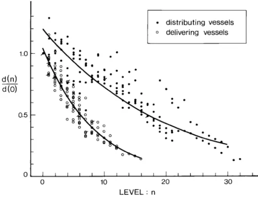
The cube law provides a useful tool for assessing the provisions for blood supply to different organs, with a useful concept in this assessment being that of “bolus speed”. A “bolus” within a blood vessel is defined as a cylindrical volume of blood which has the same diameter as the luminal diameter of the vessel and a length equal to that diameter (Fig. 1.8.1). The volume of a bolus is thus proportional to the cube of the diameter of the vessel in which
bolus volume =
d
34
π
d
34
π
bolus speed = q / (
)
d
d
Fig. 1.8.1.The concept of bolus speed. A “bolus” within a blood vessel is defined as a cylindrical volume of fluid which has the same diameter as the lumen diameter of the vessel and a length equal to that diameter. Under the branching “cube law”, whereby the diameter of a vessel is optimally proportional to the cube root of the flow rate which the vessel carries, the bolus speed is the same throughout the vascular network.
corresponding volumes of their boluses are denoted byV1, V2, then
V2 V1 =
d2 d1
3
(1.8.1)
If theflow ratesthrough the two vessels are denoted byq1, q2, respectively,
then the cube law proposes that the ratio of the diameters of the two vessels is related to the ratio of the two flows by
d2 d1 =
q2 q1
1/3
(1.8.2)
Combining the two results (Eqs.1.8.1,2) we then have
V2 V1 =
q2
q1 (1.8.3)
Now, the volumetric flow rate through a vessel is the product of the volume of a bolus in that vessel and the number of boluses passing through the vessel per unit time. We shall refer to the latter as the “bolus speed”. That is, if the
bolus speed is denoted byS, then
q=V S (1.8.4)
and for the two vessels discussed above we then have
q2 q1 =
V2S2
1.8 Coronary Flow Reserve 29
Results in Eqs.1.8.3,5 lead clearly to
S2=S1 (1.8.6)
which is a useful expression of the cube law in terms of bolus speed, that is: The relation between diameter and flow in blood vessels of different size should optimally be such that the bolus speed is the same in all vessels. Thus the provisions for blood supply to an organ, on this basis, may be deduced from the diameters of the main supplying arteries to that organ, on the assumption that the bolus speed in these arteries is the same as that in the aorta or in the vascular system in general.
To apply this result we consider a human cardiovascular system in which
systemic flow rate through the aorta isqa = 5l/min and aortic diameter is
a= 2.5cm. The volumeVa of a bolus in the aorta is thenπ(2.5)3/4 and the
corresponding bolus speedSa is given by
Sa = qa Va =
5000/60
π(2.5)3/4 = 6.79b/s(boluses per second) (1.8.7)
This value of the bolus speed can be used as a reference for flow in other parts of the cardiovascular system.
In particular, when we consider a human heart supplied by two main coronary arteries, which for the purpose of discussion are assumed to have equal diameters and carry equal flow rates, if the diameter of each artery is
taken as 3.5 mm, and if the same bolus speed is assumed to exist in these
arteries as it does in the aorta, then the volume of a bolus in each of the two
vessels isπ(0.35)3/4 and the combined flow rate to the heart would be given
by
q= 2×π(0.35) 3
4 ×6.79 = 27.44ml/min (1.8.8)
This coronary flow rate is considerably lower, by almost an order of magni-tude, than usual estimates [83, 66, 128, 100, 183, 26, 112]. If the two coronary
arteries are each taken to be 4mmin diameter, then the corresponding flow
rate to the heart would be 40.96 ml/min, still considerably lower than
es-timated. (Under normal conditions it is approximatly 5 percent of systemic
blood flow, or 250ml/min[66]).
These simple calculations indicate clearly that coronary flow reserve in the human heart is not based on having supplying vessels that are “larger than normal”. In fact, the reverse appears to be the case. At a base flow rate of 250ml/min, if the two main coronary arteries were to have the same bolus speed as exists in the aorta, then the diamter of each vessel, using Eq. 1.8.2, would be approximately given by
d= 25
125 5000
1/3
Diameters of the two main supplying coronary arteries in the human heart
are usually in the range of 3−5mm[16, 73, 133].
Thus the diameters of the main supplying coronary arteries appear to be
matched decidedly to thelowerend of the flow range. Coronary flow reserve,
which makes it possible for blood flow to increase by a factor of 6 or more [83, 42, 66, 64, 129, 128, 100, 183, 26, 112], must therefore be based on a different design paradigm. From a purely fluid dynamic standpoint, flow rate through the coronary vascular tree can only increase by increasing the driving pressure or by decreasing the overall resistance of the tree. Since the driving pressure (for flow to the tree as a whole) can only change by a relatively small amount, it is clear that a large increase in flow can only be achieved by a change of resistance to flow. Design provisions for coronary flow reserve in the human heart must therefore be based on having wide control over the
diameters of theresistance vesselswithin the coronary network.
While in steady flow the resistance to flow in a tube or a vascular tree
is determined predominantly by the tube or vessel radii, in pulsatile flow in general, and in the coronary circulation in particular, the situation is far more complex and is not clearly predetermined. In pulsatile flow the resistance to flow, then more appropriately referred to as impedance, depends on the
fre-quency of pulsation, and hence on the harmonic composition of the driving
pressure wave, since the harmonic components of the wave propagate at dif-ferent frequencies. Also, elasticity of the conducting vessels turns the flow in each vessel into a propagating wave, with consequent wave reflections. The relation between pressure and flow in the presence of wave reflections, and hence the “resistance” to flow, are not clearly predetermined as they are in steady flow, nor easy to formulate mathematically [9, 135, 141, 221].
The complex architecture of the coronary network compounds the diffi-culty, not so much because of its degree of complexity but because of an insufficient amount of architectural data. Also, elasticity of the coronary ves-sels gives the system the ability to change its volume to an extent and in a manner which are not fully known. This property of the coronary network, generally referred to as its “capacitance”, in combination with the pulsatile nature of the flow introduces an element of inflation and deflation which fur-ther complicates the relation between pressure and flow. Finally, most of the coronary vasculature is deeply imbedded within the cardiac muscular tissue, and as this tissue contracts and relaxes in the pumping process, the effects on pressure and flow within the vessels are far from known or fully under-stood. Thus, while the nature of resistance to coronary blood flow is highly complex and not fully understood, it is clear that the design provisions for coronary flow reserve are based entirely on having substantial control over that resistance. Coronary blood flow can increase by a factor of 6 or more not by having supplying vessels that are designed to carry such high flow but by having resistance vessels under strict dynamic control. We may say that
coronary flow reserve is not part of thestaticdesign of the coronary network
1.9 Design Conflict? 31
1.9 Design Conflict?
Provisions for coronary flow reserve discussed in the previous section are clearly geared, by design, to changes in flow demand associated with normal physiological function. The sudden increase in coronary blood flow required at the onset of vigorous physical exercise, for example, is accomplished by the facility of coronary flow reserve. But the integrity of that facility is seriously compromised in the presence of obstructive coronary artery disease. The rea-sons for this are somewhat convoluted and have the appearance of a “design conflict” between the availability of coronary flow reserve and the problem of long term fluid dynamic changes associated with obstructive vascular disease. The reason for this is that as disease gradually obstructs the supplying vessels, thus increasing the resistance to flow and therefore decreasing flow rate through the vessels, the facility for coronary flow reserve counteracts by dilating resistance vessels to restore the overall resistance in the system and thereby restore flow rate. But some of the capacity to further dilate the resistance vessels and further increase the flow rate has now been lost. That is, some of the coronary flow reserve has been “used up”. And more is used up, in the same way, as the severity of obstructive disease increases, to a point where the capacity to increase coronary flow rate is completely lost.
Thus, the facility of coronary flow reserve does not appear to be aimed
by designto deal with the long-term fluid dynamic effects of coronary artery disease. It is indeed well established that coronary flow reserve diminishes in the presence of atherosclerotic coronary artery disease [83, 66, 128, 100, 183, 26, 112]. Are there other facilities or mechanisms in the coronary circulation that are aimed at the long term effects of coronary artery disease?
It is known that in the presence of obstructive coronary artery disease the coronary circulation can develop collateral flow routes aimed at counteracting the fluid dynamic restrictions imposed by the obstruction. While the precise mechanisms for this development are not fully understood or agreed upon, its association with the presence and severity of coronary artery disease is fairly well established [16, 172]. Thus, the development of collateral pathways is a mechanism that appears aimed by design to deal with the long-term fluid dynamic effects of obstructive coronary artery disease.
The most important difference between this mechanism and that of
coro-nary flow reserve is in thetime scale at which the two mechanisms operate.
While the time scale of coronary flow reserve is in the order of seconds or min-utes, the time scale of collateral pathways is in the order of weeks, months, or perhaps even years [15, 172, 128].
that tissue are met by the facility of flow reserve, the other facility is not triggered.
That is, as coronary flow reserve responds to ischemic events caused by
obstructive vascular disease, it masks the effects of that disease from the
facility of collateral pathways. The effects of the obstruction therefore do not trigger the more permanent measure of developing collateral pathways. That is until coronary flow reserve has been completely depleted.
An analogy which has been used for this conundrum is that of a bank account with a large reserve that acts to mask any deficits between the totals of deposits and debits each month [217]. A monthly deficit is “covered” by the large balance and therefore goes unreported and does not trigger any long-term remedies. But in the process the size of the large reserve has diminished by the amount of the deficit and will continue to diminish if monthly deficits continue [217].
Both in the coronary circulation and in the bank analogy the grounds for a design conflict are clear. A reserve that has the purpose of dealing with short-term (acute) deficits acts inadvertently to mask long-short-term (chronic) ones, and in both cases a resolution of the conflict can only be achieved by removing the “masking effect” of the reserve. In the bank analogy this would mean to challenge the reserve on a regular basis so as to moniter its size and take remedial action if the reserve is declining. In the coronary circulation this would mean to challenge the coronary flow reserve on a regular basis to create near-ischemic conditions that would trigger collateral pathways development. In the bank analogy the challenge to the reserve may take the form of regularly timed “conditional” spending sprees. In the coronary circulation it may take the form of regularly timed vigorous physical exercise. The benefits to the coronary circulation of regular physical exercise are indeed well known. The conundrum of coronary flow reserve is a context in which these benefits can be explained.
1.10 Summary
There are several billion cells within the human body, which consume food
(nutrients, metabolic products) and dispose of waste productson an individual
basis. The cardiovascular system achieves this mammoth task not by storing
these products in one location but by having them carried to and from every cell by means of a circulating fluid, namely blood. The most important element of the cardiovascular system is therefore not so much the place where nutrients
and metabolic products come from but the pump that circulates the fluid
1.10 Summary 33
this is not the case because of regulatory mechanisms. More commonly, heart failure is caused by coronary artery disease which disrupts cradiac blood sup-ply by obstructing some of the conducting vessels. “Heart disease” is the term most widely used to describe this course of events but the term is somewhat a misnomer because in the overwhelming majority of cases the failure is due only to a lack of blood supply to the heart for its own metabolic needs and hence a lack of the fuel it needs to perform its function as a pump. In this book the term “heart disease” is reserved for only the small proportion of cases where the heart is diseased in the true sense of a genetic or infectious
disorder. In all other cases the term “heart failure” is used to meanfailure of
the heart as a pump caused by lack of blood supply. While this usage of these terms differs from their common usage in the clinical setting, the intention here is to emphasize the strictly biomedical engineering view of the coronary circulation to be adopted in this book. According to this view, blood supply to the heart is a highly dynamic system which can be disrupted by not only a problem in the lines of supply but also by a problem in the dynamics of the
system. Indeed, the dynamics of blood supply to the heart is the principal
subject of this book.
Blood supply to the heart comes via two branches of the aorta, known as the left main and right coronary arteries. These two vessels and their branches first circle the heart in the manner of a “crown”, hence the name “coronary”, then establish branches and sub-branches to every part of the heart. The resulting vasculature, which we refer to collectively as the “coronary network”, is likely one of the most compact and complex within the human body.
The overall functional picture of the coronary network consists of main
arteries circling the heart once in a horizontal plane along the atrioventricular groove and once in a vertical plane along the interventricular groove. This picture is important because it does not vary from heart to heart, therefore
representing an invariant feature of the coronary network, a feature of its
functional design. While there are wide variations in the anatomical details of the coronary arteries, this design feature of the network rarely varies.
As they reach the back of the heart, the left circumflex and the right coronary arteries move toward each other and terminate short of actually meeting. The point at which this occurs is a measure of the extent to which the right coronary artery participates in blood supply to the left side of the heart, an important functional aspect of the coronary network usually referred to as left/right dominance.
While there are wide variations in the “details” of coronary vasculature
from heart to heart, some underlying functional design can be identified.
According to this design the heart appears to be divided into individual zones, each being circled by “distributing” vessels that bring blood supply to that zone and penetrated by branches that act as “delivering” vessels. The most important functional issue in each case is the extent to which a particular zone depends on blood supply from the left main and/or from the right coronary arteries.
Intensity of the pumping action of the heart varies considerably, depending on the metabolic activity of the rest of the body, the range extending from a base level when the body is in a resting state to a considerably higher level when it is at maximal activity. Energy required to support this pumping action is therefore highly variable, and coronary blood flow must be able to change over a wide range. This capacity of the coronary circulation is known
as “coronary flow reserve”, and it is facilitatednotby having vessels that are
larger than normal but by having control over the resistance to flow within the coronary network.
It would seem that coronary flow reserve is clearly aimed at responding to immediate changing demands for blood flow (within seconds) while the
mechanism of collateral vasculature is likely aimed at compensating forgradual