Functional Genetics of Suberin: The Role of CYP86A33 and StKCS6 in potato tuber periderm
69
0
0
Texto completo
(2) Functional genetics of suberin. Functional genetics of suberin: the role of CYP86A33 and StKCS6 in potato tuber periderm. Functional genetics of suberin: the role of CYP86A33 and StKCS6 in potato tuber periderm Functional genetics of suberin: the role of CYP86A33 and StKCS6 in potato tuber periderm. Functional genetics of suberin: the role of CYP86A33 and StKCS6 in potato tuber periderm. genetics. the role of CYP86A33 and StKCS6 in potato tuber periderm. Functional genetics of suberin: the role of CYP86A33 and StKCS6 in potato tuber periderm. Functional genetics of suberin: the role of CYP86A33 and StKCS6 in potato tuber periderm. Functional genetics of suberin: the role of CYP86A33 and StKCS6 in potato tuber periderm. Functional genetics of suberin: the role of CYP86A33 and StKCS6 in potato tuber periderm. Functional genetics of suberin: the role of CYP86A33 and StKCS6 in potato tuber periderm. Functional genetics of suberin: the role of CYP86A33 and StKCS6 in potato tuber periderm. Functional genetics of suberin: the role of CYP86A33 and StKCS6 in potato tuber periderm. Functional genetics of suberin: the role of CYP86A33 and StKCS6 in potato tuber periderm. Olga Serra Figueras.
(3) Functional genetics of suberin. Functional genetics of suberin: the role of CYP86A33 and StKCS6 in potato tuber periderm. Functional genetics of suberin: the role of CYP86A33 and StKCS6 in potato tuber periderm Functional genetics of suberin: the role of CYP86A33 and StKCS6 in potato tuber periderm. Functional genetics of suberin: the role of CYP86A33 and StKCS6 in potato tuber periderm. genetics. the role of CYP86A33 and StKCS6 in potato tuber periderm. Functional genetics of suberin: the role of CYP86A33 and StKCS6 in potato tuber periderm. Functional genetics of suberin: the role of CYP86A33 and StKCS6 in potato tuber periderm. Functional genetics of suberin: the role of CYP86A33 and StKCS6 in potato tuber periderm. Functional genetics of suberin: the role of CYP86A33 and StKCS6 in potato tuber periderm. Functional genetics of suberin: the role of CYP86A33 and StKCS6 in potato tuber periderm. Functional genetics of suberin: the role of CYP86A33 and StKCS6 in potato tuber periderm. Functional genetics of suberin: the role of CYP86A33 and StKCS6 in potato tuber periderm. Functional genetics of suberin: the role of CYP86A33 and StKCS6 in potato tuber periderm. Olga Serra Figueras.
(4)
(5) FUNCTIONAL GENETICS OF SUBERIN: THE ROLE OF CYP86A33 AND StKCS6 IN POTATO TUBER PERIDERM Memòria redactada per Olga Serra i Figueras, inscrita al Programa de Doctorat en Ciències: Química i Física de les Molècules i els Materials, Biotecnologia i Ciències de la Salut de la Universitat de Girona, per adquirir el grau de Doctor Europeu per la Universitat de Girona. Aquest treball s’ha realitzat al Laboratori del Suro del Departament de Biologia sota la direcció de Mercè Figueras Vall∙llosera i Marisa Molinas de Ferrer.. Les directores de tesi. Mercè Figueras i Vall∙llosera. Marisa Molinas de Ferrer. Autora. Olga Serra i Figueras.
(6)
(7) Aquesta tesi ha estat realitzada amb el suport del Ministerio de Ciencia y Tecnología (beca FPI i projecte finançat AGL2003‐00416), el Ministerio de Educación y Ciencia (dues estades breus, projecte finançat AGL2006‐ 07342 i HA2007‐0032) i l’European Forest Genomic Network (beca STSM)..
(8)
(9) AGRAÏMENTS La tesi que tens a les teves mans mostra la culminació del treball científic que vaig iniciar fa més de quatre anys al Laboratori del Suro. No obstant, per tal de fer realitat aquesta tesi ha calgut l’esforç d’alguns, la paciència d’uns altres i el recolzament de molts. Vull començar agraint a la Mercè que acceptés dirigir una segona tesi i a la Marisa que em donés l’oportunitat de realitzar-la al seu laboratori. A les dues els hi aprecio el gran entusiasme i compenetració en la direcció de la tesi fent que hagi pogut extreure’n el millor de cadascuna. A més els hi estimo de manera molt especial la confiança que han tingut en mi i en la meva opinió. No oblido tampoc el seu suport incondicional i la seva lluita aferrissada davant l’inesperat i amarg final de tesi. Amb igual apreci, vull agrair a n’en Marçal que em cedís la beca FPI i compartís el projecte amb mi per tal que jo pogués iniciar la tesi. Ell ha estat el meu company de cuinetes, amb qui he compartit tant els resultats desastrosos com els petits avenços, qui m’ha donat suport moral i qui mai m’ha negat l’ajuda durant aquests quatre anys. Ha estat un gran plaer tenir-te de company de poiata, hem estat un gran equip! A la Salo i a n’en Lukas els hi agraeixo que m’acollissin als seus laboratoris i juntament amb en Benni que m’ajudessin enormement. Sense ells aquesta tesi hauria estat molt diferent. A la Caro també li dec molt, ja que alguns resultats d’aquesta tesi també li pertanyen. No em voldria deixar a l’Imma i la Sara, a les quals els hi agraeixo particularment la laboriosa cura de les patates. A més, també vull donar les gràcies a la gent del meu laboratori, Gemma i Elisabeth i a la d’altres laboratoris del Dpt. de Biologia per haver-me aconsellat i deixat els seus equips i material. A en Tomàs, en Josep, l’Eduard i la Gisela també els hi he d’agrair que m’ajudessin en la recollida de les mostres de suro. A més, tampoc voldria oblidar la gent dels Serveis que m’han fet la feina molt més fàcil, al CRG en Lauro i en Juanjo, i a Girona en Vicenç, la Carme i en Jordi. I tampoc oblido la gent del laboratori blau de l’IBMB de Barcelona, entre ells l’Eva, la Vicky i la Montse, que tant amablement em van deixar ocupar en època de Northerns. A més, també agraeixo a la Pilar els consells que ens ha sabut donar per cultivar les patatetes. I d’altra banda li dono les gràcies a n’en Pere que li donés personalitat a aquesta tesi, regalant-me el seu temps i plasmant el seu art a la portada. D’una manera molt especial reconec que, encara que les condicions de treball no són les més idònies, Espais Comuns és un dels millors llocs on he estat mentre feia aquesta tesi. I no és el lloc, sinó la gent amb qui l’he compartit que el fan tant excepcional. El vostre companyerisme,.
(10) humilitat i el toc de freakisme m’han transformat els moments de crisi a rialles. A vosaltres, els qui teniu l’esperit d’Espais Comuns també us he d’agrair aquesta tesi: Marçal, Pere, Olaya, Gela, Mire, Alexandra, Jess, Ariadna, Anna (Gaxi), Núria, Marc L, Lluís S., Gisela, Marc Y., Marta, Montse, Sònia, Vicky, Àlex, Eva, Sílvia, Marga, Noe, Gerard, Roger, Laia M., Arantxa, Ari, Dolors, Magalí, Clara, Mariona, Anna V. i probablement algun que em deixi... Igualment, també vull donar les gràcies a tota la gent que em van fer les estades a Madrid i a Bonn més agradables i gratificants. Entre ells a Bonn: la Caro, en Kosala, l’Alexander, la Pannaga, en René, la Martina, en Patrik, l’Andrea i la Tina, i a Madrid: la Cristina, en Carlos, en Miguel, en Luka, en Juanma, en Jean-Micheal, l’Abe, l’Anna i molts més. Als meus amics també els hi he d’agrair que escoltessin i intentessin entendre, encara que de vegades sorpresos, les incoherències de fer un doctorat. A més, voldria agrair a tota la gent que s’ha bolcat donant-me ànims i mostrant-me el seu suport en aquesta dissortada, esgotadora i penosa recta final de tesi. I ja acabant, agraeixo als meus pares i germans la confiança i fe cega que tenen en mi, a l’igual que les mostres d’orgull i de suport que m’han demostrat abans i durant aquests anys. I finalment, a n’en Lluís que com a espectador més proper de tot plegat ha tingut la paciència i mà esquerra per aguantar els mals humors i allunyaments que ha comportat aquesta tesi. Pel suport, comprensió, equilibri i felicitat que em doneu, us dedico aquesta tesi..
(11) CONTENTS SUMMARY (RESUM / RESUMEN) INTRODUCTION 1. Periderm, a plant lipophilic barrier 1.1. Periderm aliphatic compounds: wax and suberin 1.1.1. Chemical composition of suberin 1.1.2. Chemical composition of waxes 1.1.3. Biosynthesis of wax and suberin aliphatic compounds 1.1.4. Macromolecular assembly of suberin compounds 1.2. Water barrier function of potato tuber periderm 1.2.1. Water permeability measures 1.2.2. Role of suberin and waxes in the water barrier function 2. Molecular tool: RNA interference-mediated silencing 2.1. The mechanism of gene silencing 2.2. RNA interference mediated silencing for functional genetics. OBJECTIVES 1. Background studies: identification of candidate genes of suberin biosynthesis 2. Functional genetics of StKCS6 and CYP86A33 by RNA interference-mediated silencing in potato tubers. CHAPTER 1. 3 9 11 12 12 13 13 16 18 18 20 21 21 22. 25 27 28. 31. CYP86A33 targeted gene silencing in potato tuber alters suberin composition, distorts suberin lamellae and impairs the periderm’s water barrier function.. CHAPTER 2. 57. Silencing of StKCS6 in potato periderm leads to reduced chain lengths of suberin and wax compounds and increased peridermal transpiration.. DISCUSSION 1. Suberin biosynthesis 1.1. VLCFAs elongation pathway 1.2. ω-Oxidation pathway 1.3. Glycerol entrance in the suberin biosynthesis 2. Suberin molecular organization and ultrastructure 3. Water diffusion path in periderm. 81 84 85 86 86 87 88. CONCLUSIONS. 91. REFERENCES. 95.
(12)
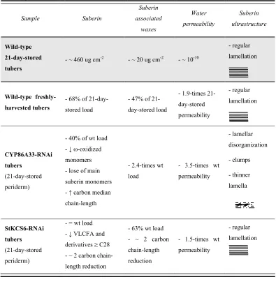
(13) 3– SUMMARY. SUMMARY The periderm, which protects the mature or secondary organs of plants, consists of the phellem or cork in its outer side. The phellem is a multilayered tissue of dead cells with suberized walls that affords protection against dehydration and pathogens. These suberized walls of phellem contain suberin and suberin associated waxes, both fractions responsible of the hydrophobic nature of periderm. Suberin is a fatty acid and glycerol-based polyester that contains phenolic compounds. The aliphatic compounds present in suberin are mainly long (C18) and very long (>C18) chain fatty acids (LCFA; VLCFA) with functionalized carboxylic or alcohol groups in their ω-terminal carbon. The suberin associated waxes are mainly derivatives of VLCFA. Thus, the ω-hydroxylation and elongation of fatty acids must be significant steps for the biosynthesis of suberin and wax compounds. To better understand the biosynthesis and function of suberin in periderm, in our laboratory we obtained a list of candidate genes for suberin biosynthesis by selecting those genes highly upregulated in phellem. In this thesis, the functional characterization of two of these genes, the putative ω-hydroxylase CYP86A33 and the putative ketoacyl-CoA synthase StKCS6, is performed by their RNA interference (RNAi)-mediated silencing in potato. In both cases, the silencing effects were analyzed at chemical, ultrastructural and physiological level. CYP86A33 is a putative ω-hydroxylase, candidate for the functionalization of the ω-terminal carbon of suberin aliphatic compounds. The CYP86A33 deficiency leads to a great reduction of the main suberin monomers, the C18:1 ω-hydroxyacid and α,ω-diacid, together with a smaller reduction of the rest of suberin compounds. All these reductions have an effect on the suberin total amount that decreases by 60%. In contrast to suberin, the wax content increases in more than 2-fold. Besides, in CYP86A33-RNAi periderms the lamellar ultrastucture of suberin is deeply distorted and the permeability to water increases. All these results demonstrate the significance of the ω-oxidized fatty acids for the deposition and assembly of suberin compounds as well as for the periderm water barrier function..
(14) 4 – FUNCTIONAL GENETICS OF SUBERIN BIOSYNTHESIS StKCS6 is a putative ketoacyl-CoA synthase, candidate for the elongation of VLCFA and derivatives of suberin and waxes of potato periderm. StKCS6 deficiency leads to a decrease of compounds with chain-length C28 and higher and an increase of those of chain-length C26 and lower. Even though this shift towards shorter chain lengths of C26 and lower in StKCS6silenced periderms, relevant changes in the relative amount of different types of compounds are not observed. Moreover, in these transgenic periderms structural defects, either in the suberin cell wall or in the cellular organization of this tissue, are not perceived. However, the StKCS6 deficiency impairs the water barrier function of periderm, suggesting that the fatty acid chainlength can contribute to the sealing properties of periderm. The results obtained in this thesis are discussed as a whole to highlight their contribution in the biosynthesis of suberin and wax compounds, in the suberin ultrastructure and in the properties of the periderm hydrophobic barrier..
(15) 5– SUMMARY. RESUM La peridermis, la qual protegeix els òrgans madurs o secundaris de les plantes, consta del fel·lema o suro a la seva part externa. El fel·lema és un teixit pluriestratificat de cèl·lules mortes amb parets cel·lulars suberificades que protegeix contra la deshidratació i els patògens. La suberina i les ceres associades que impregnen les parets suberificades del fel·lema són responsables de la naturalesa hidrofòbica del periderm. La suberina és un polièster d’àcids grassos i glicerol que conté compostos fenòlics. Els compostos alifàtics que formen la suberina són principalment àcids grassos de cadena llarga (C18) o molt llarga (> C18) que contenen grups carboxílics i alcohòlics funcionals a l’extrem ω-terminal. Les ceres associades a la suberina són bàsicament derivats d’àcids grassos de cadena molt llarga. Així, la ω-hidroxilació i l’elongació d’àcids grassos són dos processos claus per a la biosíntesi dels components de la suberina i de les ceres. Per tal de poder entendre millor la ruta biosintètica de la suberina i la seva funció en la peridermis, en el nostre laboratori varem obtenir gens candidats per la biosíntesi de la suberina mitjançant la selecció d’aquells gens que estaven sobreexpresats en el fel·lema. En aquesta tesi es presenta la caracterització de dos d’aquests gens, la hipotètica ω-hidroxilasa d’àcids grassos CYP86A33 i la hipotètica ketoacyl-CoA sintasa StKCS6, mitjançant el seu silenciament per RNA d’interferència (RNAi) en patata. En ambdós casos es van analitzar els efectes del silenciament a tres nivells: químic, estructural i fisiològic. CYP86A33 pel caràcter d’ω-hidroxilasa d’àcids grassos és candidat per la funcionalització del carboni terminal ω dels compostos alifàtics de la suberina. La deficiència de CYP86A33 comporta una gran reducció de la quantitat dels monòmers principals de la suberina, l’àcid gras ω-hidroxilat i l’α,ω-diàcid C18:1, juntament amb una reducció més petita de la resta de compostos de la suberina. Totes aquestes reduccions provoquen que la quantitat de suberina dipositada en les parets cel·lulars es vegi reduïda en un 60%. Pel que fa a les ceres, contràriament a la suberina, la seva quantitat es veu incrementada en més de dues vegades. En la peridermis de les plantes silenciades en CYP86A33 s’observa una greu alteració de la.
(16) 6 – FUNCTIONAL GENETICS OF SUBERIN BIOSYNTHESIS disposició lamel·lar de la suberina que va acompanyada d’un increment en la permeabilitat a l’aigua. Tots els resultats en conjunt demostren la rellevància dels àcids grassos ω-oxidats en la deposició i l’assemblatge de la suberina, així com també en la funció barrera de la peridermis. StKCS6 pel seu caràcter d’elongasa d’àcids grassos és candidata per síntesi dels àcids grassos o derivats de cadena molt llarga de la suberina i les ceres de la peridermis. La deficiència en StKCS6 comporta que els compostos de la peridermis de cadena igual o més llarga de 28 carbonis es redueixin i que els de cadena igual o més curta de 26 s’incrementin. A pesar d’aquest escurçament de cadenes, no s’aprecien canvis rellevants en la quantitat relativa dels diferents tipus de compostos. A més, tampoc s’observen canvis ultraestructurals de la paret de suberina ni en l’organització cel·lular de la peridermis. No obstant, la deficiència de StKCS6 danya la funció barrera de la peridermis, el que suggereix que la llargada de la cadena dels àcids grassos pot contribuir a les propietats impermeabilitzants de la peridermis. Els resultats obtinguts en aquesta tesi es discuteixen en conjunt per posar de relleu la seva contribució en la biosíntesi dels compostos de suberina i ceres, en la ultraestructura de la suberina i en les propietats de la barrera hidrofòbica de la peridermis..
(17) 7– SUMMARY. RESUMEN La peridermis, la cuál protege los órganos maduros (secundarios) de las plantas, contiene en su parte externa felema o corcho. El felema es un tejido pluriestratificado de células muertas con paredes celulares suberificadas que protege de la deshidratación y los patógenos. La suberina y las ceras asociadas que impregnan las paredes suberificadas del felema son responsables de la naturaleza hidrofóbica de la peridermis. La suberina es un poliéster de ácidos grasos y glicerol que contiene compuestos fenólicos. Los compuestos alifáticos que forman la suberina son principalmente ácidos grasos de cadena larga (C18) y muy larga (>C18) que contienen grupos carboxílicos o alcohólicos funcionales en su carbono ω-terminal. Las ceras asociadas a la suberina son básicamente derivados de ácidos grasos de cadena muy larga. Así, la ωhidroxilación y la elongación de ácidos grasos deberían ser procesos claves para la biosíntesis de los componentes de la suberina y de las ceras. Para poder entender mejor la ruta biosintética de la suberina y su función en la peridermis, en nuestro laboratorio obtuvimos una colección de genes candidatos para la biosíntesis de la suberina mediante la selección de aquellos genes sobreexpresados en el felema. En esta tesis se presenta la caracterización funcional de dos de éstos genes, la hipotética ω-hidroxilasa CYP86A33 y la hipotética ketoacyl-CoA sintasa StKCS6, mediante su silenciamiento por RNA de interferencia (RNAi) en patata. En ambos casos se analizaron los efectos del silenciamiento a tres niveles: químico, estructural y fisiológico. CYP86A33 por su carácter de ω-hidroxilasa de ácidos grasos es candidato para la funcionalización de los grupos ω de los compuestos alifáticos de la suberina. La deficiencia de CYP86A33 comporta una gran reducción de la cantidad de los monómeros principales de la suberina, el ácido graso ω-hidroxilado y el α,ω-diácido C18:1, acompañada de una reducción más pequeña en el resto de componentes de la suberina. Todas esas reducciones conllevan que la cantidad de suberina depositada en las paredes celulares se ve reducida en un 60%. En referencia a las ceras, contrariamente a la suberina, su cantidad se ve incrementada en más de dos veces. Además, en la peridermis de las plantas silenciadas se observa una gran alteración.
(18) 8 – FUNCTIONAL GENETICS OF SUBERIN BIOSYNTHESIS de la disposición lamelar de la suberina acompañada de un incremento en la permeabilidad al agua. Todos los resultados en conjunto demuestran la relevancia de los ácidos grasos ωoxidados en la deposición y ensamblaje de la suberina, así como también en la función barrera de la peridermis. StKCS6 por su carácter de elongasa de ácidos grasos es candidato para la síntesis de los ácidos grasos o derivados de cadena muy larga de la suberina y las ceras de la peridermis. La deficiencia en StKCS6 conlleva que los compuestos de la peridermis de cadena igual o más larga de 28 carbonos se reduzcan y que los de cadena igual o más corta de 26 carbonos se incrementen. A pesar de este acortamiento de cadenas, no se aprecian cambios relevantes en la cantidad relativa de los diferentes tipos de compuestos. Además, tampoco se observan cambios ultraestructurales en la pared de suberina ni en la organización celular de la peridermis. No obstante, la deficiencia de StKCS6 daña la función barrera de la peridermis lo que sugiere que la longitud de la cadena de los ácidos grasos puede contribuir a las propiedades impermeabilizantes de la peridermis. Los resultados obtenidos en esta tesis se discuten en conjunto para poner de manifiesto su contribución en la biosíntesis de los compuestos de suberina y ceras, en la ultraestructura de la suberina y en las propiedades de la barrera hidrofóbica de la peridermis..
(19) INTRODUCTION.
(20)
(21) 11 – INTRODUCTION. 1. Periderm, a plant lipophilic barrier One of the features that allowed plants to colonize the land was the development of lipophilic barriers, such as the cuticle and the periderm, to avoid water loss. The cuticle is a continuous non-cellular hydrophobic thin membrane (0.1-10 µm thick) deposited in the outermost cell walls of the epidermis of above-ground organs (Riederer and Schreiber, 2001). The periderm is found in subterraneous root organs (exodermis and tuber skin) and also develops to replace the cutinized epidermis when it is damaged by wounding or by secondary growth (like tree trunks in woody species) (Lendzian, 2006). The periderm is produced by a secondary meristematic layer called phellogen or cork cambium. Phellogen normally arises outside the vascular cambium in the subepidermal tissue of stems or in the pericycle of roots. It generates the phellem or cork centrifugally and the cork parenchyma or phelloderm (not always present) centripetally. All together, phellogen, phellem and phelloderm make up the periderm (Lendzian, 2006) (Figure 1).. Figure 1. Periderm structure. Scanning electron microscope micrograph showing potato tuber periderm made up of phellem (PM), phellogen (PG) and phelloderm (PD), which are regularly arranged in contrast with the parenchymatic cortical cells (C). The barrier properties of periderm are provided by the phellem multilayered tissue and specifically by their apoplastic lipid compounds: suberin and suberin associated waxes. As phellem cells develop, suberin and suberin associated waxes are deposited in their cell walls, become isolated from the water and nutrient supply and subsequently die, thereby forming the protective barrier (Sabba and Lulai, 2002). Suberin is also deposited in the endodermis of roots (Casparian bands), in the bundle sheath cells of grass leaves and in the hilum area (funiculus) of seeds. Although a true periderm is not developed in these tissues, the hydrophobic nature of suberin confers water barrier properties as well..
(22) 12 – FUNCTIONAL GENETICS OF SUBERIN BIOSYNTHESIS 1.1. Periderm aliphatic compounds: wax and suberin Most of chemical suberin studies have been focused in the phellem or cork of both cork oak (Quercus suber) bark and potato (Solanum tuberosum) tuber periderm, probably due to the large amount of suberin they contain. Suberin is an unknown biopolymer in many aspects. Although the monomeric chemical composition of suberin seems to be more clearly understood with the recent elucidation of glycerol as a key compound (Moire et al., 1999), very little is known about the enzymes involved in their biosynthesis. Besides, other features like monomer transport to the apoplast, monomer esterification, macromolecular organization and regulation of the whole suberization process remain to be elucidated. Due to the lack of information about suberin and taking into account that cutin shares similar features with suberin (as both are plant polyesters of a hydrophobic nature), cutin has often been helpful to better understand suberin. For this reason, multiple comparisons between the two plant polyesters will be made in this work. 1.1.1. Chemical composition of suberin Suberin consists of an aliphatic polyester esterified to some phenolics and cross-linked with an aromatic lignin-like domain bounded to cell wall carbohydrates (Bernards, 2002). The chemical composition of suberin was described after the breaking of ester linkages or transesterification (Kolattukudy and Agrawal, 1974; Franke et al., 2005; Graça and Santos, 2007). Generally, this depolymerization releases glycerol, hydroxycinnamic acids and a mixture of aliphatic compounds ranging from C16 to C32 with hydroxy and carboxylic acid functionalities (Graça and Santos, 2007). The aliphatic compounds of suberin are long and very long chain fatty acids (LCFA, VLCFAs) and derivatives such as α,ω-diacids, ω-hydroxyacids, primary alcohols and mid-chain-oxidized fatty acids. Although the same profile of α,ω-diacids, ω-hydroxyacids and glycerol seems to be prevalent in suberized tissues, suberin composition varies between plant species and tissues where it occurrs (Graça and Santos, 2007). Compared with potato suberin, the suberin from the cork oak bark is more abundant in saturated and mid-chain oxidized fatty acids and derivatives (Graça and Santos, 2007) and the suberin from Arabidopsis is shorter since it rarely contains VLCFAs and derivatives longer than C26 (Franke et al., 2005). In contrast to the chemical composition of suberin, cutin polyester rarely contains VLCFA and derivatives. Besides, the.
(23) 13 – INTRODUCTION typical α,ω-diacids of suberin are not usual cutin compounds, although they are found in Arabidopsis cutin (Franke et al., 2005). The aromatic compounds obtained after depolymerization are a mixture of hydroxycinnamic acids, especially ferulic acid, and derivatives. The aromatic fraction of suberin has been compared to lignin by some authors (Kolattukudy 1980, Bernards, 2002). Although both suberin and lignin contain monolignols, other hydroxycinnamic acids and derivatives such as feruloyltyramine have only been described in suberin (Bernards et al., 1995). 1.1.2. Chemical composition of waxes Waxes are present in the cuticle, in suberized tissue and in the pollen seed coat. Unlike the insoluble polyesters cutin and suberin, waxes are referred to as the “soluble lipid fraction” or “extractives” for their solubility in organic solvents like chloroform. Although waxes are characterized to be represented by VLCFA and derivatives, their chemical composition is highly variable depending on the organ and plant species analyzed. Waxes also include triterpenoids and minor secondary metabolites, such as sterols and flavonoids in the cuticles of Arabidopsis (Kunst and Samuels, 2003) and tomato fruit (Vogg et al., 2004) as well as in the periderm of cork oak bark (Castola et al., 2005). It is noteworthy that the VLCFA and derivatives composition of suberin associated waxes greatly differs from that of cutin associated waxes. Whereas in suberin associated waxes the main compounds are free fatty acids, primary alcohols, alkanes and alkyl esters of ferulic acid (ferulic acid esters), in cutin associated waxes the major compounds include alkanes and ketones. Cutin associated waxes also contain alkyl esters but they are obtained by the reaction of very long chain alcohols with fatty acids (Lai et al., 2007), not with ferulic acid. Recently, monoacylglycerols have been reported as minor compounds in suberin associated waxes in roots, but not in cutinized tissues (Li et al., 2007b). 1.1.3. Biosynthesis of wax and suberin aliphatic compounds The wax and suberin aliphatic monomers are basically fatty acids and their derivatives. The ubiquitous C16 and C18 (also C18:1) fatty acids synthesized by the plastid enzyme machinery act as precursors for the rest of the compounds. After being exported from the plastids, C16 and C18 fatty acids are supposed to be elongated and modified by endoplasmic reticulum (ER).
(24) 14 – FUNCTIONAL GENETICS OF SUBERIN BIOSYNTHESIS enzymes to produce all the wax and suberin aliphatic compounds. It is important to know that these fatty acid precursors are also used to synthesize the cutin polyester, the membrane glycerolipids and the storage lipids (triacylglycerols). It has recently been shown that a glycerol-phosphate acyltransferase 5 (GPAT5) is a key enzyme for suberin deposition in the cell walls (Beisson et al., 2007; Li et al., 2007a). However, the subcellular localization and the molecular mechanism of GPAT5 remain unknown. Fatty acid elongation Fatty acid elongation is required to produce the VLCFA and derivatives compounds of suberin and suberin associated waxes. The C16 and C18 fatty acids (LCFA) synthesized in the plastids bear an acyl carrier protein (ACP). Prior to the elongation of these precursors to VLCFA, three steps are required (Schnurr et al., 2004): the release of the ACP by an acyl-ACP thioesterase (like FATB; Bonaventure et al., 2003), the activation to a CoA-thioester by a long-chain acylCoA synthetase (LACS) and the transfer to the ER. The elongation of LCFA to VLCFA wax and suberin precursors is carried out by an ER membrane-bound fatty acid elongase (FAE) complex (Figure 2). The FAE sequentially adds two carbon moieties to the growing acyl chain through a four-step reaction series catalyzed by four distinct enzymes: condensation of malonyl-CoA with the long-chain acyl-CoA, reduction to β-hydroxyacyl-CoA, dehydratation to enoyl-elongated acyl-CoA and a second reduction of the enoyl-CoA (Samuels et al., 2008). The condensing reaction is carried out by the 3-ketoacyl-CoA synthase (KCS), which is the rate-limiting step (Cassagne et al., 1994; Suneja et al., 1991) and the most sensitive to substrate chain length (Todd et al., 1999). It is thought that multiple individual elongase systems are required to elongate a fatty acid from C18 to C32 (Samuels et al., 2008). Several studies have been carried out regarding plant KCS involved in the biosynthesis of cutin associated waxes such as KCS6/CER6/CUT1 (Millar et al., 1999; Jenks et al., 1995), KCS1 (Todd et al., 1999) and FDH (Lolle et al., 1997) from Arabidopsis; LsCER6 from tomato (Lycopersicum esculentum) (Vogg et al., 2004; Leide et al., 2007) and GhCER6 from cotton (Gossypium hirsutum) (Qin et al., 2007). Moreover, a wide-study expressing known and putative Arabidopsis KCSs in yeast has shown the variable saturated and unsaturated fatty acid product range of each KCS (Trenkamp et al., 2004), which could explain the relative high number of KCSs in Arabidopsis (21). Despite identifying all these KCSs for the biosynthesis of cutin associated waxes, until now only KCS1 (Todd et al., 1999) and very recently KCS2 (Franke et.
(25) 15 – INTRODUCTION al. 2008) have been involved in elongating suberin monomers in Arabidopsis. In this thesis (Chapter 2), we report on the phenotypic effects that StKCS6 deficiency causes to the aliphatic compounds of suberin and their associated waxes in potato tuber periderm. Once VLCFAs are produced, they will be further modified to obtain the fatty acid derivatives of suberin and/or suberin associated waxes. It is expected that alcohols and alkyl esters are produced via an acyl-reduction pathway, whereas alkanes are produced via a decarbonylation pathway (Kunst and Samuels, 2003). In contrast with suberin associated waxes, in the wax fraction of cuticle further modifications in alkanes are needed to yield the ketones and secondary alcohols. Suberin ω-hydroxyacids and the corresponding α,ω-diacids are produced via a ω-oxidation pathway.. Figure 2. Fatty acid elongation and ωoxidation steps to yield suberin compounds. The fatty acid is elongated by the FAE complex in which the KCS is the enzyme that condenses the malonyl-CoA with the growing fatty acid. Cytochromes P450 are the enzymes that catalyze the oxidation of the ω-terminal group of the fatty acids. This is a part of the figure extracted from the proposed model for the suberin biosynthetic pathway (Franke and Schreiber, 2007).. ω-Oxidation of fatty acids ω-Hydroxyacids and α,ω-diacids, together with glycerol, are the major monomers released after suberin polyester transesterification. Their synthesis was demonstrated years ago in potato tuber disks, in which radiolabeled oleic acid (C18:1) was incorporated into ω-hydroxyoleic acid and the corresponding diacid (Dean and Kolattukudy, 1977). The oxidation of the fatty acid in the ω-position (terminal position) is catalyzed by NADPH-dependent cytochromes P450 monooxygenases (Figure 2), especially from the CYP86, CYP94 and CYP92 subfamilies (Benveniste et al., 1998; Kandel et al., 2006; Le Bouquin et al., 2001). Although the conversion of ω-hydroxyacid to the corresponding α,ω-diacid normally involves a two-reaction.
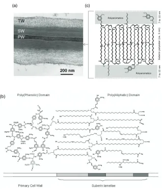
(26) 16 – FUNCTIONAL GENETICS OF SUBERIN BIOSYNTHESIS step catalyzed by ω-hydroxyacid and ω-oxoacid dehydrogenases (Kolattukudy, 2001), the tobacco CYP94A5 and the Arabidopsis CYP94C1 have been shown to directly catalyze the oxidation of fatty acids to the corresponding α,ω-diacid (Kandel et al., 2007; LeBouquin et al., 2001). In this thesis, the effects of CYP86A33 downregulation on the suberin and periderm function are presented in Chapter 1. 1.1.4. Macromolecular assembly of suberin compounds Suberin is deposited in the secondary wall of suberized cells. Kolattukudy (1980), based on the chemical depolymerization of suberin and the resemblances between suberin and cutin and lignin, proposed a model where the aliphatic compounds were structured in a matrix like cutin and linked to the phenolic domain, which was polymerized by oxidative coupling like lignin. This phenolic domain would covalently link the aliphatic suberin structure to the carbohydrates of the cell wall. However, this model did not elucidate the lamellation (alternated opaque and translucent lamellae) of suberin observed by a transmission electron microscope (TEM) (Figure 3a). Studies using the cotton green-lint mutant (Lg), whose fibres are suberized with a large proportion of waxes, helped to understand the suberin macromolecular structure. On one hand, treatment with a VLCFA synthesis inhibitor showed shorter electron translucent lamellae suggesting that the aliphatic compounds are “stacked” perpendicular to the lamellae plane (Schmutz et al., 1996). On the other, the treatment with an aromatic compound synthesis inhibitor was associated with the disappearance of electron opaque lamellae, which were replaced by empty spaces, demonstrating that the polyaromatics are relevant for the formation of these lamellae (Schmutz et al., 1993). Moreover, finding glycerol in suberin (Moire et al.,1999; Schmutz et al., 1993) and the studies based on partial suberin depolymerization (Graça and Santos, 2006a; Graça and Santos, 2006b; Graça and Pereira, 2000) have also shed light on the poorly understood macromolecular structure of suberin. Bernards (2002) proposed a model in which aliphatic monomers, corresponding to the translucent lamellae, were regularly oriented perpendicular to their lamellae plane and esterified with aromatic compounds and glycerol located in the opaque lamellae (Figure 3b). Glycerol, as a key compound, would provide the alcohol groups for ester linkages, permitting the growth of the polyester in a three dimensional network. This regular opaque and translucent lamellae.
(27) 17 – INTRODUCTION. Figure 3. Suberin macromolecular structure under TEM and proposed models for suberin macromolecular assembly. (a) Suberized cell wall from two cells of potato tuber phellem separated by the middle lamella (not seen) of the polysaccharide primary wall (PW). The polysaccharide tertiary wall (TW) is also observed. The suberized secondary wall (SW) shows the typical opaque and translucent lamella alternating structure. (b) Tentative model proposed by Bernards (2002) for the assembly of suberin compounds in a macromolecular structure. The polyphenolic domain is shown restricted to the primary cell wall and covalently attached to carbohydrate units. The polyaliphatic domain is represented by a glycerol-based polyester. In the model, the predominantly aliphatic zones would yield the translucent bands observed in TEM micrographs, while the phenolic rich zones would yield the opaque bands (C: carbohydrate; P: phenolic; S: suberin). (c) Model proposed by Graça and Santos (2007) to explain the suberin lamellae structure seen under TEM. They proposed that glycerol and α,ω-diacids compounds constituted the framework of the suberin assembly..
(28) 18 – FUNCTIONAL GENETICS OF SUBERIN BIOSYNTHESIS structure was named the polyaliphatic domain despite the presence of some aromatic monomers. The polyphenolic domain was formed exclusively by aromatic compounds which were suggested to link the polyaliphatic domain to the cell wall carbohydrates. Graça and Santos (2007), taking into account all their results from suberin partial depolymerizations, suggested a model in which the basis of the suberin macromolecular structure was constituted by α,ω-diacids anchored at both sides to glycerol (Figure 3c). Ferulic acid would assure the linkage between the polyaromatics (opaque lamellae) and aliphatics (translucent lamellae). The several lamellae of aliphatic polyester would be connected by ω-hydroxyacids, crossing through the polyaromatic lamellae. In the translucent lamellae, intra and intermolecular bounds were postulated between the mid-chain-oxidized monomers. However, despite the efforts to understand the molecular structure of suberin, conclusive evidence linking the suberin chemical composition and ultrastructure is lacking. 1.2. Water barrier function of potato tuber periderm The cork tissue of periderm contains aerenchymatous cork areas called lenticels, which are paths for the “regulated” water vapour and the oxygen and carbon dioxide exchange between inside and outside. In fact, lenticels are formed in the periderm by the divisions of cells apparently continuous with the phellogen, located below the stomata of the original epidermis replaced by the periderm (Tyner et al., 1997). In contrast, the unregulated water loss goes through the cork layer of the periderm, thus determining the state of the periderm water barrier function (Lendzian et al., 2007). 1.2.1. Water permeability measures The use of well-defined enzymatically isolated periderms is a good strategy to measure the transfer of molecules via the periderm controlling the presence/absence of lenticels (Schreiber et al., 2005; Vogt et al., 1983) in a way that is similar to the one widely used for cuticles (Riederer and Schreiber, 2001). The treatment of periderms with fungal cellulases and pectinases leads to the isolation of the heavily suberized cells that are forming the phellem. The suberin deposited in the cells protects the polysaccharide cell wall from the hydrolytic enzymes without changing the structure of the phellem (Vogt et al., 1983). Therefore, in a strict sense, the term “isolated periderm” or “periderm membranes” should be replaced by “isolated phellem” as phellogen and phelloderm become digested. However, the term periderm.
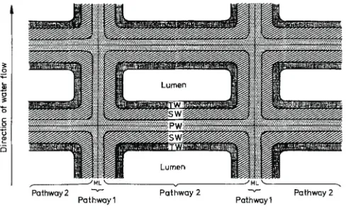
(29) 19 – INTRODUCTION is still being used as it has been done in the past by different authors (Schreiber et al., 2005; Stark et al., 1994; Vogt et al., 1983). The water permeability of the potato tuber periderm is determined by gravimetric methods using stainless steel or brass transpiration chambers of a specific diameter. The isolated periderms are mounted on these water-filled chambers with the physiological inner side of the periderm facing the inner side of the chamber (Figure 4). The permeability values are calculated based on the loss of the chamber weight caused by the water evaporated across the periderm.. Figure 4. Diagram of the longitudinal section of the typical transpiration chamber used to measure the water permeability across isolated cuticles or periderms by gravimetric methods. (a) Periderm mounted on the transpiration chamber filled by water. (b) Once mounted, the transpiration chambers were turned upside down to ensure the direct contact between the water and the periderm, kept in closed boxes containing dry silica gel and stored at a constant temperature. The water is forced to pass through the periderm by evaporation to the silica gel.. Using modified transpiration chambers and tritiated water, it was possible to describe two pathways for water transport across the periderm (Figure 5) (Vogt et al., 1983). Pathway 1 is characterized to have fast water diffusion and low permeability values and could be represented by middle lamellae and a primary wall orientated in a radial direction parallel to the direction of water flow. Pathway 2 is characterized to have slow water diffusion and high permeability and could be represented by the cell walls formed by middle lamellae, the primary wall, the suberized wall and the tertiary wall together with the wax fraction, orientated perpendicular to the water flow (Vogt et al., 1983). Using the gravimetric method, which takes measurements at day intervals, it is not possible to distinguish the water diffusion through.
(30) 20 – FUNCTIONAL GENETICS OF SUBERIN BIOSYNTHESIS pathways 1 and 2, and therefore the measured permeability represents the total permeability of both pathways (Schreiber et al., 2005).. Figure 5. Schematic drawing of a cross-section of a periderm membrane indicating the two different pathways of water movement. The drawing is not to scale. Extracted from Vogt et al., (1983).. 1.2.2. Role of suberin and waxes in the water barrier function Periderm membranes consist of two components: i) the extractable or organic solvent-soluble fraction known as wax and ii) the non-extractable or insoluble material formed by suberin and polysaccharides (Lendzian, 2006). Until now, the wax and suberin contribution to maintaining the water barrier properties of potato tuber periderm has seemed ambiguous and very biased depending on the potato variety studied (Lendzian, 2006). Likewise, the role of the chainlength profile and the substance class distribution of the aliphatic periderm compounds, as well as their physical assembly, have not yet been determined. Waxes have been proposed as space fillers, supporting the three-dimensional network of suberin based on the shrinkage produced in periderms when this wax fraction is removed. However, more evidence is needed because in potato periderm, where the wax fraction is very small, the shrinkage is still very high (Lendzian, 2006). Waxes have also been identified as key compounds in maintaining the periderm water barrier function. After wax extraction, the water permeability of tuber periderm membranes increases by a factor of 100 (Schreiber et al., 2005; Vogt et al., 1983), although this increased permeability was not observed in all the potato varieties (Lendzian, 2006)..
(31) 21 – INTRODUCTION Besides, the importance of wax in potato tuber periderm has been demonstrated using trichloroacetate as an inhibitor of fatty acid chain elongation. After treatment, the periderm experienced a great decrease of wax compounds and an inhibition of the development of diffusion resistance of the tissue to water vapour (Soliday et al., 1979). Regarding the insoluble material formed by suberin and polysaccharides, as far as we know, no one has provided any direct evidence correlating these compounds to the periderm water barrier function. In potato, the immature/mature periderm is defined based on its susceptibility to the excoriation of the skin (skinning) (Lulai and Orr, 1993). During the storage period, the periderms become resistant to excoriation, and reach the mature stage when the transpiration rate is minimum and constant (Schreiber et al., 2005). At this stage of maturity, measures of periderm water permeability were around 10-10 m s-1 (Schreiber et al., 2005; Vogt et al., 1983). This decreased water permeability at maturity is attributed to the huge amount of suberin and waxes deposited in the cell wall during this storage period, especially during the first days of storage (Schreiber et al., 2005). However the deposition of suberin and waxes cannot by itself explain the 100-times higher permeability seen in wound periderms, which amounts to 50-60% of the total suberin and wax load of native periderm (Schreiber et al., 2005). Other factors, such as the physical assembly of all periderm components, might therefore contribute to the periderm water barrier properties.. 2. Molecular tool: RNA interference-mediated silencing 2.1. The mechanism of gene silencing Gene silencing was initially perceived as an unpredictable and inconvenient side effect of introducing transgenes into plants (Napoli et al., 1990; Van der Krol et al., 1990). Since the discovery of the gene silencing phenomenon in 1998 (Fire and Mello 1998), silencing pathways have been characterized. At present it is widely accepted that many eukaryotes benefit from a highly abundant class of single-stranded small RNAs (20-30 bp) that trigger gene silencing through RNA-RNA and possibly also RNA-DNA interactions, hence called silencing RNAs (sRNAs). These sRNAs participate in a wide range of processes such as transcriptional silencing of heterochromatin, post-transcriptional regulation of mRNA and silencing of invading viral genomes or transgenes (Brodersen and Voinnet, 2006; Lippman and.
(32) 22 – FUNCTIONAL GENETICS OF SUBERIN BIOSYNTHESIS Martienssen, 2004; Vaucheret, 2006). sRNAs are derived from the RNA cleavage (Dicer family) of longer, partially or fully double-stranded RNA (dsRNA) molecules, produced by inter or intra-molecular interaction. There are two types of sRNA: i) micro RNAs (miRNAs) produced by longer single-stranded imperfect fold-back mRNAs which mediate posttranscriptional control of endogenous transcripts and ii) short-interfering RNAs (siRNAs) produced by longer double-stranded perfectly complementary RNAs. The siRNAs come from viral replication or through the action of RNA-dependent RNA polymerase on single-stranded plant RNA. They mediate the post-transcriptional regulation of endogenous/exogenous (including viral and transgene) transcripts and transcriptional silencing of transposable elements (Ossowski et al., 2008). 2.2. RNA interference mediated silencing for functional genetics. From a practical perspective, complementary long RNA able to produce double-stranded RNA was reported to efficiently silence an endogenous loci and a transgene in tobacco (Waterhouse et al., 1998). Since then, gene silencing has been used as a specific and powerful technique to knock down the expression of target genes and subsequently evaluate their physiological role in plants. Now we know that the classical use of long antisense RNAs, resulting in the downregulation of the complementary mRNA is mediated by sRNAs as well as the unexpected silencing in which a sense transgene overexpression results in the downregulation of the transgene and the homologous endogenous gene (sense PTGS) (Ossowski et al., 2008). A more recently developed and most efficient silencing method in plants is the use of the inverted repeat (ir) post-transcriptional gene silencing (PTGS), also called the RNA interference (RNAi) or hairpin RNAi (hpRNAi) approach (Watson et al., 2005). The hpRNAi, controlled by a promoter and a terminator, consists of two transgene sequences oriented inversely and separated by a spacer region, normally a short intron. In planta, the transcription of this hpRNAi leads to a dsRNA which is processed by Dicer into heterogeneous siRNAs. One strand of the siRNAs associates itself with Argonaute (AGO), the catalytic subunit of RNAinduced silencing complexes (RISCs). AGO binds by complementarity both small and longer endogenous mRNA and cuts the target transcript in the corresponding position 10/11 of the sRNA. Due to the heterogeneity of the siRNAs, the AGO-dependent cleavage is found in many sites of the target transcript. The resulting fragments of the cleavage are degraded or used as primers for the RNA-dependent RNA polymerase to synthesize new dsRNAs which will be.
(33) 23 – INTRODUCTION processed as the hpRNAi, hence the amplification of the process leading to strong RNAimediated silencing (Ossowski et al., 2008). One of the advantages of the RNAi-mediated silencing is that the hpRNAi construct is genetically dominant and therefore the silencing effects can be screened in the same plant or first progeny without needing to produce homozygous lines (Helliwell and Waterhouse, 2003). Thus, RNAi-mediated silencing has been identified as the appropriate strategy for functional genetic approaches in non-model polyploid plants with restricted capacity for sexual reproduction such as the potato..
(34)
(35) OBJECTIVES.
(36)
(37) 27 – OBJECTIVES. The research in our laboratory is focused on understanding the molecular processes that govern suberized tissues. Despite the importance of suberized tissues for land plants, an easily detected phenotype for suberin deficiency has not been reported, indicating that forward genetic approaches are inappropriate to identify genes involved in the suberization process. A more suitable strategy would be to identify genes putatively involved in suberized tissues and, afterwards, confirm their real role using a reverse genetic approach. Specifically, we followed a two-step strategy: i) the isolation of candidate genes for suberin biosynthesis through suppression subtractive hybridization (SSH) libraries (presented as background studies in this section) and ii) the development of a reverse genetic approach in potato to downregulate these target genes and subsequently analyze the effects in the chemical composition, structure and water barrier function of the tuber periderm (presented in the Chapters 1 and 2). 1. Background studies: identification of candidate genes of suberin biosynthesis The periderms of cork oak (Quercus suber) bark and potato (Solanum tuberosum) tuber skin are paradigms of suberized tissues due to the large amount of suberin that their cork or phellem cells synthesize. These periderms were used to underscore genes involved in the suberin biosynthesis and periderm formation by constructing two SSH libraries (Diatchenko et al., 1996). The SSH technique was used to obtain expressed sequence tags (ESTs) in the periderm (tester) but not in the control tissue (reference or driver) and to enrich ESTs of rare transcripts. The first SSH library was constructed using the cork bark of field-grown cork oak and the proliferative mass of in vitro-grown cork oak embryo as tester and driver tissues respectively. To distinguish the candidates of periderm among those involved in the suberin aliphatic or aromatic fraction, the library was printed on a microarray and cohybridized with cork and wood (lignified tissue) cDNA (Soler et al., 2007). At the same time, a second SSH library containing ESTs preferentially expressed in potato tuber skin (or periderm) was constructed, using as driver tissue the parenchymatous tissue of potato tubers..
(38) 28 – FUNCTIONAL GENETICS OF SUBERIN BIOSYNTHESIS 2. Functional genetics of StKCS6 and CYP86A33 by RNA interference-mediated silencing in potato tubers Potato tuber periderm, a model to study suberin biosynthesis and function Arabidopsis is the model plant for molecular studies in dicotyledonous because it is a relatively short life-cycle plant with fully-sequenced genome and large molecular resources available. It was recently identified as a valuable model for suberin functional studies due to the development of methods to detect and analyze suberin in roots (Franke et al., 2005). To date, however, the lack of a well-developed direct methodology to measure the efficiency of the suberin barrier in Arabidopsis for water transport is not available, probably due to the smallness of Arabidopsis roots (Schreiber et al., 2005) and the complex devices needed. Moreover, the study of periderm features cannot be approached in Arabidopsis because it is difficult to find stem periderm areas free of cuticle and root periderm areas free of endodermis. In contrast, the methodology to study the water barrier transport of potato tuber periderm has been fully developed (Schreiber et al., 2005; Vogt et al., 1983). The potato, although it does not have the same molecular tools as Arabidopsis, is an outstanding model for functional genetic studies of suberin biosynthesis and function, since sufficient amounts of tuber periderm can be easily obtained for chemical, ultrastructural and physiological studies. Finally, it is also significant that potato is an important crop plant whose tubers are of great economic interest. The potato, like many crop plants, is polyploid (The NSF Potato Genome Project, http://www.potatogenome.org/nsf5/potato_biology/). The use of mutagenesis is therefore not suitable because polyploidy hinders the obtaining of homozygous progeny. On the contrary, RNA interference (RNAi)-mediated silencing technology has allowed stable knockdown of target genes in traditionally non-model plants such as the potato. These plants are of more agronomic interest and more appropriate to study certain processes, such as periderm suberification. CYP86A33 and StKCS6 genes, candidates for suberin biosynthesis The hydrophobic nature of suberin is attributed to the aliphatic domain. The main monomers released after suberin depolymerization are long chain and very long chain fatty acids (LCFA; VLCFAs), some of them functionalized in the ω or terminal carbon. Thus, pivotal reactions to.
(39) 29 – OBJECTIVES yield these main suberin compounds must be the fatty acid ω-hydroxylation catalyzed by cytochrome P450 monooxygenases and the elongation of LCFA catalyzed by the ER membrane-bound fatty acid elongase complex. This elongation pathway is also crucial for the biosynthesis of suberin associated waxes, as these waxes are mainly composed of VLCFAs and derivatives. Taking into account the importance of ω-oxidation and elongation steps in suberin biosynthesis, the specific goal of the present work is to use functional genetic approaches to study genes suspected of being involved in these enzymatic reactions in potato. With this purpose in mind, two genes encoding a putative cork oak fatty acid ω-hydroxylase member of the CYP86A subfamily (CYP86A32 from now on) and a putative potato ketoacyl-CoA synthase (StKCS6 from now on) were selected from the cork oak and potato SSH libraries respectively. CYP86A32 was strongly up-regulated in cork versus wood (Soler et al., 2007) and its expression pattern during the cork growing season was consistent with a function for suberin biosynthesis (Soler et al., 2008). The transcript profiles in potato tissues of the putative potato orthologous gene of CYP86A32 (CYP86A33 from now on) and the StKCS6 gene were consistent with suberin biosynthesis and these genes were consequently characterized by a reverse genetic approach. To analyse the CYP86A33 and StKCS6 gene function in suberin and periderm, RNAi-mediated silencing technology was used. The two selected genes were respectively downregulated in potato tuber periderm and the resulting phenotype was analyzed in three independent silenced lines. A diagram showing the different steps of the proposed strategy is shown below:.
(40) 30 – FUNCTIONAL GENETICS OF SUBERIN BIOSYNTHESIS Specific objectives involving CYP86A33 and StKCS6 The main objective of this thesis is to better understand the function of ω-oxidation pathway in periderm through CYP86A33 gene deficiency, and VLCFA elongation pathway through StKCS6 gene deficiency, with emphasis on their role in periderm aliphatic compound biosynthesis, ultrastructure and water barrier function. The specific objectives are: 1. Study the role of CYP86A33 in suberin (Chapter 1). 1.1.. Analyze the expression pattern of CYP86A33.. 1.2.. Isolate the CYP86A33 full-length coding sequence.. 1.3.. Demonstrate by chemical analysis that CYP86A33 is an enzyme involved in suberin biosynthesis.. 1.4.. Analyze the function of CYP86A33 in suberin macromolecular structure by transmission electron microscopy (TEM).. 1.5.. Study the effects of CYP86A33 downregulation on periderm water barrier by gravimetric methods.. 2. Study the role of StKCS6 in potato periderm (Chapter 2). 2.1.. Analyze the expression pattern of StKCS6.. 2.2.. Isolate the StKCS6 full-length coding sequence.. 2.3.. Demonstrate that StKCS6 is involved in the biosynthesis of VLCFA and derivatives of suberin and wax by chemical analyses.. 2.4.. Analyze the function of StKCS6 in the suberin macromolecular structure by TEM.. 2.5.. Study the effects of StKCS6 downregulation on the periderm water barrier by gravimetric methods.. 3. Discuss the significance of our finding in relation to the global knowledge of suberin biosynthesis and periderm function..
(41) CHAPTER 1 CYP86A33 targeted gene silencing in potato tuber alters suberin composition, distorts suberin lamellae and impairs the periderm’s water barrier function Manuscript submitted to Plant Physiology, July 2008..
(42)
(43) 33 – CYP86A33 DEFICIENCY IMPAIRS PERIDERM FUNCTION. CYP86A33 targeted gene silencing in potato tuber alters suberin composition, distorts suberin lamellae and impairs the periderm’s water barrier function.. ABSTRACT Suberin is a cell wall lipid-polyester found in the cork cells of the periderm offering protection against dehydration and pathogens. Its biosynthesis and assembly, as well as its contribution to the sealing properties of the periderm, are still poorly understood. Here, we report on the isolation of the coding sequence CYP86A33 and the molecular and physiological function of this gene in potato (Solanum tuberosum) tuber periderm. CYP86A33 was downregulated in potato plants by RNA interference (RNAi)-mediated silencing. Periderm from CYP86A33 silenced plants revealed a significant decrease in its suberin load (~60%) and greatly reduced levels of C18:1 ω-hydroxyacid (~70%) and α,ω-diacid (~90%) in comparison with wild-type. The typical regular ultrastructure of suberin, consisting of dark and light lamellae, disappeared and the thickness of the suberin layer was clearly reduced. In addition, the water permeability of the periderm isolated from CYP86A33 silenced lines was 3.5-times higher than that of the wild-type. Thus, our data provide convincing evidence for the involvement of CYP86A33 in establishing suberin structure and function..
(44) CHAPTER 2 Silencing of StKCS6 in potato periderm leads to reduced chain lengths of suberin and wax compounds and increased peridermal transpiration Manuscript submitted to Journal of Experimental Botany, August 2008.
(45)
(46) 59 – StKCS6 DEFICIENCY IMPAIRS VLCFAS ELONGATION IN PERIDERM. Silencing of StKCS6 in potato periderm leads to reduced chain lengths of suberin and wax compounds and increased peridermal transpiration. ABSTRACT Very long chain aliphatic compounds occur in the suberin polymer and associated wax. Up to now only few genes involved in suberin biosynthesis have been identified. Here we report on the isolation of a potato (Solanum tuberosum) 3-ketoacyl-CoA synthase (KCS) gene and the study of its molecular and physiological relevance by means of a reverse genetic approach. This gene, called StKCS6, was stably silenced by RNA interference (RNAi) in potato. Analysis of the chemical composition of silenced potato tuber periderms indicated that StKCS6 down-regulation has a significant and fairly specific effect on the chain length distribution of very long-chain fatty acids (VLCFAs) and derivatives, occurring in the suberin polymer and peridermal wax. All compounds with chain lengths of C28 and higher were significantly reduced in silenced periderms, whereas compounds with chain lengths of C26 and lower accumulated. Thus, StKCS6 is preferentially involved in the formation of suberin and wax lipidic monomers with chain lengths of C28 and higher. As a result, peridermal transpiration of the silenced lines was about 1.5-times higher than that of wild-type. Our results convincingly show that StKCS6 is involved in both suberin and wax biosynthesis and that a reduction of the monomeric carbon chain-lengths leads to increased rates of peridermal transpiration..
(47) DISCUSSION.
(48)
(49) 83– DISCUSSION. The main function of periderm suberin and their associated waxes is to protect plant secondary (mature) organs against dehydration (Bernards, 2002). However the suberin/wax chemical and physical properties that confer this physiological role to periderm remain unclear. Moreover, no direct relationship has been established between the composition, ultrastructure and sealing properties of suberin. The biosynthetic pathways proposed for suberin and wax compounds have been hypothesized on the basis of suberin and wax chemical composition (Bernards, 2002; Franke et al., 2005; Kolattukudy, 2001). However, most steps of these pathways remain unconfirmed, probably due to technical difficulties inherent to the nature of suberized tissues. To shed some light on suberin biosynthetic pathways, in this thesis we report on the study of two genes involved in the two major steps of suberin monomer biosynthesis: fatty acid ω-oxidation (CYP86A33) and elongation of long chain fatty acids (LCFA) (StKCS6). Our approach aims to characterize these genes by RNA interference (RNAi)-mediated silencing in potato tuber periderm. CYP86A33 and StKCS6 were selected based on their identification in two suppression subtractive hybridization (SSH) periderm libraries. The cork oak CYP86A32, the putative ortholog of potato CYP86A33 and Arabidopsis CYP86A1, was isolated from the cork oak bark SSH library (Soler et al., 2007) and the potato StKCS6 from the potato tuber SSH library. In this thesis, we demonstrate that CYP86A33 is responsible for the biosynthesis of the ω-oxidized suberin major compounds and StKCS6 for the biosynthesis of the longest very long chain fatty acids (VLCFAs) and derivatives in potato tuber periderm. Moreover, it has been demonstrated that CYP86A33 is fundamental in organizing the suberin lamellar ultrastructure and both CYP86A33 and StKCS6 are critical to maintaining the water barrier function afforded by suberin polyester and their associated waxes. Therefore, using potato periderm as a model has made it possible to directly relate changes in suberin and wax chemical composition with the suberin ultrastructure and the water barrier function of periderm. Very recently and during the course of research done in this thesis, other authors have approached the molecular genetics of suberin biosynthesis with a strategy based on the analysis of Arabidopsis mutants for suberin candidate genes. This alternative approach has revealed that four Arabidopsis genes encode suberin related-enzymes: CYP86A1 (At5g58860), encoding a fatty acid ω-hydroxylase (Höfer et al., 2008; Li et al., 2007a); GPAT5 (At3g11325), encoding.
(50) 84 – FUNCTIONAL GENETICS OF SUBERIN BIOSYNTHESIS a glycerol-phosphate acyltransferase (Beisson et al., 2007); and KCS1 (At1g01120) (Todd et al., 1999) and KCS2 (At1g04220) (Franke et al., 2008) encoding ketoacyl-CoA synthases. All together, our results in potato and the results obtained in Arabidopsis represent a major advance in the knowledge of the biosynthesis and assembly of suberin. Moreover, our results are also related with the physiological role of periderm. The following sections are focussed on how our work and that of the other authors have contributed to a better understanding of the chemical and physiological properties of suberin.. 1. Suberin biosynthesis The phenotypic effects in CYP86A33 and StKCS6 deficient periderms are consistent with the role expected for these enzymes in the ω-oxidation and elongation pathways of suberin aliphatic precursors previously suggested (Bernards, 2002; Franke et al., 2005; Kolattukudy, 1981). Figure 1 summarizes the biosynthesis of the suberin and suberin associated waxes showing the reactions that the potato and Arabidopsis suberin related-enzymes are probably catalyzing..
(51) 85– DISCUSSION 1.1. VLCFAs elongation pathway The downregulation of StKCS6 reports results in periderm consistent with those seen for cuticle waxes in the mutants of its putative orthologs in Arabidopsis (Millar et al., 1999; Jenks et al., 1996) and tomato (Leide et al., 2007; Vogg et al., 2004). In contrast to AtKCS6/CER6/CUT1, which was designated as wax-specific KCS (Samuels et al., 2008), our results demonstrate that StKCS6 is involved in the synthesis of very long chain linear compounds of suberin and suberin associated waxes. The deficiency in StKCS6 affects the elongation of C26 VLCFAs and derivatives to longer homologous compounds, which correspond to 15% and 65% of suberin and wax monomers of wild-type tuber periderm respectively. Despite the reduction in the longest VLCFAs and derivatives, the total load of each type of compound fraction was not greatly affected due to the simultaneous accumulation of homologous series of shorter chained compounds. This increase in shorter chain wax compounds, putatively synthesized by the acyl-reduction pathway (primary alcohols and ferulic acid esters), suggests that the enzymes of this pathway are not rate-limiting; hence shorter fatty acids can be transformed to primary alcohols and subsequently to ferulic acid esters by the acyl-reduction pathway (Figure 1). Moreover, the fact that StKCS6 deficiency causes the same effect in the chain-length profile in both suberin and wax fractions indicates that suberin and wax compounds could share the elongation pathway. However, it is not known whether wax biosynthesis is independent of that of suberin, or that waxes might be suberin precursors that have not been polymerized or might be derived by some suberin post-. Figure 1. General biosynthetic scheme for the aliphatic suberin and suberin associated wax compounds. The biosynthetic pathways leading to the fatty acids and derivatives which will be incorporated into the suberin and suberin associated waxes are based on previous publications and on cutin wax biosynthesis. The layout shows the direction of the main pathways, but only the enzymes known to be involved in suberin are depicted: StKCS6, KCS1, KCS2, CYP86A33, CYP86A1 and GPAT5. Fatty acid precursors are exported from the plastids to the endoplasmic reticulum to be modified. The elongation process controlled by KCSs yields the VLCFA precursors. It has been suggested that KCS1 and KCS2 act on fatty acids below C26 and StKCS6 on C26 VLCFAs. These elongated fatty acids, as well as the C18:0 and C18:1 fatty acids, can be ω-oxidized by NADPH-dependent cytochrome P450 monooxygenases, such as CYP86A33 and CYP86A1, to yield the ω-hydroxyacid and subsequently the α,ω-diacid compounds. Likewise, the acyl-reduction and the decarbonylation pathway use the same VLCFA precursors to produce primary alcohols, ferulic acid esters and alkanes. All these compounds can be incorporated as suberin and/or suberin associated waxes. The putative reactions that GPAT5 might catalyze are highlighted (#) (for more details see the text)..
(52) 86 – FUNCTIONAL GENETICS OF SUBERIN BIOSYNTHESIS deposition hydrolytic event (Li et al., 2007b). Taking into account that in CYP86A33 silenced periderms the reduction in suberin content is accompanied by a wax increase, the latter hypothesis seems unlikely. In Arabidopsis, the elongases KCS1 (Todd et al., 1999) and KCS2 (Franke et al., 2008) have been involved to the elongation of suberin compounds below C26. 1.2. ω-Oxidation pathway The CYP86A33 downregulation reports a deficit of C18:1 ω-hydroxyacids and α,ω-diacids, the two major suberin compounds (~50% of wild-type suberin), suggesting that CYP86A33 is a suberin fatty acid ω-hydroxylase. So far, only one other cytochrome P450 from the same 86A subfamily, CYP86A1, has been demonstrated to be a suberin effector (Höfer et al., 2008; Li et al., 2007a). Based on the fact that CYP86A1, the Arabidopsis putative orthologous gene of CYP86A33, is not able to produce α,ω-diacids (Benveniste et al., 1998), it is quite probable that CYP86A33 cannot yield α,ω-diacids either. However, without demonstrating the in vitro biochemical activity of CYP86A33, the ability to completely oxidize the ω-terminal group cannot be entirely ruled out. Although it seems that the elongation pathway occurs before the ω-oxidation pathway (as shown in Figure 1), there are some intriguing results to show that the ω-oxidized monomers could be elongated after being oxidized (Höfer et al., 2008; Pollard et al., 2008). This is supported because cyp86a1 plants are defective in C18:0 and C18:1 ω-oxidized monomers (Höfer et al., 2008; Li et al., 2007a) whereas in vitro results have shown that the C18:0 fatty acid was not a substrate for CYP86A1 and that the greatest activity was obtained towards the C16:0 fatty acid instead of the C18:1 fatty acid substrates (Benveniste et al., 1998). 1.3. Glycerol entrance in the suberin biosynthesis The finding that GPAT5 is acting on the formation of suberin fatty acid precursors (Beisson et al., 2005; Li et al., 2007a) was significant for suberin biosynthesis. GPATs catalyzes the transfer of an acyl group to the sn-1 position of glycerol 3-phosphate to form the 2lysophosphatidic acid (Murata and Tasaka, 1997). Hence three different possibilities emerge for GPAT5 activity and glycerol incorporation into suberin (# in Figure 1): i) formation of fatty acid-glycerol units to be further oxidized; ii) formation of oxidized fatty acid-glycerol units to be polymerized; or iii) addition of acyl chains to a growing glycerol-containing lipid polyester.
Figure


+3
Documento similar
In the preparation of this report, the Venice Commission has relied on the comments of its rapporteurs; its recently adopted Report on Respect for Democracy, Human Rights and the Rule
The draft amendments do not operate any more a distinction between different states of emergency; they repeal articles 120, 121and 122 and make it possible for the President to
Therefore, these aspects would confirm that improvements possibly would arise from gains in impulse at swim start obtained specifically on lower limbs with the experimental
H I is the incident wave height, T z is the mean wave period, Ir is the Iribarren number or surf similarity parameter, h is the water depth at the toe of the structure, Ru is the
We seek to characterize the transport in a time-dependent flow by identifying coherent structures in phase space, in particular, hyperbolic points and the associated unstable and
The dissertation is organized according to the following specific objectives: (i) gain a better understanding of the role of different parameters in enabling the
SECTION 3 - The role of RINGO proteins in the brain 123 RingoA mRNA is expressed in neural stem cells and proliferating progenitor cells 123 Analysis of neural stem cells
In the previous sections we have shown how astronomical alignments and solar hierophanies – with a common interest in the solstices − were substantiated in the
