Analysis of CK2-dependent regulation of Myo5-induced actin polymerization during the endocytic uptake in S. Cerevisiae / Análisis de la regulación mediada por CK2 de la polimerización de actina inducida por Myo5 durante la internalización endocítica en “S
213
0
0
Texto completo
(2) Analysis of CK2-dependent regulation of Myo5induced actin polymerization during the endocytic uptake in S. cerevisiae. Isabel M. Fernández Golbano Barcelona 2012.
(3) Análisis de la regulación mediada por CK2 de la polimerización de actina inducida por Myo5 durante la internalización endocítica en S. cerevisiae. Memòria presentada per Isabel M. Fernández Golbano per optar al grau de doctora per la Universitat de Barcelona. Programa de Doctorat de Biologia Cel·lular Departament de Biologia Cel·lular. Institut de Biologia Molecular de Barcelona (IBMB-CSIC). Doctoranda:. Directora:. Tutor:. Isabel M. Fernández Golbano. Maribel Geli Fernández-Peñaflor. Albert Martínez.
(4) Index. Index Abbreviations. i v. 1. Introduction. 1. 1.1. Molecular mechanism for actin remodeling in Saccharomyces cerevisiae 1.1.1. The monomer & the filament: biochemistry of treadmilling and the polarity of the actin filament 1.1.2. Actin-binding proteins that regulate actin polymerization/depolymerization 1.1.2.1. Actin nucleators 1.1.2.1.1. The formins: Bni1, Bnr1 1.1.2.1.2. The Arp2/3 complex 1.1.2.1.2.1. Nucleation promoting factors: Las17, Myo3 and Myo5, Pan1, Abp1 1.1.2.2. G-actin binding proteins: Pfy1, Srv2, Twf1, Vrp1 1.1.2.3. Capping proteins: Cap1/Cap2 and Aip1 1.1.2.4. Actin depolymerizing/Severing proteins: Cof1, Aim7 1.1.3. Organization and stabilization of actin filaments 1.1.3.1. The tropomyosins: Tpm1/Tpm2 1.1.3.2. The actin crosslinking proteins: Sac6, Scp1, Iqg1, Abp140 1.1.3.3. Linkers of actin to membranes: Sla2 1.1.4. Actin-dependent molecular motors 1.1.4.1. Type II myosins: Myo1 1.1.4.2. Type V myosins: Myo2 and Myo4 1.1.4.3. Type I myosins: Myo3 and Myo5. 3 3 5 8 8 9 12 18 21 22 24 24 25 26 27 30 30 31. 1.2. Physiological functions of actin in Saccharomyces cerevisiae 1.2.1. Cell division: assembly and contraction of the cytokinetic ring 1.2.2. Polarized secretion and organelle inheritance: the actin cables 1.2.3. The role of actin in endocytosis 1.2.3.1. Endocytic vesicle budding from the plasma membrane 1.2.3.1.1. The classical clathrin and actin-dependent endocytic pathway in yeast: the cortical actin patches. 1.2.3.1.1.1. Assembly of the endocytic coat 1.2.3.1.1.2. Actin-driven membrane deformation 1.2.3.1.1.3. Vesicle scission 1.2.3.1.1.4. Uncoating 1.2.3.1.2. Evidence for an actin-dependent but clathrin-independent endocytic pathway in yeast 1.2.3.2. Post internalization roles of actin in the endocytic traffic: retrograde traffic of endosomes, endosome motility and vacuole fusion. 33 34 35 35 36. 2. Antecedents and objectives. 51. 2.1. Antecedents 2.1.1. The assembly of Myo5-induced actin foci in vitro recapitulates the assembly of actin structures required for endocytic budding in vivo 2.1.1.1. Assembly of Myo5-induced actin foci is temperature and cytosol-dependent 2.1.1.2. Assembly of Myo5-induced actin foci requires the Myo5 TH2, SH3 and acidic domains and the presence of the Arp2/3 complex and Vrp1, but does not require Las17 or Pan1 2.1.1.3. The composition of the Myo5-induced actin foci recapitulates that of the endocytic actin patches in vivo 2.1.2. The assembly of Myo5-induced actin foci is down-regulated by phosphorylation 2.1.3. Myo5 S1205 is phosphorylated by CK2 in vitro. 53. 2.2. Objectives. 61. i. 36 40 42 45 47 48 49. 53 53. 54 56 57 58.
(5) Index. 3. Results. 63. 3.1. Analysis of Myo5 S1205 phosphorylation by CK2 3.1.1. Phosphorylation of Myo5 at S1205 in vitro is Cka2-dependent but Cka1- and Ckb1/Ckb2independent 3.1.1.1. Depletion of Cka2, but not Cka1 or Ckb1 and Ckb2, prevents phosphorylation of the Myo5 S1205 by yeast extracts 3.1.1.2. Overexpression of CKA2, but not CKA1, strongly increases phosphorylation of the Myo5 S1205 in vitro 3.1.2. A non-cytosolic CK2 activity predominantly phosphorylates Myo5 S1205. 65. 3.2. Analysis of the regulatory role of Myo5 S1205 phosphorylation by Cka2 in Myo5-induced actin polymerization 3.2.1. The formation of Myo5-induced actin foci is down or up-regulated by mutations that mimic the constitutively phosphorylated or unphosphorylated Myo5 S1205 states, respectively 3.2.2. Cka2 downregulates the formation of Myo5-induced actin foci 3.2.2.1. Depletion of Cka2, but not Cka1 up-regulates the formation of Myo5-induced actin foci 3.2.2.2. Overexpression of CKA2, but not CKA1, down-regulates the formation of Myo5induced actin foci. 65 65 67 69. 71 71 72 72 73. 3.3. Analysis of the influence of the Myo5 S1205 phosphorylation on the Myo5 interactome 3.3.1. Mutants mimicking the constitutive phosphorylated and unphosphorylated Myo5 S1205 states show reciprocal differential affinities for the Myo5-coactivator Vrp1 and the clathrin adaptor Sla1 3.3.2. Sla1 is an inhibitor of Myo5-induced actin patch assembly 3.3.2.1. The Myo5/Sla1 interaction is direct and requires the Myo5 TH2 domain and the two N-terminal SH3 domains of Sla1 3.3.2.2. Depletion of Sla1 or disruption of the Myo5/Sla1 interaction enhances Myo5induced actin polymerization. 75. 3.4. Analysis of the regulatory role of the Myo5 S1205 phosphorylation by Cka2 in the endocytic uptake 3.4.1. Phosphorylation of Myo5 S1205 delays the internalization of the endocytic coat and the dissociation of Myo5 from the plasma membrane 3.4.1.1. Mutations mimicking the constitutive phosphorylated or unphosphorylated Myo5 S1205 states have a limited influence on the ligand-induced Ste2 internalization rate 3.4.1.2. The Myo5-S1205D mutation significantly delays the internalization of the endocytic coat and the dissociation of Myo5 from the plasma membrane 3.4.2. Overexpression of CKA2, but not CKA1, delays the internalization of the endocytic coat and the dissociation of Myo5 from the plasma membrane 3.4.3. Cka2 has endocytic functions others than the phosphorylation of Myo5-S1205 3.4.3.1. Depletion of Cka2, but not of Cka1, significantly accelerates the internalization of Ste2 3.4.3.2. Depletion of Cka2 up-regulates the assembly of endocytic patches and slightly accelerates their maturation, independently of Myo5 phosphorylation at Ser1205. 80. 4. Discussion. 97. 75 77 77 79. 81. 81 83 88 91 91 94. 4.1. Phosphorylation at Myo5 S1205 by CK2 regulates the NPA of type-I myosins 99 4.2. The molecular mechanism explaining the down-regulation of myosin-I induced actin polymerization by CK2 103 4.3. Mammalian and pathogenic NPFs are also modulated by CK2-dependent phosphorylation 109 4.4. Cka2 might regulate the assembly and disassembly of the endocytic coat 112 4.5. A particulate-associated non-canonical CK2 phosphorylates Myo5 114 5. Conclusions. 117. ii.
(6) Index. 6. Materials and methods. 121. 6.1. Cell culture 6.1.1. Cell culture of Escherichia coli 6.1.2. Cell culture of Saccharomyces cerevisiae. 123 123 123. 6.2. Genetic techniques 6.2.1. Transformation of Escherichia coli 6.2.2. Transformation of Saccharomyces cerevisiae 6.2.3. Generation of yeast strains 6.2.3.1. Generation of yeast strains by mating, sporulation and tetrad dissection 6.2.3.2. Generation of yeast strains by homologous recombination 6.2.3.3. Scoring of genetic markers 6.2.3.3.1. Scoring for auxotrophies and temperature sensitivity 6.2.3.3.2. Scoring of the mating type 6.2.3.3.3. Halo assay for the detection of bar1 mutants 6.2.3.3.4. Scoring of synthetic lethality after contra-selection of cells bearing plasmids expressing URA3 in a ura3 mutant background 6.2.3.4. Construction of strains generated for this study 6.2.4. Serial dilution cell growth assays. 124 124 124 124 124 125 125 125 125 126. 6.3. DNA and RNA techniques and plasmid construction 6.3.1. Standard molecular biology techniques: amplification and purification of plasmids in E. coli, enzymatic restriction of DNA, PCR, agarose gels, purification of DNA fragments, and DNA sequencing 6.3.2. Purification of DNA from S. cerevisiae 6.3.2.1. Extraction and purification of plasmid DNA 6.3.2.2. Extraction and purification of genomic DNA 6.3.3. Construction of plasmids generated for this study 6.3.4. Primers. 133. 6.4. Biochemistry techniques 6.4.1. SDS-PAGE, immunoblots, and antibodies 6.4.2. Protein extraction from yeast 6.4.2.1. Quick yeast protein extract 6.4.2.2. Low speed pelleted (LSP) yeast protein extract 6.4.3. Protein purification 6.4.3.1. Purification of HA-tagged proteins from yeast by affinity chromatography 6.4.3.2. Purification of recombinant GST-fusion proteins from E. coli by affinity chromatography 6.4.3.3. Purification of 35S-radiolabelled -factor by ion exchange chromatography 6.4.4. Analysis of protein-protein interactions 6.4.4.1. Pull down assays 6.4.4.2. Immunoprecipitation of proteins from yeast extracts 6.4.4.3. Yeast two hybrid assay. 140 140 141. 6.5. In vitro phosphorylation assays. 146. 6.6. In vitro actin polymerization assay. 147. 6.7. Subcellular fractionation. 148. 6.8. Live cell fluorescence imaging of yeast cells. 148. 6.9. In vivo protein transport assays 6.9.1. Ste2 internalization assays using 35S--factor 6.9.1.1. Ligand-induced Ste2 internalization 6.9.1.2. Constitutive Ste2 internalization 6.9.2. Maturation of Carboxypeptidase Y (CPY) assay. 149 149 149 150 150. iii. 126 126 133. 133 134 134 134 135 139. 141 142 142 143 144 145 145 145 146.
(7) Index. 7. Bibliography. 153. 8. Resumen del proyecto. 179. 8.1. Introducción 8.1.1. Mecanismos moleculares de la remodelación del citoesqueleto de actina 8.1.2. Funciones fisiológicas de la actina en S. cerevisiae 8.1.2.1. La función de la actina en endocitosis 8.1.2.1.1. Formación de vesículas endocíticas en la membrana plasmática 8.1.2.1.2. Funciones de la actina en el tráfico endocítico tras la internalización. 181 181 184 185 185 188. 8.2. Resultados 8.2.1. Antecedentes 8.2.2. Análisis de la fosforilación de la S1205 de Myo5 por CK2 8.2.3. Análisis del papel regulatorio de la fosforilación de la S1205 de Myo5 por CK2 en la polimerización de actina inducida por miosina 8.2.4. Análisis de la influencia de la fosforilación de la S1205 de Myo5 en el interactoma de Myo5 8.2.5. Análisis del papel regulatorio de la fosforilación de la S1205 de Myo5 por CK2 en la internalización endocítica. 188 188 189. 8.3. Discusión 8.3.1. La fosforilación de la S1205 de Myo5 por CK2 regula la actividad NPA de la miosina 8.3.2. Los mecanismos moleculares que explican la inhibición de la NPA de Myo5 por CK2 8.3.3. Diferentes NPFs también se modulan por fosforilación dependiente de CK2 8.3.4. Cka2 podría regular la asociación/disociación del ‘coat’ endocítico 8.3.5. Una actividad CK2 no canónica y asociada a partículas fosforila Myo5. 193 193 195 196 197 198. 8.4. Conclusiones. 199. 9. Appendix: Publications. 201. iv. 190 190 191.
(8) Abbreviations. aa ADP AMPR ATP Ci DNA g GFP GST GTP HEPES His IgG kDa Leu min mRFP OD600 ORF PCR Pi PIP2 PMSF ProtA RT SDC ts Trp Ura WT YPD. amino acid adenosine 5’-diphosphate ampicillin resistance gene adenosine 5’-triphosphate Curie deoxyribonucleic acid gravity green fluorescent protein glutathion-S-transferase guanosine 5’-triphosphate N-[2-Hydroxyethyl]piperazine-N’-[2-ethanesulfonic acid] histidine immunoglobulin G kilodalton leucin minute(s) monomeric red fluorescent protein optical density at 600nm open reading frame polymerase chain reaction inorganic phosphate phosphatidylinositol 4,5-bisphosphate phenylmethanesulfonyl fluoride protein A of Staphylococcus aureus room temperature synthetic dextrose complete medium temperature-sensitive tryptophan uracil wild type yeast peptone dextrose medium. v.
(9) vi.
(10) 1. INTRODUCTION. 1.
(11) 2.
(12) 1. Introduction. Actin filaments, intermediate filaments, and microtubules constitute the cytoskeleton in eukaryotic cells, which serves to organize the cytoplasm, to generate force, to determine the cell shape, and to provide structural integrity. The actin cytoskeleton is composed of linear polymers of actin subunits that assemble forming a double-stranded helix. An important property of actin is its ability to polymerize and depolymerize rapidly, producing movement in the presence and in the absence of motor proteins. For this reason, the actin cytoskeleton plays an essential role in dynamic processes such as muscle contraction, cell migration, intracellular transport, cellular division, and a number of morphogenetic processes. Actin is conserved in all eukaryotic organisms and several studies have identified homologs in prokaryotes as well (reviewed in (Wickstead and Gull, 2011)), indicating the importance of actin during evolution. Powerful molecular and genetic methods, well-established biochemical techniques, and development of live cell imaging make the budding yeast Saccharomyces cerevisiae particularly powerful to study the relation between molecular mechanisms of actin regulation and its biological function. Compared with higher eukaryotes, S. cerevisiae has a simple actin cytoskeleton, but at least one member for each class of actin regulators have been identified in yeast (most of them showing a high degree of homology to their counterparts in mammals), suggesting that the molecular mechanisms that control the remodeling of the actin cytoskeleton are conserved from yeast to mammals. The fact that a number of findings in S. cerevisiae were applicable to higher eukaryotes validates S. cerevisiae as a powerful model system for studying actin-based cellular processes (Ayscough and Drubin, 1996; Engqvist-Goldstein and Drubin, 2003). This introduction has been divided into two main sections: the first part briefly describes the biochemical activities of the highly conserved actin-binding proteins known to regulate actin dynamics and organization, while the second introduces the actin structures found in S. cerevisiae and the major biological functions of actin in this organism -with special attention to the internalization step of endocytosis, which is the focus of this thesis-. 1.1. Molecular mechanism for actin remodeling in Saccharomyces cerevisiae 1.1.1. The monomer & the filament: biochemistry of treadmilling and the polarity of the actin filament Actin is a highly conserved ATPase of 42 kDa found in all eukaryotic cells, being one of the most abundant protein in nearly all organisms. Unicellular organisms have 1 or 2 copies of the actin gene, whereas higher eukaryotes have several genes encoding for different isoforms. -actin is found in muscle and is associated with muscle contraction, whereas - and -actin are expressed in non-muscle cells. -Actin mainly polymerizes at the cell leading edge and -actin is mainly associated with stress fibers. Actin can be found in two states: a globular monomeric form (namely G-actin) or forming filaments (F-actin), which are assembled in a double helical pattern in a head-to-tail manner, which confers polarity to the filament. Based on the arrowhead pattern observed upon decoration with myosin fragments (Huxley, 1963), one end of the. 3.
(13) 1. Introduction. filament is called the barbed- and the other the pointed-end. Actin filaments are strongly oriented with respect to the cell surface, barbed end outwards (Woodrum et al., 1975). Formation of a new actin filament starts with the assembly of two or three ATP-charged actin subunits to form the nucleus in a process called nucleation. This step is thermodynamically unfavorable and it is the rate limited step in actin filament formation (Pollard and Cooper, 1986; Sept and McCammon, 2001). However, once the nucleus is assembled, the affinity of an actin monomer for the end of the filament increases. Thus, in the most simple situation, without the contribution of regulatory proteins, actin subunits are added in a rate directly proportional to the concentration of G-actin (K = C·k+ - k-, where k+ is the association rate constant, k- is the dissociation rate constant, and C is the actin monomer concentration) (Oosawa, 1975). As filament grows, C decreases until it reaches the critical concentration (Cc = k- / k+). At steady state, undergoing addition and loss of actin subunits at both barbed- and pointed-ends, the critical concentration for ATP-actin is much lower at the barbed than at the pointed end, thus filament growth is favorable at this end (Pollard, 1986; Wegner, 1976; Woodrum et al., 1975). Once an ATP-actin monomer is added to the growing end, it promotes irreversible and fast hydrolysis of ATP, leaving actin bound to ADP + Pi. Phosphate is then released more slowly to produce ADP-actin, which shows less affinity to the neighbor subunit and consequently is dissociated from the pointed end (Carlier and Pantaloni, 1988). Then, the free ADP-actin undergoes nucleotide exchange to be recycled for a new round of polymerization at the barbed end (Goldschmidt-Clermont et al., 1992). This turnover cycle is called treadmilling, or head to tail polymerization (Wegner, 1976) (Figure 1).. Figure 1. Actin treadmilling. ATP-bound actin monomers (white) preferentially associate with the fast-growing barbed end of the filament. Once the monomer interacts with the filament, ATP-hydrolysis is triggered, leaving actin bound to ADP+Pi (light gray); the release of Pi generates ADP-bound actin (dark gray) that dissociates from the slow-growing pointed end of the filament. Nucleotide exchange allows the restitution of the assembly-competent pool of ATPbound actin monomers.. 4.
(14) 1. Introduction. S. cerevisiae has a single actin isoform, Act1, which shows 87 % and 91 % sequence identity with mammalian - and -actin, respectively. The crystal structure of the yeast actin has revealed minor differences with the mammalian isoforms (Kabsch et al., 1990; Vorobiev et al., 2003). However, careful examination indicates functional differences between yeast actin and actin from other species. Complementation studies have shown that yeast cells cannot survive with muscle actin as the sole actin isoform, and although the yeast actin can be replaced with a vertebrate non-muscle -actin, cells exhibited an altered morphology, slower growth and temperature-sensitive lethality (Karlsson et al., 1991). Budding yeast actin can also substitute for actin from metazoan in a wide range of biochemical assays such as activation of an ATPase activity, or actin-myosin motility assays, although it does it less efficiently (Greer and Schekman, 1982; Kron et al., 1992). Yeast actin also displays some differences in the polymerization behavior with respect to mammalian actin. Interestingly, polymerization of budding yeast actin is faster than that of muscle actin (Buzan and Frieden, 1996; Kim et al., 1996). Moreover, the turnover cycle of an actin filament slightly differs in S. cerevisiae. Addition of new ATP-bound subunits to the barbed end of the filament is followed by ATP hydrolysis, but instead of retaining the Pi, yeast actin releases it concomitantly with hydrolysis of the bound ATP (Melki et al., 1996; Yao and Rubenstein, 2001). This difference is significant since ADP-actin is less stable than ADP-Pi-bound actin (Orlova and Egelman, 1992). 1.1.2. Actin-binding proteins that regulate actin polymerization/depolymerization The mechanism of treadmilling predicts a subunit flux from the barbed towards the pointed end at steady state, a flow that was visualized directly at individual actin filaments by fluorescence microscopy (Fujiwara et al., 2002). Remarkably, growth observed in pure actin filaments (of about 1.1 nm/s (Fujiwara et al., 2002)) is two orders of magnitude slower than actin movement observed in vivo (Theriot and Mitchison, 1991); thus, regulatory proteins are indispensable to explain the physiological behavior of actin filaments. As seen by the reconstitution of actinbased motility using purified proteins (Loisel et al., 1999), efficient actin-driven, motorindependent motility requires the presence of proteins that accomplish each of the following molecular. functions:. 1). stabilization. of. the. actin. nucleus. to. enable. explosive. actin. polymerization, 2) monomer binding to either concentrate G-actin close to the growing filament ends or to prevent its polymerization, 3) capping of the actin filament to prevent growth toward unproductive directions and, 4) actin severing to produce new barbed ends or to recycle actin monomers. A high number of proteins can perform each of these functions, most of them conserved among all eukaryotes (Pollard et al., 2000). In S. cerevisiae the number of proteins that regulate actin treadmilling is reduced to only 16 highly conserved actin-associated proteins (Table 1). Some of these proteins are essential for viability whereas others display functional redundancy. In addition to the regulation of actin treadmilling, actin filament stabilization and actin-dependent molecular motors play a fundamental role on actin dynamics (see below). In the following sections, the biochemical activities of these relevant actin regulators are briefly described (see also an illustrated overview in Figure 2).. 5.
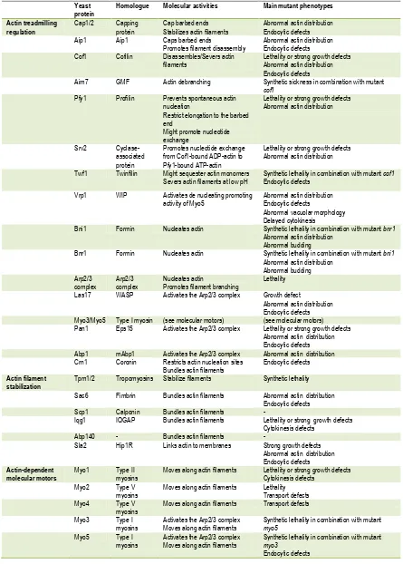
(15) 1. Introduction. Columna1 Actin treadmilling regulation. Actin filament stabilization. Actin-dependent molecular motors. Yeast protein Cap1/2. Homologue. Molecular activities. Main mutant phenotypes. Aip1. Capping protein Aip1. Cof1. Cofilin. Cap barbed ends Stabilizes actin filaments Caps barbed ends Promotes filament disassembly Disassembles/Severs actin filaments. Aim7. GMF. Actin debranching. Pfy1. Profilin. Srv2 Twf1. Cyclaseassociated protein Twinfilin. Prevents spontaneous actin nucleation Restrict elongation to the barbed end Might promote nucleotide exchange Promotes nucleotide exchange from Cof1-bound ADP-actin to Pfy1-bound ATP-actin Might sequester actin monomers Severs actin filaments at low pH. Abnormal actin distribution Endocytic defects Abnormal actin distribution Endocytic defects Lethality or strong growth defects Abnormal actin distribution Endocytic defects Synthetic sickness in combination with mutant cof1 Lethality or strong growth defects Abnormal actin distribution. Vrp1. WIP. Activates de nucleating promoting activity of Myo5. Bni1. Formin. Nucleates actin. Bnr1. Formin. Nucleates actin. Arp2/3 complex Las17. Arp2/3 complex WASP. Nucleates actin Promotes filament branching Activates the Arp2/3 complex. Myo3/Myo5 Pan1. Type I myosin Eps15. (see molecular motors) Activates the Arp2/3 complex. Abp1 Crn1. mAbp1 Coronin. Tpm1/2. Tropomyosins. Activates the Arp2/3 complex Restricts actin nucleation sites Bundles actin filaments Stabilize filaments. Sac6. Fimbrin. Bundles actin filaments. Scp1 Iqg1. Calponin IQGAP. Bundles actin filaments Bundles actin filaments. Abp140 Sla2. Hip1R. Bundles actin filaments Links actin to membranes. Myo1. Type II myosins Type V myosins Type V myosins Type I myosins Type I myosins. Moves along actin filaments. Myo2 Myo4 Myo3 Myo5. Moves along actin filaments Moves along actin filaments Activates the Arp2/3 complex Moves along actin filaments Activates the Arp2/3 complex Moves along actin filaments. Lethality or strong growth defects Abnormal actin distribution Synthetic lethality in combination with mutant cof1 Endocytic defects Abnormal actin distribution Endocytic defects Abnormal vacuolar morphology Delayed cytokinesis Synthetic lethality in combination with mutant bnr1 Abnormal actin distribution Abnormal budding Synthetic lethality in combination with mutant bni1 Abnormal actin distribution Abnormal budding Lethality Growth defect Abnormal actin distribution Endocytic defects (see molecular motors) Lethality or strong growth defects Abnormal actin distribution Endocytic defects Abnormal actin distribution Endocytic defects Synthetic lethality Abnormal actin distribution Endocytic defects Lethality or strong growth defects Cytokinesis defects Strong growth defects Abnormal actin distribution Endocytic defects Lethality or strong growth defects Cytokinesis defects Lethality Transport defects Transport defects Synthetic lethality in combination with mutant myo5 Synthetic lethality in combination with mutant myo3 Endocytic defects. Table 1. S. cerevisiae actin regulators. List of the most relevant actin regulators identified in budding yeast, including the name of mammalian homologs and an overview of their biochemical activities and mutant phenotypes. See text for further details.. 6.
(16) 1. Introduction. Figure 2. Overview of the molecular activities of S. cerevisiae actin regulators. This figure illustrates the main functions of the most relevant actin regulators in budding yeast. Briefly, the formins Bni1 and Bnr1, and the Arp2/3 complex -with the assistance of nucleation promoting factors- initiate the assembly of ATP-bound actin filaments adjacent to the plasma membrane, with the fast growing barbed end oriented towards the cell surface. Formins assemble unbranched actin filaments while the Arp2/3 complex forms Yshaped actin branches. Crn1 restricts actin nucleation to sites of dynamic actin assembly. The capping proteins Cap1/2 prevents further polymerization. Aim7 promotes filament debranching. Old actin filaments (formed by ADP-bound actin) are severed and/or dissociated by Cof1, while Aip1 caps the severed oligomer to prevent rapid re-polymerization and stimulate actin disassembly. The ADP-bound actin monomers might be sequestered by Twf1, or be delivered to Srv2 that, together with Pfy1, promotes nucleotide exchange to finally refill the pool of ATP-actin monomers bound to Pfy1. The actin filaments are stabilized by Tpm1/2, and bundled by Sac6, Scp1, or Iqg1 (not depicted). Sla2 and type-I myosins can link the actin polymers to membranes, and myosins move/slide along actin filaments. Arrows represent growth/force directions. See text for details. This illustration has been adapted from (Pollard, 2007) and (Gandhi et al., 2009).. 7.
(17) 1. Introduction. 1.1.2.1. Actin nucleators The spontaneous initiation of a new filament assembly requires the assembly of a trimeric nucleus. Since the actin dimer intermediate is very unstable, proteins that bypass or promote this step are very important for efficient actin dynamics (Pollard and Borisy, 2003). So far, five types of actin nucleators have been identified: proteins of the formin family, Spir proteins, Cordon-bleu, Leiomodin, and the Arp2/3 complex (Ahuja et al., 2007; Chereau et al., 2008; Mullins et al., 1998; Pruyne et al., 2002; Quinlan et al., 2005; Sagot et al., 2002b); of those, only formins and the Arp2/3 complex are conserved in all species. Each type of actin nucleators work by distinct mechanisms and trigger building of specialized actin-based structures (Campellone and Welch, 2010). The molecular mechanisms that control the activity of the conserved actin nucleators, the formins and the Arp2/3 complex, are discussed in the next two sections and depicted in Figure 4. 1.1.2.1.1. The formins: Bni1, Bnr1 Proteins of the formin family are conserved in yeast, plants and animals. Formins assemble structures composed of unbranched actin filaments including stress fibers, filopodia, the cytokinetic contractile ring or polarized actin cables. Yeast cells typically have 2 or 3 genes encoding formin proteins, while mammals have around 15 classified in 7 subfamilies (for a review see (Chesarone et al., 2010)). The conserved active region of formins is located at the C-terminus, and consists of the formin homology domains FH1 and FH2 (Figure 3). The Nterminus confers regulatory roles and varies considerably between different organisms (Chesarone et al., 2010). S. cerevisiae contains only 2 genes codifying for formins, BNI1 and BNR1. Deletion of each gene has no effect on viability, but deletion of both genes is lethal, indicating functional redundancy (Imamura et al., 1997). Analysis of mutants point out to an essential role of Bni1 and Bnr1 in cell polarity, cytokinesis and the formation of long filamentous actin cables required for polarized secretion and organelle inheritance, and indicate that both the FH1 and FH2 domains are required for the biological function of formins (Evangelista et al., 1997; Evangelista et al., 2002; Pruyne et al., 2002; Sagot et al., 2002a; Sagot et al., 2002b; Zahner et al., 1996). Formins do not seem to bind actin monomers and how exactly they activate actin nucleation is still unclear. It has been proposed that the FH2 domain, which in vitro is sufficient to catalyze nucleation, might bind and stabilize spontaneous formed actin seeds that otherwise would disassembly (Pring et al., 2003; Sagot et al., 2002b). In addition, nucleation-cofactors and monomer-binding sequences outside the FH2 domain might also be required for some formin to efficiently promote actin polymerization. For example, in S. cerevisiae the formin-binding protein Bud6 recruits actin monomers to nucleate actin dimers that might be captured by the FH2 domain (Graziano et al., 2011; Moseley et al., 2004). Once an actin seed is formed, the FH2-dimer switches between an open state, which allows addition of new monomers at the filament barbed end (about 100 monomers per second), and a closed state, which prevents elongation of the filaments but also binding of Capping Protein to the fast growing end. The FH1 domain contributes to FH2-dependent elongation by concentrating actin. 8.
(18) 1. Introduction. subunits through its multiple profilin-actin binding domains (Figure 4)(Kovar et al., 2006; Kovar and Pollard, 2004; Pruyne et al., 2002; Sagot et al., 2002b; Vavylonis et al., 2006). Nucleation and processive capping are the two conserved activities found for most formins, but some members of the family have been reported to bundle, severe, and depolymerize actin filaments. Whether these effects on the actin dynamics are functionally relevant is still unknown (Chesarone et al., 2010).. Figure 3. Domain organization of yeast formins. Domain organization of the yeast formins Bni1 and Bnr1. Both proteins show a similar organization. GDB: GTPase binding domain; FH1: formin homology domain 1; FH2: formin homology domain 2; DAD: diaphanous autoregulatory domain.. Formins are spatially and temporally regulated by different mechanisms. Five of the seven subfamilies of mammalian formins are autoinhibited by interactions between two domains, the diaphanous inhibitory domain DID and the diaphanous autoregulatory domain DAD, located at the N- and C-terminus, respectively. Binding of Rho GTPases and/or other factors and posttranslational modifications can disrupt the DID-DAD interaction (Chesarone et al., 2010). In S. cerevisiae, the formin Bni1 seems to be autoinhibited by an interaction between the N-terminal region and the DAD domain (Wang et al., 2009). Phosphorylation by the kinases Prk1 and Fus3 release the formin from autoinhibition and regulate its activity and its proper localization (Matheos et al., 2004; Wang et al., 2009). In addition to Fus3, other proteins that regulate the localization of yeast formins are Spa2, Bud6, and distinct members of the Rho family (Dong et al., 2003). Bud14, on the other hand, inhibits the activity of the Bnr1-FH2 domain and displaces the formin from the newly formed actin filament (Chesarone et al., 2009). 1.1.2.1.2. The Arp2/3 complex The Arp2/3 complex was first isolated from Acanthamoeba castellanii (Machesky et al., 1994), and since then it has been purified from several organisms including budding yeast and humans (Welch et al., 1997; Winter et al., 1997). Arp2/3 is a highly conserved complex of seven subunits: Arp2, Arp3, Arpc1 (Arc40 in budding yeast), Arpc2 (Arc35), Arpc3 (Arc18), Arpc4 (Arc19), and Arpc5 (Arc15). Two of the subunits, Arp2 and Arp3, are actin-related proteins actually Arp2 was first identified based of its sequence similarity to actin (Lees-Miller et al., 1992; Schwob and Martin, 1992)- and it has been recently demonstrated that because of their similarity to actin, they provide the two first ‘actin’ subunits of the new filament (Egile et al.,. 9.
(19) 1. Introduction. 2005; Rouiller et al., 2008). The Arp2/3 complex not only nucleates new actin filaments by mimicking an actin dimer, but it also anchors them to preexisting filaments with an angle of approximately 70º. As a result, the pointed end is capped with the old filament and the new filament grows towards the barbed end direction, forming a characteristic Y-shaped branch with the Arp2/3 complex at the junction (Figure 4) (Mullins et al., 1998)(Svitkina and Borisy, 1999). This tight coupling of nucleation and cross-linking of actin filaments is biased towards the barbed end due to the affinity of Arp2/3 for ATP-actin or ADP-Pi-actin (Amann and Pollard, 2001; Ichetovkin et al., 2002).. Figure 4. Actin nucleation and elongation by formins and the Arp2/3 complex. (A) Formins (green) might initiate or stabilize actin nucleation from free actin monomers (white) provided by Bud6 (red), and remain associated with the growing barbed end while allowing the addition of new actin subunits. Profilin-bound actin monomers (yellow and white, respectively) associate with the formins to facilitate the delivery of actin to the barbed end of the filament (arrow). (B) Nucleating promoting factors (blue) bind actin monomers (white) and the Arp2/3 complex (green) through its WH2 and acidic domains. Binding to the side of a preformed filament (mother filament) completes activation and allows growth of the daughter filament with an angle of 70 º with respect to the mother filament. See text for further details.. Purified Arp2/3 is able to nucleate actin in vitro, although highly inefficiently (Mullins et al., 1998; Welch et al., 1998); however, the addition of regulatory factors (see next section) enhance Arp2/3 mediated actin polymerization several orders of magnitude (Machesky et al., 1999; Rohatgi et al., 1999; Welch et al., 1998). The crystal structure of the bovine purified complex shows that Arp2 and Arp3 are too far apart to form an actin dimer, suggesting that they are in an inactive state and that regulatory proteins might promote a conformational change to close up the complex in order to trigger nucleation (Robinson et al., 2001). Recent studies have shown that the binding of regulatory proteins collectively known as nucleating. 10.
(20) 1. Introduction. promoting factors (NPFs) induce such a large conformational change (Goley et al., 2004; Rodal et al., 2005). Actin filaments also stimulate actin nucleation by increasing the affinity of regulatory proteins to the Arp2/3 complex (Marchand et al., 2001). In addition, the Arp2/3 complex must also be activated by ATP binding to the actin homologous proteins Arp2 and Arp3 (Dayel et al., 2001; Goley et al., 2004; Le Clainche et al., 2001; Martin et al., 2005). However, the role of ATP hydrolysis in the Arp2/3 complex is far from being understood. It has been proposed that ATP hydrolysis either occurs rapidly and is required for nucleation or that it occurs after nucleation and is necessary for debranching the dendritic network (Dayel and Mullins, 2004) (Le Clainche et al., 2003). Recent work performed in S. cerevisiae with an arp2 mutant that cannot hydrolyze ATP revealed that ATP hydrolysis occurs simultaneously with actin nucleation but is actually required for filament debranching (Martin et al., 2006). Another level of regulation has been reported recently. LeClaire et al. observed that phosphorylation on either threonine or tyrosine conserved residues in the Arp2 subunit is necessary for the nucleating activity of the complex (LeClaire et al., 2008). Genetic analysis of the Arp2/3 complex in S. cerevisiae indicates that depletion of any subunit except Arc18 causes severe growth defects or lethality, depending on the yeast background (Huang et al., 1996; Schwob and Martin, 1992; Winter et al., 1997; Winter et al., 1999b). Generation of mutants early pointed out a role for the Arp2/3 complex in the organization of the actin cytoskeleton and in the uptake step of endocytosis (Moreau et al., 1997; Moreau et al., 1996; Munn and Riezman, 1994; Pan et al., 2004; Winter et al., 1997; Winter et al., 1999b). The mutational analysis of the yeast Arp2/3 complex has provided some important insights into the Arp2/3 nucleation mechanism (Balcer et al., 2010; D'Agostino and Goode, 2005; Daugherty and Goode, 2008). In vitro, yeast Arp2/3 complex has more basal nucleation activity than the mammalian Arp2/3 in the absence of NPFs, but addition of NPFs further enhances actin polymerization (Rodal et al., 2005; Wen and Rubenstein, 2005). Besides the classical NPFs, coronin (Crn1) also regulates the Arp2/3 complex by a completely different mechanism. Crn1 is a multifunctional protein (Figure 5) that interacts with the Arc35 subunit of the complex and inhibits spontaneous nucleation in vitro (Humphries et al., 2002). However, Crn1 exhibits high affinity for the newly assembled actin filaments, loaded with ATPactin, and in contact with those, the inhibition of the Arp2/3 complex by Crn1 is released. Thereby, coronin restricts the Arp2/3 activity to sites where new forming filaments are abundant (Gandhi et al., 2009; Humphries et al., 2002). In addition to this role, Crn1 bundles actin filaments, and synergizes with cofilin and Aip1 to sever ADP-bound actin filaments (Brieher et al., 2006; Gandhi et al., 2009; Goode et al., 1999). Deletion of the single gene encoding coronin in budding yeast, CRN1, causes a defect in endocytosis (Burston et al., 2009). Phosphorylation of a serine located in the N-terminus of human coronin 1B by PKC weakens coronin-Arp2/3 interaction (Cai et al., 2005). Yeast Crn1 is phosphorylated at multiple sites in vivo but no biological function has been assigned to any of these post-translational modifications (Albuquerque et al., 2008; Chi et al., 2007; Ficarro et al., 2002; Li et al., 2007; Smolka et al., 2007).. 11.
(21) 1. Introduction. Figure 5. Domain organization of yeast coronin. WD: WD40 repeats –a region rich in tryptophan and aspartic acid; CC: potential coiled-coil domain.. 1.1.2.1.2.1. Nucleation promoting factors: Las17, Myo3 and Myo5, Pan1, Abp1 Arp2/3 is activated by a collection of regulatory proteins that are known as nucleation promoting factors (NPFs). The first NPF to be identified was ActA, a protein found in the surface of the intracellular pathogenic bacteria Listeria monocytogenes that activates the Arp2/3 complex to produce actin comet tails required for bacterial motility in the host cytoplasm (Welch et al., 1998). Since then, several proteins that activate the Arp2/3 complex have been identified including the well-studied NPF WASP (from Wiskott-Aldrich syndrome protein), whose mutation produces a human immunodeficiency and bleeding disorder (for further information see (Lappalainen, 2007)). Although the domain organization of different NPFs is remarkably diverse, they all share some common characteristics. The organization of representative NPFs is shown in Figure 6. The main feature for all type of nucleator promoting factors is the presence of an Arp2/3-binding sequence called the CA domain, which consists in a central o connecting region (C) plus an acidic sequence that includes a conserved tryptophan (A). The CA region is necessary and sufficient to bind the Arp2/3 complex but is not sufficient to activate it (Rohatgi et al., 1999). In order to activate the Arp2/3 complex, an NPF must bind either monomeric or filamentous actin (classifying the NPFs in group I or group II, respectively). Class I and II NPFs activate the Arp2/3 complex by slightly different mechanisms: Class I NPFs contains one or two monomer-binding WH2 domains, also called W or V domains from WASP homology 2, or Verprolin homology- that is found usually just before the CA domain. Although most class I NPFs contains the triple domain WCA, some members might lack one of the typical sequences or they can even be located in a separate molecule (Lechler et al., 2001). Typical members of this type of NPFs are the Wiskott-Aldrich syndrome protein (WASP) family, including budding yeast WASP homolog Las17 or Scar/WAVE proteins (Machesky et al., 1999; Rohatgi et al., 1999; Winter et al., 1999a; Yarar et al., 1999); proteins from intracellular pathogens such as ActA from Listeria, RickA from Rickettsia, or p78/83 from a baculovirus (Goley et al., 2006; Gouin et al., 2004; Jeng et al., 2004; Welch et al., 1998); the capping protein Arp2/3 complex and myosin-I linker CARMIL (Jung et al., 2001); WHAMM -from WASP Homolog associated with actin, membranes, and microtubules- (Campellone et al., 2008); the WASP and Scar homologue (WASH) (Derivery et al., 2009; Gomez and Billadeau, 2009); the p53 cofactor JMY (Zuchero et al., 2009); and yeast type I myosins (Geli et al., 2000; Lechler et al., 2001; Lee et al., 2000; Sun et al., 2006).. 12.
(22) 1. Introduction. Figure 6. Domain organization of nucleation promoting factors. S. cerevisiae NPFs are underlined. Class I NPFs contain the monomer-binding WH2 domain (W) and the Arp2/3 binding regions Connecting (C) and Acidic (A). Note that CARMIL does not contain the C region and Myo3/Myo5 does not include the W domain, which are provided by co-activators. Class II NPFs interact with the Arp2/3 complex via the acidic domain and to F-actin through the tandem repeat (TR) of cortactin, the coiled-coil (CC) of Pan1, and the actin-depolymerizing factor homology (ADFH) of Abp1. Other domains are required to bind signaling molecules and/or actin regulators. WH1: WASP-homology 1, verprolin-binding domain; B: basic region, PIP2-binding region; GDB: GTPase binding domain; PRD: proline-rich domain, SH3-binding region; SHD: Scar homology domain; WHD1: WASH homology domain 1; WHD2: WASH homology domain 2; N: N-terminal region; LRR: Leucine-rich repeat; TH1: Tail homology 1, lipid-binding region; TH2: Tail homology 2; SH3: Src homology 3, PRD-binding domain; SS: signal sequence; TM: transmembrane; LR: long repeat; EH: Eps15 homology; black bars: IQ motifs, calmodulin-binding regions.. 13.
(23) 1. Introduction. Arp2/3 activation by class I NPFs requires both the CA and the WH2 domain. Constructs consisting on the WCA domain from WASP-related proteins can activate the Arp2/3 complex, independently of other factors (Machesky et al., 1999; Rohatgi et al., 1999; Winter et al., 1999a). The CA region is thought to bind the Arp2/3 complex and tether the WH2 domain so that the first actin subunit can be positioned to start a new filament. Moreover, it promotes an activating conformational change in the Arp2/3 complex that depends on conserved residues in the C region (Goley et al., 2004; Rodal et al., 2005). Class I NPFs have low affinity for the Arp2/3 complex, thus after transiently interact and stimulate the complex they might dissociate to start a new round of activation. Class II NPFs do not bind G-actin. Instead, they bind filamentous actin through F-actin binding domains. This is an important difference as class II NPFs are far less powerful activators in vitro that class I NPFs (Higgs and Pollard, 2001; Sun et al., 2006). Members of this group of NPFs are cortactin (Uruno et al., 2001; Weaver et al., 2001), yeast Abp1 (Goode et al., 2001), and yeast Pan1 (Duncan et al., 2001). It is not clear how class II activate the Arp2/3 complex, but they lack the connecting region and cannot trigger the Arp2/3 activating conformational change (Goley et al., 2004). Their mechanism of activation might involve the enhancement of the Arp2/3 complex association with the mother filament, which is itself an activator of the complex (Higgs et al., 1999; Machesky et al., 1999). Cortactin remains associated at the branch point, apparently to inhibit branch dissociation (Egile et al., 2005). Five NPFs are present in S. cerevisiae: Las17 and the type I myosins Myo3 and Myo5 (see also section 1.1.4.3), which are class I NPFs, and Abp1 and Pan1, which are class II NPFs and therefore have a lower nucleation promoting activity (NPA). The S.cerevisiae protein Las17 was first found based on its high sequence homology with the Wiskott-Aldrich syndrome protein WASP (Symons et al., 1996). It has a similar organization to WASP (Figure 6), except that Las17 does not contain the GTPase binding domain (GBD), a domain that has been shown to promote WASP auto-inhibition (see below) (Miki et al., 1998; Rohatgi et al., 1999). The WH1 domain of Las17 interacts with Vrp1 while the proline-rich regions of Las17 interact with SH3-domain containing proteins that regulate its activity (see below). Downstream from the poly-proline region, the WH2 domain binds to monomeric actin. At the C-terminus is located the central and the acidic domains (CA), which interact with the Arp2/3 complex (Winter et al., 1999). Purified WCA is able to activate the Arp2/3 complex in vitro (Winter et al., 1999) albeit with less efficiency than the full length protein, suggesting that other regions in Las17 might contribute to efficient Arp2/3 activation (Rodal et al., 2003). Deletion of LAS17 causes severe actin cytoskeleton defects (Li, 1997) and prevents endocytic internalization (Madania et al., 1999; Naqvi et al., 1998). Interestingly, deletion of Las17-WCA domain alone does not cause any detectable phenotype (Winter et al., 1999), indicating that different domains are important for Las17 function in actin organization and that other factors can fulfill its function in actin assembly. Indeed, mutation of the acidic domains of the Arp2/3 activators Pan1 and the myosins-I Myo3 and Myo5 show synthetic defects with deletion of the. 14.
(24) 1. Introduction. acidic domain of Las17, suggesting functional redundancy (Duncan et al., 2001; Evangelista et al., 2000; Lechler et al., 2000; Sun et al., 2006). Mammalian WASP and N-WASP proteins are autoinhibited by an intramolecular interaction between the GBD and WCA domain, which prevents binding to the Arp2/3 complex. Some WASP ligands, including the Rho-family GTPases Cdc42 and Rac cooperate with PIP2 to release this auto-inhibition by binding to the GBD and displacing the WCA domain, which can then bind and activate the Arp2/3 complex (Takenawa and Suetsugu, 2007). The activity of WASP, N-WASP, and WAVE proteins are also modulated by intermolecular interactions. The N-terminal region of WASP/N-WASP contains a WH1 domain that mediates their interactions with verprolins (WIP, WICH/WIRE, or CR16, see section 1.1.2.2). The binding of verprolins to the NPF seems to have different functions, from maintaining it in the inactive conformation to recruiting the NPF to localized regions where massive actin polymerization is needed; there, a myriad of regulatory mechanisms that are still being characterized release the WIP-WASP inhibitory complex (Takenawa and Suetsugu, 2007). Although Las17 shares high sequence homology with WASP, it does not contain an obvious GBD domain and does not seem to interact with Cdc42 directly (Lechler et al., 2001). This is an important functional difference, because full length Las17 is extremely active in vitro while purified WASP is inactive in the absence of Cdc42 or PIP 2 (Rodal et al., 2003)(Higgs and Pollard, 2000). Both, inhibition of actin assembly mediated by Las17 and the release of such inhibition is achieved by interactions with SH3-domain containing proteins (Figure 7). Four proteins have been shown to date to inhibit Las17-induced actin assembly: Sla1, Bbc1, Syp1, and Abp1 (D'Agostino and Goode, 2005; Rodal et al., 2003)(Boettner et al., 2009). Sla1 and Bbc1 both bind Las17 in vivo, and they inhibit Las17 in vitro by binding to different regions of Las17 (Li, 1997; Rodal et al., 2003; Tong et al., 2002). In contrast, Abp1 attenuates Las17 actin assembly by competing for the Arp2/3 complex (D'Agostino and Goode, 2005). Bzz1 also binds Las17 (Soulard et al., 2002; Tong et al., 2002), but this interaction serves to relieve Las17 inhibition mediated by Sla1 (Sun et al., 2006). Myosins are a family of actin-activated molecular motors that have a crucial role in actindependent processes in eukaryotic cells (see also section 1.1.4). In S. cerevisiae, type-I myosins are recruited to the plasma membrane where both motor and nucleating promoting activities plays an important function in endocytic internalization (see section 1.2.3.1.1, and references therein). The domain organization of budding yeast myosins-I is shown in Figure 6. Briefly, Myo3 and Myo5 contain an N-terminal motor domain that carries an actin-based ATPase (Sun et al., 2006), a neck that contains 2 IQ motifs that bind calmodulin (Geli et al., 1998), a basic tail homology 1 (TH1) domain that probably interacts to acidic phospholipids (Pollard et al., 1991), and a C-terminal extension (Cext) that includes the domains required for their nucleation promoting activity: a TH2 domain that contains poly-proline motifs, an SH3 domain, and a central and acidic domains (CA) at the very C-terminus that directly binds to the Arp2/3 complex (Evangelista et al., 2000; Lechler et al., 2000)(Geli et al., 2000). Whether budding yeast were directly capable of activating actin polymerization was initially a matter of debate since Myo3 and Myo5 do not contain the WH2 domain required for G-actin binding. Yet, it was. 15.
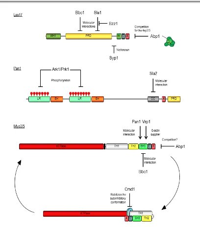
(25) 1. Introduction. subsequently demonstrated that the yeast myosins-I interact with the WIP homologue Vrp1 which contain two WH2 domains- through the SH3 domain located at the myosin C-terminal extension (Evangelista et al., 2000; Geli et al., 2000; Vaduva et al., 1997). Robust evidence now accumulates demonstrating that binding of Vrp1 to the myosins is necessary and sufficient to the develop their potent NPA: 1) our lab has demonstrated that a Myo5 construct consisting on the TH2, SH3 and acidic domains immobilized in Glutathione-Sepharose beads is able to induce the formation of actin patch-like structures in the presence of the Arp2/3 complex and Vrp1 (Geli et al., 2000; Idrissi et al., 2002)(see also section 5.1); 2) a chimeric protein containing the WH2 domain of Vrp1 and the A domain of Myo3 was able to directly promote actin polymerization in vitro (Lechler et al., 2001); and 3) more recently, Myo5/Vrp1-induced actin polymerization was observed using purified components (Sun et al., 2006). In fission yeast, although type I myosin alone is able to weakly induce actin polymerization (Lee et al., 2000), this activity is also enhanced by Vrp1 (Sirotkin et al., 2005). Importantly, the NPF activity of the Myo5/Vrp1 complex is very powerful, being comparable to Las17 activity (Sun et al., 2006). As mentioned above, deletion of the acidic domains of Myo3 and Myo5 is synthetically defective with deletions of the acidic domain of Las17 (Evangelista et al., 2000; Lechler et al., 2000; Sun et al., 2006). Thus, although the NPA of Myo5 and Las17 have different functions (see section 1.2.3.1.1.2, and references therein), in the absence of the myosin-I NPA, Las17 can take over and viceversa (Evangelista et al., 2000; Lechler et al., 2000). In addition to its activity as NPFs, type-I myosins have other essential functions that cannot be fulfilled by any other proteins since deletion of both Myo3 and Myo5 results in lethality or strong defects in growth, depending on the yeast strain used (Geli and Riezman, 1996; Goodson et al., 1996). The nucleating promoting activity of the yeast myosins-I is regulated by intra- and intermolecular interactions (Figure 7). An autoinhibitory interaction between the TH1 domain and the C-terminal extension of the protein, containing the SH3 and CA domains, prevents binding of the co-activator Vrp1 to the myosin SH3 domain. This inhibitory interaction is stabilized by binding of calmodulin to Myo5. Calmodulin is thought to work as a clamp linking the myosin neck adjacent to the TH1 domain and the C-terminal extension. Calmodulin dissociation from the myosin neck at the plasma membrane induced by a still unknown factor, releases the autoinhibitory interaction (Grotsch et al., 2010). In addition, the Myo5 NPA can be regulated by other interacting proteins. Both Vrp1 and Bbc1 interact with the SH3 domain of Myo5, but while Vrp1 is required to activate the NPF activity of Myo5, Bbc1 negatively regulates this function (Anderson et al., 1998; Mochida et al., 2002; Sun et al., 2006). Another SH3binding protein, Pan1 (see below) has been recently proposed to enhance Myo5-mediated actin polymerization (Barker et al., 2007). Bzz1 also interacts with Myo5 but the functional significance of this interaction is not clear (Soulard et al., 2005; Soulard et al., 2002) (see discussion). Abp1 inhibits Arp2/3 complex activation by Myo5/Vrp1, and although the molecular mechanism that triggers this inhibition has not been demonstrated, it might involve the competition for the Arp2/3 complex (D'Agostino and Goode, 2005)(Sun et al., 2006). The. 16.
(26) 1. Introduction. regulation of Myo5 NPA by phosphorylation (see below) was the subject of this study and is discussed along the dissertation.. Figure 7. Mechanisms of regulation of S. cerevisiae NPFs. The molecular mechanisms that regulate the nucleating promoting activities of Las17, Pan1, and the myosins Myo3 and Myo5 are depicted. See text for further details.. Pan1 was found in various screens to be required for endocytosis and for normal actin cytoskeleton organization (Tang and Cai, 1996; Wendland et al., 1996). Pan1 contains two long repeat domains at the N-terminus, each embodying an Eps15-Homology domain (EH) (see the domain organization of Pan1 in Figure 6). Through these domains, Pan1 interacts with cargo adaptors and other endocytic proteins (Tang et al., 1997; Tang et al., 2000; Wendland and. 17.
(27) 1. Introduction. Emr, 1998; Wendland et al., 1999). At the central region a coiled-coil domain contains a WH2like region, which differs from others WH2 domains since it is unable to interact with G-actin. Nevertheless, the coiled-coil domain binds F-actin with high affinity (Toshima et al., 2005). At the C-terminus of the protein an acidic domain interacts with the Arp2/3 complex (Duncan et al., 2001) and a poly-proline region binds to type-I myosins (Barker et al., 2007). Purified Pan1 activates the Arp2/3 complex (Duncan et al., 2001), although much less efficiently than Myo5/Vrp1 or Las17 (Sun et al., 2006). Deletion of Pan1-WA domain does not cause any detectable phenotype, but it shows synthetic defects with deletions of the acidic domains or Las17 and the type-I myosins (Sun et al., 2006; Toshima et al., 2005). The NPA of Pan1 is regulated by two mechanisms: phosphorylation by the Ark1/Prk1 kinases and inter-molecular interactions (Figure 7). Pan1 contains 15 consensus sites for the kinase Prk1, a kinase required for endocytosis (Sekiya-Kawasaki et al., 2003; Zeng and Cai, 1999). Mutation of all 15 potential phosphorylation residues into non-phosphorylatable amino acids increases the nucleation promoting activity of Pan1, while addition of the kinase inhibits Pan1mediated actin polymerization (Toshima et al., 2005). In addition, the actin-binding protein Sla2 (see section 1.1.3.3) binds to the WH2-like (CC) domain of Pan1 and also inhibits its NPA in vitro (Toshima et al., 2007). Abp1 was the first actin-associated protein described in yeast (Drubin et al., 1988). Its domain organization is shown in Figure 6. Abp1 contains an ADF-H domain required for F-actin binding (Goode et al., 2001), a proline-rich region of unknown interaction partners, and a SH3 domain that interacts with the kinase Prk1 (Fazi et al., 2002). Moreover, it contains two acidic domains, both required for the activation of the Arp2/3 complex (Goode et al., 2001). Abp1 has been shown to co-fractionate with the Arp2/3 complex and to activate the complex in vitro (Goode et al., 2001). However, increasing evidence indicate that Abp1 might negatively regulate actin polymerization in vivo, as it inhibits the NPF activity of Las17 and Myo5/Vrp1, which are much powerful Arp2/3 activators than Abp1 (D'Agostino and Goode, 2005; Sun et al., 2006). 1.1.2.2. G-actin binding proteins: Pfy1, Srv2, Twf1, Vrp1 A. high. number. of. actin-monomer-binding. proteins. (>25. in. mammals). control. actin. polymerization and depolymerization, but only six classes are conserved from yeast to mammals: ADF/cofilin (Cof1 in budding yeast), profilin (Pfy1), Srv2/CAP, twinfilin (Twf1), verprolins (Vrp1), and WASP/WAVE (Las17). Paradoxicallythymosins, which are the major actin-sequestering proteins in vertebrate cells, have not been found in invertebrates or yeast (Safer et al., 1991; Safer et al., 1990). The biochemical functions of Pfy1, Srv2, Twf1, and Vrp1 are explained in this section and their domain organization shown in Figure 8; the general features of Las17 were described above and that of Cof1 are explained in section 1.1.2.4. Profilin is an evolutionarily conserved small protein that binds G-actin, forming a 1:1 complex with the monomer. It is essential to maintain the normal actin distribution in yeast cells, when not critical for viability (depending on the yeast background), and it seems to function in a post-. 18.
(28) 1. Introduction. internalization step of endocytosis (Haarer et al., 1990; Idrissi et al., 2002; Magdolen et al., 1988; Wolven et al., 2000). In vitro, Pfy1 promotes rapid actin turnover in the presence of cofilin (Wolven et al., 2000). Actin monomers bound to profilin are considered the main source of actin for rapid assembly at the barbed ends because it has higher affinity for ATP- than for ADP-bound G-actin; in addition, profilin can bind to actin and to actin nucleators (or NPFs) simultaneously, and with higher affinity than to each one separately (Moseley and Goode, 2006). Moreover, because the profilin-binding site is located in the actin-barbed end (Schutt et al., 1993), profilin inhibits both spontaneous actin nucleation and elongation at the pointed end (Pantaloni and Carlier, 1993; Pollard and Cooper, 1984; Pring et al., 1992). Pfy1 binds to short series of proline residues located in the FH1 domain of yeast Bni1 and Bnr1 formins (Evangelista et al., 1997; Imamura et al., 1997), and while profilin is not required for the processivity of formins, it increases formin-dependent actin filament assembly and elongation in vitro (Kovar et al., 2006; Sagot et al., 2002b). Another assigned role of profilin was to promote nucleotide exchange of monomeric actin. However, yeast profilin is almost 2 orders of magnitude less efficient than human profilin in promoting this exchange (Eads et al., 1998); moreover, it requires Srv2 to catalyze this exchange in the physiological substrate cofilin-bound ADP-actin, and indeed Srv2 alone can promote this turnover (Balcer et al., 2003; Quintero-Monzon et al., 2009). Profilin, like other actin-binding proteins, interacts with PIP2 (Lassing and Lindberg, 1985). The physiological significance of this interaction is not well understood, but PIP2 can partially dissociate profilin-actin and profilin can inhibit PIP2 hydrolysis (see (Lappalainen, 2007), chapter 3, and references therein). ROCK-mediated phosphorylation of profilin might also modulate its interaction with actin, at least in vitro (Shao et al., 2008). It is not known whether yeast profilin is regulated by the same mechanisms. Another evolutionary conserved G-actin binding protein is a multifunctional molecule called CAP (Cyclase-associated protein), also named Srv2 in budding yeast. The essential protein Srv2 was first identified in budding yeast for its role in the stimulation of adenylate cyclase, a function independent of its role in actin filament organization and only observed for the budding and fission yeast CAP proteins (Fedor-Chaiken et al., 1990; Field et al., 1990; Hubberstey and Mottillo, 2002). Shortly after, a role for Srv2 in actin regulation was demonstrated (Gerst et al., 1991; Hubberstey and Mottillo, 2002; Vojtek et al., 1991). Profilin only weakly catalyzes nucleotide exchange of cofilin-bound ADP-actin. This activity has recently been attributed to Srv2 based on several observations: 1) it binds to ADF/cofilin-ADP-actin complexes; 2) it promotes ADF/cofilin dissociation; 3) it strongly catalyzes nucleotide exchange on cofilin-bound ADP-G-actin; and 4) it has low affinity for the ATP-actin, and therefore, it might release the monomer to profilin for a new round of polymerization (Balcer et al., 2003; Bertling et al., 2007; Gandhi et al., 2010; Mattila et al., 2004; Moriyama and Yahara, 2002; Quintero-Monzon et al., 2009). Srv2 is linked to actin filaments through the actin-binding protein Abp1 (Balcer et al., 2003; Lila and Drubin, 1997). The association of actin and Srv2/CAP might also be regulated by PIP2, since PIP2 partially dissociate actin from CAP, at least in some organisms (Gottwald et al., 1996). Srv2 is phosphorylated in vivo but whether this phosphorylation regulates the activity of the protein is unknown (Albuquerque et al., 2008).. 19.
(29) 1. Introduction. Figure 8. Domain organization of yeast actin-monomer binding proteins. Domain organization of yeast profilin (Pfy1), the yeast adenylyl cyclase-associated protein (Srv2), twinfilin (Twf1) and verprolin (Vrp1). Different G-actin binding domains are depicted. PRF: profilin domain; G-A: G-actin binding site; W: WH2 domain; ADF-H: actin-depolymerizing factor homology domain. Other domains are required to bind signaling molecules and/or regulators. P: proline-rich domain, SH3-binding region; AC: adenylyl cyclase binding region.. Twf1 has two ADF-H domains, and like cofilin, it is required for endocytic uptake and rapid actin turnover. However its mechanism of function is still not well understood (Burston et al., 2009; Goode et al., 1998; Moseley et al., 2006). Twinfilin binds ADP-G-actin with higher affinity than to ATP-G-actin and inhibits nucleotide exchange, suggesting an actin-sequestering role (Goode et al., 1998; Palmgren et al., 2001). Still, Twf1 stimulates rapid actin disassembly, possibly due to its role in severing at low pH (Moseley et al., 2006). Twf1 also interacts with capping protein, which might localize twinfilin-bound actin monomers to sites of active actin assembly (Falck et al., 2004). This interaction seems to prevent twinfilin’s severing activity (Moseley et al., 2006). A capping activity found for mammalian twinfilin is not conserved for yeast Twf1 (Helfer et al., 2006). Twinfilin, like other actin-interacting proteins, binds directly PIP2 and this binding prevents its interaction with actin (Palmgren et al., 2001). Phosphorylation of Twf1 in vivo has also been reported, but still no regulatory role has been assigned to this post-translational modification (Albuquerque et al., 2008). Verprolins are proline-rich proteins that have been identified in most eukaryotic organisms. The members of this family include WIP, WIRE/WICH, and CR16 in vertebrate cells (Lappalainen, 2007). Besides its high content in prolines, verprolins contain two WH2 domains, one of the most abundant and functional diverse actin binding fold (Vaduva et al., 1997). Yeast Vrp1 shares some homology with the WASP-interacting protein WIP. Deletion of the VRP1 causes cytoskeletal disorganization, a delayed cytokinesis, and endocytic defects (Donnelly et al., 1993; Munn et al., 1995; Naqvi et al., 2001). Human WIP is able to rescue these phenotypes, indicating functional similarities (Vaduva et al., 1999). Verprolins are important effectors of the regulation of actin dynamics, although its precise role is still not well understood. In mammals, verprolins maintain the nucleating promoting factor N-WASP in an inactive conformation, but. 20.
(30) 1. Introduction. also recruit N-WASP and Arp2/3 to activate localized bursts of actin polymerization and regulate actin dynamics in a N-WASP independent manner (Kato and Takenawa, 2005; Kinley et al., 2003; Martinez-Quiles et al., 2001; Moreau et al., 2000). In S. cerevisiae, Las17 binding serves to recruit Vrp1 to sites with active actin dynamics, but the effect of this interaction in the Las17 actin nucleating activity is not well understood (Naqvi et al., 1998; Sun et al., 2006). Vrp1 also associates with another Arp2/3 activator, Myo5 (and probably also Myo3), and activates its nucleating activity, probably by providing the actin monomers binding activity (see above, section 1.1.2.1.2.1) (Evangelista et al., 2000; Geli et al., 2000; Sun et al., 2006). Vrp1 is phosphorylated in vivo, but the biological role of this post-translational modifications are still not known (Albuquerque et al., 2008; Smolka et al., 2007) 1.1.2.3. Capping proteins: Cap1/Cap2 and Aip1 Since actin polymerization and depolymerization occur at the end of the filament, regulation of the dynamics and organization of filaments can be achieved by blocking the availability of filament ends and therefore, by preventing further assembly of actin monomers. Paradoxically, although capping proteins prevents both the addition and removal of subunits and therefore, they limit the length of F-actin, they enhance actin-based motility in vivo and in vitro (Hug et al., 1995; Loisel et al., 1999). The widely accepted actin funneling hypothesis postulate that the number of free actin monomers increases when most actin filaments are capped, resulting in the fast elongation of the few uncapped barbed ends (Carlier and Pantaloni, 1997). However, a recent study indicates that the capping protein CP (also known asactinin or capZ in higher eukaryotes) does not influence the kinetics of filament elongation but the rate of Arp2/3dependent actin polymerization (Akin and Mullins, 2008). The authors propose an alternative hypothesis that postulates that capping protein modifies the architecture of the actin network rather than its kinetics. Two barbed end capping proteins are conserved from yeast to humans, indicating a general mechanism for actin regulation: capping protein (CP) and AIP1. The domain organization of the yeast CP subunits and Aip1 are shown in Figure 9. In addition to CP and Aip1, Eps8 and tropomodulins are capping-proteins only present in metazoan (Di Fiore and Scita, 2002; Weber et al., 1994). Yeast capping protein (CP) is a heterodimer composed of Cap1 and Cap2 subunits, each encoded by one gene. Disruption of CP results in abnormal actin distribution and endocytic defects (Amatruda and Cooper, 1992; Amatruda et al., 1992; Burston et al., 2009; Kaksonen et al., 2005), and analysis of a set of CP mutants indicate that the actin distribution phenotype correlates with the capping activity of the dimer (Kim et al., 2004). Interestingly, nematode CP can substitute the deletion of yeast CP in vivo, indicating that CP function is conserved across evolution (Waddle et al., 1993). By binding to the filament barbed end CP regulates the filament length and modifies the architecture of the actin network (Kaksonen et al., 2005; Kim et al., 2006; Moseley and Goode, 2006). Several proteins are able to bind and inhibit CP in metazoan (either by preventing its binding to actin or by a direct uncapping activity) including CARMIL, V1/myotrophin, CKIP-1, and CD2AP (Cooper and Sept, 2008), but no protein has been found to. 21.
(31) 1. Introduction. inhibit yeast CP to date. Moreover, the activity of CP from several organisms, including yeast, is inhibited by PIP2 (Amatruda and Cooper, 1992). Since PIP2 is enriched at the plasma membrane (Di Paolo and De Camilli, 2006), capping of barbed ends might be prevented near the plasma membrane to locally regulate actin turnover. Moreover, in vivo phosphorylation of both CP subunits from a number of organisms including S. cerevisiae has been reported, but how phosphorylation regulates the activity of capping protein is not yet known (Albuquerque et al., 2008; Li et al., 2007; Malik et al., 2009; Olsen et al., 2010; Raijmakers et al., 2010). The actin-interacting protein Aip1 was first identified as an actin-interacting protein by yeast two hybrid (Amberg et al., 1995). Shortly later, it was reported to stimulate cofilin-mediated actin disassembly in vitro. Deletion of the AIP1 gene leads to formation of atypical actin filaments and endocytic defects {Rodal, 1999 #485}(Burston et al., 2009; Okada et al., 2006). Aip1 caps the barbed ends of ADF/cofilin-severed filaments in vitro, preventing its further elongation and/or filament re-annealing (Okada et al., 2002) (Balcer et al., 2003), and helping to convert the short actin oligomers into monomeric actin (Okreglak and Drubin, 2010). Proteomic analysis indicates that yeast Aip1 is phosphorylated in vivo, but whether this phosphorylation regulates its activity is still unknown (Albuquerque et al., 2008).. Figure 9. Domain organization of yeast capping proteins. Domain organization of the yeast subunits of heterodimeric capping protein Cap1 and Cap2, and of yeast Aip1. Aip1 contains eleven WD40 repeats, a region rich in tryptophan and aspartic acid (WD).. Two other conserved proteins, formins and the Arp2/3 complex, also cap actin filaments at their barbed and pointed end, respectively, but their major role is the nucleation of actin filaments and its function was discussed in section 1.1.2.1. 1.1.2.4. Actin depolymerizing/Severing proteins: Cof1, Aim7 Actin depolymerization and severing of filaments are crucial to dynamize filamentous actin, because it increases the availability of actin monomers and provides new barbed ends for the new rounds of actin polymerization. The members of the gelsolin/villin family (gelsolin, villin, adseverin, advillin, supervillin, and flightless I) are actin-severing and barbed-end actin-capping factors widely expressed in metazoan and in plants, but only cofilin (a member of the ADF/cofilin family, see domain organization in Figure 10) is conserved from yeast to mammals.. 22.
(32) 1. Introduction. ADF and cofilin are the best characterized members of a family of essential and conserved proteins collectively known as the ADF/cofilin family. Members of this protein family have in common the presence of a single actin-depolymerizing factor homology domain (ADF-H). The gene COF1 encodes the only member of the ADF/cofilin family present budding yeast, and its deletion is lethal (Iida et al., 1993; Moon et al., 1993). Analysis of temperature-sensitive cofilin alleles indicate that Cof1 is required for endocytic uptake and stimulates the actin turnover in vivo and in vitro by the disassembly of actin in a molecular mechanism not yet fully understood (Idrissi et al., 2002; Lappalainen and Drubin, 1997). Growing evidence indicates that Cof1 binding weakens the longitudinal interaction between the actin subunits and shifts the helical twist of the filament (Bobkov et al., 2004; McGough et al., 1997). Cofilin inhibits nucleotide exchange as long as it remains bound to the monomer (Nishida, 1985). Therefore, other interaction partners -Aip1, Srv2, and Pfy1- are required for the recycling of Cof1-bound actinmonomers (Balcer et al., 2003; Quintero-Monzon et al., 2009). Alternative proposed functions for members of the ADF/cofilin are the dissociation of actin branches produced by Arp2/3 (Blanchoin et al., 2000), and actin nucleation when cofilin is present at high concentrations (Andrianantoandro and Pollard, 2006). Due to the essential roles of cofilin, its activity must be tightly regulated. In fact, multiple mechanisms that regulate ADF/cofilins have been described. Phosphorylation of a serine located in the N-terminus prevents its binding to both monomeric and filamentous actin. In mammals, the members of the LIM and TES family of kinases and the members of the SSH family of phosphatases and CIN are responsible for the phosphorylation and dephosphorylation of ADF/cofilin, respectively, via a complex control of several signaling pathways (see (Van Troys et al., 2008). Budding yeast cofilin is also phosphorylated in vivo (Albuquerque et al., 2008), but replacement of the corresponding residue does not cause any detectable phenotype, suggesting that phosphorylation of Cof1 does not play a major regulatory role in yeast (Lappalainen et al., 1997). Protonation/deprotonation of the only histidine in human cofilin regulates its binding to actin and to phosphatidylinositol PIP2, which competes with F-actin to bind cofilin (Bamburg, 1999; Frantz et al., 2008). Yeast cofilin does not seem to be regulated by changes in pH, but it is also regulated by PIP 2 (Ojala et al., 2001). The activity of ADF/cofilin is also modulated by interacting proteins. Aip1 (section 1.1.2.3) and Srv2 (see section 1.1.2.2) cooperate with ADF/cofilin to promote actin disassembly; coronin (section 1.1.2.1.2) collaborate with cofilin to mediate disassembly at old actin regions -rich in ADP-actinwhile it prevents Cof1-mediated disassembly at newly assembled actin regions -rich in ATPactin- (Gandhi et al., 2009). Tropomyosins (see section 1.1.3.1.) are coiled-coil dimers that bind along the length of actin filaments; in general they stabilize actin filaments and prevent the depolymerizing/severing activity of ADF/cofilin, though at least one isoform of tropomyosin seems to recruit ADF/cofilin to dynamic actin structures (Bryce et al., 2003). In contrast, tropomyosin cannot protect yeast F-actin from depolymerization/severing caused by yeast Cof1 (Fan et al., 2008). Recently, based on structural homology, the glia maturation factor (GMF) was identified as a member of the ADF/cofilin superfamily, although the actin-binding residues of GMT and ADF/cofilins are not conserved (Goroncy et al., 2009). GMF has an homolog in yeast, the altered. 23.
Figure
+7


Documento similar
H I is the incident wave height, T z is the mean wave period, Ir is the Iribarren number or surf similarity parameter, h is the water depth at the toe of the structure, Ru is the
If the concept of the first digital divide was particularly linked to access to Internet, and the second digital divide to the operational capacity of the ICT‟s, the
In support of our hypothesis of the role of aldolase A in actin polymerization processes in sperm, we not only found that aldolase A and actin directly interact with each other, (Figs
1. S., III, 52, 1-3: Examinadas estas cosas por nosotros, sería apropiado a los lugares antes citados tratar lo contado en la historia sobre las Amazonas que había antiguamente
Since such powers frequently exist outside the institutional framework, and/or exercise their influence through channels exempt (or simply out of reach) from any political
In the previous sections we have shown how astronomical alignments and solar hierophanies – with a common interest in the solstices − were substantiated in the
Díaz Soto has raised the point about banning religious garb in the ―public space.‖ He states, ―for example, in most Spanish public Universities, there is a Catholic chapel
Peter (being copies), one sees with how much felicitousness. Jusepe de Ribera works in this way, for among al1 the great paintings the Duke of Alcalá has, [Ribera's] figures
