The role of insula in language: an
unsettled question
AL FR E D O AR DI L A
Miami Institute of Psychology, Florida, USA
Abstract
In this paper some of Benson’s ideas about the role of the insula in language are developed. It is proposed that the insula is involved in two diåerent aspects of language. On one hand, the insula should be regarded as a part of the brain language area. Damage to the insula frequently results in aphasia. Among the various language disturbances associated with damage in the left insula are Broca’s aphasia, conduction aphasia, and the word-deafness component of Wernicke’s aphasia. Apraxia of speech and mutism have been also reported associated with insula damage. Then on the other hand, recent studies of anatomical connections of the insula point to an important viscero-limbic role and it has been suggested that the insula may in¯uence verbal motivation and verbal aåect.
Introduction
` Unfortunately, the problem of insular aphasia, which would be so important for our considerations, has not so far been clari®ed by clinical observation. Meynert, de Boyer, Wernicke himself and others maintain that the insula belongs to the speech area, while Bernard and others, following Charcot, emphatically deny such a relation. Nothing decisive concerning this problem emerged from Naunyn’s survey. Although it seems highly probable that lesions of the insula cause speech disorders (not only because of anatomical contiguity to the so-called center), it is nevertheless impossible to state whether the speech disorder is of a speci®c type and if so of what type.’ (Freud 1891, pp. 12±13).
The discussion regarding the participation of the insula in language has extended for over one century. Nonetheless, a ®nal answer is not available yet. For a long time, it was supposed that the insula might be participating in language processes. However, when Dejerine (1914) proposed the concept of brain language area, the insula was not explicitly included. The brain language zone suggested by Dejerine included the left frontal (posterior part of the foot of F3, the frontal operculum, and the immediate surrounding zone, including the foot of F2, andprobablyextending to the anterior insula), temporal (encompassing the posterior ®rst and second temporal gyri), and parietal (the angular gyrus) areas. The concept of brain language zone was largely accepted by virtually all researchers in the aphasia area (e.g. Head 1920, Nielsen 1936, Pen®eld and Roberts 1959, Luria 1966, Benson and Geschwind 1971, Goodglass and Kaplan 1972, Albertet al. 1981). It was assumed
Address correspondence to : Alfredo Ardila, Ph.D., Miami Institute of Psychology, 8180 NW 36 Street, Miami, Florida 33166, USA.
Journal of AphasiologyISSN 0268-7038 print}ISSN 1464-5041 online’ 1999 Taylor & Francis Ltd http:} }www.tandf.co.uk}JNLS}aph.htm
that the brain language zone corresponded to the perisylvian area of the left hemisphere. During the following decades few direct references to the role of the insula in language are found. Only recently, the potential participation of the insula in language processes has again attracted attention.
D. Frank Benson was intensively interested in the role played by the insula in language. The reason was evident: the insula is situated in the core of the brain language area. The major aphasic syndromes (perisylvian aphasias) are quite frequently associated with insula damage. The anterior segment of the insula extends to and interfaces with Broca’s area while its posterior elements adjoin Wernicke’s area. The left insula is notably larger than the right in most humans (Mesulam and Mufson 1985). Both the asymmetry and the location in the epicentre of the human language area (Luria 1970, Benson 1979, Benson and Ardila 1996) suggest that the insula may be active in language. The role of the insula in speech and language processing has long been noted (Wernicke 1874, Freud 1891).
For over 6 years I was with Benson reviewing and analysing relevant research studies concerning the potential participation of the insula in language. An attempt was made to integrate available literature. The ®nal paper, unfortunately, was published only after Benson’s death (Ardila et al. 1997). Our personal inter-pretations of the participation of the insula in language, however, were not totally coincidental. While Benson emphasized the motivational and aåective role of the insula in linguistic processes, I personally preferred to consider the insula to be a true language areaÐin fact, the epicentre of linguistic processes. Benson’s bias derived from the strong anatomical connections existing between the insula and the viscero-limbic system. Mine, on the other hand, emphasized the strategic location of the insula, and the numerous studies demonstrating that damage to the insula is quite frequently involved in the major aphasic syndromes. This diåerence, however, was a matter of perspective. We both agreed with each other’s interpretation: the insula is located in the epicentre of the persylvian language region, and the insula quite likely plays a signi®cant motivational and aåective role in language.
Re-reviewing the classical literature, and considering recent research reports, my conclusion is that both sets of claims were correct. Indeed, the insula may play a signi®cant role in major aphasic syndromes, and in consequence, the insula should be regarded as a part of the cortical language area. In addition, the insula also appears to play a crucial role in language motivation and verbal aåect.
In this paper I will attempt to integrate available literature, emphasizing both aspects : language and motivation
}
aåect. This paper represents an extension of our previously published paper (Ardilaet al. 1997).The insula as a part of the brain language area
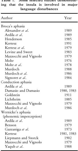
Table 1. Some selected studies demonstrat-ing that the insula is involved in major
language disturbances
Author Year
Broca’s aphasia
Alexanderet al. 1989
Ardilaet al. 1989
Henderson 1985
Kertesz 1991
Kerteszet al. 1979
Levine and Sweet 1983
Mazzocchi and Vignolo 1979
Mohr 1976
Mohret al. 1978
Murdoch 1988
Murdochet al. 1986
Signoretet al. 1984
Conduction aphasia
Ardilaet al. 1989
Damasio and Damasio 1980, 1983
Goldstein 1911
Lichtheim 1885
Mazzocchi and Vignolo 1979
Murdochet al. 1986
Wernicke’s aphasia (phonemic imperception)
Ardilaet al. 1989
Benson 1979
Gazzanigaet al. 1973
Kertesz 1981, 1983
Liepmann and Storck 1902
Mazzocchi and Vignolo 1979
Yaqubet al. 1988
An extensive body of clinical research has corroborated that the insula is quite frequently involved in the major aphasic syndromes. This body of clinical research has been previously reviewed (Ardilaet al. 1997) and it is summarized in table 1. These studies suggest a possible role for the insula in language processing and indicate that damage to the insula may frequently be a source of aphasia. Both the older autopsy-based studies and more recent brain image correlations suggest that anterior insula damage is often present in cases of moderate to severe Broca’s aphasia, middle insula damage is frequently correlated with repetition defects (conduction aphasia), and posterior insula damage co-occurs with the word deafness features of Wernicke’s aphasia.
that a similar language syndrome followed isolated extreme capsule damage and postulated that insular damage without extreme capsule involvement would not produce aphasia. Starksteinet al. (1988) observed crossed aphemia associated with a right insular lesion. Fifer (1993) described a patient with a lesion involving the right insula and adjacent white matter. The patient presented with a unilateral auditory processing disorder when speech materials were presented to the left ear. Habibet al. (1995) reported a case of bilateral insular damage, extending to a small part of the striatum on the left side, and to the temporal pole on the right. The patient presented mutism for about 1 month, did not respond to any auditory stimuli, and made no eåort to communicate.
As a matter of fact, mutism has been frequently observed in cases of insular pathology. Transient mutism is found in cases of left inferior motor cortex damage extending to the insula (Schiået al. 1983, Alexander et al. 1989), whereas lasting mutism appears to be associated with bilateral lesions of the frontal operculum and anterior insula (Sussmanet al. 1983, Cappaet al. 1987, Groswaseret al. 1988, Pineda and Ardila 1992). A case of crossed transient mutism in a right-handed patient has been reported. An ischemic lesion of the right insula with mild extension to the frontal operculum was demonstrated (Starksteinet al. 1988). Alexanderet al. (1989) point out that left cortical and sub-cortical opercular lesions may result in a total speech loss associated with a right hemiparesis. Right hemiparesis rapidly recovers, while the oral apraxia responsible for the mutism improves only slowly. Articulation is slow and eåortful, and syntactic errors are observed.
Shuren (1993) observed a patient who developed impaired speech initiation as a result of a left anterior insular infarct. The author proposed that dominant hemisphere anterior insular lesions impair the speech initiation loop. A possible interactive role of the left insula in speech initiation and language motivation could thus be conjectured. That is, disrupted motivation to speak will ipso facto aåect initiation.
Impairment in motor organization of speech represents a central defect in Broca’s aphasia (Benson and Ardila 1996). Dronkers (1996) showed that the left precentral gyrus of the insula is involved in motor planning of speech. Twenty-®ve stroke patients with a disorder in motor planning of articulatory movements, which Dronkers labelled as `apraxia of speech ’, were compared with 19 individuals without such de®cits. A robust double dissociation was observed. All patients with articulatory planning impairments presented lesions including the anterior insula. This area was completely spared in all patients without these articulatory defects. Hence, anterior insula represents the crucial brain area in motor planning and organization of speech. Verbal articulatory disruptions in some cases may be so severe as to result in mutism (Alexander et al. 1989, Pineda and Ardila 1992).
Table 2 summarizes some reports of insular damage associated with selective forms of language disturbances.
Contemporary neuroimaging technique studies have supported the hypothesis regarding an active involvement of the insula in linguistic processes. Activation of the insula has been demonstrated during word generation task performance (Baker
et al. 1997, McCarthyet al. 1993) and naming (Priceet al. 1996). Lexical knowledge
Table 2. Relatively restricted insular damage associated with selective forms of language disturbances
Author Characteristics
Alexanderet al. (1987) Aphasia with mildly paraphasic production and agraphia. Dronkers (1996) Motor planning of speech.
Habibet al. (1995) Mutism. Patient did not respond to any auditory stimuli. Disruption of the motivational mechanisms preluding to the
motoric aspects of human communication. Shuren (1993) Impaired speech initiation.
Starksteinet al. (1988) Aphemia.
lenticular nucleus. The insula would accordingly not participate in some unique and isolated language dimension, but rather would be active in diåerent linguistic aspects. These contemporary cerebral blood ¯ow studies in consequence support the assumption that the insula may be considered as a crucial brain language area. Activation of the insula has also been reported during phonological decision tasks (Rumseyet al. 1997). This ®nding, however, is not totally unexpected, considering that insular damage has been frequently observed in cases with phoneme imperception (Liepmann and Storck 1902, Mazzocchi and Vignolo 1979, Kertesz 1981, 1983, Yaqubet al. 1988).
It may therefore be concluded that contemporary neuro-imaging studies lend support to the assumption of a signi®cant participation of the insula in language. Futhermore, the insula would be involved not in a single linguistic activity, but in several verbal processes simultaneously. The anterior portion of the insula would be involved in the organization and planning of language articulation, while the middle and posterior portions would be involved with lexical knowledge, word retrieval, language understanding, and phonological discrimination.
Interestingly, it has even been suggested that the insula may be involved early in Alzheimer’s disease and that atrophy of the insula may partially contribute to the cognitive de®cits typical of early Alzheimer’s disease (Foundaset al. 1997). Naming di¬culties, word retrieval defects, semantic paraphasias, and a general decrease in lexical knowledge represent early linguistic defects in cases of Alzheimer’s disease (Cummings and Benson 1992).
Taken together, all these observations support an active participation of the insula in linguistic processes.
Role of the insula in language motivation and aåect
The insula has been further associated with a signi®cant number of processes. These include : oesophageal sensation (Aziz et al. 1997), tactual memories (Bonda
et al. 1996), vestibular projections (Bottini et al. 1994), spatial and temporal
auditory processing (Gri¬ths 1997), gustatory processing (Kabayakawa et al. 1996), and pain perception (Xuet al. 1997). Interestingly, the insula possesses not only contralateral motor and sensory representation but also ipsilateral motor and sensory representation (see Flynn et al. in this issue).
area 6 in the frontal lobe. The insula has been found to connect as well with the temporal pole and the superior temporal sulcus. Signi®cant projections to the cingulate gyrus, amygdaloid nucleus, perirhinal cortex, entorhinal and periamyg-daloid cortex have been observed (Augustine 1996). The insula in consequence maintains a complex system of interconnections not only with classical cortical language regions in the temporal and frontal lobe, but with a variety of limbic structures as well, including the cingulate gyrus and the perirhinal and entorhinal cortex. The assignment of a very complex role to the insula is accordingly fully justi®ed, not only in purely linguistic terms but also in terms of aåect and motivation.
Moreover, it can be proposed that the insula represents a major source of autonomic-visceral in¯uence on the sensory-motor association cortex. Among the areas of dominant hemisphere sensory-motor cortex under this in¯uence would be neuroanatomical structures crucial to language. From this it could be conjectured that the left insula would have a more direct in¯uence on motivation and emotion as expressed in spoken
}
written language (verbal aåect). The converse, a right insula in¯uence on non-verbal aåect, may also be considered. The insula may represent a crucial element participating in several distinct networks involved in verbal and non-verbal communication (Mesulam 1985). The anatomy and connections of the insula are revised in another paper in this issue (see Flynnet al.Anatomy of the Insula).
Recent reports have supported the hypothesis that the insula may play a signi®cant role in verbal aåect and linguistic motivation. Habibet al. (1995) have proposed that the insula may represent one key component of a ®nely tuned attentional system whose function would be not only to select the relevant information from the continuous ¯ow of auditory inputs, but also to trigger an adequate inter-hemispheric balance according to the verbal or non-verbal nature of the current stimulus. Habibet al. (1995) have proposed that bilateral damage to the insula would disrupt the motivational mechanisms that lead to the motoric production of human communication, by depriving them of connections with various limbic structures. Anatomical studies suggest that the insula could well act as the cortical representation of the limbic (autonomic) nervous system and, as such, may provide a direct input from the limbic-emotion system that could in turn in¯uence the aåective tone and content of language output.
Conclusion
The brain language zone should be reconsidered to include not only the perisylvian area of the left hemisphere but also the insula.
References
Albert, M. L., Goodglass, H., Helm, N. A., Rubens, A. B. and Alexander, M. P. 1981,Clinical
Aspects of Dysphasia(New York : Springer-Verlag).
Alexander, M. P., Benson, D. F. and Stuss, D. T. 1989, Frontal lobes and language.Brain and
Language,37, 656±691.
Alexander, M. P., Naeser, M. A. and Palumbo, C. L. 1987, Correlations of subcortical CT lesions
sites and aphasia pro®les.Brain,110, 961±991.
Ardila, A., Benson, D. F. and Flynn, F. G. 1997, Participation of the insula in language.
Aphasiology,11, 1159±1170.
Ardila, A., Rosselli, M. and Pinzon, O. 1989, Alexia and agraphia in Spanish-speakers: CT
correlations and interlinguistic analysis. In A. Ardila and F. Ostrosky (Eds)Brain Organization
of Language and Cognitive Processes(New York : Plenum Press), pp. 147±176.
Augustine, J. R. 1996, Circuitry and functional aspects of the insular lobe in primates.Brain Research
Review,22, 229±244.
Aziz, Q., Andersson, J. L., Valind, S., Sundin, A., Hamdy, S., Jones, A. K., Foster, E. R.,
Langstrom, B. and Thompson, D. G. 1987, Identi®cation of human brain loci processing
esophageal sensation using positron emission tomography.Gastroenterology,113, 50±59. Baker, S. C., Frith, C. D. and Dolan, R. J. 1997, The interaction between mood and cognitive
function studied with PET.Psychological Medicine,27, 565±578.
Benson, D. F. 1979,Aphasia,Alexia and Agraphia(New York : Churchill Livingstone).
Benson, D. F. and Ardila, A. 1996,Aphasia : A Clinical Perspective(New York : Oxford University
Press).
Benson, D. F. and Geschwind, N. 1971, The aphasias and related disturbances. In A. B. Baker and
L. H. Bake (Eds)Clinical Neurology, vol 1 (New York : Harper and Row), pp. 112±140.
Bernheim, F. 1900,De l’Aphasie Motrice(Paris : These de Paris).
Bonda, E., Petrides, M. and Evans, A. 1996, Neural systems for tactual memories.Journal of
Neurophysiology,75, 1730±1737.
Bottini, G., Sterzi, R., Paulesu, E., Vallar, G., Cappa, S. F., Erminio, F., Passingham, R. E.,
Frith, C. D. and Frackowiak, R. S. 1994, Identi®cation of the central vestibular projections in man : a positron emission tomography activation study.Experimental Brain Research,99, 164±169.
Cappa, S. F., Guidotti, M., Papagno, C. and Vignolo, L. A. 1987, Speechlessness with occasional vocalization after bilateral opercular lesions : a case study.Aphasiology,1, 35±39.
Cummings, J. L. and Benson, D. F. 1992,Dementia : A Clinical Approach(London: Butterworths),
2nd edition.
Damasio, H. and Damasio, A. R. 1983, Localization of lesions in conduction aphasias. In A. Kertesz
(Ed.)Localization in Neuropsychology (New York : Academic Press), pp. 231±244.
Dejerine, J. 1914,Semiologie des Aåections du Systeme Nerveux(Paris : Masson).
Dronkers, N. N. 1996, A new brain region for coordinating speech articulation.Nature, 384,
159±161.
Fifer, R. C. 1993, Insular stroke causing unilateral auditory processing disorder: Case report.Journal
American Academy of Audiology,4, 364±369.
Foundas, A. L., Leonard, C. M., Mahoney, S. M., Agee, O. F. and Heilman, K. M. 1997, Atrophy
of the hippocampus, parietal cortex, and insula in Alzheimer’s disease : A volumetric magnetic resonance imaging study.Neuropsychiatry,Neuropsychology,and Behavioral Neurology,10, 81±89. Freud, S. 1891,On Aphasia : A Critical Study(London: Imago Publishing Co. Ltd.).
Gazzaniga, M. S., Glass, A. A., Sarno, M. T. and Posner, J. B. 1973, Pure word deafness and
hemispheric dynamics: a case history.Cortex,9, 136±143.
Goldstein, K. 1911, Uber die amnestische and centrale aphasie.Archiv fuer Psychiatrie und Neurologie,
Goodglass, H. and Kaplan, E. 1972,The Assessment of Aphasia and Related Disorders(Philadelphia, PA : Lea and Febiger).
Griffiths, T. D., Rees, A., Witton, C., Cross, P. M., Shakir, R. A. and Green, G. G. R. 1997,
Spatial and temporal auditory processing de®cits following right hemisphere infarction. A psychophysical study.Brain,120, 785±794.
Groswaser, Z., Korn, C., Groswaser-Reider, I. and Solzi, P. 1988, Mutism associated with
buccofacial apraxia and bihemispheric lesions.Brain and Language,34, 157±168.
Habib, M., Daquin, G., Milandre, L., Royere, M. L., Rey, M., Lanteri, A., Slamanon, G. and
Khalil, R. 1995, Mutism and auditory agnosia due to bilateral insular damageÐrole of the
insula in human communication.Neuropsychologia,33, 327±339.
Head, H. 1920,Aphasia and Kindred Disorders(Cambridge: Cambridge University Press).
Henderson, V. W. 1985, Lesion localization in Broca’s aphasia: implications of Broca’s aphasia
without hemiparesis.Archives of Neurology,42, 1210±1212.
Kabayakawa, T., Endo, H., Ayabe-Kanamura, S., Kumagai, T., Yamagushi, Y., Kikuchi, Y.,
Takeda, T., Saito, S. and Ogawa, H. 1996, The primary gustatory area in human cerebral
cortex studied by magnetoencephalography.Neuroscience Letter,212, 155±158.
Kertesz, A. 1981, The anatomy of jargon. In J. Brown (Ed.)Jargonaphasia(New York : Academic
Press), pp. 63±112.
Kertesz, A. 1983, Localization of lesions in Wernicke’s aphasia. In A. Kertesz (Ed.)Localization in
Neuropsychology(New York : Academic Press), pp. 209±230.
Kertesz, A. 1991, Language cortex.Aphasiology,5, 207±234.
Kertesz, A., Harlock, W. and Coates, R. 1979, Computer tomography localization, lesion size, and
prognosis in aphasia and non-verbal impairment.Brain and Language,8, 34±50.
Levine, D. N. and Sweet, E. 1983, Localization of lesions in Broca’s aphasia. In A. Kertesz (Ed.)
Localization in Neuropsychology(New York : Academic Press), pp. 185±208.
Lichtheim, L. 1885, On aphasia.Brain,7, 433±484.
Liepmann, H. and Storck, E. 1902, Ein Fall von reiner Sprachtaubheit.Manuschfrift Psychiatrie und
Neurologie,17, 289±311.
Luria, A. R. 1966,Higher Cortical Functions in Man(New York : Basic Books). Luria, A. R. 1970,Traumatic Aphasia(The Hague: Mouton).
Mazzocchi, F. and Vignolo, L. A. 1979, Localization of lesion in aphasia: clinical-CT correlations
in stroke patients.Cortex,15, 627±654.
McCarthy, G., Blamire, A. M., Rothman, D. L., Gruetter, R. and Shulman, R. G. 1993,
Echo-plantar magnetic resonance imaging studies of frontal cortext activating during word generation in humans.Proceedings of the National Academy of Sciences,90, 4952±4956.
Mesulam, M. M. 1985, Patterns in behavioral neuroanatomy: association areas, the limbic system
and hemispheric specialization. In M. M. Mesulam (Ed.) Principles of Behavioral Neurology
(Philadelphia, PA : F. A. Davis Company), pp. 1±70.
Mesulam, M. M. and Mufson, E. J. 1985, The insula of Reil in man and monkey. Architectonics,
connectivity and function. In A. Peters and E. G. Jones (Eds)Cerebral Cortex, vol 4 (New York : Plenum Press), pp. 179±226.
Mohr, J. P. 1976, Broca’s area and Broca’s aphasia. In H. Whitaker and H. A. Whitaker (Eds)Studies
in Neurolinguistics, vol 1 (New York : Acacemic Press), pp. 201±236.
Mohr, J. P., Pessin, M. S., Finklestein, S., Funkestein, H. H., Duncan, G. D. and Davis, K. R. 1978, Broca aphasia: pathologic and clinical aspects.Neurology,28, 311±324.
Murdoch, B. E. 1988, Computerized tomography scanning: its contributions to the understanding
of the neuroanatomical basis of aphasia.Aphasiology,2, 437±462.
Murdoch, B. E., Thompson, D., Fraser, S. and Harrinson, L. 1986, Aphasia following non
hemorrhagic lesions in the left striato-capsular region. Australian Journal of Human
Communication Disorders,14, 5±21.
Nielsen, J. M. 1936,Agnosia,Apraxia and Aphasia : Their Value in Cerebral Localization(New York :
Hafner).
Nielsen, J. M. and Friedman, A. P. 1942, The quadrilateral space of Marie.Bulletin of Los Angeles
Neurological Society,8, 131±136.
Penfield, W. and Roberts, L. 1959, Speech and Brain Mechanisms (Princeton, NS : Princeton
University Press).
Pineda, D. and Ardila, A. 1992, Lasting mutism associated with buccofacial apraxia.Aphasiology,
Price, C. J., Moore, C. J., Humphreys, G. W., Frackowiak, R. S. and Friston, K. J. 1996, The neural regions sustaining object recognition and naming.Proceedings Royal Society of London,
263, 1501±1507.
Rousseaux, M., Steinling, M., Griffie, G., Quint, S., Cabaret, M., Lesoin, F., Mazingue, M.
and Destee, A. 1990, Correlation of thalamic aphasia and cerebral blood ¯ow. Revue de
Neurologie,146, 345±353.
Rumsey, J. M., Horwitz, B., Donohue, B. C., Nace, K., Maisog, J. M. and Andreason, P. 1997,
Phonological and orthographic components of word recognition. A PET-rCBF study.Brain,
120, 739±759.
Schiff, H. B., Alexander, M. P., Naeser, M. A. and Galaburda, A. M. 1983, Aphemia :
clinical-anatomical correlations.Archives of Neurology,40, 720±727.
Shuren, J. 1993, Insula and aphasia.Journal of Neurology,240, 216±218.
Signoret, J. L., Lhermitte, J., Abelent, R. and Lavorel, R. 1984, Rediscovery of Leborgne’s
brain : anatomical description with CT scan.Brain and Language,22, 303±309.
Starkstein, S. E., Berthier, M. and Leiguarda, R. 1988, Bilateral opercular syndrome and crossed
aphemia due to a right insular damage: A clinicopathological study.Brain and Language,34, 253±261.
Sussman, N. M., Gur, R. C., Gur, R. F. and O’Connor, M. J. 1983, Mutism as a consequence of
callosotomy.Journal of Neurosurgery,59, 514±519.
Wernicke, C. 1874,Der Aphasische Symtomenkomplex(Breslau : Cohn and Weigert).
Xu, X., Fukuyama, H., Yazawa, S., Mima, T., Hanakawa, T., Magata, Y., Kanda, M., Jujiwara, N., Shindo, K., Nagamine, T. and Shibasaki, H. 1997, Functional localization of pain perception in the human brain studied by PET.Neuroreport,8, 555±559.