Otras secciones de este sitio: ☞ ☞ ☞ ☞
☞ Índice de este número ☞
☞ ☞ ☞
☞ Más revistas ☞
☞ ☞ ☞
☞ Búsqueda
Others sections in this web site:
☞ ☞ ☞ ☞
☞ Contents of this number ☞
☞ ☞ ☞
☞ More journals ☞
☞ ☞ ☞ ☞ Search
Artículo:
Hypopituitarism
Derechos reservados, Copyright © 2005: Sociedad Mexicana de Nutrición y Endocrinología, AC
Revista de Endocrinología y Nutrición
Suplemento
Supplement 1
Julio-Septiembre
July-September 2 0 0 5
Volumen
Volume 1 3
edigraphic.com
Hipófisis
Hipófisis
Hipófisis
Hipófisis
Hipófisis
Hypopituitarism
Mark E. Molitch*
Mark E. Molitch*
Mark E. Molitch*
Mark E. Molitch*
Mark E. Molitch*
* Division of Endocrinology, Metabolism and Molecular Medicine. Northwestern University Feinberg School of Medicine.
Hypopituitarism indicates the diminished production of one or more anterior pituitary hormones. Although the recog-nition of complete or panhypopituitarism is usually straight-forward, the detection of partial or selective hormone deficiencies is more challenging. Pituitary hormone defi-ciencies can be caused by loss of hypothalamic stimula-tion (tertiary hormone deficiency) or by direct loss of pi-tuitary function (secondary hormone deficiency). The distinction between hypothalamic and pituitary causes of hypopituitarism is important for establishing the correct diagnosis and but less so when applying and interpret-ing the relevant diagnostic endocrine tests. With improved procedures for testing the hypothalamic-pituitary axis, it is apparent that hypothalamic causes of hypopituitarism are more common than previously appreciated. When hypopituitarism is accompanied by diabetes insipidus or hyperprolactinemia, one should particularly consider hy-pothalamic causes of pituitary dysfunction.
CAUSES OF HYPOPITUITARISM
A variety of congenital and acquired causes of hypopitu-itarism have been described (Table I). Sporadic and fa-milial forms of panhypopituitarism occur, but the underly-ing genetic or developmental defects have not been elucidated. Congenital combined deficiencies of GH, PRL, and TSH are caused by mutations in the gene encoding Pit-1, a pituitary-specific transcription factor that is involved in the development of somatotroph, lactotroph, and thy-rotroph cell lineages. Different types of Pit-1 mutations are inherited in an autosomal dominant or recessive pat-tern. When gonadotrophs are also deficient, mutations may be present in the genes for transcription factors that are active earlier in the development of the pituitary lin-eages, such as Lhx3 and Prop-1.
Gene mutations have been found at several steps lead-ing to pituitary hormone secretion, includlead-ing those for the hypophysiotropic releasing factor receptors for GnRH, GHRH, and TRH; those for the pituitary hormone structures for GH, ACTH, and the a subunits of FSH, TSH, and LH; and those for the target organ receptors for GH, ACTH, TSH, and LH. Best studied are mutations of the GH gene, which include large deletions and point mutations; some of these can be inher-ited in an autosomal dominant manner, apparently because the mutant hormone impairs GH biosynthesis and normal function of the somatotroph cell. Mutations of the other types described earlier generally cause autosomal recessive forms of selective hormone deficiencies.
Mark E. Molitch. Hypopituitarism S48
edigraphic.com
Mutations responsible for these developmental defects are the subject of active investigation. One possible mu-tation that has been found is in the Hesx1 gene (also called Rpx, for Rathke’s pouch homeobox), which is a member of the paired-like class of homeobox genes ex-pressed in the thickened layer of oral ectoderm that gives rise to Rathke’s pouch. Many other transcription factors have been described that are expressed sequentially during embryogenesis in Rathke’s pouch that are impor-tant in the ultimate development of the normal pituitary cell lineages. Mutations have been found in many of these transcription factors genes, including Pit-1 and Prop-1 (discussed above); Lhx3 and Lhx4 which give similar clin-ical findings to Prop-1 mutations but have additional cer-vical spine and skull base abnormalities; and Sox3, which gives rise to isolated GH deficiency and variable mental retardation and facial abnormalities . Combined pituitary hormone deficiency has an incidence of about 1 in 8,000 births and about 10% have an affected relative; it ap-pears that more than half of these cases are due to Pit-1 or Prop-1 deficiencies.
Neoplastic lesions, particularly pituitary adenomas, are the most common cause of acquired hypopituitarism. Pi-tuitary adenomas cause hypopituitarism in several differ-ent ways. In some cases, there is direct destruction or compression of the normal pituitary. Compression of the pituitary stalk can impair blood supply to the pituitary as well as decrease input from hypothalamic hormones. Hemorrhage into tumors can lead to pituitary infarction. When tested carefully, most patients with macroadenom-as have partial deficiencies of one or more pituitary hor-mones, most often involving GH and gonadotropins. A mild degree of hyperprolactinemia is characteristic of dis-orders that cause stalk compression, and hyperprolactine-mia further impairs gonadotropin secretion. A variety of other neoplasms that occur near the sella, such as cran-iopharyngiomas, can also cause hypopituitarism.
Radiation causes hypopituitarism primarily because of its effects on hypothalamic function, although high-dose radiation (e.g., proton beam) can also cause direct pitu-itary damage. The sellar region is subjected to radiation in the treatment of pituitary adenomas, craniopharyngi-omas, optic glicraniopharyngi-omas, meningicraniopharyngi-omas, dysgermincraniopharyngi-omas, and neoplasms of the oropharynx. Importantly, the effects of radiation can be delayed as much as several years, and patients at high risk should be evaluated at about yearly intervals for radiation-induced hypopituitarism. Although GH and gonadotropin deficiencies develop first in most patients, ACTH or TSH deficiencies occasionally occur first, emphasizing the need to evaluate each of the major axes. Empty sella syndrome can occur as a primary or as an acquired condition. It is caused by defects in the diaphrag-ma sellae that allow herniation of the arachnoid membrane
Table 1. Causes of hypopituitarism. Genetic defects
Hypophysiotropic hormone gene defects
Hypophysiotropic hormone receptor gene defects GHRH receptor defect
GnRH receptor defect TRH receptor defect
Pituitary hormone gene defects
Gonadotropins: LH β- and FHS β-subunit gene defects Growth hormone: defects in GH gene
Thyrotropin: defects in TSH β-subunit
Multiple hormone (GH, PRL, TSH) defects: due to mutation in Pit-1 gene and Propl gene
Pituitary hormone receptor genetic defects
Growth hormone receptor defects: GH insensitivity syndrome (Laron-type dwarfisin)
ACTH receptor defects: congenital insensitivity to ACTH LH receptor defects
FSH receptor defects TSH receptor defects
Congenital embryopathic defects Anencephaly
Midline cleft defects: septo-optic dysplasia, basal encephalo-cele, cleft lip and palate
Pituitary aplasia
Kallmann’s syndrome (GnRH defect with anosmia)
Acquired defects
Tumors: pituitary adenomas, craniopharyngiomas, dysgermi-nomas, meningiomas, gliomas, metastatic tumors, hamarto-mas, Rathke’s cleft cysts
Irradiation
Trauma: surgery, external blunt trauma Empty sella syndrome
Vascular
Pituitary apoplexy Sheehan’s syndrome Internal carotid aneurysm Vasculitis
Inflammatory/infiltrative diseases Sarcoidosis
Langerhans’ cell histiocytosis (histiocytosis X, eosinophilic granuloma)
Tuberculosis, syphilis Meningitis
Lymphocytic hypophysitis, infundibulohypophysitis Metabolic
edigraphic.com
into the hypophyseal fossa. In long-standing cases, sellar enlargement occurs, probably because of persistent trans-mission of intracranial pressure. With appropriate imaging studies, the pituitary gland can be seen as a flattened rim of tissue along the floor of the sella. Primary empty sella oc-curs most commonly in women and may be associated with features of benign intracranial hypertension. Pituitary func-tion in patients with primary empty sella syndrome is usual-ly normal, although 15% have mild hyperprolactinemia, prob-ably because of stretching of the pituitary stalk. Acquired forms may occur as a result of surgery, radiation, or pitu-itary infarction (usually of an adenoma).
Pituitary apoplexy is usually caused by hemorrhage into a tumor with associated infarction. In the absence of a tumor, predispositions to apoplexy include trauma,
preg-nancy, anticoagulation, sickle cell anemia, and diabetes mellitus. Pituitary infarction in the peripartum period is referred to as Sheehan’s syndrome and is usually associ-ated with significant obstetric hemorrhage and hypov-olemia. Although Sheehan’s syndrome may manifest acutely with vascular collapse, it more commonly has a subacute manifestation consisting of postpartum inability to lactate, amenorrhea, and symptoms of adrenal insuf-ficiency. Sheehan’s syndrome is now infrequent, owing to improvements in obstetric care.
Infiltrative diseases such as sarcoidosis, histiocytosis, and tuberculosis usually cause hypopituitarism by infil-trating the hypothalamus and stalk rather than the pitu-itary. In lymphocytic hypophysitis, there is massive infil-tration of the pituitary by lymphocytes and plasma cells
Table II. Test of pituitary insufficiency.
Hormone Test Interpretation
Growth hormone Insulin tolerance test: Regular insulin If hypoglycemia occurs (glucose < 40 mg/dL), (GH) (0.5-0.15 U/kg) is given IV and blood is GH should increase to > 5 µg/L.*
drawn at -30, 0, 30, 45, 60, and 90 min for measurement of glucose and GH
Arginine-GHRH test: GHRH 1µg/kg IV bolus Normal response is GH > 4.1 µg/L followed by 30 min finsuion of L-arginine (30g)
Adrenocorticotropic Insulin tolerance test: Regular insulin If hypoglycemia occurs (glucose < 40 mg/dL),
Hormone (ACTH) (0.5-0.15 U/kg) is given IV and blood is cortisol should increase by > 7 µg/dL or to >20 µg/dL. drawn at -30, 0, 30, 45, 60, and 90 min for
measurement of glucose and cortisol.
Metyrapone test: Metyrapone (30 mg/kg-max. A normal response is 11-deoxycortisol > 7.5 µg/dL or 2 g) at midnight with measurements of plasma ACTH > 75 pg/mL. Plasma cortisol should fall below 4 11-deoxycortisol and cortisol at 8 AM. µg/dL to ensure an adequate response.
ACTH can also be measured. A 3-day test is also available. Basal cortisol should be > 5-6 µg/dL before test.
ACTH stimulation test: ACTH 1-24 (Cosyntropin), A normal response is cortisol > 18 µg/dL. In suspected 0.25 mg IM or IV. Cortisol is measured at hypothalamic-pituitary deficiency, a low dose (1 µg) 0, 30, and 60 min. test may be more sensitive.
Thyroid-stimulating Basal thyroid function tests: free T4, free T3, Low free thyroid hormone levels in the setting of TSH
hormone (TSH) TSH. levels that are not appropriately increased.
Luteinizing hormone Basal levels of LH, FSH, testosterone, estrogen Basal LH and FSH should be increased in postmeno-(LH), follicle-stimulating pausal women. Low testosterone levels in
hormone (FSH) conjunction with low or low-normal LH and FSH are
consistent with gonadotropin deficiency.
Mark E. Molitch. Hypopituitarism S50
edigraphic.com
sustraídode-m.e.d.i.g.r.a.p.h.i.c cihpargidemedodabor
with destruction of the parenchyma; it is believed to have an autoimmune basis. The lesion that develops is usually large, and patients present with either symptoms or signs of hypopituitarism or those of a mass lesion (i.e., visual field defects and/or headaches). Some patients may have mild hyperprolactinemia and diabetes insipidus. There seems to be a particular prediliction for damage to the corticotroph cells, resulting in ACTH/adrenal insufficiency. Almost all cases have been reported in women, and most present during or after pregnancy. Because of the pre-sentation as a mass lesion during pregnancy, such lesions may be confused with prolactinomas, but the mild PRL elevation points to a nonsecretory lesion rather than a prolactinoma. MRI cannot reliably differentiate pituitary adenoma from hypophysitis, although hypophysitis usu-ally manifests with a diffuse enlargement of the pituitary that enhances, rather than as a focal lesion. Diagnosis is usually made by biopsy, but the lesion may be suspect-ed clinically if it manifests during or just after pregnancy. Careful pituitary function testing is mandatory, because many of the patients in the reported cases went undiag-nosed and died of adrenocortical insufficiency. Although
the prognosis is not clear, a number of cases have re-solved spontaneously. An entity with similar histologic find-ings involving the stalk and posterior pituitary, referred to as infundibuloneurohypophysitis, can cause diabetes insipidus. The causes and interrelationships between these entities remain unknown.
The pituitary may undergo damage because of iron deposition in patients with hemochromatosis and amy-loid fibrils in patients with systemic amyamy-loidosis. Function-al, reversible hypopituitarism of varying degrees occurs in patients with severe systemic illness, severe psychosocial and emotional deprivation, and severe weight loss -particularly in those with anorexia nervosa.
DIAGNOSIS AND TREATMENT
The diagnosis of hypopituitarism rests on the stimu-lation tests that are summarized in Table II. Children di-agnosed with GH deficiency during childhood should be retested as adults when considering continuation of GH therapy, unless the GH deficiency was documented to be due to a proven genetic defect or structural lesions
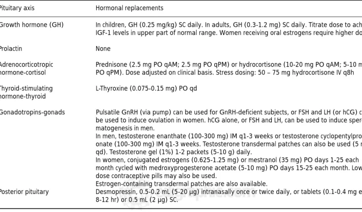
Table III. Hormone replacement in hypopituitarism. Pituitary axis Hormonal replacements
Growth hormone (GH) In children, GH (0.25 mg/kg) SC daily. In adults, GH (0.3-1.2 mg) SC daily. Titrate dose to achieve IGF-1 levels in upper part of normal range. Women receiving oral estrogens require higher doses
Prolactin None
Adrenocorticotropic Prednisone (2.5 mg PO qAM; 2.5 mg PO qPM) or hydrocortisone (10-20 mg PO qAM; 5-10 mg hormone-cortisol PO qPM). Dose adjusted on clinical basis. Stress dosing: 50 – 75 mg hydrocortisone IV q8h
Thyroid-stimulating L-Thyroxine (0.075-0.15 mg) PO qd hormone-thyroid
Gonadotropins-gonads Pulsatile GnRH (via pump) can be used for GnRH-deficient subjects, or FSH and LH (or hCG) can be used to induce ovulation in women. hCG alone, or FSH and LH, can be used to induce sper-matogenesis in men.
In men, testosterone enanthate (100-300 mg) IM q1-3 weeks or testosterone cyclopentylpropi-onate (100-300 mg) IM q1-3 weeks. Testosterone transdermal patches can also be used (5 mg qd). Testosterone gel (1%) 1-2 packets (5-10 g) daily.
In women, conjugated estrogens (0.625-1.25 mg) or mestranol (35 mg) PO days 1-25 each month cycled with medroxyprogesterone acetate (5-10 mg) PO days 15-25 each month. Low-dose contraceptive pills may also be used.
Estrogen-containing transdermal patches are also available.
Posterior pituitary Desmopressin, 0.5-0.2 mL (5-20 µg) intranasally once or twice daily, or tablets (0.1-0.4 mg every 8-12 hr) or 0.5 mL (2 µg) SC.
edigraphic.com
:rop odarobale FDP
VC ed AS, cidemihparG
arap
acidémoiB arutaretiL : cihpargideM
sustraídode-m.e.d.i.g.r.a.p.h.i.c
causing multiple hormone deficiencies. A recent study evaluated the relative performance of the GHRH-argin-ine test, the ITT, arginGHRH-argin-ine alone, clonidGHRH-argin-ine, levodopa, and the combination of arginine plus levodopa in the diag-nosis of adult GH deficiency. The overall performance of the GHRH-arginine test, with 95% sensitivity and 91% specificity at a GH cutoff of 4.1 µg/L at the central labo-ratory used, compared well to the ITT, which had an optimal GH cutoff of 5.1 µg/L (96% sensitivity and 92% specificity). However, if a patient has deficiencies in three or more other axes, they have a greater than 95% chance of being GH deficient. Measurement of IGF-I alone is not sufficient to make a diagnosis of GH deficiency, as many patients with defective GH responses have IFG-I level in the low-normal range.
Therapy for hypopituitarism depends on the nature and severity of the hormone deficiencies as well as on the desired clinical endpoints. The goal is to replace hormones in a physiologic manner, with efforts to avoid the consequences of overreplacement. In patients with acquired forms of hypopituitarism (e.g., pituitary tu-mors, radiation treatment), it is not uncommon to en-counter a mixture of partial hormone deficiencies. It is generally prudent to provide hormone replacement if partial deficiency is suspected, because patients may experience symptoms over a number of years before an unequivocal diagnosis of hormone deficiency is made. Examples of hormonal replacement paradigms are provided in Table III. Adjustment of hormone doses is done primarily based on clinical findings; it should be remembered that the TSH level is not helpful for adjusting thyroxine doses in patients with central hy-pothyroidism. Even when conventional hormone re-placement (adrenal, thyroid, gonadal) is carried out appropriately, there is an approximately twofold ex-cess risk of death reported in patients with hypopitu-itarism.
Although untreated GH deficiency has been hypoth-esized to be the cause of this excess risk, this has not been proven. The benefits of GH therapy are less clear than those for the other pituitary hormones and include improvements in body composition, bone and quality of life; although there are few adverse effects, treat-ment involves daily injections. GH in adults is generally started at a dose of 0.2 – 0.3 mg daily and adjusted upwards gradually with the goal of achieving an IGF-1 level in the upper part of the normal range. Women receiving oral estrogens usually require substantially higher doses of GH than men. It should be emphasized that for GH treatment, long-term clinical outcomes stud-ies on hard endponts such as fractures, clinical heart disease, cancer, and mortality are still lacking at present.
SUGGESTED READING SUGGESTED READING SUGGESTED READING SUGGESTED READING SUGGESTED READING
1. Agha A, Rogers B, Sherlock M et al. Anterior pituitary dys-function in survivors of traumatic brain injury. J Clin Endo-crinol Metab 2004; 89: 4929-4936.
2. Aimaretti G, Ambrosio MR, Di Somma C et al. Traumatic brain injury and subarachnoid haemorrhage are conditions at high risk for hypopituitarism: screening study at 3 months after the brain injury. Clin Endocrinol (Oxf) 2004; 61(3): 320-326.
3. Arafah BM. Reversible hypopituitarism in patients with large non-functioning pituitary adenomas. J Clin Endocrinol Metab 1986; 62: 1173-1179.
4. Arafah BM. Medical management of hypopituitarism in pa-tients with pituitary adenomas. Pituitary 2002; 5: 109-117. 5. Bates AS, Van’t Hoff W, Jones PJ, Clayton RN. The effect of hypopituitarism on life expectancy. J Clin Endocrinol Metab 1996; 81: 1169-1172.
6. Biller BMK, Samuels MH, Zagar A et al. Sensitivity and spec-ificity of six tests for the diagnosis of adult growth hormone deficiency. J Clin Endocrinol Metab 2002; 87: 2067-2079. 7. Cohen LE, Radovick S. Molecular basis of combined
pitu-itary hormone deficiencies. Endocr Rev 2002; 3: 431-442. 8. Feigl GC, Bonelli CM, Berghold A, Mokry M. Effects of gam-ma knife radiosurgery of pituitary adenogam-mas on pituitary function. J Neurosurg 2002; 97(5 suppl): 415S-421S. 9. Gama R, Smith MJ, Wright J, Marks V. Hypopituitarism in
primary haemochromatosis: recovery after iron depletion. Postgrad Med J 1995; 71: 297-298
10. Hartman ML, Crowe BJ, Biller BMK, Ho KY, Clemmons DR, Chipman JJ. Which patients do not require a growth hor-mone (GH) stimulation test for the diagnosis of adult GH deficiency? J Clin Endocrinol Metab 2002; 87: 477-485. 11. Kaltsas GA, Powles TB, Evanson J et al.
Hypothalamo-pi-tuitary abnormalities in adult patients with Langerhans cell histiocytosis: clinical, endocrinological, and radiological fea-tures, and response to treatment. J Clin Endocrinol Metab 2000: 85; 1370-1376.
12. Lamberts SWJ, deHerder WW, van der Lely AJ. Pituitary insufficiency. Lancet 1998; 352: 127-134.
13. Littley MD, Shalet SM, Beardwell CG, Ahmed SR, Apple-gate G, Sutton ML. Hypopituitarism following external ra-diotherapy for pituitary tumours in adults. Q J Med 1989; 70(262): 145-160.
14. Rosen T, Bengtsson BA. Premature mortality due to car-diovascular disease in hypopituitarism. Lancet 1990; 336: 285-288.
15. Tomlinson JW, Holden N, Hills RK et al. The West Midlands Prospective Hypopituitary Study Group. Association be-tween premature mortality and hypopituitarism. Lancet 2001; 357: 425-431.
16. Vance ML. Hypopituitarism. N Engl J Med 1994; 330: 1651-1662.
17. Webb SM, Rigla M, Wägner A, Oliver B, Bartumeus F. Re-covery of hypopituitarism after neurosurgical treatment of pituitary adenomas. J Clin Endocrinol Metab 1999; 84: 3696-3700.