Published Ahead of Print 17 September 2010.
10.1128/AEM.01135-10.
2010, 76(21):7243. DOI:
Appl. Environ. Microbiol.
Berrocal and Mauricio Bittner
Pamela Machuca, Leslie Daille, Enrique Vinés, Liliana
Fusobacterium nucleatum
for the Periodontal Pathogen
Isolation of a Novel Bacteriophage Specific
http://aem.asm.org/content/76/21/7243
Updated information and services can be found at:
These include:
REFERENCES
http://aem.asm.org/content/76/21/7243#ref-list-1
at:
This article cites 36 articles, 10 of which can be accessed free
CONTENT ALERTS
more»
articles cite this article),
Receive: RSS Feeds, eTOCs, free email alerts (when new
http://journals.asm.org/site/misc/reprints.xhtml Information about commercial reprint orders:
http://journals.asm.org/site/subscriptions/ To subscribe to to another ASM Journal go to:
on August 10, 2012 by UNIVERSIDAD ANDRES BELLO
http://aem.asm.org/
0099-2240/10/$12.00 doi:10.1128/AEM.01135-10
Copyright © 2010, American Society for Microbiology. All Rights Reserved.
Isolation of a Novel Bacteriophage Specific for the Periodontal
Pathogen
Fusobacterium nucleatum
䌤
Pamela Machuca,
1Leslie Daille,
1Enrique Vine
´s,
1Liliana Berrocal,
1and Mauricio Bittner
1,2*
Laboratorio de Microbiologia y Biotecnologia Oral, Departamento de Ciencias Biologicas, Facultad de Ciencias Biologicas, Universidad Andres Bello, Santiago, Chile,1and Facultad de Odontologia, Universidad Andres Bello, Santiago, Chile2
Received 11 May 2010/Accepted 4 September 2010
Fusobacterium nucleatumis a periodontal pathogen that has been directly associated with the development and progression of periodontal disease, a widespread pathology that affects the support tissues of the tooth. We isolated a new bacteriophage (Fnp⌽02) that specifically infects this bacterium. Transmission electron micros-copy showed that the virion is composed of an icosahedral head and a segmented tail. The size of the phage genome was estimated to be approximately 59 kbp of double-stranded DNA. The morphological features and the genetic characteristics suggest that Fnp⌽02 is part of theSiphoviridaefamily. Using one-step growth and adsorption experiments, the latent period, burst size, and adsorption rate were estimated to be 15 h, 100 infectious units per cell, and 7.5ⴛ10ⴚ10ml minⴚ1, respectively. A small fragment of phage DNA was cloned and sequenced, showing 93% nucleotide identity with the phage PA6 ofPropionibacterium acnesand amino acid identity with fragments of two proteins (Gp3 and Gp4) of this phage. To our knowledge, Fnp⌽02 is the first phage described to infectFusobacterium nucleatumand provides the base for future exploration of phages in the control of periodontal disease.
The term “periodontal disease” refers to a wide set of patho-logical alterations of the periodontal tissue. The most common clinical manifestations are known as gingivitis and periodonti-tis, and both are widely distributed around the world (18). Periodontitis is a multifactorial inflammatory-based infection of the supporting tissues of the tooth. It is essentially charac-terized by the progressive destruction of the periodontal liga-ment and the alveolar bone, leading to the loss of the affected tooth (2). Periodontitis is caused by bacteria or bacterial groups embedded in a biofilm or dental plaque that protects them against antimicrobial agents (18). The bacterial species involved in periodontal disease are predominantly Gram-neg-ative anaerobes, and although they are usually isolated from affected patients, they are also isolated from healthy individu-als, but in a lesser proportion and frequency (26).
Fusobacterium nucleatum is an anaerobic, Gram-negative, long bacillus and a member of the microflora in the oral cavity. F. nucleatumis considered a periodontal pathogen because it is frequently isolated from lesions, produces a high number of tissue irritants, and has the ability to form coaggregates with other periodontal pathogens, acting as a bridge between early and late colonizers in the surface of the enamel (4, 11). Three different subspecies ofF. nucleatumhave been related to the pathology of periodontal disease,F. nucleatumsubsp. nuclea-tum, subsp. polymorphum, and subsp. vincentii, all of which have been associated with lesions of periodontitis but also have been isolated in high numbers from successfully treated pa-tients (9).
Bacteriophages are viruses that can infect and kill only
bac-teria and have been used for many years as powerful tools for the study of bacterial genetics and, given their specificity, used in the identification and characterization of microorganisms (phage typing). Nevertheless, phages were originally described as therapeutic elements to treat human and animal infections (34). This application, known as phage therapy, has regained interest in the past years, particularly in an era when antibiotic resistance and biofilm-based infections are permanent issues (25). Bacteriophages are denominated “temperate” when their genetic material is integrated within the bacterial genome with no immediate lysis of the bacterium until, under certain con-ditions, the expression of the viral genome is induced and the production of new virus particles lyses the host cell; they are called “lytic” or “virulent” when, immediately after the infec-tion, they redirect the bacterial metabolism to the production of new phages, which are released during the bacterial lysis (22, 36). There are many examples of the use of bacteriophages at a clinical (14, 32) and commercial level (20). Specifically in the dentistry area, several bacteriophages that infect diverse oral bacteria have been isolated from saliva and dental plaque (12, 13, 23, 37).
AlthoughF. nucleatumis an important periodontal patho-gen, reports of bacteriophages for this microorganism do not exist. In this work, we isolated and characterized a new bacte-riophage for F. nucleatum from a saliva sample, designated Fnp⌽02, and to our knowledge this is the first bacteriophage for this bacterium.
MATERIALS AND METHODS
Bacterial strains and growth conditions.Bacterial strains used in this study are listed in Table 1. TheF. nucleatumsubsp. polymorphumclinical isolate strain used as the host for the isolation, dilution, and propagation of the Fnp⌽02 phage was called Fnp.F. nucleatumstrains were cultured anaerobically in brain heart infusion (BHI) broth (Merck) or BHI agar at 37°C for 3 days. All bacteria from the oral flora were incubated under the same conditions except black-pigmented bacteria, which were cultivated in BHI agar with sheep blood (5%)
supple-* Corresponding author. Mailing address: Laboratorio de Microbio-logia y BiotecnoMicrobio-logia Oral, Departamento de Ciencias Biologicas, Uni-versidad Andres Bello, Echaurren 237, Santiago, Chile. Phone: (562) 770-3027. Fax: (562) 464-1630. E-mail: [email protected].
䌤Published ahead of print on 17 September 2010.
7243
on August 10, 2012 by UNIVERSIDAD ANDRES BELLO
http://aem.asm.org/
mented with 10g/ml hemin-menadione (BBL; BD Ltd.) for at least 5 days at the same temperature. All aerobically grown bacteria used in the study were incubated in Luria-Bertani (LB) broth (10 g/liter tryptone, 5 g/liter yeast extract, and 5 g/liter NaCl) or LB agar at 37°C for 24 to 48 h. Chloramphenicol (20
g/ml) was added when necessary. Bacterial growth was monitored by measuring optical density at 600 nm (OD600).
Isolation ofF. nucleatumstrains.Samples from the saliva and tongues of healthy individuals and patients with periodontal disease were obtained from the Dental Clinic of the Universidad Andres Bello, Chile. All samples were grown in a selective medium forF. nucleatumcalled CVE (10 g/liter soy Trypticase, 5 g/liter yeast extract, 5 g/liter NaCl, 0.2% glucose, 0.02% tryptophan, 5% sheep blood, 5g/ml crystal violet, and 4g/ml erythromycin) (35) and incubated under anaerobic conditions for 4 days at 37°C. Then, the blue colonies were picked and cultured for their identification. Initially, all clinical isolates were confirmed asF. nucleatumby PCR (5) and RAPID32A (bioMe´rieux, France). To determine which subspecies we were working with, we used specific PCR primers described in the literature (17, 29) to identifyF. nucleatumsubsp.vincentiiand
F. nucleatumsubsp.nucleatumand designed PCR primers based on a reported sequence to identifyF. nucleatumsubsp.polymorphum(FnpF, 5⬘-CCAGGAGG AATAGGGGTAGG-3⬘; FnpR, 5⬘-GCCATTTCAGCTTCAACTCC-3⬘). The
PCR program used for the determination ofF. nucleatumsubsp.nucleatum, subsp.polymorphum, and subsp.vincentiiwas as follows: initial denaturation for 5 min at 94°C; 35 cycles of 94°C for 30 s, 50°C for 30 s, and 72°C for 30 s; and a final extension at 72°C for 10 min. A 100-l reaction mixture contained 0.5 UTaq
polymerase (1.5 mM MgCl2, 2 mM each deoxynucleoside triphosphate [dNTP],
1M each primer, and 0.1 to 0.5g of template DNA) (Invitrogen, Carlsbad, CA). All PCRs were performed in a Multigene thermocycler (Labnet Inc.).
Isolation of bacteriophages.Saliva samples from 25 healthy individuals and from 85 periodontally affected patients who had not received antibiotics within the previous 3 months and drainage samples from dental chairs were used for the bacteriophage screening. One milliliter of each saliva sample was cleared of debris and bacteria by centrifugation at 15,000⫻gfor 10 min. Supernatant fluids were collected and kept at 4°C until used. To enrich bacteriophages in saliva samples, we inoculated 100l of the clear supernatant on a mid-log culture of the Fusobacterium nucleatum Fnp strain (Public Health Institute, Santiago, Chile) in BHI broth (Oxoid, Basingstoke, United Kingdom) and incubated it anaerobically for 48 h. After this period, bacteria were harvested (15,000⫻gfor 3 min), and the supernatant was recovered and filtered (0.45-m Millipore filter). Five microliters of this enriched saliva sample was spotted onto double-layered plates containing 100l of a stationary-phase culture ofF. nucleatum
TABLE 1. Host range of phage Fnp⌽02
Bacterium type and species No. of strains
used Reference or source
c Phage
sensitivityd Fusobacteria
Fusobacterium nucleatumsubsp.polymorphum(Fnp)a ISP ⫹⫹
Fusobacterium nucleatumsubsp.polymorphum 3 Clinical isolates, this work ⫹⫹
Fusobacterium nucleatumsubsp.nucleatumATCC 25586 ATCC ⫹
Fusobacterium nucleatumsubsp.nucleatum 3 Clinical isolates, this work ⫹
Fusobacterium nucleatumsubsp.vincentii 3 Clinical isolates, this work ⫹
Fusobacterium necrophorumATCC 25286 ATCC ⫺
Fusobacteriumspeciesb 8 Clinical isolates, this work ⫺
Gram-positive bacteria (oral cavity)
Propionibacterium acnes 4 Clinical isolates, this work ⫺
Actinomyces naeslundii 2 Clinical isolates, this work ⫺
Bacteroides urealyticus 2 Clinical isolates, this work ⫺
Bacteroides vulgatusATCC 8482 ATCC ⫺
Eubacterium limosum 4 Clinical isolates, this work ⫺
Eubacterium lentum 4 Clinical isolates, this work ⫺
Gram-negative bacteria (oral cavity)
Aggregatibacter actinomycetemcomitans 5 Clinical isolates, this work ⫺
Porphyromonas gingivalisATCC 33277 ATCC ⫺
Porphyromonas endodontalis 2 Clinical isolates, this work ⫺
Prevotella nigrescens 4 Clinical isolates, this work ⫺
Prevotella intermedia 3 Clinical isolates, this work ⫺
Peptostreptococcus anaerobius Clinical isolates, this work ⫺
Streptococcus mutansATCC 25175 ATCC ⫺
Streptococcus sanguinis 2 Clinical isolates, this work ⫺
Other Gram-negative bacteria
Escherichia coliDH5␣ Laboratory stock ⫺
Pseudomonas aeruginosa ISP ⫺
Pseudomonas oxytoca ISP ⫺
Proteus mirabilis ISP ⫺
Proteus vulgaris ISP ⫺
Klebsiella pneumoniae ISP ⫺
Salmonella entericaserovar Typhimurium ATCC 14028 ATCC ⫺
Salmonella entericaserovar Typhi TY2 ISP ⫺
Other Gram-positive bacteria
Staphylococcus aureusATCC 43330 ATCC ⫺
Staphylococcus epidermidisATCC 14990 ATCC ⫺
Bacillus cereus ISP ⫺
Streptococcus pyogenes ISP ⫺
aStrain used for isolation of Fnp⌽02.
bIdentified asFusobacteriumby Rapid32A, but no match for species. cISP, Public Health Institute, Santiago, Chile.
d⫹⫹, sensitive;⫹, low sensitivity;⫺, insensitive.
7244 MACHUCA ET AL. APPL. ENVIRON. MICROBIOL.
on August 10, 2012 by UNIVERSIDAD ANDRES BELLO
http://aem.asm.org/
mixed with 7 ml of top agar (agar, 0.7%). Plates were incubated anaerobically for 4 days at 37°C. For the enriched samples that inhibited bacterial growth, the clear zone was picked and propagated in a new culture. This lysate was serially diluted, spotted onto double-layered plates, and incubated as described above. Two lysates designated Fnp⌽02 and Fnp⌽13 were obtained. Fnp⌽02 was selected for further studies.
Determination of the phage host range.For the determination of the phage host range, 68 different strains were tested against the bacteriophage. These strains are listed in Table 1 and were grown as detailed above. Five microliters of the phage suspension (1⫻107plaque-forming units [PFU]/ml) were spotted
onto the plate, which had previously been inoculated with the specific bacteria and incubated for 24 to 48 h for aerobic bacteria and 7 to 10 days for anaerobic bacteria. Bacterial sensitivity to the bacteriophage was established by bacterial lysis at the place where the phage was spotted.
Electron microscopy of bacteriophages.To perform this study, 50 ml of a lysate (6.75⫻107
PFU/ml) was centrifuged at 30,000⫻gfor 3 h at 4°C in a Sorvall RC90 ultracentrifuge (AH-629/17 rotor), and the bacteriophage pellet was resuspended in 10l of distilled water. This phage suspension was dropped onto the 300- by 300-mesh grid, which had been treated by coating with one drop of 0.1% bacitracin. After 3 min, the phage particles were stained with 2% uranyl acetate for 10 s and then examined under a JEM-1200 EX II electron microscope (JEOL, Peabody, MA) at an operating voltage of 120 kV. The virion size was estimated from the negatively stained images. To observe the phage along with bacteria, exponentially growing cells of theF. nucleatumFnp strain (100 ml) were collected by centrifugation (15,000⫻g, 10 min). The pellet was resuspended in 1 ml of BHI medium and then inoculated with 100l of phage lysate (1⫻107
PFU/ml). The samples were treated, and ultrathin sections were stained with 2% uranyl acetate for 10 s and then examined under the same conditions as de-scribed for the lysate.
Growth experiments.First, exponentially growing cultures of theF. nucleatum
Fnp strain (2⫻107
CFU/ml) were inoculated with the virus (1.25⫻107
PFU/ml) at multiplicities of infection (MOIs) of 1, 0.1, 0.01, and 0.001. The mixtures were incubated, and the changes in the bacterial culture were monitored over time by measuring optical density (OD600). The same procedure was done with theF.
nucleatumsubsp.nucleatumATCC 25586 strain and theF. necrophorumATCC 25286 strain as a negative control of infection.
One-step growth.To determine the latent period, eclipse period, rise period, and burst size, we used the procedure described by Sillankorva et al. (30) with some modifications. Briefly, 10 ml of an exponential-phase culture ofF. nuclea-tumwas harvested by centrifugation (15,000⫻gfor 5 min) and resuspended in fresh BHI medium to an OD600of 0.1 (ca. 1⫻107CFU/ml). A 5-ml aliquot of
this suspension was taken, 5l of phage suspension (ca. 1.25⫻107
PFU/ml) was added to an MOI of 0.01, and phage was allowed to adsorb for 5 min at room temperature. The mixture was then centrifuged as described above, and the pellet was resuspended in 10 ml of fresh BHI medium and maintained under anaerobic conditions. Two samples were taken every 1 h over a period of 45 h. The first sample was plated immediately without any treatment, and the second set of samples was plated after treatment with 1% (vol/vol) chloroform to release intracellular phages. The number of viral particles (PFU) was determined by spotting serial dilutions on double-layer BHI plates containing theF. nucleatum
Fnp strain. Phage plaques were counted after the incubation time at 37°C.
Adsorption rate.The determinations were done with the procedure described by Sillankorva et al. (30) with small modifications. Briefly, bacteria in an expo-nential-phase culture ofF. nucleatumwere diluted in BHI medium to an optical density (OD600) of 0.1 (1⫻107PFU/ml). Then, 10 ml of the bacterial suspension
and 100l of the phage suspension (1.25⫻107PFU/ml) were mixed at an MOI
of 0.1. The mixture was incubated at room temperature, and samples were collected every minute during a total period of 10 min. Samples were treated with chloroform, diluted, and plated on BHI plates. Phage plaques were counted after the incubation time at 37°C under anaerobic conditions.
Purification of bacteriophage DNA and restriction analysis.Phage DNA was isolated from 50 ml lysate (6.75⫻107PFU/ml) by using the Qiagen lambda
midikit (Promega, Madison, WI) according to the manufacturer’s instructions. We tested the susceptibility of the nucleic acid to 17 restriction enzymes: BamHI, HindIII, KpnI, and PstL (Invitrogen) and BstEII, BfuCI, DraI, DpnI, EcoRI, EcoRV, HpaII, HaeIII, NotI, Sau3AI, SpeI, XbaI, and PvuI (Promega, Madison, WI). The DNA digestion mixtures were analyzed by electrophoresis at 50 V for 3.5 h in a 1.5% Tris-acetate-EDTA (TAE) agarose gel stained with ethidium bromide using a 1-kb DNA ladder (New England Biolabs) and lambda mix marker (New England Biolabs) as molecular size markers. The genome size was determined using the same molecular size markers, and the restriction pattern was obtained with HindIII.
Sequencing of a fragment of phage genome.Phage DNA digested with HindIII was used for cloning into the pSU19 vector (CamrLacZ⫹). The ligation mix was
used to transformEscherichia coliDH5␣competent cells by electroporation, and the clones were selected in Luria-Bertani plates supplemented with chloram-phenicol (20g/ml) and X-Gal (5-bromo-4-chloro-3-indolyl--D -galactopyrano-side) (40g/ml). The DNA fragment was amplified with primer M13-F (5⬘-CG CCAGGGTTTTCCCAGTCACGAC-3⬘) and M13-R (5⬘-TCACACAGGAAAC AGCTATGAC-3⬘). The PCR product was purified from an agarose (0.8%) gel by using the Wizard SV gel and PCR clean-up system (Promega) and was sent for sequencing to Macrogen (Seoul, South Korea). The sequence was analyzed with the NCBI BlastX bioinformatic tool (http://blast.ncbi.nlm.nih.gov/Blast.cgi) for the nucleotide analysis and the NCBI open reading frame (ORF) finder (http: //www.ncbi.nlm.nih.gov/projects/gorf/) for the identification of ORFs. Similarity analyses of the ORF sequences were performed using BlastP (http://blast.ncbi .nlm.nih.gov/Blast.cgi) and ClustalW2 (http://www.ebi.ac.uk/clustalw).
Nucleotide sequence accession number.The partial sequence for bacterio-phage Fnp⌽02 described in this work was submitted to GenBank and was assigned the accession number HQ014662.
RESULTS
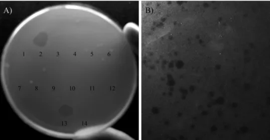
Isolation of a new bacteriophage forF. nucleatum.We ana-lyzed saliva samples from 25 healthy individuals and 85 pa-tients with periodontal disease from the Dental Clinic at the Universidad Andres Bello in June 2009. Two samples, en-riched as described in Materials and Methods, showed a halo of growth inhibition of the Fnp strain in a double-layered BHI plate (Fig. 1A). Lysates were obtained from the inhibition halos as described in Materials and Methods; the first lysate was called Fnp⌽02 and came from a sample from a healthy 24-year-old man, while the second was called Fnp⌽13 and was obtained from a sample of drainage from a hydraulic dental chair (Model Sagi 0.1, DentoLabs, Mexico). Only Fnp⌽02 was selected for further studies. To confirm that we found a bac-teriophage, the sample was propagated and diluted in order to obtain isolated lysis plaques (Fig. 1B). The lysis plaques showed a heterogeneous morphology with diameters of ap-proximately 1 to 2 mm; because of this, 15 of them were chosen for a new propagation. Every time this procedure was re-peated, we observed a variety of plaque morphologies, suggest-ing that we isolated a ssuggest-ingle phage with no characteristic plaque morphology. Finally, the clearest plaque was picked and propagated, and a lysate was obtained and preserved at 4°C for further studies.
Host specificity of Fnp⌽02. Of all the bacterial strains tested, Fnp⌽02 was able to infect and lyse onlyFusobacterium nucleatum, including our clinical isolates. Interestingly, we ob-served some differences in the phage effectiveness to lyse some strains, with F. nucleatum subsp. polymorphum being more sensitive to the infection thanF. nucleatumsubsp. nucleatum and subsp. vincentii.
Physical properties of Fnp⌽02.The transmission electron microscopy presented in Fig. 2A revealed that Fnp⌽02 pos-sessed an icosahedral head with a diameter of approximately 83 nm and a flexible tail that was 211 nm long. At the same time, we sawF. nucleatumcells after the phage infection, which revealed the propagation of virus-like particles (Fig. 2B and C). In particular, we observed bacterial lysis and the liberation of phage particles from the bacterium ends.
Infection of F. nucleatum with Fnp⌽02. The effect of Fnp⌽02 infection on F. nucleatum subsp. polymorphum was observed through the inoculation of a bacterial culture in early exponential phase (OD600at 0.15) at different MOIs (2, 1, 0.1,
on August 10, 2012 by UNIVERSIDAD ANDRES BELLO
http://aem.asm.org/
0.01, and 0.001). In all the infections, we saw a slow lysis of the bacterial culture, and for all the MOIs tested, the phage was unable to cause a complete bacterial lysis after 80 h of incu-bation, so the culture reached an OD600of 0.1 when the
high-est MOI was used (Fig. 3). The lysis ofF. nucleatumsubsp. nucleatumby Fnp⌽02 was lower than the lysis ofF. nucleatum subsp.polymorphumat the same MOI; as a negative control, Fnp⌽02 was incubated withF. necrophorum, causing no lysis (data not shown).
One-step growth.This analysis was performed to identify the different phases of the phage infection process. After infection ofF. nucleatumFnp with Fnp⌽02, the phage growth parame-ters—latent period, eclipse period, rise period, and burst size—were determined, comparing free and total phages. The system showed that the latency and eclipse periods of Fnp⌽02 were 15 and 7 h, respectively. Fnp⌽02 showed a burst size of
⬃100 phage per infected cell, measured along the 10-h rise period at 37°C (Fig. 4).
Adsorption efficiency.In the adsorption analysis of Fnp⌽02 toF. nucleatumFnp, we observed the rapid phage-bacterium interaction, with an 87% phage adsorption in only 3 min, as shown in Fig. 5. The adsorption process seems to be one rapid step, keeping the number of free phage constant until 5 min. The adsorption rate represents the affinity level between the phage and bacteria and was determined according to Barry and Goebel (3) in a 3-min period, resulting in an adsorption con-stant of 7.5⫻10⫺10ml min⫺1for phage Fnp⌽02.
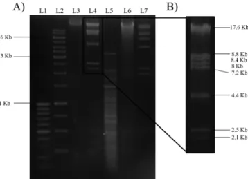
Genome analysis of Fnp⌽02. The phage genetic material was treated with restriction enzymes BamHI, HindIII, KpnI, PstI, BstEII, BfuCI, DraI, DpnI, EcoRI, EcoRV, HpaII, HaeIII, NotI, PvuI, Sau3AI, SpeI, and XbaI, of which HindIII and DraI were able to cut the viral genome, generating 8
FIG. 1. Bacteriophage isolation. (A) Double-layered plate showing growth inhibition ofF. nucleatumfrom a saliva sample from a healthy 24-year-old man (no. 2) and a drainage sample (no. 13). (B) Fragment of a double-layered plate with Fnp showing the plaque morphology of phage Fnp⌽02.
FIG. 2. Transmission electron micrographs. (A) Morphology of phage Fnp⌽02; (B) Fnp cell after inoculation with Fnp⌽02; (C) higher magnification of the Fnp cell lysis and the liberation of new phage particles (arrows).
7246 MACHUCA ET AL. APPL. ENVIRON. MICROBIOL.
on August 10, 2012 by UNIVERSIDAD ANDRES BELLO
http://aem.asm.org/
fragments and more than 10 fragments, respectively (Fig. 6). The genome size of Fnp⌽02 was determined from an electro-phoretic digestion pattern with HindIII. Thus, the sum of the sizes of all fragments gave us a genome size of⬃59 kbp (Fig. 6A and B). A small fragment (⬃500 bp) of the HindIII digest was cloned into pSU19 vector and sequenced. The sequence obtained (GenBank accession no. HQ014662), shown in Fig. 7A, presented a high identity (approximately 90.5%) with the Propionibacterium acnesphage PA6, a member of an unclassi-fied genus within the Siphoviridae family (Fig. 7A). Neither identity nor homology was found with any other reported phage sequence. Southern hybridization, using the lambda c1
gene as a probe, revealed no orthologous gene in Fnp⌽02 (data not shown). All of these results suggest that our phage could be considered a member of the unclassified group within theSiphoviridaefamily. The Fnp⌽02 genome section that we analyzed contains two small fragments that align with ORFs that have a consecutive disposition in the genomic context of phage PA6 (Fig. 7B). We found that both fragments had an amino acid similarity with two proteins from phage PA6. As shown in Fig. 7C, the first ORF fragment encodes a small peptide of 54 amino acids that has 98% identity with a segment of Gp3, a 441-amino-acid protein that has a putative structural function. The second ORF fragment encodes a 72-amino-acid
FIG. 3. Growth curves in the presence or absence of Fnp⌽02. Representative curves are based on results from three independent assays. The arrow indicates the infection with Fnp⌽02. Phage was added at different MOIs to Fnp in early exponential growth phase (OD600⫽0.1) as described
in Materials and Methods. As a control, an Fnp culture was inoculated with an autoclaved phage suspension.
FIG. 4. One-step growth curves of phage Fnp⌽02 infection with Fnp at an MOI of 0.001. Shown are the number of PFU per infected cell in untreated cultures (䉫) and in chloroform-treated cultures (f). The phage growth parameters are indicated in the figure: E, eclipse period; L, latent period; B, burst size. Representative curves based on results from three independent assays.
on August 10, 2012 by UNIVERSIDAD ANDRES BELLO
http://aem.asm.org/
polypeptide that shows 84% identity and 94% similarity to the Gp4 protein, a 251-amino-acid protein with a putative termi-nase function.
DISCUSSION
Periodontal disease is a chronic pathology that does not have an effective or definitive treatment. Currently, the dental ther-apy involves the mechanical removal of the dental plaque and the use of antimicrobial agents, trying in both cases to reduce the bacterial biofilm associated with tissue destruction. This treatment controls the progression of the disease for a limited time period, but the tissue damage is irreversible and may start again with the rapid recolonization by pathogenic bacteria. This deficient therapy, together with the increasing concern about drug-resistant pathogenic bacteria, has rekindled inter-est in the alternative treatment of bacterial infections (15, 16, 24). Phage therapy is a rediscovered option that uses bacterio-phages to kill and otherwise control the bacterial populations in the infected hosts. Renewed interest in phage therapy has risen from recent theoretical studies on phages and also ex-perimental work using phages associated with bacteria with clinical relevance (14, 20, 22, 32). One of these impressive studies was done by Paisano et al. (23), where a specific phage for Enterococcus faecalis was able to completely eradicate a bacterial infection from human dentine.
Although several bacteriophages have been isolated for a number of oral bacteria (8, 10, 12, 13, 23, 33), there have been no reports on the isolation of a phage for fusobacteria until now.F. nucleatum, together with a bacterial consortium, gen-erates the well-known dental plaque, a biofilm conformation that eventually leads to the onset of periodontal disease. The interaction that occurs between bacteria in these biofilms has been described in several studies (6, 31), but the effect of oral phages in the ecology of the dental plaque is not well under-stood.
As a first approach to explore the possibility for phage ther-apy as an alternative treatment for periodontal disease, we isolated a phage specific forFusobacterium nucleatum, a key bacterium in the development of periodontal disease.
Fnp⌽02 was isolated from a saliva sample from a healthy
individual, confirming that oral bacteriophages can be isolated from samples from the oral cavitities of people with and with-out periodontal disease (6, 8, 12, 13, 33, 37). This is because in a bacterium-phage system, the phage could be found wherever its target bacterium is found, and oral pathogens can be found in both healthy and periodontally diseased individuals (26). In the analysis of host specificity of Fnp⌽02, we observed that the phage has a narrow range, evidenced by a difference in the sensitivities seen for F. nucleatum subsp. nucleatum and F. nucleatumsubsp.vincentii compared to that ofF. nucleatum subsp.polymorphum. This different efficiency suggests that the phage receptor presents structural differences between subspe-cies, causing a decreased affinity and a less efficient adsorption phase (34). Based on these results, we can define Fnp⌽02 as a phage specific for the speciesF. nucleatum. The narrow host range of Fnp⌽02 is an interesting feature because it means that this phage would be harmless to the rest of the oral flora in the mouth, suggesting that a specific phage therapy for periodontal disease could be developed.
The genetic material of Fnp⌽02 was found to be double-stranded DNA, since it shows susceptibility to restriction en-zymes. Fnp⌽02 viral DNA could be digested only by 2 of the 17 enzymes tested, which showed us that the phage DNA was highly resistant to restriction enzymes, a well-characterized feature in the genetic material of phages, which usually contain methylated bases and methylated DNA as well as modified nucleotides (19). This result suggests that the Fnp⌽02 genome has a high level of methylation that protects the DNA. The genome size (⬃59 kbp) is within the normal range of the Siphoviridaefamily, which has an average size of 50 kbp (1, 7) and a range of 22 to 121 kbp (21). These features, along with the Fnp⌽02 virion structure, allowed us to classify Fnp⌽02 within the Siphoviridae family. Complete sequencing of the bacteriophage genome is under way, and in the future we may
FIG. 6. Restriction digest patterns electrophoresed on a 1.5% aga-rose gel and stained with ethidium bromide. (A) Fnp⌽02 genomic DNA digested with restriction enzymes. Lanes: L1, 100-bp DNA lad-der marker (Fermentas Inc., Canada); L2, 1-kb ladlad-der marker (Fer-mentas Inc., Canada); L3, undigested DNA; L4, DNA/HindIII; L5, DNA/DraI; L6, DNA/XbaI; and L7, lambda DNA/HindIII. (B) Higher magnification of the bands of the digestion of Fnp⌽02 with HindIII used for genome size determination. Selected sizes of the marker are indicated in panel A.
FIG. 5. Adsorption of Fnp⌽02 to Fnp: representative adsorption curve based on results from three separate experiments. Adsorption was carried out at an MOI of 0.01, and the supernatant was titrated at various time points to determine the amount of phage unadsorbed.
7248 MACHUCA ET AL. APPL. ENVIRON. MICROBIOL.
on August 10, 2012 by UNIVERSIDAD ANDRES BELLO
http://aem.asm.org/
determine if Fnp⌽02 is a lytic or temperate phage. In the latter case, analysis of the lysogeny activity of Fnp⌽02 will be the focus of a future study, especially due to the absence of genetic tools for the study of this bacterium today.
TheSiphoviridaefamily includes a temperate virus, such as the “lambda-like” virus, and lytic viruses, such as “T1-like” and “T5-like” viruses, in addition to other viruses that are not yet classified (1). The features of plaque lysis and lysis efficiency of Fnp⌽02 did not allow us to assess whether Fnp⌽02 is a lytic or a temperate phage, because the lysis plaques show heteroge-neous turbidity and size. This may occur if the phage infects cells at different times during the bacterial growth phase or due to the absence of certain culture conditions under which this
specific phage could produce more homogenous plaque char-acteristics (27). On the other hand, the bacteriophage was unable to reach complete lysis of the culture ofF. nucleatum subsp. polymorphum, and a variable recovery of CFU from lysates at different MOIs was obtained. These observations could be indicative of a temperate phage or the fact that the optimal conditions for infection were not present (28). This issue can be resolved only through the analysis of the complete sequence of the phage genome.
About the life cycle parameters of Fnp⌽02, we observed a difference between the phases of latent time, eclipse time, and rise period versus the burst size of the phage. The first three features have higher values than those traditionally found in
FIG. 7. Amino acid and nucleotide analysis of a short sequence of Fnp⌽02. (A) ClustalW analysis showing the nucleotide identity of Fnp⌽02 with PA6 phage. Asterisks show nucleotide identity. The stop codon of thegp3gene is in boldface, and the start codon of thegp4gene is underlined. (B) Schematic disposition of the nucleotide identity of PA6 and Fnp⌽02 phage and percentages of identity of the nucleotide alignments. (C) BlastX analysis comparing the amino acid identities of PA6 Gp3 and Gp4 proteins against a fragment of the Fnp⌽02 protein sequence. The numbers indicate the positions of the amino acid sequences in the proteins (first-line sequences) and the DNA fragment (third-line sequences). The second-line sequences are the consensus sequences, where plus signs correspond to amino acids of the same family and empty spaces represent the absence of identity.
on August 10, 2012 by UNIVERSIDAD ANDRES BELLO
http://aem.asm.org/
theSiphoviridaefamily (21); this is not surprising, since it has been shown that these parameters are highly influenced by the metabolic rate of the bacterial host. In this context,F. nuclea-tumhas a long generation time of approximately 5.6 h, affect-ing the length of the phage life cycle. On the other hand, the burst size of Fnp⌽02 (⬃100 phages per infected cell) is found within the normal range of theSiphoviridaefamily (3, 21).
Finally, when a small fragment of the genome of Fnp02 was analyzed, we found nucleotide and amino acid identity with phage PA6. This phage, similar to the Fnp02 phage, belongs to the Siphoviridae family and is a member of an unclassified genus. The PA6 genome, at approximately 30 kbp, is smaller than the calculated Fnp⌽02 genome, and no evident genes for a lysogenic cycle have been found in it (10). The genome size of Fnp02 compared with that of the PA6 phage suggests that the genes for a lysogenic cycle could be present. In summary, Fnp⌽02 is a bacteriophage that specifically attacks the periodontal pathogen Fusobacterium nucleatum and seems to belong to theSiphoviridaefamily. Our laboratory is currently studying the potential use of this new bacterio-phage as a biological control agent to reduce proliferation ofF. nucleatumin pathogenic oral biofilms as a first step to explore the development of phage therapy to improve the current treatment of periodontal disease.
ACKNOWLEDGMENTS
This work was supported by FONDECYT grant 11060181 and Uni-versidad Andres Bello grants DI 17/06R and DI 48/09R (to M.B.).
REFERENCES
1.Ackermann, H. W.1998. Tailed bacteriophages: the order Caudovirales. Adv. Virus Res.51:135–201.
2.Armitage, G. C.2004. Periodontal diagnoses and classification of periodontal diseases. Periodontol. 200034:9–21.
3.Barry, G. T., and W. F. Goebel.1951. The effect of chemical and physical agents on the phage receptor of Phase-liShigella sonnei. J. Exp. Med.94:
387–400.
4.Bolstad, A. I., H. B. Jensen, and V. Bakken.1996. Taxonomy, biology, and periodontal aspects ofFusobacterium nucleatum. Clin. Microbiol. Rev.9:55–71. 5.Boutaga, K., A. J. van Winkelhoff, C. M. Vandenbroucke-Grauls, and P. H. Savelkoul.2005. Periodontal pathogens: a quantitative comparison of anaer-obic culture and real-time PCR. FEMS Immunol. Med. Microbiol.45:191– 199.
6.Bradshaw, D. J., P. D. Marsh, G. K. Watson, and C. Allison.1998. Role of
Fusobacterium nucleatumand coaggregation in anaerobe survival in plank-tonic and biofilm oral microbial communities during aeration. Infect. Im-mun.66:4729–4732.
7.Brussow, H., and F. Desiere.2001. Comparative phage genomics and the evolution ofSiphoviridae: insights from dairy phages. Mol. Microbiol.39:
213–222.
8.Delisle, A. L., R. K. Nauman, and G. E. Minah.1978. Isolation of a bacte-riophage forActinomyces viscosus. Infect. Immun.20:303–306.
9.Dzink, J. L., M. T. Sheenan, and S. S. Socransky.1990. Proposal of three subspecies ofFusobacterium nucleatumKnorr 1922:Fusobacterium nuclea-tum subsp.nucleatumsubsp. nov., comb. nov.;Fusobacterium nucleatum
subsp.polymorphumsubsp. nov., nom. rev., comb. nov.; andFusobacterium nucleatumsubsp.vincentiisubsp. nov., nom. rev., comb. nov. Int. J. Syst. Bacteriol.40:74–78.
10.Farrar, M. D., K. M. Howson, R. A. Bojar, D. West, J. C. Towler, J. Parry, K. Pelton, and K. T. Holland.2007. Genome sequence and analysis of a
Propionibacterium acnesbacteriophage. J. Bacteriol.189:4161–4167. 11.Feuille, F., J. L. Ebersole, L. Kesavalu, M. J. Stepfen, and S. C. Holt.1996.
Mixed infection withPorphyromonas gingivalisandFusobacterium nucleatum
in a murine lesion model: potential synergistic effects on virulence. Infect. Immun.64:2094–2100.
12.Haubek, D., K. Willi, K. Poulsen, J. Meyer, and M. Kilian.1997. Presence of bacteriophage Aa phi 23 correlates with the population genetic structure of
Actinobacillus actinomycetemcomitans. Eur. J. Oral Sci.105:2–8.
13.Hiroki, H., A. Shiki, M. Totsuka, and O. Nakamura.1976. Isolation of bacteriophages specific for the genusVeillonella. Arch. Oral Biol.21:215– 217.
14.Housby, J. N., and N. H. Mann.2009. Phage therapy. Drug Discov. Today
14:536–540.
15.Jeffcoat, M. K., K. S. Bray, S. G. Ciancio, A. R. Dentino, D. H. Fine, J. M. Gordon, J. C. Gunsolley, W. J. Killoy, R. A. Lowenguth, N. I. Magnusson, S. Offenbacher, K. G. Palcanis, H. M. Proskin, R. D. Finkelman, and M. Flashner.1998. Adjunctive use of a subgingival controlled-release chlorhexi-dine chip reduces probing depth and improves attachment level compared with scaling and root planing alone. J. Periodontol.69:989–997.
16.Jong, R. A., and W. A. Van der Reiiden.2010. Feasibility and therapeutic strategies of vaccines againstPorphyromonas gingivalis. Expert Rev. Vaccines
9:193–208.
17.Kim, H. S., S. K. Song, S. Y. Yoo, D. C. Jin, H. S. Shin, C. K. Lim, M. S. Kim, J. S. Kim, S. J. Choe, and J. K. Kook.2005. Development of strain-specific PCR primers based on a DNA probe Fu12 for the identification of Fuso-bacterium 128nucleatumsubsp. nucleatumATCC 25586T. J. Microbiol.
43:331–336.
18.Kinane, D., P. Bouchard, and Group E of European Workshop on Peri-odontology.2008. Periodontal diseases and health: consensus report of the Sixth European Workshop on Periodontology. J. Clin. Periodontol.
35(Suppl. 8):333–337.
19.Krueger, D. T., and T. A. Bickle.1983. Bacteriophage survival: multiple mechanisms for avoiding the deoxyribonucleic acid restriction systems of their hosts. Microbiol. Rev.47:345–360.
20.Levin, B. R., and J. J. Bull2004. Population and evolutionary dynamics of phage therapy. Nat. Rev. Microbiol.2:166–173.
21.Maniloff, J., and H. W. Ackermann.1998. Taxonomy of bacterial viruses: establishment of tailed virus genera and the order Caudovirales. Arch. Virol.
143:2051–2063.
22.Matsuzaki, S., M. Rashel, J. Uchiyama, S. Sakurai, T. Ujihara, M. Kuroda, M. Ikeuchi, T. Tani, M. Fujieda, H. Wakiguchi, and S. Imai.2005. Bacte-riophage therapy: a revitalized therapy against bacterial infectious diseases. J. Infect. Chemother.11:211–219.
23.Paisano, A. F., B. Spiera, S. Cai, and A. C. Bombana.2004. In vitro antimi-crobial effect of bacteriophages on human dentin infected withEnterococcus faecalisATCC 29212. Oral Microbiol. Immunol.19:327–330.
24.Raghavendra, M., A. Korengol, and S. Bhola.2009. Photodymanic therapy: a targeted therapy in periodontics. Aust. Dent. J.54(Suppl. 1):S102–S109. 25.Ripp, S.2010. Bacteriophage-based pathogen detection. Adv. Biochem. Eng.
Biotechnol.118:65–83.
26.Rylev, M., and M. Kilian.2008. Prevalence and distribution of principal periodontal pathogens worldwide. J. Clin. Periodontol.35(Suppl. 8):346– 361.
27.Santos, S. B., C. M. Carvalho, S. Sillankorva, A. Nicolau, E. C. Ferreira, and J. Azeredo.2009. The use of antibiotics to improve phage detection and enumeration by the double-layer agar technique. BMC Microbiol.9:148. 28.Shao, Y., and I. N. Wangi.2008. Bacteriophage adsorption rate and optimal
lysis time. Genetics180:471–482.
29.Shin, H. S., M. J. Kim, H. S. Kim, S. N. Park, D. K. Kim, D. H. Baek, C. Kim, and J. K. Kook.2010. Development of strain-specific PCR primers for the identification ofFusobacterium nucleatumsubsp.fusiformisATCC 51190(T) and subsp.vincentiiATCC 49256(T). Anaerobe16:43–46.
30.Sillankorva, S., P. Neubauer, and J. Azevedo.2008. Isolation and character-ization of T7-like lytic phage forPseudomonas fluorescens. BMC Biotechnol.
8:80.
31.Socransky, S. S., A. D. Haffajee, M. A. Cugini, C. Smith, and R. L. Kent, Jr.
1998. Microbial complexes in subgingival plaque. J. Clin. Periodontol.25:
134–144.
32.Tang, K. H., K. Yusoff, and W. S. Tan.2009. Display of hepatitis B virus PreS1 peptide on bacteriophage T7 and its potential in gene delivery into HepG2 cells. J. Virol. Methods159:194–199.
33.Tylenda, C. A., C. Calvert, P. E. Kolenbrander, and A. Tylenda.1985. Isolation ofActinomycesbacteriophage from human dental plaque. Infect. Immun.49:1–6.
34.Waldor, M. K., D. I. Friedman, and S. L. Adhya.2005. Phages, their role in bacterial pathogenesis and biotechnology. ASM Press, Washington, DC. 35.Walker, C. B., D. Ratliff, D. Muller, R. Mandell, and S. S. Socransky.1979.
Medium for selective isolation ofFusobacterium nucleatumfrom human periodontal pockets. J. Clin. Microbiol.10:844–849.
36.Weinbauer, M. G.2004. Ecology of prokaryotic viruses. FEMS Microbiol. Rev.28:127–181.
37.Yeung, M. K., and C. S. Kozelsky.1997. Transfection ofActinomycesspp. by genomic DNA of bacteriophages from human dental plaque. Plasmid37:
141–153.
7250 MACHUCA ET AL. APPL. ENVIRON. MICROBIOL.